Publisher’s version / Version de l'éditeur:
Vous avez des questions? Nous pouvons vous aider. Pour communiquer directement avec un auteur, consultez la première page de la revue dans laquelle son article a été publié afin de trouver ses coordonnées. Si vous n’arrivez pas à les repérer, communiquez avec nous à [email protected].
Questions? Contact the NRC Publications Archive team at
[email protected]. If you wish to email the authors directly, please see the first page of the publication for their contact information.
https://publications-cnrc.canada.ca/fra/droits
L’accès à ce site Web et l’utilisation de son contenu sont assujettis aux conditions présentées dans le site LISEZ CES CONDITIONS ATTENTIVEMENT AVANT D’UTILISER CE SITE WEB.
European Chemical Bulletin, 7, 3, pp. 106-114, 2018-05-27
READ THESE TERMS AND CONDITIONS CAREFULLY BEFORE USING THIS WEBSITE. https://nrc-publications.canada.ca/eng/copyright
NRC Publications Archive Record / Notice des Archives des publications du CNRC :
https://nrc-publications.canada.ca/eng/view/object/?id=acb4ce95-ff71-4bca-be0c-5948788eb912 https://publications-cnrc.canada.ca/fra/voir/objet/?id=acb4ce95-ff71-4bca-be0c-5948788eb912
NRC Publications Archive
Archives des publications du CNRC
This publication could be one of several versions: author’s original, accepted manuscript or the publisher’s version. / La version de cette publication peut être l’une des suivantes : la version prépublication de l’auteur, la version acceptée du manuscrit ou la version de l’éditeur.
For the publisher’s version, please access the DOI link below./ Pour consulter la version de l’éditeur, utilisez le lien DOI ci-dessous.
https://doi.org/10.17628/ecb.2018.7.106-114
Access and use of this website and the material on it are subject to the Terms and Conditions set forth at
Potential environmental impact of nanoenergetics
POTENTIAL ENVIRONMENTAL IMPACT OF
NANOENERGETICS
Nancy N. Perreault
[a]*Keywords: Nanomaterial, nanothermite, environmental fate, nanotoxicity.
Nanoenergetics has the potential to become the next generation of explosives and propellants. Nano-explosives often show better performances in terms of energy release, ignition and mechanical properties compared to their bulk counterparts. In addition to monomolecular explosives such as nano-TNT and nano-RDX, diverse energetic nanocomposites have been developed, including the nanometer-sized versions of conventional thermites (nanothermites) and those using carbon-based nanomaterials (e.g., fullerenes and carbon nanotubes). While the unique characteristics of nanomaterials allow groundbreaking applications, they also result in distinct environmental fate, transport and toxicity. The high surface to volume ratio and reactivity of nanomaterials make them highly dynamic in environmental systems. Once released in the environment, nanoenergetic chemicals will undergo transportation and transformation processes including dissolution, aggregation, adsorption, photolysis and biotransformation, which will also affect their persistence and bioavailability. This review was conducted to better understand the potential environmental fate and ecological impact of the use of nanoenergetics.
* Corresponding Authors Fax: 514-496-6265
E-Mail: [email protected]
[a] National Research Council Canada, 6100 Royalmount Ave., Montréal, Québec H4P 2R2
Introduction
Over the past decades, remarkable progress has been made in military science and technology to design energetic materials with higher burning rate, density and reactivity, lower sensitivity and smaller critical diameter. While explosives of coarse size are reaching their capacity limit, the emergence of nanotechnology brings new possibilities to meet the increasing military demands for high performance, safety, and environmentally friendly munitions. Nanomaterials, which are made of components with at least one dimension below 100 nm, have attractive characteristics allowing to decrease the sensitivity of explosives to impact, friction and shock waves and to increase energy release and burning rate.Increase in burning rate due to the addition of nanoenergetics to propellants is due primarily to the i) increased reactivity of nanomaterials compared to bulk materials (micrometer-sized particles), ii) increased rate of diffusion of the reactants, and iii) increased rate of energy release due to the much shorter combustion time of nanoparticles.1 Nanoenergetics has the potential to become the next generation of explosives and propellants.
Many high explosives have been synthesized at the nanometer size including nano-nitramines (RDX, HMX, CL-20), nano-TATB (2,4,6-triamino-1,3,5-trinitrobenzene), nano-NTO (5-nitro-2,4-dihydro-3H-1,2,4-triazole-3-one) and nano-TNT (2,4,6-trinitrotoluene). Compared to their micron-sized counterparts, the shock sensitivities of nano-nitramine explosives were shown to be decreased by 59.9% (RDX), 56.4% (HMX) and 58.1% (CL-20).2
Nanothermites, mixtures of a metal (e.g. Al) and metal oxide (e.g. Fe2O3) at the nanoscale, are much more reactive than traditional thermites. Nanothermites are the most intensively investigated nanoenergetics and the only one
currently in use by the military. Nano-aluminum is the most common component of metal-based nanomaterials and considered as a potential replacement for the conventional aluminum powders and flakes widely used in explosives and propellants.
Carbon-based nanomaterials, including single- or multi-walled nanotubes and fullerene C60, have become important due to their unique combinations of chemical and physical properties (e.g. thermal and electrical conductivity, high mechanical strength) and are used for many industrial applications. Carbon-based nanomaterials can be functionalized with energetic groups or filled with high energetic molecules, mixed with nanothermites or doped with metal oxide catalysts for use as nanoenergetics. Carbon nanotubes (CNTs) are long, thin, hollow cylinders, composed of one (single-walled carbon nanotubes, SWCNTs) or many (multi-walled carbon nanotubes, MWCNTs) concentric layers of graphenic carbon. Fullerene C60 has a cage-like fused-ring structure (truncated icosahedron) that resembles a soccer ball, comprising 60 carbon atoms. The use of functionalized carbon nanomaterials in energetic compositions greatly improves their combustion performances, thermal stability and sensitivity. Choi et al.3 obtained a burning rate exceeding more than 103 times the value of bulk RDX when using 7-nm RDX annular shell around a MWCNT. When energetic materials 4-nitrobenzenediazonium nitrate, 4-nitroaniline, 2,4-dinitroaniline and RDX were loaded on CNTs, the exothermic reaction speeds of all energetic materials increased by approximately 100 times compared with those of the energetic materials alone.4 CNT-supported metal oxide nanoparticles significantly enhanced thermal decomposition of nitrocellulose-absorbed nitroglycerin, RDX, ammonium perchlorate and N-guanylurea-dinitramide (FOX-12). Moreover, the encapsulation of FOX-7 (1,1-diamino-2,2-dinitroethylene), RDX and HMX inside a CNT was shown to stabilize these energetic chemicals, thereby reducing their sensitivity.5 Because of heat energy coming from the high density of covalent energy stored by the carbon-carbon bonds in the C60, fullerenes are of interest for use in nanoscale explosives. A study showed that fullerene C60 can enhance the burning rate of double-based
propellants, broaden the plateau region and lower the concentration of NOx in gas products.6 Fullerenes C60 were also shown to reduce the friction and impact sensitivities of HMX.7
Environmental Fate
Nanoenergetics will enter the environment either in the pristine state from spills during manufacture, transport and storage of munitions, or will be disseminated in their post-combustion (or post-detonation) state. Poda et al.8 found that residues of Al/Fe2O3-based nanothermite comprise particles, generally larger than 75 m, consist of a mixture of FeO, Al2O3 and FeAl2O4. In the same study, Al/Bi2O3 formulations fully transformed to small (˂ 5 m) particles consisting of metallic Bi and Al2O3. These products are largely resistant to wetting, settle quickly, and evidence suggests that transport in aqueous environments would be limited. Due to the particle size ranges found, it has been speculated that aerosolization should be a major transport route for the nanothermite residues. The high surface to volume ratio and reactivity of nanomaterials make them highly dynamic in environmental systems and energetic nanomaterials are likely to undergo transportation and transformation processes including dissolution, aggregation, adsorption, photolysis and biotransformation.
Transport
Solubility and dissolution
Transport is positively correlated with solubility. Theoretically, the solubility of a particle increases as size decreases. Furthermore, the rate of dissolution is dependent on particle surface area. Nanoparticles should thus dissolve faster than larger particles. The dissolution behaviour of metallic nanoparticles will affect their overall stability in the aquatic environment and their potential ecotoxicity, since toxicity has often been attributed to the release of ionic species rather than being induced by the nanoparticles themselves.9 Most metal-based nanoparticles have very low solubility, however, their solubility is significant (e.g. in the mg L-1 range) from an environmental and toxicological perspective. For example, zinc oxide is classified as insoluble in water with a solubility of ~1 mg L-1 at pH 8; nevertheless, one gram of ZnO nanoparticles added to one litre of water at pH 7.8 will generate ~10 mg L-1 of dissolved Zn. While only representing dissolution of less than 2 % of ZnO, the Zn is well in excess of the 5 mg L-1 that would be toxic to most aquatic biota.
Pristine C60 fullerene and CNTs are insoluble in water. Solubility of C60 fullerene was estimated to be as low as 10-8 ng L-1 at 25 C, equivalent to ten C
60 molecules per ml of water.10 Direct solubility is however not the most relevant measure of the availability of fullerenes to organisms, particularly to aquatic organisms. Indeed, although C60 is not soluble in water or other polar solvents, water-soluble colloidal aggregates (nano-C60 or nC60) readily form when pristine C60 is added to water. The aggregates have reported diameters of 5 to 500 nm and allow for C60 concentrations up to 100 mg L-1, eleven orders of magnitude more than the estimated solubility.11 Recent developments in chemical
modification and functionalization of CNTs have greatly improved the solubility and dispersion of CNTs in water. The solubility of monomolecular nano-explosives such as RDX and TNT has not been reported yet.
Aggregation and adsorption
Environmental fate and transport of nanoparticles can be significantly impacted by aggregation in water and adsorption onto the soil matrix and other particles. Because of their high surface area, nanoparticles have a tendency to sorb to sediments and soil particles, which greatly reduce their transport and bioavailability. Although assumption is often made than nanoparticles will be more mobile in porous media due to their small size, they may in fact be less mobile in porous media such as groundwater aquifer if they readily sorb to mineral surfaces. Thus, all other factors being equal (ionic strength and composition, media grain size and hydrodynamics), smaller particles should be less mobile due to their relatively large diffusivity that produces more frequent contacts with the surfaces of the porous media.12 On the other hand, dissolved organic matter ubiquitous in aquatic ecosystems also adsorbs onto nanoparticles and this should favour their transport in aquatic environments.
Particles up to a few microns in size, that are present as individual particles, do not readily settle from solution. However, nanoparticles have the propensity to form larger micron-sized water-stable aggregates. Aggregation of nanoparticles typically leads to sedimentation and thus, to their removal from the aqueous phase and their immobilization. Aggregates of nanoparticles that settle can be expected to accumulate in sediments of lakes or rivers in the absence of nanoparticle degradation. Aggregation was shown to increase as the ionic strength of a solution increases and as the concentration of divalent cations such as Ca2+ and Mg2+ increases. Nanoparticles will thus tend to aggregate more in seawater (high ionic strength) than in freshwater. A study tested the stability of three metal oxide nanoparticles (TiO2, ZnO and CeO2) in aqueous samples.13 In the low organic matter concentration and high ionic strength of seawater, the rate of sedimentation of the three nanoparticles was very high. At high nanoparticle concentrations, the concentration of suspended particles decreased by more than 80 % in less than 100 min; the rate decreased as nanoparticle aggregates were removed from solution, lowering the concentration of suspended particles. Metal oxide nanoparticles entering seawater are thus expected to be removed from the water column in a few hours. This suggests that marine organisms in the water column may only be significantly affected if the loading is frequent or continuous; benthic organisms may be at higher risk of exposure, although the exposure would be in the form of relatively large aggregates (> 1 μm) of nanoparticles.
A completely different behaviour occurred when the metal oxide nanoparticles were dispersed in freshwater, with high organic matter concentration and low ionic strength.13 In freshwater, the size of the aggregates remained stable at slightly above 300 nm for the three metal oxides, with a very low rate of sedimentation. The significant amount of organic molecules that can be adsorbed onto the nanoparticle surfaces provided an additional barrier to aggregation. In the presence of NOM at realistic concentrations (1–30 mg of carbon L-1), nanoparticles are
stabilized and aggregation is limited.14,15 Thus, under these conditions, aquatic organisms within the water column (e.g. fish, algae, filter-feeders) will be exposed to small nanoparticle aggregates for a longer time. Eventually, within a few days the particles will sediment out, exposing benthic organisms as well. Chekli et al.16 evaluated that 20 % of dissolved organic matter was adsorbed to nanoparticles of Fe2O3 in river and lake waters; adsorption was lower in groundwater (10 %) and seawater (5 %). As expected, the size of Fe2O3 aggregates was higher in ground- and seawater than in river and lake waters.
Nanoparticle mobility will be significantly reduced in terrestrial compared to aquatic systems. Adsorption of nanoparticles by soil minerals and soil organic matter has not been evaluated, but is likely to be a function of particle size, shape and surface properties (specific surface area and surface charge). Most soil particles have a negative charge: small hydrophilic nanoparticles (< 20 nm) with net negative surface charges are likely to be mobile, while large hydrophilic positively-charged particles are likely to be sorbed by soil. Strongly hydrophobic nanoparticles should be strongly retained by soil organic matter.17 In addition, aggregation in soil leads to entrapment of particles in pores through which the dispersed nanoparticles could have passed, thus restricting mobility.
Transformation Abiotic transformation
Redox reactions can alter the surface chemistry of nanoparticles and therefore affect their mobility, toxicity and ultimate fate in the environment. Despite the limited studies of redox transformations, most metal and metal oxide nanoparticles are expected to weather slowly overtime, similar to chemical weathering of minerals in the environment. Studies on the photochemical transformation of nanoparticles compared to their bulk counterparts are not available in the literature. Photochemical transformation may be an important fate process of fullerene C60 in surface waters due to its strong absorption within the solar spectrum. When exposed to light, the cluster size of nC60 was shown to decrease as the irradiation time increased.18 Water-soluble products were formed and, with continued light exposure, these intermediates eventually mineralized, volatilized, or converted to other products. The decay rate of C60 in small clusters (150-nm diameter) was greater than for C60 in larger clusters (500 nm), with half-lives of 19 and 41 h, respectively. This suggests that release of C60 into surface waters will result in photochemical production of currently unknown products. Unfunctionalized SWCNTs are not, or only slowly, photo-transformed under sunlight, however, they can undergo indirect phototransformation through reactions with hydroxyl radicals.19 A carboxylated SWCNT solution was also shown to produce ROS when exposed to sunlight or when irradiated in a solar simulator, which in turn oxidized CNTs and modified their surface.20
Biotransformation
Unlike organic pollutants, metals and metal oxides cannot be degraded. Therefore, biodegradation processes do not apply to nanothermite constituents. Biodegradation studies
of nano-explosives are nonexistent in the literature and very few studies are available on the biotransformation of nanoparticles in general. Nano-sized explosives have a larger surface area compared to their micron-sized counterparts and this could possibly allow faster degradation by microorganisms.
A very limited number of microorganisms and enzymes have been found to transform or degrade nanomaterials. The physical and chemical nature of CNTs, fullerenes and their derivatives make them inert, stable and difficult to degrade. Nevertheless, some fungi and fungal enzymes were successful at transforming carbon-based nanomaterials. Lignin peroxidase (LiP) secreted by the mushroom
Sparassis latifolia degraded carboxylated SWCNTs.21 In addition, manganese peroxidase (MnP) from the white rot fungus Phanerochaete chrysosporium was reported to decompose SWCNTs.22 Another white rot fungus, Trametes versicolor, showed a weak ability to degrade SWCNTs.23 Phlebia tremellosa and T. versicolor mineralized fullerol, a
water-soluble hydroxylated derivative of fullerene C60, after 32 weeks of decay.24 C
60 itself was shown to be resistant to mineralization in sandy loam soil for at least 11 months and in silt loam for at least 2 years.25 In addition, three bacteria (Burkholderia kururiensis, Delftia acidovorans and
Stenotrophomonas maltophilia) were reported to form a
consortium that degrade MWCNTs to CO2 with several intermediate products such as methoxynaphthalene, 2-naphthol, cinnamaldehyde and isophthalic acid.26 Although individual bacteria in this community could weakly degrade MWCNTs, they were much more efficient degraders in combination. Chouhan et al.27 isolated bacteria from a soil contaminated with nanomaterials and showed that the bacteria were adaptive and tolerant to the nanomaterials and thus could well survive in the contaminated soil. The bacteria were also able to transform MWCNTs by an oxidation process. The persistence of nanoenergetics in the environment will highly depend on their ability to be biodegraded. However, CNTs and fullerenes are not readily biodegradable and their fate upon release in the environment may end up being similar to polychlorinated biphenyls and other types of persistent organic pollutants (POPs), which also tend to be associated with particles in the environment.12
Potential ecotoxicity of nanomaterials and
nanoenergetics
The fundamental properties of matter change at the nanoscale, therefore toxicity of nanoparticles cannot be predicted from the known properties of larger-sized particles of the same substance. The majority of studies have arrived at a similar conclusion: smaller particles induce a higher degree of cell death. For example, studies clearly indicated that tissue distribution of Al2O3 was size-dependent, and that metal oxide nanoparticles (Al2O3, Fe2O3) were able to cause more size- and dose-dependent toxicity in vivo compared to bulk materials.28,29 Other factors influencing toxicity include shape, chemical composition, surface structure, surface charge, aggregation and solubility. As yet, little work has been conducted to determine the effects of nanoparticles to the biota in aquatic and terrestrial habitats. The current understanding of the toxicity mechanisms and impacts of nanomaterials to different ecosystems and trophic levels,
including bacteria, algae, crustaceans, fish, plants and soil invertebrates are summarized.
Mechanisms of nanotoxicity
Living organisms may be exposed to nanoparticles via several routes, including inhalation of airborne particles (respiratory tract), ingestion (gastrointestinal tract) and dermal contact (skin). Because of their small size, nanoparticles are often more readily taken up than larger particles. Nanoparticles can cross biological membranes and access cells, tissues and organs than larger-sized particles normally cannot. Inhalation of nanoparticles may become the major route for unwanted exposure to nanothermites and nano-explosives residues. Studies over the last 25 years have suggested that smaller particles made of toxicity, low-solubility materials are more potent at inducing lung inflammation than the same materials at larger particle sizes. Furthermore, smaller particles exhibit enhanced penetration across the lung epithelium and into the interstitium of the lung tissue. On the other hand, nanoparticles naturally tend to aggregate into larger particles that can be microns in size, thereby reducing the likelihood of free nanoparticles being respired. However, surface modifications designed to limit particle-particle interactions may reduce the tendency for nanoparticle aggregation and increase the potential for inhalation and deposition within the lungs. Inhaled particles of different sizes exhibit different fractional depositions within the human respiratory tract. Although inhaled ultrafine particles (< 100 nm) deposit in all regions, tracheobronchial deposition is highest for particles < 10 nm in size, whereas alveolar deposition is highest for particles approximately 10–20 nm in size.30,31 Fullerenes, CNTs, metal and metal oxide nanoparticles were all shown to induce pulmonary inflammation and fibrosis in in vivo and in vitro studies. Once nanomaterials reach the blood stream, they are transported around the body and taken up by the organs and tissues. CNTs, for example, have been found to rapidly enter and disseminate in the organism, initially accumulating in lungs and brain and later reaching the liver and kidney via the bloodstream.32 Many nanoparticles are capable of crossing the blood–brain barrier, potentially damaging the central nervous system and inducing neurotoxic effects.33
Reactive oxygen species (ROS) and free radical production has been identified as a primary mechanism of nanoparticle toxicity. Nanomaterial-induced ROS may result in oxidative stress, inflammation, and consequent damage to cellular constituents.34 ROS can damage cell membrane, particularly through lipid peroxidation, as well as mitochondria, proteins and DNA, leading to cytotoxicity, genotoxicity and cancer. Another problematic phenomenon is the adsorption of proteins on nanoparticle surface that leads to the formation of nanoparticle-protein complexes commonly referred to as the nanoparticle-protein corona. Nanoparticles induce changes in the structure of adsorbed proteins and therefore impair their function. SWCNTs was shown to induce loss of structure and catalytic activity of enzymes.35 Strong protein binding onto metal oxides is well documented, so adsorption unto metal oxide nanoparticles is likely. Nanoparticles can also induce conformational changes in proteins that can lead to fibril formation. Linse et al.36 showed that a range of nanoparticles, including CNTs, are capable of inducing fibrillation of β2-microglobulin due
to increased protein localization on the nanoparticle surface. Fibrillation of proteins is associated with diseases such as Parkinson’s and Alzheimer’s.
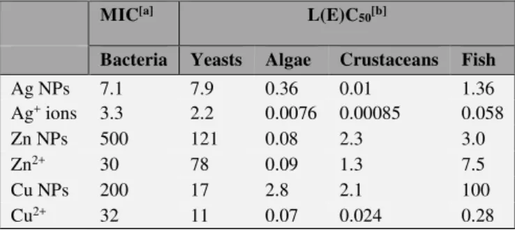
Dissolution is an essential factor to consider when evaluating toxicity of metal-containing nanoparticles. Toxicity of metal-based nanoparticles, such as ZnO, CuO and Ag, partly results from the release of metal ions.9 Table 1 presents toxicity values37 for some organism groups. In most cases, the free metal ions are significantly more toxic. Yet, the issue is relatively complex as also other nano-specific factors may complement the effect of dissolved metal ions and the distinct role of each factor remains unknown for the moment.38
As mentioned earlier, although fullerenes are highly insoluble in water, they are modified during mixing in water to form stable aggregates, known as nC60, enabling C60 concentrations up to 100 mg L-1. This ability to be dispersed in water increases their mobility in aquatic environments and makes them available to aquatic organisms.
Table 1. Toxicity of Ag, ZnO and CuO nanoparticles (NPs) and their respective ions to bacteria, yeasts, algae, crustaceans and fish. Adapted from Ivask et al.38
MIC[a] L(E)C
50[b]
Bacteria Yeasts Algae Crustaceans Fish
Ag NPs 7.1 7.9 0.36 0.01 1.36 Ag+ ions 3.3 2.2 0.0076 0.00085 0.058 Zn NPs 500 121 0.08 2.3 3.0 Zn2+ ions 30 78 0.09 1.3 7.5 Cu NPs 200 17 2.8 2.1 100 Cu2+ ions 32 11 0.07 0.024 0.28 Values selected and summarized from Bondarenko et al.37 [a]
Minimal inhibitory concentration (mg L-1); [b] Half-lethal or
half-effective concentration (mg L-1)
Aquatic ecotoxicity
Nanomaterials can enter natural water systems by numerous direct (through aerial deposition, effluents, dumping and run-off) and indirect routes (e.g. via river systems). Most of the currently available ecotoxicological data regarding nanoparticles are limited to species used in regulatory testing or freshwater species, and there are discrepancies in the reported effects of nanoparticles on aquatic organisms. One of the key problems encountered when assessing aquatic ecotoxicology is the protocol used to prepare the nanoparticles. For CNTs and fullerenes, there is a general consensus that they have poor aqueous solubility and that some combination of chemical dispersants, stirring or sonication is necessary to maintain them in aqueous solution. A number of ecotoxicology studies have used the organic solvent tetrahydrofuran (THF) to disaggregate nanoparticles such as C60 prior to treatment of organisms. It was demonstrated that even after filtration and evaporation, THF remains trapped between the aggregated C60 particles,39 suggesting that studies using THF investigated the effects of C60 combined with THF rather than the effects of C60 per se. THF is classified by many regulatory bodies as a neurotoxin, and so could in part explain some of the effects observed in the fish. For these reasons, it is worth considering some ecotoxicological studies with caution.
Uptake and toxicity from primary producers (algae), microscopic invertebrates, up through the trophic chain is probable. Sorption onto aquatic organisms has been reported as a dominant toxicity mechanism of nanoparticles as it affects the molting behavior and swimming speed of daphnids,40 and alter filtering efficiency. Nanoparticle coating of gills can affect respiration, cause hypoxia in blood and spleen and reduce the locomotion of fish. Nanoparticles were also shown to cover algal cell surface, increasing the cellular weight by more than 2 fold, affecting the algae’s ability to float, and reducing sunlight availability for photosynthesis.41
Effects on freshwater organisms
The early studies on freshwater invertebrates focused on crustaceans, with Daphnia magna being the most studied test species. It has been shown that daphnids take up significant amounts of both fullerene C60 aggregates (nC60) and nano-iron from aqueous media.42 Tissue levels of > 2 mg L-1 were reached when daphnids were exposed for 72 h to 30 mg L-1 of nC
60 stirred in water. nC60 could not be prepared at high enough concentration levels to cause 50 % mortality (LC50) at 48 or 96 h, and no mortality was observed before 5 to 6 days of exposure. Mortality was observed at concentrations of 1, 2.5, and 5 mg L-1, with the highest mortality (40 %) achieved at 2.5 mg L-1. Exposure for 21 days to 2.5 and 5 mg L-1 fullerenes resulted in a significant delay in D. magna molting and significantly reduced offspring production, which could have negative impacts at the population level. The ingested C60 did not visibly bind to the daphnid exoskeleton and antennae.
Exposure of D. magna to nano-iron only resulted in significant mortality at 62.5 mg L-1 (50 % level achieved in 24 h), which is considered a very large dose in environmental toxicology term. In addition, these organisms exhibited significant binding of the nano-iron particles to their exoskeleton surface and antennae as well as significant ingestion of the particles.
Exposure of up to 500 µg nano-Ag L-1 for 48 h did not cause mortality in D. magna;43 in contrast, the crustacean was extremely sensitive to free Ag ion (Ag+ added as AgNO3), with a measured 48 h 50 % lethal concentration of 2.51 µg L-1, suggesting that the acute toxicity of Ag nanoparticles was caused by the release of Ag+ into solution. TiO2 nanoparticles were shown to transfer through food chain from Daphnia to zebrafish.44 Overall, toxicity studies on D. magna indicate that the lethality of the nanoparticles tested is relatively low, but that there may still be cause for concern.
C60 fullerene prepared with THF has been reported to cause significant oxidative damage in vivo in largemouth bass (Micropterus salmoides).45 Fish exposed to 0.5 and 1 mg L-1 nC
60 for 48 h exhibited signs of lipid peroxidation in the brain. Zhu et al.46 reported that nC
60 generated by water stirring had no impact on lethality within 6 to 18 h of exposure in adult fathead minnow (Pimephales promelas); however, as for the largemouth bass, lipid peroxidation was observed. Exposure of zebrafish to nC60 caused negative developmental effects that were mitigated after treatment with an antioxidant, supporting the notion that nC60 exerts oxidative stress.47 On the other hand, a critical review of
evidence (2007–2011) by Henry et al.48 suggested that aqueous nC60 has minimal potential to produce ROS and that oxidative stress in fish is not induced by environmentally relevant exposure to nC60. A detailed study by Smith et al.49 demonstrated that SWCNTs was a respiratory toxicant in rainbow trout and caused cellular defects indicative of systemic pathologies.
Effects on marine organisms
Few ecotoxicological studies on marine bacteria, diatoms and other algae, marine invertebrates and fish have been reported. Studies of conventional explosives such as TNT, RDX and HMX have shown that the dissolution rate, transformation rate and sorption testing were generally in close agreement under saline and freshwater conditions. However, nanoparticles in seawater and freshwater have different aggregation behaviour; this will undoubtedly have an impact on the toxicity of nanoenergetics towards marine versus freshwater receptors, even at high dilutions.
It was proposed that marine bivalves, such as Mytilus
edulis, might take up nanoparticles using endocytosis.50 Indeed, such modes of uptake may be especially relevant in marine ecosystems, because aggregation of nanoparticles is favoured and should occur on the surface of the organisms. Aggregation was shown to significantly enhance the uptake of 100-nm particles of polystyrene in two marine bivalve species51.
A study by Wei et al.52 on the marine green alga Dunaniella tertiolecta also supports this theory by showing
that only large aggregates (2 μm range) of functionalized MWCNT were able to induce cytotoxic effects. Crustaceans and molluscs are well known for their ability to sequester toxic metals in granules in the hepato-pancreas and other tissues; it might therefore be possible for them to do the same with metal nanoparticles, and this would make these organisms potent bio-accumulators of this type of nanoparticles. In this regard, in M. edulis exposed to SiO2 (3–7 μm length), endocytosis resulted in entry of SiO2 nanoparticles in gill and digestive gland cells and their distribution in mitochondria, lysosomes and nuclei.53 Accumulation and oxidative stress were also reported in the digestive glands of M. edulis exposed to gold citrate nanoparticles (13 nm) and in the digestive glands of
Crassostrea gigas exposed to C60-fullerene in vitro and in vivo.54,55 Another mussel species (M. galloprovincialis) exposed to C60-fullerene also exhibited oxidative stress, a number of alterations in hemocytes (invertebrate immune cells) and a decrease in lysosomal stability in the digestive glands.56,57 Similar effects were reported in M. edulis hemocytes exposed to C60-fullerene in the same concentration range,58 while no effects were observed after exposure to CNT. Studies thus suggest that the major targets of nanoparticle toxicity in marine bivalve molluscs are the immune and digestive systems.
Terrestrial ecotoxicity
Very few data exist by which to assess the potential environmental risk of nanoparticles to soil biota, and this is seen as a key knowledge gap by regulators. In fact, most toxicity studies with soil organisms have been performed
using simple aqueous media instead of soil, and persistence of the nanoparticles in the test media were rarely assessed.
Effects on soil microorganisms
Soil properties are important in determining the toxic effects of nanoparticles. Organic matter content, pH and texture influence the type of microorganisms living in the soil and nanoparticle bioavailability. Tong et al.59 examined the toxicity of nC60 to soil microorganisms using soil respiration, microbial biomass, phospholipid fatty acid analysis and enzyme activities as endpoints. They found no effect of nC60 to any endpoint in the soil medium used (silty clay loam, 4 % organic matter, pH 6.9). This was attributed to the strong binding of nC60 to soil organic matter. A similar set of experiments examined the effect of nC60 added to a neutral soil with low organic carbon content (1.5 %). No effect was found on soil respiration, biomass C and protozoan abundance, but a reduction in bacterial abundance was observed.60 Metal nanoparticles are generally more toxic for soil microorganisms than carbon-based nanoparticles. Metal and metal oxide nanoparticles can negatively affect microbial activity, abundance and diversity even at concentrations below 1 mg kg-1. For example, silver nanoparticles were shown to reduce some enzyme activities in microorganisms, while copper- and zinc-based nanoparticles reduced bacterial growth and biomass.61
Effects on soil invertebrates
Earthworms play an important role in soil biological functioning and organic matter dynamics and therefore many nanoecotoxicology studies have focused on earthworms. Petersen et al.62 exposed 14C-labeled MWCNTs and SWCNTs to Eisenia fetida in two different soils and concluded that they were not readily absorbed into organism tissues. CNTs and C60 did not affect the hatchability, growth and survival of earthworms when provided in food, but were toxic to reproduction (cocoon production) at high concentration, i.e., 495 mg kg-1 CNT and 1000 mg kg-1 of C60.63 Exposure of earthworms to up to 10,000 mg kg-1 of Al2O3 nanoparticles resulted in 100 % survival;64 earthworms avoided the nanoparticle-amended soil at 5000 mg kg-1 of Al
2O3. Bioaccumulation in earthworm tissues increased as the Al2O3 particle size decreased.64 Roh et al.65 evaluated genotoxicity, survival, growth and reproduction of nano-Ag on the soil nematode Caenorhabditis elegans. Silver nanoparticles exerted considerable toxicity, decreasing the reproduction potential and increasing enzyme induction and protein formation. Ma et al.66 reported that ZnO nanoparticles did not cause significant effect on lethality, behavior, reproduction and transgene expression of
C. elegans.
Effects on terrestrial plants
Nanoparticles absorbed by plants may enter the food chain and cause serious alterations in humans and animals. Some nanoparticles can enter the plants via the root cell walls.67,68 Nanoparticles with sizes smaller than the pore diameter may pass through the plant cell wall while others may increase the permeability of cell walls under stress and then penetrate the cells. They may also cross membranes using embedded
transport carrier proteins or through ion channels. Airborne nanoparticles that accumulate over leaf surface can penetrate through leaf stomata are then be translocated to various tissues.69 Accumulation of nanoparticles on photosynthetic surfaces may reduce sunlight availability and hence reduce photosynthetic rate. In the cytoplasm, nanoparticles can bind different organelles and interfere with normal metabolic processes, possibly via the production of ROS.70
Nanotoxicity studies on plants have been conducted with various species and nanoparticles. In early studies, Yang and Watts reported inhibition of root elongation as an effect of 13-nm sized Al2O3 in maize, cucumber, soybean, cabbage and carrot.71 Later, Lin and Xing, using larger particles of Al2O3 (60 nm), reported no phytotoxicity to radish, rape, ryegrass, lettuce and cucumber, while the root elongation was reduced by 35 % in maize.72 Other studies indicated that 100-nm and 150-nm Al2O3 particles had no adverse effect on the growth of Phaseolus vulgaris and Lolium perenne, and Arabidopsis thaliana, respectively.73,70 One of the possible reasons for conflicting root elongation results is the variability among the applied nanoparticle size. In a study by Yanik and Vardar,74 different concentrations of 13-nm Al2O3 inhibited wheat root growth consistent with the study of Yang and Watts.71 Their results also confirmed that toxicity was closely associated with a decrease in particle size. Asztemborska et al.75 investigated the effects of Al
2O3 particle size (nano or micro) on bioaccumulation by four different plant species. The most effective uptake and transport through the plants was observed for Al2O3 nanoparticles.
Zhu et al.76 evaluated uptake, translocation and accumulation of Fe3O4 nanoparticles in pumpkin and lima bean. The results varied depending on the test-media and the plant species. Fe3O4 nanoparticles were detected in roots, stems and leaves in pumpkin plant when grown in liquid medium; they were not detected in lima bean. No uptake was observed when plants were grown in soil and reduced uptake when grown on sand; this may be due to the adherence of nanoparticles to soil and sand grains.
Lin et al.77 investigated the uptake and translocation of carbon nanomaterials by rice plants (Oryza sativa) and they found that fullerene C70 could be easily taken up by roots and transported to shoots. Their study also suggested that C70 can be transported downward from leaves to roots through phloem if C70 enters into plants through plant leaves. Similar results were not observed for MWCNTs even at high concentration (800 mg L-1),77 which could be due to the relatively larger size of MWCNTs compared to fullerenes.
Conclusion
The use of nanomaterials opens a vast potential for innovative applications in military areas. Several high explosives have been successfully synthesized at the nanometer size including RDX, HMX, CL-20, TNT, TATB, NTO, and PETN, for which the environmental fate and ecotoxicity have been studied only at the micrometer size. The environmental fate and ecological impact of nanoparticles/nanomaterials are far from being completely understood.
Extrapolations from their micrometer-sized counterparts must be made with caution since the extremely small size of nanoparticles results in novel properties and reactivity. Nanoenergetics will contaminate the environment either in the pristine or combustion state. It was shown that post-detonation residues of nanothermites settle quickly and thus their transport in aqueous environments should be limited. Most nanoparticles tend to form aggregates that should sediment and accumulate in soils and sediments rather than remaining in suspension in water or in the atmosphere. Aggregation and adsorption of nanoparticles by soil minerals will also limit their mobility. Consequently, the highest concentrations of nanoenergetic residues in the environment should be at or near their source of release.
Due to the extent of environmental contamination associated with the use of traditional explosives such as TNT, RDX and HMX, the military industry is now seeking more environmentally friendly alternatives; however, the physical and chemical nature of CNTs, fullerenes and their derivatives, currently tested in energetic nanocomposites, make them difficult to degrade. Nanoenergetic residues could possibly persist for long periods of time in the environment and accumulate in the food chain. Data in the literature diverge regarding toxicity of nanoparticles depending on the concentrations and the tests used. As particle size decreases, some metal-based nanoparticles are showing increased toxicity to ecological receptors, even if the same material is relatively inert in its bulk form (e.g. Ag, Al2O3 and Fe2O3).
Overall, the behavior of nanomaterials in the environment remains largely unknown, even for first-generation nanoparticles such as metals and metal oxides (Al, Ag, Fe2O3, Fe3O4, CeO2) and carbonaceous materials (CNTs, C60) currently being investigated for their use in energetic materials. A better understanding of the interactions of nanoenergetic components with diverse environmental matrices and evaluation of the ecotoxicological effects of low level exposures will help in performing appropriate environmental risk assessment and to guide military munitions suppliers, munitions acquirement managers, site managers and environmental officers as regards to the future use of nanoenergetics.
Acknowledgements
This work was supported by Defence Research Development Canada (Valcartier), Department of National Defence. The author particularly thanks Dr. Sonia Thiboutot.
References
1Berner, M. K., Zarko, V. E., Talawar M. B., Combust. Explos. Shock Waves, 2013, 49(6), 625-647.
https://doi.org/10.1134/S0010508213060014
2Liu, J., Jiang, W., Yang, Q., Song, J., Hao, G., Li, F. S., Def. Technol., 2014, 10(2), 184-189.
https://doi.org/10.1016/j.dt.2015.07.002
3Choi, W., Hong, S., Abrahamson, J. T., Han, J. H., Song, C., Nair,
N., Baik, S., Strano, M. S., Nat. Mater., 2010, 9(5), 423-429.
https://doi.org/10.1038/nmat2714
4Um, J. E., Yeo, T., Choi, W., Chae, J. S., Kim, H. S., Kim, W. J., Sci. Adv. Mater., 2016, 8(1), 164-170.
https://doi.org/10.1166/sam.2016.2622
5Smeu, M., Zahid, F., Ji, W., Guo, H., Jaidann, M., Abou-Rachid,
H., J. Phys. Chem., 2011, 115(22), 10985-10989.
https://doi.org/10.1021/jp201756p
6Li, S. F., Gao, F., Zhao, F. Q., Li, S. W., J. Propul. Technol., 2000, 21(3), 75-78.
7Jin, B., Peng, R., Chu, S., Huang, Y., Wang, R., Propellants, Explos., Pyrotech., 2008, 33(6), 454-458.
https://doi.org/10.1002/prep.200700255
8Poda, A. R., Moser, R. D., Cuddy, M. F., Doorenbos, Z., Lafferty,
B. J., Weiss, C. A., Harmon, A., Chappell, M. A., Steevens, J. A., J. Nanomater. Mol. Nanotechnol., 2013,
2(1).https://doi.org/10.4172/232 4-8777.1000105
9Franklin, N. M., Rogers, N. J., Apte, S. C., Batley, G. E., Gadd, G.
E., Casey, P. S., Environ. Sci. Technol., 2007, 41(24), 8484-8490.https://doi.org/10.1021/es071445r
10Heymann, D., Fullerene Sci. Technol., 1996, 4(3), 509-515. https://doi.org/10.1080/10641229608001567
11Fortner, J. D., Lyon, D. Y., Sayes, C. M., Boyd, A. M., Falkner, J.
C., Hotze, E. M., Alemany, L. B., Tao, Y. J., Guo, W., Ausman, K. D., Colvin, V. L., Hughes, J. B., Environ. Sci.
Technol., 2005, 39(11), 4307- 4316.
https://doi.org/10.1021/es048099n
12Lowry, G. V., Wiesner, M. R., Nanotoxicology: Characterization, Dosing and Health Effects, Eds: N.A. Monteiro-Riviere, C.L.
Tran, Informa Healthcare USA, Inc., New York, 2007, pp. 369-389. https://doi.org/10.3109/9781420045154-23
13Keller, A. A., Wang, H., Zhou, D., Lenihan, H. S., Cherr, G.,
Cardinale, B. J., Miller, R., Ji, Z., Environ. Sci. Technol.,
2010, 44(6), 1962-1967. https://doi.org/10.1021/es90298 7d 14Chowdhury, I., Hong, Y., Walker, S. L., Colloids Surf., A, 2010,
368, 91-95. https://doi.org/10.1016/j.colsurfa.2010.07.019 15Liu, J., Legros, S., Ma, G., Veinot, J. G. C., von der Kammer, F.,
Hofmann, T., Chemosphere, 2012, 87(8), 918-924.
https://doi.org/10.1016/j.chemosphere.2012.01.045
16Chekli, L., Zhao, Y. X., Tijing, L. D., Phuntsho, S., Donner, E.,
Lombi, E., Gao, B. Y., Shon, H. K., J. Hazard. Mater., 2015,
284, 190-200. https://doi.org/10.1016/j.jhazmat. 2014.11.003 17Batley, G. E., McLaughlin, M. J., CSIRO Niche Manufacturing
Flagship Report 2008, Lucas Heights, NSW, 2008.
18Hou, W. C., Jafvert, C. T., Environ. Sci. Technol., 2009, 43(14),
5257-5262. https://doi.org/10.1021/es900624s
19BeigzadehMilani, S., Doctoral dissertation, Purdue University,
2015.
20Chen, C. Y., Jafvert, C. T., Environ. Sci. Technol., 2010, 44(17),
6674-6679. https://doi.org/10.1021/es101073p
21Chandrasekaran, G., Choi, S. K., Lee, Y. C., Kim, G. J., Shin, H.
J., J. Ind. Eng. Chem., 2014, 20, 3367-3374. https://doi.org/ 10.1016/j.jiec.2013.12.022
22Zhang, C., Chen, W., Alvarez, P. J., Environ. Sci. Technol., 2014, 48(14), 7918-7923. https://doi.org/10.1021/ es5011175 23Parks, A. N., Chandler, G. T., Ho, K. T., Burgess, R. M.,
Ferguson, P. L., Environ. Toxicol. Chem., 2015, 34(2), 247-251. https://doi.org/10.1002/etc.2791
24Schreiner, K. M., Filley, T. R., Blanchette, R. A., Bowen, B. B.,
Bolskar, R. D., Hockaday, W. C., Masiello, C. A., Raebiger, J. W., Environ. Sci. Technol., 2009, 43(9), 3162-3168.
https://doi.org/10.1021/es801873q
25Avanasi, R., Jackson, W. A., Sherwin, B., Mudge, J. F.,
Anderson, T. A., Environ. Sci. Technol., 2014, 48(5), 2792-2797. https://doi.org/10.1021/es405306w
26Zhang, L., Petersen, E. J., Habteselassie, M. Y., Mao, L., Huang,
Q., Environ. Pollut., 2013, 181, 335-339.
27Chouhan, R. S., Qureshi, A., Yagci, B., Gülgün, M. A., Ozguz,
V., Niazi, J. H., Chem. Eng. J., 2016, 298, 1-9.
https://doi.org/10.1016/j.cej.2016.04.019
28Prabhakar, P. V., Reddy, U. A, Singh, S. P, Balasubramanyam,
A., Rahman, M. F., Indu Kumari, S., Agawane, S. B., Murty, U. S. N., Grover, P., Mahboob, M., J. Appl. Toxicol., 2012,
32(6), 436-445. https://doi.org/10.1002/jat.1775
29Kumari, M., Rajak, S., Singh, S. P., Kumari, S. I., Kumar, P. U.,
Murty, U. S., Mahboob, M., Grover, P., Rahman, M. F., J.
Nanosci. Nanotechnol., 2012, 12, 2149-2159.
https://doi.org/10.1166/jnn.2012.5796
30Asgharian, B., Price, O. T., Inhal. Toxicol., 2007, 19(13),
1045-1054. https://doi.org/10.1080/08958370701626501
31Oberdörster, G., Stone, V., Donaldson, K., Nanotoxicology, 2007, 1, 2-25. https://doi.org/10.1080/17435390701314761 32Albini, A., Pagani, A., Pulze, L., Bruno, A., Principi, E., Congiu,
T., Gini, E., Grimaldi, A., Bassani, B., De Flora, S., de Eguileor, M., Int. J. Nanomedicine, 2015, 10, 6133.
33Feng, X., Chen, A., Zhang, Y., Wang, J., Shao, L., Wei, L., Int. J. Nanomedicine, 2015, 10, 4321-4340.
34Nel, A., Xia, T., Mädler, L., Li, N., Science, 2006, 311(5761),
622-627. https://doi.org/10.1126/science.1114397
35Karajanagi, S. S., Vertegel, A. A., Kane, R. S., Dordick, J. S., Langmuir, 2004, 20(26), 11594-11599.
https://doi.org/10.1021/la047994h
36Linse, S., Cabaleiro-Lago, C., Xue, W.-F., Lynch, I., Lindman, S.,
Thulin, E., Radford, S. E., Dawson, K. A., Proc. Natl. Acad.
Sci., 2007, 104(21), 8691-8696.
https://doi.org/10.1073/pnas.0701250104
37Bondarenko, O., Juganson, K., Ivask, A., Kasemets, K., Mortimer,
M., Kahru, A., Arch. Toxicol., 2013, 87(7), 1181-1200.https://doi.org/10.1007/s00204-013-1079-4
38Ivask, A., Juganson, K., Bondarenko, O., Mortimer, M., Aruoja,
V., Kasemets, K., Blinova, I., Heinlaan, M., Slaveykova, V., Kahru, A., Nanotoxicology, 2014, 8, 57-71.
https://doi.org/10.3109/17435390.2013.855831
39Brant, J., Lecoanet, H., Hotze, M., Wiesner, M., Environ. Sci. Technol., 2005, 39(17), 6343-6351.
https://doi.org/10.1021/es050090d
40Noss, C., Dabrunz, A., Rosenfeldt, R. R., Lorke, A., Schulz, R.,
Three-dimensional analysis of the swimming behavior of
Daphnia magna exposed to nanosized titanium dioxide, PloS One, 2013, 8, e80960.
https://doi.org/10.1371/journal.pone.0080960
41Navarro, E., Baun, A., Behra, R., Hartmann, N. B., Filser, J.,
Miao, A. J., Quigg, A., Santschi, P. H., Sigg, L.,
Ecotoxicology, 2008, 17(5), 372-386.
https://doi.org/10.1007/s10646-008-0214-0
42Oberdörster, E., Zhu, S., Blickley, T. M., McClellan-Green, P.,
Haasch, M. L., Carbon, 2006, 44(6), 1112-1120.
https://doi.org/10.1016/j.carbon.2005.11.008
43Zhao, C. M., Wang, W. X., Environ. Toxicol. Chem., 2011, 30(4),
885-892. https://doi.org/10.1002/etc.451
44Zhu, X., Chang, Y., Chen, Y., Chemosphere, 2010, 78, 209-215. https://doi.org/10.1016/j.chemosphere.2009.11.013
45Oberdörster, E., Environ. Health Perspect., 2004, 112(10),
1058-1062. https://doi.org/10.1289/ehp.7021
46Zhu, S., Oberdörster, E., Haasch, M. L., Mar. Environ. Res., 2006, 62, S5-S9. https://doi.org/10.1016/j.marenvres.2006.04.059 47Zhu, X., Zhu, L., Li, Y., Duan, Z., Chen, W., Alvarez, P. J. J.,
Environ. Toxicol. Chem., 2007, 26(5), 976–979. https://doi.org/10.1897/06-583.1
48Henry, T. B., Petersen, E. J., Compton, R. N., Curr. Opin. Biotechnol., 2011, 22(4), 533-537.
https://doi.org/10.1016/j.copbio.2011.05.511
49Smith, C. J., Shaw, B. J., Handy, R. D., Aquat. Toxicol., 2007, 82(2), 94-109. https://doi.org/10.1016/j.aquatox.2007.02.003 50Moore, M. N., Environ. Int., 2006, 32(8), 967-976.
https://doi.org/10.1016/j.envint.2006.06.014
51Ward, J. E., Kach, D. J., Mar. Environ. Res., 2009, 68(3),
137-142. https://doi.org/10.1016/j.marenvres.2009.05.002
52Wei, L., Thakkar, M., Chen, Y., Ntim, S. A., Mitra, S., Zhang, X., Aquat. Toxicol., 2010, 100(2), 194-201.
https://doi.org/10.1016/j.aquatox.2010.07.001
53Koehler, A., Marx, U., Broeg, K., Bahns, S., Bressling, J., Mar. Environ. Res., 2008, 66(1), 12-14.
https://doi.org/10.1016/j.marenvres.2008.02.009
54Tedesco, S., Doyle, H., Redmond G., Sheehan, D., Mar. Environ. Res., 2008, 66(1), 131-133.
https://doi.org/10.1016/j.marenvres.2008.02.044
55Tedesco, S., Doyle, H., Blasco, J., Redmond, G., Sheehan, D., Comp. Biochem. Physiol., Part C: Toxicol. Pharmacol., 2010, 151(2), 167-174.
56Canesi, L., Ciacci, C., Betti, M., Fabbri, R., Canonico, B.,
Fantinati, A. Marcomini, A., Pojana, G., Environ. Int., 2008,
34(8), 1114-1119.
https://doi.org/10.1016/j.envint.2008.04.002
57Canesi, L., Fabbri, R., Gallo, G., Vallotto, D., Marcomini, A.,
Pojana, G., Aquat. Toxicol., 2010, 100(2), 168-177.
https://doi.org/10.1016/j.aquatox.2010.04.009
58Moore, M. N., Readman, J. A., Readman, J. W., Lowe, D. M.,
Frickers, P. E., Beesley, A., Nanotoxicology, 2009, 3(1), 40-45. https://doi.org/10.1080/17435390802593057
59Tong, Z., Bischoff, M., Nies, L., Applegate, B., Turco, R. F., Environ. Sci. Technol., 2007, 41(8), 2985-2991.
https://doi.org/10.1021/es061953l
60Johansen, A., Pedersen, A. L., Karlson, U., Hansen, B. J.,
Scott-Fordsmand, J. J., Winding, A., Environ. Toxicol. Chem.,
2008, 27(9), 1895-1903. https://doi.org/10.1897/07-375.1 61Simonin, M., Richaume, A., Environ. Sci. Pollut. Res., 2015,
22(18), 13710-13723. https://doi.org/10.1007/ s11356-015-4171-x
62Petersen, E. J., Huang, Q., Weber Jr., W. J., Environ. Sci. Technol., 2008, 42(8), 3090-3095.
https://doi.org/10.1021/es071366f
63Scott-Fordsmand, J. J., Krogh, P. H., Schaefer, M., Johansen, A., Ecotoxicol. Environ. Saf., 2008, 71(3), 616-619.
https://doi.org/10.1016/j.ecoenv.2008.04.011
64Coleman, J. G., Johnson, D. R., Stanley, J. K., Bednar, A. J.,
Weiss, C. A., Boyd, R. E., Steevens, J. A., Environ. Toxicol.
Chem., 2010, 29(7), 1575-1580.
https://doi.org/10.1002/etc.196
65Roh, J. Y., Sim, S. J., Yi, J., Park, K., Chung, K. H., Ryu, D. Y.,
Choi, J., Environ. Sci. Technol., 2009, 43(10), 3933-3940. https://doi.org/10.1021/es803477u
66Ma, H., Bertsch, P. M., Glenn, T. C., Kabengi, N. J., Williams, P.
L., Environ. Toxicol. Chem., 2009, 28(6), 1324-1330.
https://doi.org/10.1897/08-262.1
67Cifuentes, Z., Custardoy, L., de la Fuente, J. M., Marquina, C.,
Ibarra, M. R., Rubiales, D., Pérez-de-Luque, A., J.
Nanobiotechnol., 2010, 8(1), 26.
https://doi.org/10.1186/1477-3155-8-26
68Hischemöller, A., Nordmann, J., Ptacek, P., Mummenhoff, K.,
Haase, M., J. Biomed. Nanotechnol., 2009, 5(3), 278-284.
https://doi.org/10.1166/jbn.2009.1032
69Hong, J., Peralta-Videa, J. R., Rico, C., Sahi, S., Viveros, M. N.,
Bartonjo, J., Zhao, L., Gardea-Torresdey, J. L., Environ. Sci.
Technol., 2014, 48(8), 4376-4385.
70Lee, C. W., Mahendra, S., Zodrow, K., Li, D., Tsai, Y. C., Braam,
J., Alvarez, P. J., Environ. Toxicol. Chem., 2010, 29(3), 669-675. https://doi.org/10.1002/etc.58
71Yang, L., Watts, D. J., Toxicol. Lett., 2005, 158(2), 122-132. https://doi.org/10.1016/j.toxlet.2005.03.003
72Lin, D., Xing, B., Environ. Pollut., 2007, 150(2), 243-250. https://doi.org/10.1016/j.envpol.2007.01.016
73Doshi, R., Braida, W., Christodoulatos, C., Wazne, M., O’Connor,
G., Environ. Res., 2008, 106(3), 296-303.
https://doi.org/10.1016/j.envres.2007.04.006
74Yanik, F., Vardar, F., Water, Air, Soil Pollut., 2015, 226(9), 296. https://doi.org/10.1007/s11270-015-2566-4
75Asztemborska, M., Steborowski, R., Kowalska, J.,
Bystrzejewska-Piotrowska, G., Int. J. Environ. Res., 2015,
9(1), 109-116.
76Zhu, H., Han, J., Xiao, J. Q., Jin, Y., J. Environ. Monit., 2008, 10(6), 713–717.https://doi.org/10.1039/ b805998e
77Lin, S., Reppert, J., Hu, Q., Hudson, J. S., Reid, M. L., Ratnikova,
T. A., Rao, A. M., Luo, H., Ke, P. C., Small, 2009, 5(10), 1128-1132. https://doi.org/10.1002/smll. 200801556
Received: 01.05.2018. Accepted: 07.06.2018.