Article
Reference
Management of locally advanced anal canal carcinoma with
intensity-modulated radiotherapy and concurrent chemotherapy
KLAUSNER, Guillaume,
et al.
Abstract
The best curative option for locally advanced (stages II-III) squamous-cell carcinomas of the anal canal (SCCAC) is concurrent chemo-radiotherapy delivering 36-45 Gy to the prophylactic planning target volume with an additional boost of 14-20 Gy to the gross tumor volume with or without a gap-period between these two sequences. Although 3-dimensional conformal radiotherapy led to suboptimal tumor coverage because of field junctions, this modality remains a standard of care. Recently, intensity-modulated radiotherapy (IMRT) techniques improved tumor coverage while decreasing doses delivered to organs at risk. Sparing healthy tissues results in fewer severe acute toxicities. Consequently, IMRT could potentially avoid a gap-period that may increase the risk of local failure. Furthermore, these modalities reduce severe late toxicities of the gastrointestinal tract as well as better functional conservation of anorectal sphincter. This report aims to critically review contemporary trends in the management of locally advanced SCCAC using IMRT and concurrent chemotherapy.
KLAUSNER, Guillaume,
et al. Management of locally advanced anal canal carcinoma with
intensity-modulated radiotherapy and concurrent chemotherapy.
Medical Oncology, 2018, vol.
35, no. 10, p. 134
DOI : 10.1007/s12032-018-1197-1
PMID : 30128811
Available at:
http://archive-ouverte.unige.ch/unige:111081
https://doi.org/10.1007/s12032-018-1197-1
REVIEW ARTICLE
Management of locally advanced anal canal carcinoma
with intensity-modulated radiotherapy and concurrent chemotherapy
Guillaume Klausner1 · Eivind Blais2 · Raphaël Jumeau1 · Julian Biau1 · Mailys de Meric de Bellefon3 ·
Mahmut Ozsahin1 · Thomas Zilli4 · Raymond Miralbell4 · Juliette Thariat5 · Idriss Troussier4
Received: 3 August 2018 / Accepted: 15 August 2018 / Published online: 20 August 2018 © Springer Science+Business Media, LLC, part of Springer Nature 2018
Abstract
The best curative option for locally advanced (stages II–III) squamous-cell carcinomas of the anal canal (SCCAC) is con-current chemo-radiotherapy delivering 36–45 Gy to the prophylactic planning target volume with an additional boost of 14–20 Gy to the gross tumor volume with or without a gap-period between these two sequences. Although 3-dimensional conformal radiotherapy led to suboptimal tumor coverage because of field junctions, this modality remains a standard of care. Recently, intensity-modulated radiotherapy (IMRT) techniques improved tumor coverage while decreasing doses delivered to organs at risk. Sparing healthy tissues results in fewer severe acute toxicities. Consequently, IMRT could potentially avoid a gap-period that may increase the risk of local failure. Furthermore, these modalities reduce severe late toxicities of the gastrointestinal tract as well as better functional conservation of anorectal sphincter. This report aims to critically review contemporary trends in the management of locally advanced SCCAC using IMRT and concurrent chemotherapy.
Keywords Squamous-cell carcinoma · Anal canal, Cancer · Chemo-radiotherapy · Intensity-modulated radiation therapy · Volumetric modulated arc therapy · Helical tomotherapy
* Idriss Troussier idrisstroussier@hotmail.com Guillaume Klausner guillaume.klausner@gmail.com Eivind Blais eivind.blais@aphp.fr Raphaël Jumeau raphael.jumeau@chuv.ch Julian Biau julian.biau@chuv.ch Mailys de Meric de Bellefon mdebellefon@hotmail.com Mahmut Ozsahin esat-mahmut.ozsahin@chuv.ch Thomas Zilli thomas.zilli@hcuge.ch Raymond Miralbell raymond.miralbell@hcuge.ch Juliette Thariat jthariat@gmail.com
1 Radiation Oncology Department, Centre Hospitalier
Universitaire Vaudois (CHUV), Rue du Bugnon 46, 1011 Lausanne, Switzerland
2 Radiation Oncology Department, Centre Hospitalier
Universitaire (CHU) La Pitié-Salpêtrière Charles-Foix, Sorbonne University, 47-83 Boulevard de l’Hôpital, 75013 Paris, France
3 Radiation Oncology Department, Institut du Cancer de
Montpellier Val d’Aurelle, Montpellier University, 208 Avenue des Apothicaires, 34298 Montpellier, France
4 Radiation Oncology Department, Hôpitaux Universitaires
de Genève (HUG), Rue Gabrielle-Perret-Gentil 4, 1211 Geneva 14, Switzerland
5 Radiation Oncology Department, François Baclesse
Center/ARCHADE, Normandy University, 3 Avenue du Général Harris, 14000 Caen, France
Introduction
Squamous-cell carcinoma of the anal canal (SCCAC) is a rare malignancy representing 0.4% of new cancer cases per year in the United States with a predominance in women [1]. Over the past three decades, SCCAC’s incidence has been rising due to human papilloma virus (HPV) and acquired human immunodeficiency virus (HIV), particularly among middle-aged men [2]. SCCAC’s management represents a double challenge: ensuring high local tumor control and prolonged survival while preserving sphincter function in order to main-tain the best quality of life.
The anal canal measures 3–5 cm and is located in the distal part of the digestive tract between the rectum and the anal mar-gin. The most common histology is squamous-cell carcinoma representing 95% of cases. Less frequently, other forms could be diagnosed: adenocarcinoma, small-cell carcinoma, undif-ferentiated carcinoma, sarcoma, lymphoma, or melanoma. The latest WHO classification distinguishes three subtypes within SCCAC: large keratinizing cell carcinoma, large non-keratiniz-ing cell carcinoma, and basaloid cell carcinoma [3]. Among all newly diagnosed SCCAC, between 25 and 35% present lymph node involvement [4]. Lymphatic pathways are the following: an ascending one to the perirectal (mesorectal), internal iliac, and pre-sacral lymph nodes for lesions arising above the den-tate line; a second one, anterior, to the external inguinal and iliac lymph nodes for lesions infiltrating below the dentate line. Only 5% of patients are metastatic at diagnosis [5].
In the 1970s and 1980s, the mainstay of treatment was sur-gery and consisted in an abdominal-perineal amputation. In the 1980s, the radiosensitivity of SCCAC was demonstrated, leading to the reduction of surgical indications in favor of definitive radiotherapy (± concurrent chemotherapy) [6–11]. Currently, the treatment of SCCAC depends upon the stage of disease and can include chemotherapy, radiation therapy, or surgery. Locally advanced SCCAC is usually treated with concurrent chemotherapy and radiotherapy [12]. Abdominal-perineal surgery is generally reserved for recurrent or residual disease following first-line chemo-radiation [12, 13]. Treat-ment should be started as soon as possible after validation in a multidisciplinary tumor board, preferably in a reference center with known expertise in treating SCCAC.
The aim of this report is to critically review contemporary trends in the management of locally advanced SCCAC (stages II and III) using modern radiotherapy techniques and concur-rent chemotherapy.
Epidemiology
Risk factors associated with SCCAC are HPV infection, HIV-positive status, anal intercourse, multiple sexual part-ners, chronic immunosuppression, age, and tobacco use [14]. The relationship between oncogenic HPV infection and incidence of SCCAC has been investigated. On the one hand, high rates of HPV-DNA up to 88% are detected in SCCAC or in most precancerous lesions. On the other hand, HPV vaccination reduces the rate of intraepithelial neopla-sia in the anal canal. These results argue for a causal issue between the oncogenic viral infection and the development of SCCAC [15, 16]. Several studies have shown that expres-sion of oncogene p16 was associated with HPV in SCCAC [17]. Early reports have suggested a more favorable outcome in patients with markers indicative of HPV infection [18,
19]. Among a HIV-positive population factors favoring the appearance of SCCAC are a persistent HPV infection and a low CD4 count [20, 21]. Unfavorable prognostic factors include male gender [22], lymph node involvement [22], tumor size superior to 5 cm [23], ulcerated tumor [24], and absence of HPV-DNA or expression of p16 [19].
Prevention of SCCAC is based on the following: detec-tion of early HPV-induced lesions, HPV vaccinadetec-tion, detection and treatment of HIV infection in high-risk pop-ulations, and smoking cessation. In a randomized trial of 4065 males aged from 16 to 26 years, a quadrivalent HPV vaccine demonstrated a significant reduction in external genital lesions compared with placebo [25]. In a subset analysis, the quadrivalent vaccine was associated with a 50% incidence reduction of intraepithelial neoplasia which has led to an approval by the US Food and Drug Admin-istration of the quadrivalent vaccine (Gardasil; Merck, Readington Township, NJ) to prevent SCCAC [26].
Pre‑therapeutic assessment
SCCAC symptoms occur lately, and are generally non-specific, reflecting tumor size and/or infiltration; 45% of patients present with anorectal bleeding, 30% with anorectal with pain or fullness, and 20% are asymptomatic. Physical exam specifically includes a digital rectal (and vaginal in women) examination specifying tumor size and location as well as adjacent anatomic structure involvement, a detailed evaluation of the sphincter function, and a perineal skin and anal margin inspection. Careful palpation of inguinal and supraclavicular lymph nodes is systematic and can be sup-plemented by an ultrasonography (US) if necessary.
A complete gynecological examination including a cer-vical smear (Papanicolaou (PAP) test) is recommended in
women to detect dysplasia/neoplasia of the cervix, vagina, or vulva. For men, a penile examination should also be systematic. Anuscopy and rectoscopy with biopsies are mandatory for histopathologic analyses, to evaluate anal canal involvement and to detect concurrent high-grade precancerous lesions. Excisional biopsies and/or tumor cytoreduction surgery are not recommended due to the risk of sphincter lesions. Last but not least, patients’ per-formance status, medical co-morbidities, and tobacco con-sumption should be assessed. Laboratory analyses should test for relevant infections such as HIV and pre-chemo-therapy evaluation such as renal and hepatic function and complete blood count [13].
Two staging classifications are commonly used based on physical examination and radiographic imaging. First, the UICC TNM classification (8th edition) takes into account tumor stage (size and invasion of adjacent structures), lymph node involvement (location), and presence of metastasis. Second, the US-TNM classification evaluates the depth of the tumor lesion by endoscopic US. This one allows a better distinction between the T2 and T3 stages [27, 28].
Radiographic imaging includes magnetic resonance imaging (MRI) of the anal canal to assess the local extent to the neighboring anatomical structures, particularly the sphincter-related musculature and the lymph nodes with a special attention for the mesorectum [29]. Computed tomog-raphy (CT) scans of the chest, the abdomen, and the pelvis can detect distant metastases. Fluorodeoxyglucose positron emission tomography (FDG-PET) is recommended for advanced tumors or for patients with a high suspicion of lymph node involvement. FDG-PET has a high sensitiv-ity (about 93%) and a moderate specificsensitiv-ity (about 76%) in immunocompetent patients that were classified negative on CT [30, 31]. HIV-positive patients, however, may present with a high incidence of false-positive FDG-PET-inguinal lymph nodes, ranging from 25 to 57% [32]. Although inflam-matory lymph nodes may present with hypermetabolic activ-ity on FDG-PET, biopsies are recommended for all suspi-cious inguinal lymph nodes in order to avoid unnecessary over-irradiation. Moreover, FDG-PET imaging has shown high sensitivity rates (93–100%) in the initial detection of primary tumor in situ [31].
The impact of FDG-PET on patient management reveals a marked trend for upstaging after identifying occult nodal disease. T2–T4 stages are more likely to get their final stage reconsidered. As a consequence, eight studies have reported on modified treatment plans (in 12.5–59.3% of patients) based on FDG-PET findings, mostly changes in radiotherapy dose or field adjustments [31].
Management should not be modified based on HIV-pos-itive status or age with good performance status. Ideally, the viral load should be < 10.000, and CD4 > 200 [33, 34] though for patients with CD4 < 250 chemotherapy should
be discussed on a case-by-case basis. The risk of a unique hematological toxicity from mitomycin C (MMC) in the elderly and HIV + patients recommends a thorough moni-toring of these frail patients during chemotherapy [35, 36].
Chemotherapy for locally advanced stages
The standard for locally advanced SCCAC is concurrent chemo-radiotherapy [13]. The reference chemotherapy regi-men consists in prescribing 2 cycles of a continuous intrave-nous infusion of 1 000 mg/m2/day of 5-fluorouracil (5-FU)
(day 1 to 4 and 29 to 32) associated with 10 mg/m2 of
intra-venous MMC (days 1 and 29) administrated in 28-day inter-vals [13, 37, 38]. Historically, the United-Kingdom
Coor-dinating Committee for Cancer Research trial conducted
the first randomized trials that proved higher local–regional control using concurrent chemo-radiotherapy with MMC and 5-FU (64%) versus radiotherapy alone (41%) [39]. The benefit of concurrent chemotherapy was confirmed after 13-year of follow-up [22]. Later on, the European
Organi-zation for Research and Treatment of Cancer (EORTC)
published similar results in terms of loco-regional control and lower colostomy rates with the adjunction of concurrent 5-FU and MMC [40]. The role of MMC was questioned because of a marked treatment-related hematologic toxicity [41]. The RTOG 87-04 trial showed lower colostomy rates and increased disease-free survival at 4 years with the asso-ciation of MMC and 5-FU compared to 5-FU alone when combined to radiotherapy even though high-grade toxic-ity rates were more prevalent with MMC [42]. Therefore, MMC prescription may be reconsidered for unfit patient or if hematologic toxicity is expected. In addition, Glynne-Jones et al. observed minimal toxicity and an acceptable compli-ance when replacing the usual 4-day intravenous perfusion of 5-FU by capecitabine (825 mg/m2/twice daily) on each
radiation day and MMC 12 mg/m2 only on day 1 [43]. For
that reason, capecitabine remains nowadays an accepted option in substitution of 5-FU in association with MMC.
Alternative chemotherapy strategies have turned out so far to be disappointing. Indeed, cisplatin (with 5-FU) has been tested as induction, concomitant, or maintenance schedules. No evidence of improvement has been observed in terms of disease-free or colostomy-free survival after induction [23, 44, 45]. Moreover, MMC-5FU is associated with bet-ter colostomy and disease-free survival rates compared to cisplatin-5FU regimens [23, 44]. Cisplatin-based concurrent chemo-radiotherapy did not exhibit lower adverse effects or better complete tumor responses, colostomy, or progression-free survival rates [46].
Epidermal growth factor receptors (EGFR) are over-expressed in 80–90% of SCCAC, whereas KRAS and BRAF mutations, determinants for an anti-EGFR antibody
resistance, are less common than in colorectal cancer [47,
48]. Although preliminary reports suggested a potential activity of anti-EGFR agent trials combining cetuximab, 5-FU, and cisplatin, such studies were condemned to an early shutdown due to unexpected severe adverse event rates [49–53]. However, a phase II trial testing Panitumumab (NCT01285778), a human monoclonal antibody targeting EGFR, completed recruitment in 2017. Results of safety and efficacy of concurrent radiotherapy with MMC, 5-FU, and Panitumumab are awaited.
Three‑dimensional conformational
radiotherapy (3D‑CRT)
In the 1990s–2000s, six phase III trials defined concurrent chemo-radiotherapy as the “gold standard” treatment for locally advanced stages [39, 40, 42, 45, 54, 55]. Radiation therapy aimed to deliver a total dose of 45 Gy in 1.8–2 Gy daily fractions to the pelvic planning target volumes (PTV), followed by an additional dose (boost) of 14–20 Gy to a reduced volume including the tumor and the macroscopi-cally involved lymph nodes. Large volumes of normal tis-sues and organs at risk were irradiated as most studies used 2D or 3D-CRT techniques. This technical limitation was associated with high rates of adverse events: > 70% of grade 3–4 acute toxicities, 15% of treatment breaks or early treat-ment completion of radiation therapy, and 10% of grade 3–4 late toxicities [42, 54].
A rest between the pelvic and the reduced boost volume irradiation phases was considered mandatory to better man-age acute grade 3–4 toxicity events from 3D-CRT (e.g., per-ineal epithelitis, rectitis, diarrhea, nausea, and hematologic toxicities) that compromised compliance. The Radiation
Therapy Oncology Group (RTOG) 92-08 study suggested a
deleterious effect on local control of a two-week gap during radiotherapy. A comparison of 2 trials found that overall survival and disease-free survival of patients treated with a gap (n = 20) were worse than those irradiated without planned interruptions (n = 46) after a follow-up of 8 years (43% vs. 73%, and 34% vs. 63%, respectively). As expected, however, acute toxicity rates were higher for patients treated without a gap (i.e., grade 3–4 hematologic (78%), dermato-logic (78%), digestive (28%), and infectious (17%) toxicities) [56]. A shorter treatment-free interval correlated best with a better 5-year loco-regional control [57]. Short potential doubling times and fast accelerated repopulation of SCCAC may explain their clinical response to continuous compared to protracted treatments. Indeed, treatment interruptions may favor tumor repopulation [58].
There is no wide consensus around the doses to be pre-scribed to both the subclinical and the gross tumor and/or nodal volumes. Indeed, dose escalation strategies, such as
the ACCORD 03 trial, that evaluated a dose escalation of the boost dose from 15 Gy to 20–25 Gy failed to demon-strate any major benefit [45]. The high-dose boost in this trial did not improve the 5-year colostomy-free survival. Besides, radiosensitive healthy organs exposed to high doses of radiation, such as the gastrointestinal tract, presented with a marked risk increase of late toxicity with dose escalation [59]. For instance, fecal incontinence which progresses over 1–3 years following radiotherapy is common among SCCAC survivors (43%) lowering patients’ quality of life [60]. Recent retrospective studies tend to confirm the absence of a relevant benefit of escalation dose over 59 Gy on overall survival or local control [61]. The RTOG 92-08 evaluated prospectively a boost of 59.4 Gy with a mandatory 2-week treatment break. In comparison to the RTOG 87-04 which delivered a boost of 50.4 Gy, no statistical difference was shown, possibly due to the limited number of patients [56].
IMRT versus 3D‑CRT
The first dosimetric studies comparing IMRT with 3D-CRT for SCCAC were published in 2005. IMRT significantly reduced doses delivered to OARs while providing satisfac-tory tumor coverage and homogeneous PTV dose distri-bution [62–64]. Unfortunately, RTOG 05-29 trial was not conclusive because the primary endpoint, a 15% reduction in acute grade-2 toxicity with IMRT compared to 3D-CRT, was not achieved. Nonetheless, skin and digestive grade-3 toxicity events with this trial were significantly reduced with IMRT, though in terms of efficacy, 2-year overall survival rates were not statistically different between the two groups [65]. With dose constraints to the pelvis (iliac bones), hema-tological toxicity was also reduced after dosimetric optimi-zation [66]. Table 1 summarizes studies comparing IMRT versus 3D-CRT for the management of locally advanced SCCAC.
IMRT reduces, in addition, late toxicity rates. Vieillot et al. observed late genito-urinary and cutaneous toxicity rates (grade-1/2) of 14% and 3%, respectively, and late gas-trointestinal toxicity rate (≥ grade-3) of 7% in a series of 39 patients [70]. Pollom et al. reported that IMRT was associ-ated with reduced hospitalization events at 3 and 6 months (hazard ratio, 0.70; 95%CI 0.58–0.84) compared to 3D-CRT [71].
Compared to 3D-CRT, arc therapy and helical tomo-therapy present the following advantages: lower doses to the organs at risk with lesser risk for acute and late toxicity events; possibility of skipping the irradiation-free gap; and reduce intermediate and high doses to normal tissues [72,
73]. Joseph et al. reported on quality of life in a prospec-tive study with patients treated with helical tomotherapy and concurrent chemotherapy. The impairment of functions and
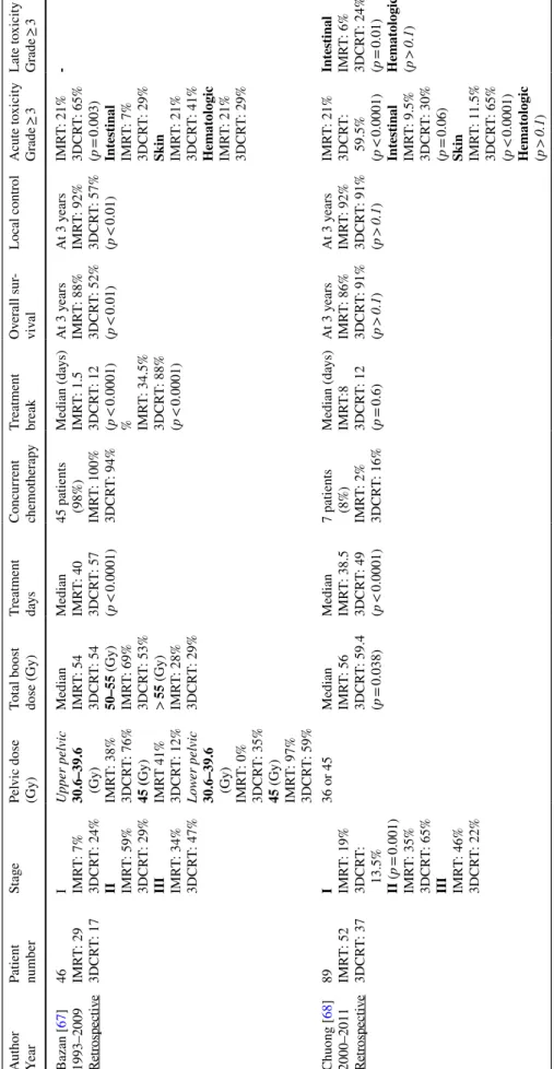
Table
1
Com
par
ison of 3-dimensional conf
or mal v ersus intensity -modulated r adio ther ap y f or t he manag ement of sq
uamous-cell anal canal car
cinoma Aut hor Year Patient number St age Pel vic dose (Gy) To tal boos t dose (Gy) Tr eatment day s Concur rent chemo ther ap y Tr eatment br eak Ov er all sur -viv al Local contr ol A cute t oxicity Gr ade ≥ 3 Late t oxicity Gr ade ≥ 3 Bazan [ 67 ] 1993–2009 Retrospectiv e 46 IMR T: 29 3DCR T: 17 I IMR T: 7% 3DCR T: 24% II IMR T: 59% 3DCR T: 29% III IMR T: 34% 3DCR T: 47% U pper pel vic 30.6–39.6 (Gy) IMR T: 38% 3DCR T: 76% 45 ( Gy) IMR T 41% 3DCR T: 12% Lo wer pel vic 30.6–39.6 (Gy) IMR T: 0% 3DCR T: 35% 45 (Gy) IMR T: 97% 3DCR T: 59% Median IMR T: 54 3DCR T: 54 50–55 (Gy) IMR T: 69% 3DCR T: 53% > 55 (Gy) IMR T: 28% 3DCR T: 29% Median IMR T: 40 3DCR T: 57 (p < 0.0001) 45 patients (98%) IMR T: 100% 3DCR T: 94% Median (da ys) IMR T: 1.5 3DCR T: 12 (p < 0.0001) % IMR T: 34.5% 3DCR T: 88% (p < 0.0001) At 3 y ears IMR T: 88% 3DCR T: 52% (p < 0.01) At 3 y ears IMR T: 92% 3DCR T: 57% (p < 0.01) IMR T: 21% 3DCR T: 65% (p = 0.003) Int es tinal IMR T: 7% 3DCR T: 29% Skin IMR T: 21% 3DCR T: 41% Hemat ologic IMR T: 21% 3DCR T: 29% -Chuong [ 68 ] 2000–2011 Retrospectiv e 89 IMR T: 52 3DCR T: 37 I IMR T: 19% 3DCR T: 13.5% II (p = 0.001) IMR T: 35% 3DCR T: 65% III IMR T: 46% 3DCR T: 22% 36 or 45 Median IMR T: 56 3DCR T: 59.4 (p = 0.038) Median IMR T: 38.5 3DCR T: 49 (p < 0.0001) 7 patients (8%) IMR T: 2% 3DCR T: 16% Median (da ys) IMR T:8 3DCR T: 12 (p = 0.6) At 3 y ears IMR T: 86% 3DCR T: 91% (p > 0.1 ) At 3 y ears IMR T: 92% 3DCR T: 91% (p > 0.1 ) IMR T: 21% 3DCR T: 59.5% (p < 0.0001) Intes tinal IMR T: 9.5% 3DCR T: 30% (p = 0.06) Skin IMR T: 11.5% 3DCR T: 65% (p < 0.0001) Hemat ologic (p > 0.1 ) Int es tinal IMR T: 6% 3DCR T: 24% (p = 0.01) Hemat ologic (p > 0.1 )
Table 1 (continued) Aut hor Year Patient number St age Pel vic dose (Gy) To tal boos t dose (Gy) Tr eatment day s Concur rent chemo ther ap y Tr eatment br eak Ov er all sur -viv al Local contr ol A cute t oxicity Gr ade ≥ 3 Late t oxicity Gr ade ≥ 3 W eber [ 69 ] 1992–2014 Retrospectiv e 103 VMA T: 17 3DCR T: 86 I VMA T: 6% 3DCR T: 13% II VMA T: 41% 3DCR T: 53.5% III VMA T: 53% 3DCR T: 33.5% 50.4 (Gy)
50.4 (Gy) (1.8 (Gy) per fractions)
– 96 patients (93%) VMA T: 94% 3DCR T: 93% – – At 2 y ears VMA T: 100% 3DCR T: 80% (p = 0.7) VMA T: 35% 3DCR T: 53.5% (p < 0.05) Intes tinal VMA T: 12% 3DCR T: 9% (p = 0.2) Skin VMA T: 29% 3DCR T: 44% (p = 0.1) Hemat ologic VMA T: 12% 3DCR T: 17% (p = 0.9) – For IMR T K ac hnic [ 65 ] 2006–2008 RTOG 0529 Phase II tr ial For 3DCR T Ajani [ 44 ] 1998–2005 RTOG 9811 Phase III tr ial IMR T: 52 3DCR T: 325 I IMR T: 0% 3DCR T: 47% II IMR T: 54% 3DCR T: 19% III IMR T: 46% 3DCR T: 10% IMR T 42–45 (Gy) 3DCR T 45 (Gy) IMR T 50.4–54 (Gy) 3DCR T 55–59 (Gy) Median IMR T: 43 3DCR T: 49 (p < 0.0001) IMR T: 100% 3DCR T: 100% Median (da ys) IMR T: 0 3DCR T: 3 (p = 0.0047) % IMR T: 49% 3DCR T: 62% (p = 0.09) – – IMR T: 83% 3DCR T: 87% (p = 0.23) Int es tinal IMR T: 21% 3DCR T: 36% (p = 0.0082) Skin IMR T: 23% 3DCR T: 49% (p < 0.0001) Hemat ologic IMR T: 58% 3DCR T: 62% (p = 0.29) – IMR T s
tatic or dynamic intensity
-modulated r adiation t her ap y, 3DCR T 3-dimensional conf or mal r adio ther ap y, VMA T v olume tric intensity -modulated ar c t her ap y
symptoms was temporary for most patients and recovered 3 months after treatment completion [74]. The potential drawbacks of arc therapy and helical tomotherapy are also important: numerous entrance gates of the beams around the body (360° rotation) with a widely spread-of integral low dose; long linac- and MLC-related quality control times; long preparation times (e.g., delegation, dosimetry, quality control); high-dose gradients within the target volume; an accurate delineation of organs at risk; and time-consuming daily IGRT. Tubiana et al. reported on the importance of
intermediate and high doses, as well as on the major impact of dose per fraction and dose distribution in the risk of sec-ond cancer induction [75].
To summarize, IMRT is a significant technical innovation that has led to better acute and medium-term tolerance of radiotherapy. It allows an optimization of the distribution of doses resulting in an improved hematological tolerance of chemo-radiotherapy. Nevertheless, it has not demonstrated any superiority in terms of efficacy compared to a conven-tional 3-dimensional technique.
Table 2 Differences between the French, American, and Australian/Asian Atlases regarding target volumes and expansion margins
CTV clinical target volume, T tumor, N lymph node(s), PTV planning target volume, GTV gross tumor volume, IGRT image-guided radiotherapy
Referential CTV T CTV N Prophylactic or “low-risk”
PTV Boost or high-risk PTV
French Intergroup [12] GTV T + anal canal + 10
(mm) Vessels + 7 (mm) excluding muscles and bones CTV T and N + 7 (mm) GTV T and N + 15 (mm) American (RTOG) [79] GTV T + canal anal + 20
(mm) Vessels + 7–8 (mm) exclud-ing muscles and bones CTV T and N + 7–10 (mm) GTV T and N + 20 (mm) Australian (AGITG) [78] GTV T + canal anal + 20
(mm) Vessels + 7 (mm) excluding muscles and bones CTV T and N + 10 (mm)CTV T and N + 5–7 (mm) in case of daily IGRT
GTV T + 20 (mm) GTV N + 10–20 (mm)
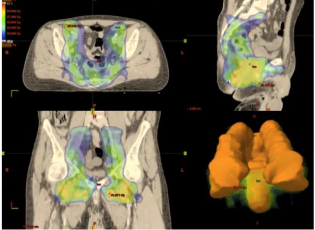
Fig. 1 Coverage of the prophylactic planning target volume by the 95% isodose (47 Gy) with intensity-modulated radiation therapy for locally
Volumes and doses using IMRT
The extent of the gross tumor volume (GTV) is determined from physical examination, imaging, and endoscopic find-ings. The delineation of target volumes may be optimized by co-registration between the planning CT scan and a FDG-PET, and/or a strict axial cross-section pelvic MRI [76]. Lymph node involvement looks related with the size and invasion extent of the primary tumor (i.e., 0–10% for T1–T2 vs. 40–50% for T3–T4) [77]. The risk of recurrence is high-est in the first three years mostly in the pelvic region (50% in the anorectal region and less often in the common iliac and the pre-sacral regions).
Three atlas guidelines are currently available for deline-ation [12, 78, 79]. Differences between these guidelines are summarized in Table 2. The superior extent of the low-risk volume (or subclinical disease treatment volume) is commonly the sacral promontory (or L5-S1 interface); the inferior one is 3 cm below the GTV. The definition of subclinically involved lymph node clinical target volume systematically includes bilateral internal and external iliac,
obturator, pre-sacral, and mesorectal lymph nodes. Ischio-rectal and common iliac regions are added in patients with advanced-stage disease (T3–T4 or N+). Historically, the risk of metachronous inguinal metastases has been reported to be low (7–8%) in clinically node-negative patients at initial staging without inguinal irradiation [80, 81].
Ortholan et al. assessed the benefit of prophylactic bilateral inguinal irradiation with 45 Gy. They were able to show a lower 5-year cumulative rate of inguinal recur-rence in the irradiated group (2% vs. 16%, p = 0.006). The benefit was particularly relevant in patients irradiated with T3–T4 tumors (0% vs. 30%, p = 0.003), though non-signifi-cant in patients irradiated with T1–T2 tumors (3% vs. 12%,
p = 0.17) [82]. Similar findings were observed in a cohort of 116 patients with T2 node-negative tumors, with only a 4.7% rate of inguinal relapses in patients treated without inguinal irradiation [83]. In summary, published data are consistent to recommend the inclusion of the inguinal regions in the low-risk irradiation volume only in patients with advanced local disease (T3–T4) or infiltration below the dentate line. No decisional consensus exists, however, for patients with
Fig. 2 Coverage of the boost planning target volume by the 95% isodose (56.4 Gy) with intensity-modulated radiation therapy for locally advanced anal canal carcinoma (T2N3, bilateral nodes)
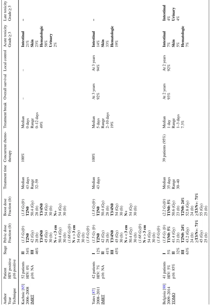
Table 3 Liter atur e r evie w of intensity -modulated r adio ther ap y wit h simult aneous integ rated boos t (SIB) modality f or t he manag ement of sq
uamous-cell anal canal car
cinoma Aut hor Year Tec hniq ue
Patient HIV positiv
e p16 positiv e St age Pel vic dose Fr action (fr) Boos t dose Fr action (fr) Tr eatment time Concur rent c hemo -ther ap y Tr eatment br eak Ov er all sur viv al Local contr ol A cute t oxicity Gr ade ≥ 3 Late t oxicity Gr ade ≥ 3 K ac hnic [ 65 ] 2006–2008 IMR T 52 patients HIV : 0% p16: N A II 54% III 46% (1.5 Gy /fr ) T2N0 42 (Gy) 28 (fr) T3/4N0 45 (Gy) 30 (fr) N + < 3 cm 50.4 (Gy) 30 (fr) (1.68 Gy /fr ) N + > 3 cm 54 (Gy) 30 (fr) (1.8 Gy /fr ) (1.8 Gy /fr )
T2N0 50.4 (Gy) 28 (fr) T3/4N0 54 (Gy) 30 (fr) N+ 54 (Gy) 30 (fr) Median 43 da ys Rang e 32–59 100% Median 0 da ys Rang e 0–12 da ys 49% – – Int es tinal 21% Skin 23% Hemat ologic 58% Urinar y 2% – Yates [ 87 ] 2008–2011 IMR T 42 patients HIV : 7% p16: N A I 12% II 41% III 45% (1.5 Gy /fr ) T2N0 42 (Gy) 28 (fr) T3/4N0 45 (Gy) 30 (fr) N + < 3 cm 50.4 (Gy) 30 (fr) (1.68 Gy /fr ) N + > 3 cm 54 (Gy) 30 (fr) (1.8 Gy /fr ) (1.8 Gy /fr )
T2N0 50.4 (Gy) 28 (fr) T3/4N0 54 (Gy) 30 (fr) N+ 54 (Gy) 30 (fr) Median 43 da ys 100% Median 5 da ys Rang e 2–10 da ys 19% At 3 y ears 92% At 3 y ears 94% Int es tinal 14% Skin 33% Hemat ologic 19% – Belgioia [ 88 ] 2009–2014 TOMO 41 patients HIV : 5% p16: 85% I 5% II 32% III 63% (1.8 Gy /fr ) T1N0: 5% 41.4 (Gy) 23 (fr) T2N0: 24% 43.2 (Gy) 24 (fr) ≥T3 /N +: 71% 45 (Gy) 25 (fr) (2.2 Gy /fr ) T1N0: 5% 50.6 (Gy) 23 (fr) T2N0: 24% 52.8 (Gy) 24 (fr) ≥T3 /N +: 71% 55 (Gy) 25 (fr) Median 35 da ys Rang e 30–40 39 patients (95%) Median 1 da y Rang e 1–3 da ys 7.3% At 2 y ears 93% At 2 y ears 92% Int es tinal 7% Skin 5% Hemat ologic 7% Int es tinal 4% Urinar y 4%
Table
3
(continued)
Aut
hor
Year Techniq
ue
Patient HIV positiv
e p16 positiv e St age Pel vic dose Fr action (fr) Boos t dose Fr action (fr) Tr eatment time Concur rent c hemo -ther ap y Tr eatment br eak Ov er all sur viv al Local contr ol A cute t oxicity Gr ade ≥ 3 Late t oxicity Gr ade ≥ 3 Joseph [ 89 ] 2008–2016 TOMO 57 patients HIV : 0% p16: N A II 48% III 52% (1.5 Gy /fr ) 45 (Gy) 30 (fr) (1.8 Gy /fr ) 54 (Gy) 30 (fr) – 56 patients (98%) Median (da ys) NA 9% At 3 y ears 91% – Int es tinal 24% Skin 10.5% Hemat ologic 46% Urinar y 3.5% Int es tinal 5% Urinar y 2% Gynecologic 9% Fr anco [ 90 ] 2007–2013 IMR T 74% VMA T 13% TOMO 13% 54 patients HIV : 6% p16: N A II 54% III 46% T2N0 42 (Gy) 28 (fr) (1.5 Gy /fr ) T3 /4N0 45 (Gy) 30 (fr) (1.5 Gy /fr ) N + < 3 cm 50.4 (Gy) 30 (fr) (1.68 Gy /fr ) N + > 3 cm 54 (Gy) 30 (fr) (1.8 Gy /fr ) T2N0 50.4 (Gy) 28 (fr) (1.8 Gy /fr ) T3 54 (Gy) 30 (fr) (1.8 Gy /fr ) T4 60 (Gy) 30 (fr) (2 Gy /fr ) Median 44 da ys Rang e 37–55 52 patients (96%) Mean 4 da ys 17% At 4 y ears 78% At 4 y ears 85% Int es tinal 8% Skin 13% Hemat ologic 11% Urinar y 2% Gr ade 2 Int es tinal 14% Urinar y 2% Gynecologic 4% Fr anco [ 91 ] 2007–2013 VMA T 39 patients HIV : 8% p16: N A I 5% II 67% III 28% T2N0 42 (Gy) 28 (fr) (1.5 Gy /fr ) T3 /4N0 45 (Gy) 30 (fr) (1.5 Gy /fr ) N + < 3 cm 50.4 (Gy) 30 (fr) (1.68 Gy /fr ) N + > 3 cm 54 (Gy) 30 (fr) (1.8 Gy /fr ) T2N0: 33% 50.4 (Gy) 28 (fr) (1.8 Gy /fr ) T3 /T4: 67% 54 (Gy) 30 (fr) (1.8 Gy /fr ) Median 43 da ys Rang e 38–54 100% Mean 2 da ys 30% At 2 y ears 85% – Int es tinal 5% Skin 18% Hemat ologic 13% Urinar y 2% –
T1–T2 tumors. Prophylactic groin irradiation will be recom-mended on a case-by-case basis and based on the location and size of the primary tumor and on whether a lymph node evaluation is undertaken.
Two clinical target volumes (CTV) may be distinguished for SCCAC irradiation: a low-risk volume including mes-orectal, pelvic, and inguinal lymph nodes; and a high-risk volume corresponding to the primary tumor and the involved lymph nodes. A margin of 7–8 mm is generally recommended around the iliac vessels, excluding muscles and bony structures. The margin around the femoral vessels may be ≥ 7 mm to include all surrounding lymph nodes. The RTOG and the Australasian Gastrointestinal Trials Group guidelines recommend an additional 10-mm margin anterior to the mesorectal CTV, accounting for rectal motion [84]. International IMRT guidelines suggest a wide range of CTV to PTV margins, ranging from 5 to 10 mm in the prophylac-tic setting [51, 65, 78].
The PTV margins surrounding the GTV may range from 10 to 20 mm [78, 79, 84]. Figures 1 and 2 illustrate the coverage of the PTV by the 95% isodose using IMRT for a T2N3 SCCAC with bilateral nodes.
Too tight margins may ease a geographical miss in areas with an increased motion; too loose margins may be highly toxic if large volumes of normal tissues or organs at risk are included in the irradiation fields. Chen et al. reported on patients treated with a tight 5-mm margin and IGRT (pelvic bones auto-match on CBCT) for verification and concluded that those margins sustained by daily IGRT controls ade-quately covered the pelvic volumes reducing simultaneously the dose to the organs at risk [85]. Durrant et al. assessed the motion around the inguinal nodes and the local tumor region using an online CBCT protocol. The estimated 3D margins needed to compensate for random displacements in the lateral, longitudinal, and vertical axes around the ingui-nal nodes and the primary tumor were, respectively, 1.5 mm, 2.7 mm, and 2.8 mm and 4.6 mm, 8.9 mm, and 5.2 mm [86]. Indeed, the ongoing PLATO trial (ISRCTN88455282) will assess the safety of individualized radiotherapy doses con-sidering reduced margins around the targets. PLATO com-prises 3 independent phase II trials which evaluate the radia-tion dose based on low-, intermediate- or high-risk SCCAC. Locally advanced SCCAC (high-risk group) are randomized between a standard dose of 53.2 Gy in 28 fractions and two escalation doses of 58.8 Gy in 28 fractions or 61.6 Gy in 28 fractions. The primary outcome is loco-regional failure at 3 years.
Three issues have to be considered before scheduling the treatment. The first one is the total dose to be delivered to the low-risk PTV, 36 or 45 Gy in most studies, followed by a sequential boost of 14–23.4 Gy to the high-risk PTV (local tumor and involved lymph nodes) [12, 37]. Although these are the most frequently followed guidelines, there is no
Table 3 (continued) Aut hor Year Tec hniq ue
Patient HIV positiv
e p16 positiv e St age Pel vic dose Fr action (fr) Boos t dose Fr action (fr) Tr eatment time Concur rent c hemo -ther ap y Tr eatment br eak Ov er all sur viv al Local contr ol A cute t oxicity Gr ade ≥ 3 Late t oxicity Gr ade ≥ 3 Tomasoa [ 92 ] 2006–2012 IMR T 106 patients HIV : N A p16: N A I 4% II 33% III 55% (1.5 Gy /fr ) 49.5 (Gy) 33 (fr) (1.8 Gy /fr ) 59.4 (Gy) 33 (fr) – 87 patients (82%) 6% At 4 y ears 77% At 4 y ears 79% Int es tinal 14% Skin 62% Hemat ologic 7% Int es tinal 4% IMR T s
tatic or dynamic intensity
-modulated r adiation t her ap y, TOMO helical t omo ther ap y, VMA T v olume tric intensity -modulated ar c t her ap y
Table 4 Lo w-dose electiv e nodal intensity -modulated r adiation t her ap y sc hedule in t he liter atur e f or sq uamous-cell car cinoma of t he anus manag ement 3D-CR T 3-dimensional conf or mal, IMR T s
tatic or dynamic intensity
-modulated r adiation t her ap y, TOMO helical t omo ther ap y, VMA T v olume tric intensity -modulated ar c t her ap y; r adio ther ap y Aut hor
Year Techniq
ue Patient St age Pel vic dose Fr action (fr) Boos t dose Fr action (fr) Tr eatment time Concur rent c hemo -ther ap y Tr eatment br eak Ov er all sur viv al Lym ph node local contr ol (LNL C) Tumor contr ol (T C) A cute t oxicity Gr ade ≥ 3 Late t oxicity Gr ade ≥ 3 Henk enber ens [ 93 ] 2009–2014 3D-CR T 63% IMR T 37% 30 patients T1 27% T2 63% T3 10% (1.8 Gy /fr ) 39.6 (Gy) 22 (fr)
T1 50 (Gy) T2 55.8 (Gy) T3 59.4 (Gy)
– 27 patients (90%) – At 3 y ears 90% At 3 y ears LNL C = 100% TC = 93% Int es tinal 3% Skin 23% Hemat ologic 7% Int es tinal 0% Urinar y 0% Lepino y [ 94 ] 1996–2013 3D-CR T 73% IMR T 27% 142 patients I 2% II 55% III 43% (1.8 Gy /fr ) 36 (Gy) 20 (fr) (1,8 Gy /fr ) 59.4 (Gy) 33 (fr) Median 59 da ys 100% Median 14 da ys Rang e 14–28 100% (planned) At 5 y ears 75% At 5 y ears LNL C = 96% TC = 81.5% Inguinal = 98.5% 4 (3%) infield 1 (1%) outfield Int es tinal 1% Skin 2% Hemat ologic 2% – Les trade [ 95 ] 2006–2014 TOMO 57% IMR T 34% VMA T 9% 35 patients T1 54% T2 46% (1.8 Gy /fr ) 36 (Gy) 20 (fr) (1.8 Gy /fr ) 59.4 (Gy) 33 (fr) Median 56 da ys Rang e 48–71 28 patients (80%) Median 10 da ys Rang e 5–26 86% (planned) At 4 y ears 93% At 4 y ears LNL C = 100% TC = 96.6% Int es tinal 11% Skin 11% Hemat ologic 3% – De Bar i [ 98 ] 2007–2015 TOMO 64% IMR T 26% VMA T 10% 151 patients I 18% II 40% III 40% (1.8 Gy /fr ) 36 (Gy) 20 (fr) (1.8 Gy /fr ) 59.4 (Gy) 33 (fr) Median 57 da ys Rang e 52–59 138 patients (91%) Median 11 da ys Rang e 5–26 81% (planned) At 4 y ears 82% At 4 y ears LNL C = 96% TC = 82% Inguinal = 100% Int es tinal 7% Skin 16.5% Int es tinal 2%
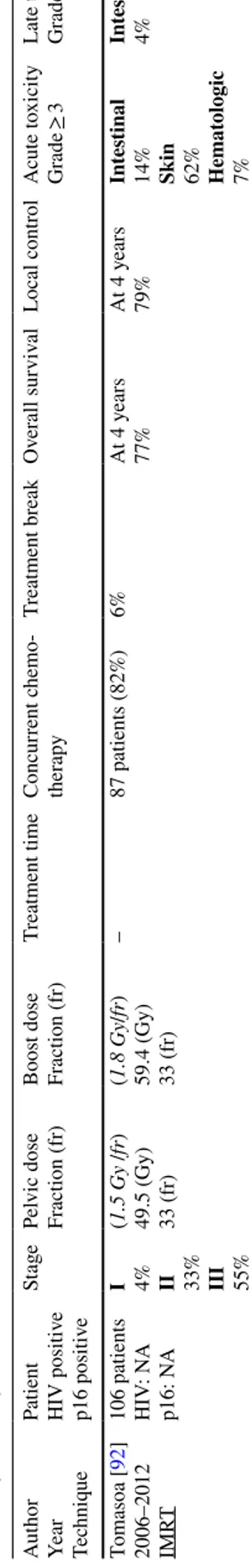
consensus regarding on the low-risk PTV dose prescription. The RTOG 9811 trial aimed to deliver 45 Gy to the prophy-lactic volume in 25 daily fractions, followed by a sequen-tial boost of 10–14 Gy in 2 Gy fractions [23]. The second issue to be considered is fractionation which is conventional (1.8 Gy/fraction/day, 5 times a week). The third issue to be taken in account is overall treatment time which should be as short as possible, ideally between 6 and 8 weeks (59–65 Gy in 33–36 fractions of 1.8 Gy).
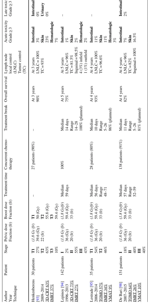
The conventional scheme is a sequential plan. Sequen-tial treatment modalities usually deliver 36–45 Gy in 20–25 fractions of 1.8 Gy, 5 times a week, to the low-risk PTV followed by a boost of 14.4–23.4 Gy in 8–13 fractions of 1.8 Gy to the high-risk region. If radiotherapy is carried out using simultaneous integrated boost (SIB) modality, the low-risk PTV shall receive a total dose ranging from 43.2 to 49.5 Gy, in 24–33 fractions of 1.5–1.8 Gy, five days a week, thus a normalized total dose (NTD1.8 Gy) of 48.2 Gy, with an alpha/beta = 10 Gy. The high-risk PTV, however, may be boosted up to a dose of 52.8–60 Gy in 24–33 fractions of 1.8–2.2 Gy, 5 times a week [67, 68, 87–97]. Table 3 pre-sents the results of published studies using SIB modalities. It should be noted that no prospective trial investigates the regional failure rates associated with less than 1.8 Gy per fraction. A sequential plan is more often used compared to SIB. Indeed, a sequential treatment is preferred as it may be more convenient to plan treatment break in case of severe acute toxicity and to determine the final boost dose based on tumor response on treatment. Finally, SIB modality needs to be validated with larger prospective studies to be considered as a valuable and validate scheme. As shown in Table 4, elective nodal irradiation from the inguinal region with doses ranging from 36 to 39.6 Gy obtained excellent control rates (96–100%) with fairly good tolerance. Further prospective and randomized studies are required to compare 36 Gy versus 45 Gy in terms of local control and toxicity.
References
1. Allemani C, Weir HK, Carreira H, Harewood R, Spika D, Wang X-S, et al. Global surveillance of cancer survival 1995–2009: analysis of individual data for 25,676,887 patients from 279 pop-ulation-based registries in 67 countries (CONCORD-2). Lancet Lond Engl. 2015;385:977–1010.
2. Johnson LG, Madeleine MM, Newcomer LM, Schwartz SM, Daling JR. Anal cancer incidence and survival: the surveillance, epidemiology, and end results experience, 1973–2000. Cancer. 2004;101:281–8.
3. Aaltonen LA, Hamilton SR, World Health Organization, Inter-national Agency for Research on Cancer, editor. Pathology and genetics of tumours of the digestive system. Lyon: IARC; 2000.
4. Boman BM, Moertel CG, O’Connell MJ, Scott M, Weiland LH, Beart RW, et al. Carcinoma of the anal canal: a clinical and pathologic study of 188 cases. Cancer. 1984;54:114–25. 5. Abramowitz L, Mathieu N, Roudot-Thoraval F, Lemarchand N,
Bauer P, Hennequin C, et al. Epidermoid anal cancer prognosis comparison among HIV+ and HIV− patients. Aliment Pharma-col Ther. 2009;30:414–21.
6. Cummings B, Keane T, Thomas G, Harwood A, Rider W. Results and toxicity of the treatment of anal canal carcinoma by radiation therapy or radiation therapy and chemotherapy. Cancer. 1984;54:2062–8.
7. Papillon J, Mayer M, Montbarbon JF, Gerard JP, Chassard JL, Bailly C. A new approach to the management of epidermoid carcinoma of the anal canal. Cancer. 1983;51:1830–7. 8. Eschwege F, Lasser P, Chavy A, Wibault P, Kac J, Rougier P,
et al. Squamous cell carcinoma of the anal canal: treatment by external beam irradiation. Radiother Oncol J Eur Soc Ther Radiol Oncol. 1985;3:145–50.
9. Touboul E, Schlienger M, Buffat L, Lefkopoulos D, Pène F, Parc R, et al. Epidermoid carcinoma of the anal canal. Results of curative-intent radiation therapy in a series of 270 patients. Cancer. 1994;73:1569–79.
10. Touboul E, Schlienger M, Buffat L, Ozsahin M, Belkacemi Y, Pene F, et al. Conservative versus nonconservative treatment of epidermoid carcinoma of the anal canal for tumors longer than or equal to 5 centimeters. A retrospective comparison. Cancer. 1995;75:786–93.
11. Nigro ND, Vaitkevicius VK, Considine B. Combined therapy for cancer of the anal canal: a preliminary report. Dis Colon Rectum. 1974;17:354–6.
12. Moureau-Zabotto L, Vendrely V, Abramowitz L, Borg C, Fran-cois E, Goere D, et al. Anal cancer: French Intergroup Clini-cal Practice Guidelines for diagnosis, treatment and follow-up (SNFGE, FFCD, GERCOR, UNICANCER, SFCD, SFED, SFRO, SNFCP). Dig Liver Dis. 2017;49:831–40.
13. Glynne-Jones R, Northover JMA, Cervantes A, On behalf of the ESMO Guidelines Working Group. Anal cancer: ESMO Clinical Practice Guidelines for diagnosis, treatment and follow-up. Ann Oncol. 2010;21:v87–92.
14. Abramowitz L, Jacquard A-C, Jaroud F, Haesebaert J, Siprou-dhis L, Pradat P, et al. Human papillomavirus genotype distribu-tion in anal cancer in France: the EDiTH V study. Int J Cancer. 2011;129:433–9.
15. De Vuyst H, Clifford GM, Nascimento MC, Madeleine MM, Franceschi S. Prevalence and type distribution of human papillomavirus in carcinoma and intraepithelial neoplasia of the vulva, vagina and anus: a meta-analysis. Int J Cancer. 2009;124:1626–36.
16. Palefsky JM, Giuliano AR, Goldstone S, Moreira EDJ, Aranda C, Jessen H, et al. HPV Vaccine against Anal HPV Infection and Anal Intraepithelial Neoplasia. N Engl J Med. 2011;365:1576–85.
17. Koerber SA, Schoneweg C, Slynko A, Krug D, Haefner MF, Her-farth K, et al. Influence of human papillomavirus and p16(INK4a) on treatment outcome of patients with anal cancer. Radiother Oncol. 2014;113:331–6.
18. Gilbert DC, Williams A, Allan K, Stokoe J, Jackson T, Linsdall S, et al. p16INK4A, p53, EGFR expression and KRAS muta-tion status in squamous cell cancers of the anus: correlamuta-tion with outcomes following chemo-radiotherapy. Radiother Oncol. 2013;109:146–51.
19. Serup-Hansen E, Linnemann D, Skovrider-Ruminski W, Høgdall E, Geertsen PF, Havsteen H. Human papillomavirus genotyping and p16 expression as prognostic factors for patients with Ameri-can Joint Committee on Cancer stages I to III carcinoma of the anal canal. J Clin Oncol. 2014;32:1812–7.
20. Silverberg MJ, Lau B, Justice AC, Engels E, Gill MJ, Goedert JJ, et al. Risk of anal cancer in HIV-infected and HIV-uninfected individuals in North America. Clin Infect. 2012;54:1026–34. 21. Gervaz P, Hirschel B, Morel P. Molecular biology of squamous
cell carcinoma of the anus. Br J Surg. 2006;93:531–8.
22. Northover J, Glynne-Jones R, Sebag-Montefiore D, James R, Meadows H, Wan S, et al. Chemoradiation for the treatment of epidermoid anal cancer: 13-year follow-up of the first ran-domised UKCCCR Anal Cancer Trial (ACT I). Br J Cancer. 2010;102:1123–8.
23. Gunderson LL, Winter KA, Ajani JA, Pedersen JE, Moughan J, Benson AB, et al. Long-term update of US GI intergroup RTOG 98-11 phase III trial for anal carcinoma: survival, relapse, and colostomy failure with concurrent chemoradiation involving fluo-rouracil/mitomycin versus fluorouracil/cisplatin. J Clin Oncol. 2012;30:4344–51.
24. Fenger C. Prognostic factors in anal carcinoma. Pathology (Phila). 2002;34:573–8.
25. Giuliano AR, Palefsky JM, Goldstone S, Moreira ED, Penny ME, Aranda C, et al. Efficacy of quadrivalent HPV vaccine against HPV Infection and disease in males. N Engl J Med. 2011;364:401–11.
26. Shridhar R, Shibata D, Chan E, Thomas CR. Anal cancer: cur-rent standards in care and recent changes in practice. CA Cancer J Clin. 2015;65:139–62.
27. Brierley J, Gospodarowicz MK, Wittekind C, editors. TNM clas-sification of malignant tumours. Chichester: John Wiley & Sons, Inc; 2017.
28. Magdeburg B, Fried M, Meyenberger C. Endoscopic ultrasonog-raphy in the diagnosis, staging, and follow-up of anal carcinomas. Endoscopy. 1999;31:359–64.
29. Roach SC, Hulse PA, Moulding FJ, Wilson R, Carrington BM. Magnetic resonance imaging of anal cancer. Clin Radiol. 2005;60:1111–9.
30. Winton E de, Heriot AG, Ng M, Hicks RJ, Hogg A, Milner A, et al. The impact of 18-fluorodeoxyglucose positron emission tomography on the staging, management and outcome of anal cancer. Br J Cancer. 2009;100:693–700.
31. Mahmud A, Poon R, Jonker D. PET imaging in anal canal cancer: a systematic review and meta-analysis. Br J Radiol. 2017;90:20170370.
32. Mistrangelo M, Pelosi E, Bellò M, Castellano I, Cassoni P, Ricardi U, et al. Comparison of positron emission tomography scanning and sentinel node biopsy in the detection of inguinal node metastases in patients with anal cancer. Int J Radiat Oncol. 2010;77:73–8.
33. Fraunholz I, Rabeneck D, Gerstein J, Jäck K, Haberl A, Weiss C, et al. Concurrent chemoradiotherapy with 5-fluorouracil and mitomycin C for anal carcinoma: are there differences between HIV-positive and HIV-negative patients in the era of highly active antiretroviral therapy? Radiother Oncol. 2011;98:99–104. 34. Seo Y, Kinsella MT, Reynolds HL, Chipman G, Remick SC,
Kin-sella TJ. Outcomes of chemoradiotherapy with 5-Fluorouracil and mitomycin C for anal cancer in immunocompetent versus immu-nodeficient patients. Int J Radiat Oncol Biol Phys. 2009;75:143–9. 35. Stadler RF, Gregorcyk SG, Euhus DM, Place RJ, Huber PJ, Sim-mang CL. Outcome of HIV-infected patients with invasive squa-mous-cell carcinoma of the anal canal in the era of highly active antiretroviral therapy. Dis Colon Rectum. 2004;47:1305–9. 36. Claren A, Doyen J, Falk AT, Benezery K, Follana P, Frin A-C,
et al. Results of age-dependent anal canal cancer treatment: a sin-gle centre retrospective study. Dig Liver. 2014;46:460–4. 37. Peiffert D, Créhange G, Vendrely V, Baumann A-S, Faivre J-C,
Huger S. Radiotherapy for anal canal cancers. Cancer Radiother. 2016;20(Suppl):183–8.
38. Houlihan OA, O’Neill BDP. Chemoradiotherapy for anal squa-mous cell carcinoma. Sureon. 2016;14:202–12.
39. UKCCCR Anal Cancer Trial Working Party. Epidermoid anal can-cer: results from the UKCCCR randomised trial of radiotherapy alone versus radiotherapy, 5-fluorouracil, and mitomycin. UKC-CCR Anal Cancer Trial Working Party. UK Co-ordinating Com-mittee on Cancer Research. Lancet Lond Engl. 1996;348:1049–54. 40. Bartelink H, Roelofsen F, Eschwege F, Rougier P, Bosset JF, Gon-zalez DG, et al. Concomitant radiotherapy and chemotherapy is superior to radiotherapy alone in the treatment of locally advanced anal cancer: results of a phase III randomized trial of the Euro-pean Organization for Research and Treatment of Cancer Radio-therapy and Gastrointestinal Cooperative Groups. J Clin Oncol. 1997;15:2040–9.
41. Ozsahin M, Santa Cruz O, Bouchaab H, Matzinger O, Tsoutsou PG. Definitive organ-sparing treatment of anal canal cancer: can we afford to question it? J Clin Oncol. 2012;30:673–4.
42. Flam M, John M, Pajak TF, Petrelli N, Myerson R, Doggett S, et al. Role of mitomycin in combination with fluorouracil and radiotherapy, and of salvage chemoradiation in the definitive non-surgical treatment of epidermoid carcinoma of the anal canal: results of a phase III randomized intergroup study. J Clin Oncol. 1996;14:2527–39.
43. Glynne-Jones R, Meadows H, Wan S, Gollins S, Leslie M, Levine E, et al. EXTRA-a multicenter phase II study of chemoradiation using a 5 day per week oral regimen of capecitabine and intrave-nous mitomycin C in anal cancer. Int J Radiat Oncol Biol Phys. 2008;72:119–26.
44. Ajani JA, Winter KA, Gunderson LL, Pedersen J, Benson AB, Thomas CR, et al. Fluorouracil, mitomycin, and radiotherapy vs fluorouracil, cisplatin, and radiotherapy for carcinoma of the anal canal: a randomized controlled trial. JAMA. 2008;299:1914–21. 45. Peiffert D, Tournier-Rangeard L, Gérard J-P, Lemanski C, Fran-çois E, Giovannini M, et al. Induction chemotherapy and dose intensification of the radiation boost in locally advanced anal canal carcinoma: final analysis of the randomized UNICANCER ACCORD 03 trial. J Clin Oncol. 2012;30:1941–8.
46. James RD, Glynne-Jones R, Meadows HM, Cunningham D, Myint AS, Saunders MP, et al. Mitomycin or cisplatin chemoradiation with or without maintenance chemotherapy for treatment of squa-mous-cell carcinoma of the anus (ACT II): a randomised, phase 3, open-label, 2 × 2 factorial trial. Lancet Oncol. 2013;14:516–24. 47. Paliga AA, Onerheim R, Gologan A, Spatz A, Vuong T.
EGFR expression in invasive anal carcinoma. J Clin Oncol. 2011;29:412–2.
48. Paliga A, Onerheim R, Gologan A, Chong G, Spatz A, Niazi T, et al. EGFR and K-ras gene mutation status in squamous cell anal carcinoma: a role for concurrent radiation and EGFR inhibitors? Br J Cancer. 2012;107:1864–8.
49. Lukan N, Ströbel P, Willer A, Kripp M, Dinter D, Mai S, et al. Cetuximab-based treatment of metastatic anal cancer: corre-lation of response with KRAS mutational status. Oncology. 2009;77:293–9.
50. De Dosso S, Martin V, Zanellato E, Frattini M, Saletti P. Molecular characterization and response to cetuximab in a patient with refractory squamous cell anal carcinoma. Tumori J. 2010;96:627–8.
51. Khan L, Choo R, Breen D, Assaad D, Fialkov J, Antonyshyn O, et al. Recommendations for CTV margins in radiotherapy planning for non melanoma skin cancer. Radiother Oncol. 2012;104:263–6.
52. Olivatto LO, Vieira FM, Pereira BV, Victorino AP, Bezerra M, Araujo CM, et al. Phase 1 study of cetuximab in combination with 5-fluorouracil, cisplatin, and radiotherapy in patients with locally advanced anal canal carcinoma. Cancer. 2013;119:2973–80.
53. Deutsch E, Lemanski C, Pignon JP, Levy A, Delarochefordiere A, Martel-Lafay I, et al. Unexpected toxicity of cetuximab combined with conventional chemoradiotherapy in patients with locally advanced anal cancer: results of the UNICANCER ACCORD 16 phase II trial. Ann Oncol Off J Eur Soc Med Oncol. 2013;24:2834–8.
54. Ajani JA, Winter K, Leonard L et al. Fluorouracil mitomycin, and radiotherapy vs fluorouracil, cisplatin, and radiotherapy for carcinoma of the anal canal. JAMA. 2008;299:1914–21. 55. James RD, Glynne-Jones R, Meadows HM, Cunningham D,
Myint AS, Saunders MP, et al. Mitomycin or cisplatin chemora-diation with or without maintenance chemotherapy for treatment of squamous-cell carcinoma of the anus (ACT II): a randomised, phase 3, open-label, 2$\times$ 2 factorial trial. Lancet Oncol. 2013;14:516–24.
56. Konski A, Garcia M, John M, Krieg R, Pinover W, Myerson R, et al. Evaluation of planned treatment breaks during radiation therapy for anal cancer: update of RTOG 92-08. Int J Radiat Oncol Biol Phys. 2008;72:114–8.
57. Weber DC, Kurtz JM, Allal AS. The impact of gap duration on local control in anal canal carcinoma treated by split-course radio-therapy and concomitant chemoradio-therapy. Int J Radiat Oncol Biol Phys. 2001;50:675–80.
58. Wong CS, Tsang RW, Cummings BJ, Fyles AW, Couture J, Brier-ley JD, et al. Proliferation parameters in epidermoid carcinomas of the anal canal. Radiother Oncol. 2000;56:349–53.
59. Khanfir K, Ozsahin M, Bieri S, Cavuto C, Mirimanoff RO, Zou-hair A. Patterns of failure and outcome in patients with carcinoma of the anal margin. Ann Surg Oncol. 2008;15:1092–8.
60. Bentzen AG, Guren MG, Vonen B, Wanderås EH, Frykholm G, Wilsgaard T, et al. Faecal incontinence after chemoradiotherapy in anal cancer survivors: long-term results of a national cohort. Radiother Oncol. 2013;108:55–60.
61. Agarwal MS, Jones DA, Mendenhall CM, Morris CG, Johns A, McAfee WJ, et al. Primary management of squamous cell carci-noma of the anal canal: A 30-year community hospital experience. Cancer Invest. 2017;35:547–51.
62. Milano MT, Jani AB, Farrey KJ, Rash C, Heimann R, Chmura SJ. Intensity-modulated radiation therapy (IMRT) in the treatment of anal cancer: toxicity and clinical outcome. Int J Radiat Oncol Biol Phys. 2005;63:354–61.
63. Chen Y-J, Liu A, Tsai PT, Vora NL, Pezner RD, Schultheiss TE, et al. Organ sparing by conformal avoidance intensity-modulated radiation therapy for anal cancer: dosimetric evaluation of cover-age of pelvis and inguinal/femoral nodes. Int J Radiat Oncol Biol Phys. 2005;63:274–81.
64. Menkarios C, Azria D, Laliberté B, Moscardo CL, Gourgou S, Lemanski C, et al. Optimal organ-sparing intensity-modulated radiation therapy (IMRT) regimen for the treatment of locally advanced anal canal carcinoma: a comparison of conventional and IMRT plans. Radiat Oncol. 2007;2:41.
65. Kachnic LA, Winter K, Myerson RJ, Goodyear MD, Willins J, Esthappan J, et al. RTOG 0529: a phase 2 evaluation of dose-painted intensity modulated radiation therapy in combination with 5-fluorouracil and mitomycin-C for the reduction of acute morbidity in carcinoma of the anal canal. Int J Radiat Oncol. 2013;86:27–33.
66. Mell LK, Schomas DA, Salama JK, Devisetty K, Aydogan B, Miller RC, et al. Association between bone marrow dosimetric parameters and acute hematologic toxicity in anal cancer patients treated with concurrent chemotherapy and intensity-modulated radiotherapy. Int J Radiat Oncol Biol Phys. 2008;70:1431–7. 67. Bazan JG, Hara W, Hsu A, Kunz PA, Ford J, Fisher GA, et al.
Intensity-modulated radiation therapy versus conventional radia-tion therapy for squamous cell carcinoma of the anal canal. Can-cer. 2011;117:3342–51.
68. Chuong MD, Freilich JM, Hoffe SE, Fulp W, Weber JM, Alm-hanna K, et al. Intensity-modulated radiation therapy vs. 3D Conformal radiation therapy for squamous cell carcinoma of the anal canal. Gastrointest Cancer Res GCR. 2013;6:39–45. 69. Weber HE, Dröge LH, Hennies S, Herrmann MK, Gaedcke
J, Wolff HA. Volumetric intensity-modulated arc therapy vs. 3-dimensional conformal radiotherapy for primary chemora-diotherapy of anal carcinoma: effects on treatment-related side effects and survival. Strahlenther Onkol. 2015;191:827–34. 70. Vieillot S, Fenoglietto P, Lemanski C, Moscardo CL, Gourgou
S, Dubois J-B, et al. IMRT for locally advanced anal cancer: clinical experience of the Montpellier Cancer Center. Radiat Oncol. 2012;7:45.
71. Pollom EL, Wang G, Harris JP, Koong AC, Bendavid E, Bhat-tacharya J, et al. The impact of intensity modulated radiation therapy on hospitalization outcomes in the SEER-Medicare population with anal squamous cell carcinoma. Int J Radiat Oncol Biol Phys. 2017;98:177–85.
72. De Bari B, Jumeau R, Bouchaab H, Vallet V, Matzinger O, Troussier I, et al. Efficacy and safety of helical tomotherapy with daily image guidance in anal canal cancer patients. Acta Oncol. 2016;55:767–73.
73. Ugurluer G, Ballerini G, Moeckli R, Matzinger O, Bourhis J, Ozsahin M. Helical tomotherapy for the treatment of anal canal cancer: a dosimetric comparison with 3d conformal radiother-apy. Tumori J. 2015;101:268–72.
74. Joseph K, Vos LJ, Warkentin H, Paulson K, Polkosnik L-A, Usmani N, et al. Patient reported quality of life after helical IMRT based concurrent chemoradiation of locally advanced anal cancer. Radiother Oncol. 2016;120:228–33.
75. Tubiana M. Can we reduce the incidence of second primary malignancies occurring after radiotherapy? A critical review. Radiother Oncol. 2009;91:4–15. discussion 1–3.
76. Krengli M, Milia ME, Turri L, Mones E, Bassi MC, Cannillo B, et al. FDG-PET/CT imaging for staging and target volume delineation in conformal radiotherapy of anal carcinoma. Radiat Oncol. 2010;5:10.
77. Davey P, Saibil EA, Wong R. Bipedal lymphography in the management of carcinoma of the anal canal. Br J Radiol. 1996;69:632–5.
78. Ng M, Leong T, Chander S, Chu J, Kneebone A, Carroll S, et al. Australasian Gastrointestinal Trials Group (AGITG) con-touring atlas and planning guidelines for intensity-modulated radiotherapy in anal cancer. Int J Radiat Oncol Biol Phys. 2012;83:1455–62.
79. Myerson RJ, Garofalo MC, El Naqa I, Abrams RA, Apte A, Bosch WR, et al. Elective clinical target volumes for conformal therapy in anorectal cancer: a radiation therapy oncology group consensus panel contouring atlas. Int J Radiat Oncol Biol Phys. 2009;74:824–30.
80. Gerard JP, Chapet O, Samiei F, Morignat E, Isaac S, Paulin C, et al. Management of inguinal lymph node metastases in patients with carcinoma of the anal canal: experience in a series of 270 patients treated in Lyon and review of the literature. Cancer. 2001;92:77–84.
81. Papillon J, Montbarbon JF. Epidermoid carcinoma of the anal canal. A series of 276 cases. Dis Colon Rectum. 1987;30:324–33.
82. Ortholan C, Resbeut M, Hannoun-Levi J-M, Teissier E, Gerard J-P, Ronchin P, et al. Anal canal cancer: management of inguinal nodes and benefit of prophylactic inguinal irradiation (CORS-03 Study). Int J Radiat Oncol Biol Phys. 2012;82:1988–95. 83. Zilli T, Betz M, Bieri S, Ris F, Roche B, Roth AD, et al. Elective
inguinal node irradiation in early-stage T2N0 anal cancer: prog-nostic impact on locoregional control. Int J Radiat Oncol Biol Phys. 2013;87:60–6.
84. Vendrely V, Galland-Girodet S, Orré M, Maire J-P. Recommanda-tions pour la délinéation des aires ganglionnaires pelviennes dans le cancer du canal anal. Cancer. 2013;17:566–70.
85. Chen Y-J, Suh S, Nelson RA, Liu A, Pezner RD, Wong JYC. Setup variations in radiotherapy of anal cancer: advantages of target volume reduction using image-guided radiation treatment. Int J Radiat Oncol Biol Phys. 2012;84:289–95.
86. Durrant L, Robinson M, Hawkins MA, Van den Heuvel F, Muir-head R. Quantifying target-specific motion in anal cancer patients treated with intensity modulated radiotherapy (IMRT). Radiother Oncol. 2016;121:92–7.
87. Yates A, Carroll S, Kneebone A, Tse R, Horvath L, Byrne C, et al. Implementing intensity-modulated radiotherapy with simul-taneous integrated boost for anal cancer: 3 year outcomes at two Sydney institutions. Clin Oncol. 2015;27:700–7.
88. Belgioia L, Vagge S, Agnese D, Garelli S, Murialdo R, Fornarini G, et al. Intensified intensity-modulated radiotherapy in anal can-cer with prevalent HPV p16 positivity. World J Gastroenterol. 2015;21:10688–96.
89. Joseph K, Nijjar Y, Warkentin H, Schiller D, Tankel K, Usmani N, et al. Prospective phase II study of tomotherapy based chemo-radiation treatment for locally advanced anal cancer. Radiother Oncol. 2015;117:234–9.
90. Franco P, Mistrangelo M, Arcadipane F, Munoz F, Sciacero P, Spadi R, et al. Intensity-modulated radiation therapy with simul-taneous integrated boost combined with concurrent chemotherapy for the treatment of anal cancer patients: 4-year results of a con-secutive case series. Cancer Invest. 2015;33:259–66.
91. Franco P, Arcadipane F, Ragona R, Mistrangelo M, Cassoni P, Munoz F, et al. Volumetric modulated arc therapy (VMAT) in the combined modality treatment of anal cancer patients. Br J Radiol. 2016;89:20150832.
92. Tomasoa NB, Meulendijks D, Nijkamp J, Cats A, Dewit L. Clini-cal outcome in patients treated with simultaneous integrated boost-intensity modulated radiation therapy (SIB-IMRT) with and without concurrent chemotherapy for squamous cell carcinoma of the anal canal. Acta Oncol Stockh Swed. 2016;55:760–6. 93. Henkenberens C, Meinecke D, Michael S, Bremer M,
Christian-sen H. Reduced radiation dose for elective nodal irradiation in node-negative anal cancer: back to the roots? Strahlenther Onkol. 2015;191:845–54.
94. Lépinoy A, Lescut N, Puyraveau M, Caubet M, Boustani J, Lakkis Z, et al. Evaluation of a 36 Gy elective node irradiation dose in anal cancer. Radiother Oncol. 2015;116:197–201.
95. Lestrade L, Zilli T, Kountouri M, Jumeau R, Matzinger O, Bourhis J, et al. Early-stage favourable anal cancer: a retrospec-tive analysis of clinical outcomes of a moderately low dose elec-tive nodal intensity-modulated radiotherapy schedule. Clin Oncol. 2017;29:e105–9.
96. Vieillot S, Azria D, Lemanski C, Moscardo CL, Gourgou S, Dubois J-B, et al. Plan comparison of volumetric-modulated arc therapy (RapidArc) and conventional intensity-modulated radiation therapy (IMRT) in anal canal cancer. Radiat Oncol. 2010;5:92.
97. Lestrade L, De Bari B, Pommier P, Montbarbon X, Lavergne E, Ardiet J-M, et al. Role of brachytherapy in the treatment of cancers of the anal canal: long-term follow-up and multivariate analysis of a large monocentric retrospective series. Strahlenther Onkol. 2014;190:546–54.
98. De Bari B, Lestrade L, Franzetti-Pellanda A, Jumeau R, Biggi-ogero M, Kountouri M, et al. Modern intensity-modulated radio-therapy with image guidance allows low toxicity rates and good local control in chemoradiotherapy for anal cancer patients. J Cancer Res Clin Oncol. 2018;144:781–89.