Behavioral impulsivity and hallucinations: Insights from Parkinson's disease
Paymon Ashourian B.A. Physics S.B. Mathematics MASSACHUSEilINITUiE0 OF Tl JoLyJUL
13 2011
LGR/RIES
University of Chicago, 2002Submitted to the Department of Brain and Cognitive Sciences in partial fulfillment of the requirements for the degree of
Doctor of Philosophy in Neuroscience at the
Massachusetts Institute of Technology September, 2011
@ Massachusetts Institute of Technology. All rights reserved.
ARCHIVES
Signature of Author
Paymon Ashourian Department of Brain and Cognitive Sciences, MIT June 7, 2011 Certified by
7
Accepted by(-67
Suzanne Corkin Professor of Behavioral Neuroscience, MIT Thesis SupervisorEarl K. Miller Picower Professor of Neuroscience Director, BCS Graduate Program
Behavioral impulsivity and hallucinations: Insights from Parkinson's disease
byPaymon Ashourian
Submitted to the Department of Brain and Cognitive Sciences on June 7, 2011 in partial fulfillment of the requirements for the degree of
Doctor of Philosophy in Neuroscience
Abstract
Parkinson's disease (PD) is an age-related degenerative disease of the brain, characterized by motor, cognitive, and psychiatric symptoms. Neurologists and neuroscientists now understand that several symptoms of the disease, including hallucinations and impulse control behaviors, stem from the dopaminergic medications used to control the motor aspects of PD. Not all patients experience these nonmotor symptoms and tools that can predict a priori which patients are likely to have an adverse response to medication do not exist. This thesis begins to fill this gap by elucidating the mechanisms underlying the adverse effects of dopaminergic medications. Converging evidence from animals and humans shows that individual differences in particular genes that affect the dopamine system may alter the response of PD patients to dopaminergic medication. We examined the hypothesis that patients taking dopamine replacement therapy who carry candidate alleles that increase dopamine signaling experience a dopamine overdose, causing unwanted psychiatric symptoms.
Thesis Supervisor: Suzanne Corkin
Thank you, PD participants Thesis committee Suzanne Corkin Clemens Scherzer Drazen Prelec Michale Fee Corkin laboratory Meredith Brown Olivia Frosch Leslie Hansen Lizabeth Jordan Colleen Koperek Cecily Koppuzha Bettiann McKay Julien Wonderlick Jeremy Young David Ziegler Scherzer laboratory Beth Hart Caroline Kan Binish Khadka Zhixiang Liao Nancy Maher Meghan McGoldrick Sarah Roderick Alison Sarokhan BCS Faculty Ann Graybiel Peter Schiller Mriganka Sur MGH neurology Alice Flaherty John Growdon Other colleagues Hisham Atallah Michael Enos Alal Eran Michael J Frank Greg Hale Denise Heintze Alex Huang Yonatan Loewenstein Danica Mijovic-Prelec Jim Mutch Ethan Myers Damon Page Jitendra Sharma Friends Lindy Blackburn Rosa Cao Yang He Lawrence Lai Henry Lin Chuie Zhou Reddit /r/Scholar Family
Mom, Dad, Mojgan, & Homa Jamshid
Table of contents
Abstract ... 2
Acknow ledgem ents...3
Table of contents...4 List o f fig u re s ... 6 L ist o f ta b le s ... 7 P re fa c e ... 9 1 Introduction ... 10 1.1 Neuropathology of PD... 11
1.2 Psychiatric com plications in PD... 13
1.2.1 Impulse control behaviors ... 13
1.2.2 Hallucinations... 14
1.3 Relation betw een psychiatric com plications and m edication use... 15
1.4 Genetics of response to dopam inergic m edication ... 17
1.5 Candidate genes...20
1 .5 .1 C O M T ... 2 0 1 .5 .2 D R .D 2 ... 2 2 1 .5 .3 D R D 3 ... 2 4 1 .5 .4 D R D 4 ... 2 4 1.6 Relevance to treatm ent of PD... 25
2 Response inhibition ... 30
2.1 Introduction ... 30
2.1.1 Neural substrates of response inhibition... 30
2.1.2 Response inhibition in PD ... ... ... 32
2.1.3 Pharmacology and genetics of response inhibition ... 32
2 .1 .4 H y p o th esis ... 3 4 2.2 M aterials and m ethods ... 35
2 .2 .1 P a rticip a n ts ... 3 5 2.2.2 Experimental design... 36 2 .2 .3 G e n o ty pin g ... 3 7 2.2.4 Statistical analysis ... 38 2 .3 R e s u lts ... 3 9 2.4 Discussion ... 41
3 Delay of gratification ... 52
3.1 Introduction ... 52
3.1.1 Neural substrates of delay discounting... 53
3.1.2 Delay discounting in PD ... 57
3.1.3 Pharmacology and genetics of delay discounting... 58
3.1.4 Hypothesis ... 61
3.2 M aterials and m ethods ... 63
3 .2 .1 P a rticip a n ts ... 6 3 3.2.2 Experimental design... 63 3.2.3 Genotyping...64 3.2.4 Statistical analysis ... 64 3.3 Results ... 65 3.4 Discussion ... 66 4 Reflection im pulsivity ... 76 4.1 Introduction ... 76
4.1.1 Neural substrates of reflection impulsivity ... 77
4.1.2 Pharmacology and genetics of reflection im pulsivity ... 78
4.1.3 Hypothesis ... 80
4.2 M aterials and m ethods ... 81
4.2.1 Participants ... 81 4.2.2 Experimental design... 81 4 .2 .3 G e n o ty pin g ... 8 2 4.2.4 Statistical analysis ... 83 4.3 Results ... 83 4.4 Discussion ... 84 5 Hallucinations...95 5.1 Introduction ... 95
5.1.1 Neural substrates of hallucinations ... 96
5.1.2 History of hallucinations in pre- and postlevodopa eras ... 97
5.1.3 Pharmacology and genetics of hallucinations...101
5.1.4 Hypothesis ... 103
5.2 M aterials and m ethods ... 104
5 .2 .1 P a rticip a n ts ... 10 4 5.2.2 Experimental and control groups...104
5.2.3 Experimental design...104 5.2.4 Genotyping...106 5.2.5 Statistical analysis ... 106 5.3 Results ... 107 5.4 Discussion ... 108 6 Conclusions ... 122 7 References... 125
List of figures
Figure 1.1 Inverted-U dopam ine response curve ... 28
Figure 1.2 Model for the development of medication-induced side effects in PD...29
Figure 2.1 Sequence of events in the Stop Signal Task ... 49
Figure 2.2 SSRT as a function of COM T genotypes ... 50
Figure 2.3 SSRT as a function of DRD2 genotypes ... 51
Figure 3.1 Probability of choosing the delayed reward ... 73
Figure 3.2 Discounting curves as a function of COMT and DRD2 genotypes...74
Figure 3.3 Log-transformed discounting rates as a function of COMT and DRD2 genotypes ... 75
Figure 4.1 Inform ation Sam pling Task... 92
Figure 4.2 Number of explored boxes as a function of COMT and DRD2 genotypes...93
List of tables
Table 1.1 Sum m ary of risk alleles ... 27
Table 2.1 Characteristics of PD patients who completed the Stop Signal Task...46
Table 2.2 Characteristics of COMT subgroups in the Stop Signal Task ... 47
Table 2.3 Characteristics of DRD2 subgroups in the Stop Signal Task... 48
Table 3.1 Characteristics of PD patients who completed the delay discounting task...70
Table 3.2 Characteristics of COMT subgroups in delay discounting task ... 71
Table 3.3 Characteristics of DRD2 subgroups in delay discounting task ... 72
Table 4.1 Characteristics of PD patients who completed the Information Sampling Task...89
Table 4.2 Characteristics of COMT subgroups in Information Sampling Task...90
Table 4.3 Characteristics of DRD2 subgroups in Information Sampling Task...91
Table 5.1 Characteristics of PD patients who completed the Queen Square Visual Hallucination In v e n to ry ... 1 1 3 Table 5.2 Frequency of form ed and benign hallucinations ... 114
Table 5.3 Frequency of formed visual and auditory hallucinations...115
Table 5.4 Overlap between formed and benign hallucinations ... 116
Table 5.5 Characteristics of patients in control, benign, and formed subgroups...117 Table 5.6 Allele frequencies of DRD2 C957T and COMTVaI158Met polymorphisms in control and
be n ig n su bg ro u ps ... 1 18
Table 5.7 Genotype frequencies of DRD2 C957T and COMTVaI158Met polymorphisms in control a nd be n ig n su bg ro u ps ... 1 19
Table 5.8 Allele frequencies of DRD2 C957T and COMTVal158Met polymorphisms in control and fo rm e d su bg ro u ps ... 120 Table 5.9 Genotype frequencies of DRD2 C957T and COMTVal158Met polymorphisms in control
Preface
This thesis is comprised of six chapters. Chapter 1, the Introduction, frames the overarching question addressed in the thesis: What are the mechanisms underlying the adverse effects of dopaminergic medications used to ameliorate motor symptoms of Parkinson's disease (PD). To tackle this question, I focus on two medication-induced side effects observed in PD: impulse
control behaviors and hallucinations. Chapters 2-4 probe three distinct dimensions of impulsivity, response inhibition, delay of gratification, and reflection impulsivity. The focus of chapter 5 is hallucinations. I present each chapter with its own introduction, methods, and discussion. Chapter 6, the Conclusion, summarizes the major findings and discusses directions for future research.
1
Introduction
Parkinson's disease (PD) is an age-related neurodegenerative disease commonly characterized by resting tremor, rigidity, slowness of movement, and postural instability. These symptoms progress relentlessly, eventually leaving most patients wheelchair bound and entirely dependent on caregivers. PD occurs worldwide and affects all races and both sexes, but with a slight predominance among males.1 Motor symptoms of the disease typically appear when
patients are in their early sixties, although up to 10% of those affected start experiencing symptoms between the ages of 30 and 60.1,2 PD prevalence increases from roughly 0.3% in the general population to 0.6% to 1% among people 65 to 69 years of age, and 1% to 3% among people older than 70.3 It is the second most common neurodegenerative disease, after Alzheimer's disease, and currently 40,000 to 70,000 new cases are diagnosed each year in the United States alone.34 The number of individuals with PD is expected to double by the year 2030 as life expectancies increase and the global population shifts in age.s
James Parkinson was the first to describe the disorder in his 1817 paper, "An Essay on the Shaking Palsy",6 although a disease named "Kampavata", described in the ancient Ayurvedic
literature of India, compiled from 4500 B.C. to 1000 B.C., bears a striking resemblance to PD.7
Parkinson reported that the "senses and intellects" remain intact in PD,6 but we now know that
Cognitive symptoms include visuospatial deficits and difficulty in tasks that require coordination of action and thought to achieve a goal.2,8 Psychiatric disorders consist of anxiety, dementia,
2,
depression, impulse control behaviors, insomnia, and hallucinations. These nonmotor aspects significantly reduce patients' quality of life, and typically do not respond to, or are worsened by, medication used to treat the motor symptoms.2
1.1
Neuropathology of PD
The major neuropathologic feature of idiopathic PD is selective loss of dopaminergic neurons in the midbrain. Dopaminergic degeneration in the early stages of PD is selective, targeting the ventrolateral tier of the substantia nigra pars compacta (SNpc) followed by the dorsolateral tier of SNpc. The ventral tegmental area (VTA) is relatively spared912 Patients who die with a diagnosis of PD show a 60% to 85% loss of tyrosine hydroxylase immunoreactive neurons in the SNpc, with 91% to 97% loss in the ventrolateral tier of SNpc and 40% to 50% loss in the
VTA. 9,13,14
SNpc and VTA are interconnected with the striatum in an inverse dorsal-ventral pattern.15 The striatum-putamen and the caudate nucleus-is located in the forebrain under the frontal lobes. It is the primary input node of the basal ganglia. Ventrolateral SNpc is interconnected with the dorsal striatum (primarily putamen), while dorsolateral SNpc and VTA are interconnected with the central (head of the caudate nucleus and rostral putamen) and ventral regions of the striatum (nucleus accumbens, and rostral/ventral caudate nucleus, and putamen), respectively.5
dopamine depletion in the dorsal striatum, moderate depletion in the central striatum, while relatively sparing dopaminergic function in the ventral striatum. 4'
Different sectors of the striatum are connected with specific regions of the cortex by functionally distinct cortico-striatal loops.19-23 The motor loop connects motor, premotor, and
supplementary motor areas with the dorsal striatum. The associative loop, which is implicated in attentional control and maintenance and manipulation of information to achieve a goal, connects dorsolateral prefrontal cortex (PFC), pre-supplementary motor area (pre-SMA), and posterior parietal cortex with the central striatum. The limbic loop, implicated in emotion, motivation, and reward processing, connects orbital/medial regions of PFC, hippocampus, amygdala, and the anterior cingulate cortex with the ventral striatum. Dopamine depletion in the dorsal striatum alters the motor loop function, causing the motor symptoms of PD.
Protective or preventive treatments for PD do not exist. The gold standard for reducing the motor symptoms is to increase dopaminergic transmission in the motor loop by giving patients the dopamine precursor levodopa or dopamine agonists. Levodopa is taken up by dopaminergic terminals and converted into dopamine by DOPA decarboxylase. Dopamine agonists, such as pramipexole, ropinirole, and bromocriptine, directly stimulate dopamine receptors. The resulting improvement in motor signs comes at a price. Use of levodopa or dopamine agonists may cause psychiatric side effects, such as hallucinations and impulse control behaviors, possibly due to over stimulation of the relatively preserved cortico-striatal
1.2
Psychiatric complications in PD
1.2.1 Impulse control behaviors
Impulsivity is "a predisposition toward rapid, unplanned reactions to internal or external stimuli without regard to the negative consequences of these reactions to the impulsive individual or to others".26
,27 Impulsive behaviors in PD may include pathologic gambling, binge eating, hypersexuality, and excessive shopping.27-31 The Diagnostic and Statistical Manual of Mental Disorders, 4th edition, text revision (DSM-IV-TR; American Psychiatric Association 2000), lists the criteria for impulse control disorders, but diagnostic criteria for excessive shopping and hypersexuality are lacking.2'3 Point prevalence of at least one impulse control behavior (pathologic gambling, binge eating, hypersexuality, or excessive shopping) is 6.9% in patients taking levodopa without a dopamine agonist, and 17.1% in patients taking levodopa and dopamine agonists.,33, Pathologic gambling is one of the more common side effects of treatment with a prevalence of 6.4% in patients taking levodopa and dopamine agonists, compared to a prevalence of 1% in the general population.27,33,35
Manifestations of impulsivity typically result in irreversible personal, social, and financial *36,37
ruin. ' One study calculated that the financial loss averages more than $100,000 for patients who pathologically gamble,36 constituting a devastating blow to families who are at retirement age and must bear the additional burden of medical expenses.
Impulsivity in PD has been studied predominantly by clinical interviews or self-report questionnaires, such as the Barratt Impulsiveness Scale (BIS-11)38 and the Eysenck Personality
Questionnaire.39 Although information gained from these questionnaires and interviews is
invaluable, it is hard to relate aspects of impulsivity measured by these methods to underlying brain function because they are too nonspecific. Importantly, these questionnaires were not developed for studying transient medication-induced impulsivity or patients with a movement disorder.
Behavioral research on the neural and chemical underpinnings of impulsivity has focused on three domains: response inhibition, the ability to inhibit a prepotent response; delay of gratification, the ability to forgo small immediate rewards for larger delayed rewards; and reflection impulsivity, the ability to collect and evaluate information before making a decision.40'41 The experiments described in Chapters 2-4 focus on each of these domains in
turn to clarify the underpinnings of medication-induced impulsivity in PD.
1.2.2 Hallucinations
According to DSM-IV-TR, a hallucination is "a sensory perception that has the compelling sense of reality of a true perception but that occurs without external stimulation of the relevant sensory organ".42 Hallucinations in PD are predominantly visual, typically fully formed
non-threatening images of people and animals. Many patients with visual hallucinations also experience auditory hallucinations.43 44 In addition, "minor" or "benign" hallucinationatory experiences, such as a sense of presence of someone when no one is there, a sense of movement, and illusions of inanimate objects appearing as living beings, are also common.434s About 30% of PD patients taking dopamine replacement therapy experience visual or auditory hallucinations.43 4s The prevalence of hallucinations increases to 40% to 75% when minor
hallucination are also considered.43 Once developed, hallucinations persist and progress, are associated with an increased risk of developing dementia, and are a primary risk factor for
46-50 nursing home placement and its associated high mortality rates.
The presence of formed and benign hallucinations in PD patients is typically assessed by self-report questionnaires because patients generally retain insight into their hallucinations, at least in the early stages of the disease.43 A positive response to a screening question is followed by a structured interview to confirm the presence of major and minor hallucinations, and to assess when hallucinations first started. Chapter 5 examines Hallucinations in PD.
1.3
Relation between psychiatric complications and medication use
Hallucinations and impulse control behaviors typically start, with variable latencies, after the introduction of dopaminergic medications or after a dose increase. They often remit when medication is decreased or discontinued.3s4344 All dopaminergic medications (levodopa, dopamine agonists) can induce psychiatric side effects, but agonists are more likely to do so than levodopa.333,s,4 4
While dopamine replacement therapy improves motor function in PD by increasing signaling in the dopamine-depleted cortico-striatal motor loop, it can have beneficial and deleterious effects on cognitive functions subserved by the associative and the limbic loops.24
,
5 1
,
52 Early
stage patients taking dopamine replacement therapy perform better than those who are off medication in tasks that engage the associative loop, such as task switching, planning and
working memory.s3 They perform worse than unmediated patients in tasks that activate the limbic loop, such as probabilistic reversal learning.2 4 The idea that dopamine replacement therapy can have an opposing impact on functions that engage the associative and limbic loops, respectively, is known as the dopamine overdose hypothesis.24 Although this hypothesis
provides an explanation for the interaction between medication status and cognitive performance at a group level, it cannot account for individual variability in cognitive performance, nor is it able to predict which patients are likely to experience cognitive deterioration while receiving dopamine replacement therapy. Our goal is to flesh out this hypothesis by elucidating the mechanisms that give rise to medication-induced side effects in PD.
Insights about the mechanisms by which dopaminergic medications cause side effects come from three findings: First, not all patients taking dopamine replacement therapy develop the side effects. Second, levodopa daily dose and levodopa-equivalent daily dose (LEDD) are similar for patients who develop the side effects and those who do not.3 3
,35,43
,44 Third, although dopamine agonists carry a higher risk for side effects, commonly prescribed dopamine agonists, such as pramipexole, ropinirole, and bromocriptine, do not differ in their association with psychiatric complications. 27,33,3,43,44 It is, therefore, likely that genetic variation plays a role in the pathogenesis of the medication-induced side effects.
1.4
Genetics of response to dopaminergic medication
Research into the genetic causes of interindividual variability in impulsivity has implicated polymorphisms in the catechol-0-methyltransferase (COMT), D2 receptor (DRD2), D3 receptor
(DRD3), and D4 receptor (DRD4) genes.s4 71 In healthy adults, increased risk for impulsivity is linked with allelic forms that reduce synaptic levels of dopamine (at least one COMT Val allele), 67,68,72-74 reduce receptor binding affinity for dopamine (presence of at least one DRD2 957C allele, presence of at least one ANKK1 TaqI Al allele,5
4-5 8 or presence of at least one DRD3 Ser alleles961
,75), or reduce receptor coupling efficacy to second messenger proteins (presence
of D4.7 allele).636s,76,7 7 In short, healthy adults with reduced dopamine signaling, conferred by the presence of one or more of the alleles noted above, show increased impulsivity.
COMT and DRD2-4 encode proteins that directly interact with anti-Parkinsonian medications: The COMT enzyme is critical for inactivation of dopamine and levodopa in the PFC and is a target of COMT inhibitors, such as entacapone and tolcapone.7 8 D
2, D3, and D4 receptors
facilitate dopamine signaling between cells, have a high affinity for commonly prescribed dopamine agonists (bromocriptine, pramipexole, pergolide, and ropinirole),79~8 1 and likely mediate the therapeutic effects of these drugs.8 2 Thus, we reasoned that these polymorphisms
may underlie the psychiatric side effects of dopamine replacement therapy in PD.
Although the impact of COMT and DRD2-4 polymorphisms in modulating the risk for impulsivity in PD has not been studied, results on the impact of COMT polymorphism on working memory and attentional control are illuminating. Healthy adults with the Met/Met genotype, who have
high endogenous synaptic dopamine levels in the PFC, perform better on tests of executive function, such as attentional control and working memory, than do healthy adults with the Val/Val genotype, who have low endogenous synaptic dopamine levels.83
-86 This finding is
reversed in medicated PD patients: PD patients with the Val/Val genotype exhibit better executive control than PD patients with the Met/Met genotype. 8
-~9 Similarly, administering d-amphetamine, which increases dopamine transmission, to healthy adults with the Val/Val genotype improves their performance on tests of executive function, while lowering the performance of individuals with the Met/Met genotype.90 Likewise, tolcapone, a COMT inhibitor commonly used in PD treatment, significantly improves the performance of healthy Val/Val carriers on a measure of attentional set shifting, but it diminishes the performance of
healthy Met/Met carriers.91
These seemingly contradictory effects of COMT polymorphism in medicated PD patients and healthy adults are consistent with an inverted-U dopamine response curve, whereby too much or too little dopamine results in cognitive dysfunction.92
,9 3 The inverted-U dopamine response
curve was derived from experimental work on animals: When researchers injected a D1
receptor agonist into the PFC of rats performing a spatial working memory task, they found that too much dopaminergic stimulation impaired spatial working memory performance.94'95 Similarly, elevated dopamine release and turnover (induced by administration of anxiogenic -carboline FG7142) in PFC of rats and monkeys resulted in impaired performance on a spatial working memory task.96
Consistent with the inverted-U hypothesis, injecting Di antagonists into the PFC of monkeys performing an oculomotor delayed-response task showed that too little dopamine signaling interfered with normal function.97 Similarly, Di receptor antagonists in rats interfered with spatial working memory.98 Electrophysiological studies in monkeys and rats have shown that the inverted-U curve arises because low to moderate increases in prefrontal dopamine levels suppress noisy task-unrelated neural firing, and thus focus task-relevant neural firing, while higher amounts of dopamine silence neuronal firing in the PFC.99 101
Investigators have hypothesized that healthy adults with reduced dopamine signaling are impulsive because they fall on the left side of the inverted-U curve (Figure 1.1). In contrast, the dopamine overdose hypothesis posits that medicated PD patients who experience cognitive side effects do so because they fall on the far right side of the curve. Differential vulnerability of PD patients to medication-induced side effects, however, suggests that not all patients are pushed to the right side of the curve by their dopaminergic medications. We hypothesize that only those patients who carry candidate alleles that increase dopamine signaling (Table 1.1) fall on the far right side of the curve. Because these patients effectively experience a dopamine overdose, they are at increased risk for developing medication-induced psychiatric side effects. We expect that PD patients with alleles that confer reduced dopamine signaling will have a low risk for psychiatric side effects on medication because they fall near the peak of the curve after dopamine replacement therapy.
In summary, we hypothesize that PD patients are at risk for medication induced psychiatric side effects if they carry candidate alleles that increase dopamine signaling (Figure 1.2). Direct
support for this hypothesis comes from studies that have examined the impact of genetic polymorphisms on hallucinations in PD. Two studies found that the frequency of DRD2 and
DRD3 genotypes that increase dopamine signaling are significantly higher in patients with
hallucinations compared to patients without hallucinations1 2,103 (but see104'105). Moreover, the
reduction of symptoms after decrease or cessation of dopaminergic treatment,45 and the
successful treatment of hallucinations with medications that decrease dopamine signaling (quetiapine, clozapine)10 6"0 7 suggest that dopamine overdose gives rise to the medication-induced negative side effects.
If our hypothesis is correct, it can shed light on the mechanisms by which dopaminergic medications give rise to psychiatric complications in PD.
1.5
Candidate genes
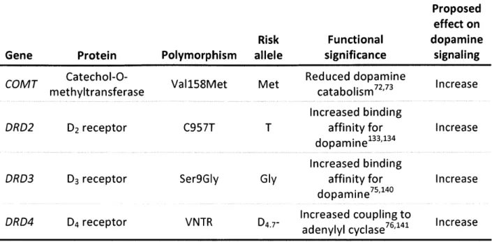
We briefly review current knowledge on select polymorphisms in COMT, DRD2, DRD3, and DRD4 that alter dopamine signaling (Table 1.1). Variations in these genes putatively determine the pre-medicated position of an individual on the inverted-U dopamine curve. This knowledge may be useful in predicting the impact of a right-ward shift on the curve due to exogenous
dopamine.8
1.5.1 COMT
The COMTVaI158Met polymorphism has a significant impact on the level of dopamine signaling in the PFC.78 In 1957, Julius Axelrod discovered COMT, an enzyme that inactivates
catecolamines, such as dopamine. 108 The COMT gene is located on chromosome 22q11 and encodes two proteins: a soluble form (S-COMT) and a membrane bound form (MB-COMT).109 MB-COMT is predominantly expressed in the brain while S-COMT is primarily present in the periphery, including blood, kidney, and liver.n3 uo-u3 In the brain, COMT is expressed intraneuronally in postsynaptic neurons and in astrocytic processes surrounding dopaminergic synapses, but the exact locus of COMT is not yet clear.114-11 6
Although COMT is ubiquitous in the brain,73
,
1 10
,
1 14 it is particularly important for inactivation of dopamine in the PFC. Dopamine transporters (DAT), which provide the primary mechanism for the clearance of dopamine from synapses in the striatum, are expressed at low levels in the PFC and only at a distance from synaptic release sites.7''11 1 8
Several studies highlighted the critical role of COMT in deactivating dopamine in the frontal cortex. 11'1 COMT knockout mice had significantly elevated dopamine levels in their frontal
cortices, but not in their striata.120
1'2 Administering levodopa to these COMT-deficient mice significantly increased the PFC levels of dopamine, and the striatal levels of DOPAC (an MAO metabolite) and levodopa, but not dopamine.119
Examination of the rate of formation of 3-methoxytyramine (created when COMT methylates dopamine) in the rat revealed that COMT accounted for roughly 60% of dopamine turnover in the PFC compared to 15% in the striatum. 2 Additionally, COMT mRNA, which encodes the COMT enzyme, was expressed in humans and rats at higher levels in the PFC than in the striatum.111 COMT appears to play a bigger role in primates than in mice and rats.1 23 For
in roughly equal amounts in the striatum and cerebrospinal fluid of rats and mice, whereas HVA dominated DOPAC by at least a factor of 12 in primates.1 23,1 2 4
A functional G to A single nucleotide polymorphism in COMT (rs4680) results in a valine (Val) to methionine (Met) amino acid change at codon 158 of MB-COMT (codon 108 of S-COMT). The Val isoform was more stable and active than the one with Met at physiological temperatures, 73, causing a 2 to 4 fold difference in COMT activity with the highest enzymatic
activity observed in Val/Val, followed by moderate activity Val/Met, and lowest activity in Met/Met individuals.73
,
1 2s The Met allele of COMT was not found in other mammals, including great apes, and thus appears to be a recent mutation in evolutionary timeline that is unique to humans.1 2 6 Decreased COMT activity putatively increases dopamine signaling in the PFC.73
Thus, healthy carriers of Val/Val likely fall on the left leg of the inverted-U curve, while Met/Met carries sit close to the peak.
1.5.2 DRD2
DRD2 C957T polymorphism alters the D2 receptor affinity for dopamine and thus may alter D2
-mediated dopamine signaling. D2 receptors are expressed at low levels across the cortex but
are abundant in subcortical regions, with the highest concentrations in the striatum and limbic structures, such as the amygdala.127-13 The DRD2 gene, which codes for dopamine receptor D2,
is located on chromosome 11q23. DRD2 C957T (rs6277) is a synonymous polymorphism (i.e., the C to T substitution does not alter the encoded amino acid due to codon redundancy) that
In vitro cell cultures showed that the T allele was associated with decreased mRNA stability, reduced receptor synthesis, and reduced dopamine-induced DRD2 up-regulation, possibly due
132 to an alteration in the folding pattern of the mRNA as a result of the C to T substitution. In contrast, subsequent studies using in vivo positron emission tomography in healthy adults showed that the T allele was related to increased striatal D2 availability, driven by enhanced D2
binding affinity with each T allele (T/T > C/T > C/C).33 ,
134 The discrepancy between the in vivo and in vitro results could have been due to the complexity of dopamine transmission regulation in the human brain. 4 Still, increased binding affinity in the presence of the T allele could potentially increase the level of dopamine signaling in the brain.
Most studies to date have focused on the impact of another D2 related polymorphism, Taq1A
(rs1800497), on cognition. PET studies showed that the Al allele of this polymorphism was associated with a 30% to 40% reduction in D2 receptor density in striatum.57 Newer reports,
however, showed that rs1800497 was located on kinase domain containing 1 (ANKK1) gene downstream from the DRD2 gene, and that ANKK1 was not expressed in the brain.135 Several
authors reported Taq1A polymorphism was in linkage disequilibrium with DRD2 C957T polymorphism (d' = 0.832 to 1, indicating strong dependence),3 2", 34 such that the Al allele of Taq1A was disproportionately over- and under-represented among C/C and T/T carriers, respectively.3 4
13 6 We chose to study the DRD2 C957T polymorphism because it is likely that Taq1A results are indirectly due to the C957T polymorphism.136
1.5.3 DRD3
D3 receptors are predominantly expressed in the nucleus accumbens, ventral tegmental area,
and limbic structures, such as amygdala127 ,137
,138 Because nucleus accumbens is a primary
target of the relatively preserved dopaminergic VTA, variations in D3 receptors may determine
whether this area will experience an overdose from exogenous dopamine.
DRD3 is located on chromosome 3q13.3 Ser9Gly (rs6280) is a C to T substitution in the first exon of DRD3 that results in a serine (Ser) to glycine (Gly) change at amino acid position 9 in the extracellular N-terminus of the receptor. The Gly/Gly variant in Chinese hamster ovary cells has a higher affinity for dopamine than the Ser/Ser and Ser/Gly variants, with no difference in affinity between the later two forms.7 5 Using a selective D
3 ligand, however, the authors found
that cells transfected with at least one Gly allele had a higher binding affinity for dopamine than those transfected with the Ser allele.75 Similarly, in an in vitro setup with human embryonic kidney cells, the Gly variant had a 4 to 5 fold increased affinity for dopamine compared to the Ser variant. 40 In addition, cAMP inhibition was increased and MAPK signal duration was prolonged with the Gly variant relative to the Ser variant, indicating that Gly variant is associated with a more robust and prolonged activation of D3-mediated signal transduction
pathways. 4
1.5.4 DRD4
A polymorphism in DRD4 impacts the level of signaling of D4 receptors. These receptors are
located on chromosome 11p15 and has a 48 base-pair variable number of tandem repeats (VNTR) polymorphism in its third exon.142 The number of repeats ranges from 2 to 11, represented as D4.2 to D4.11, respectively, causing a 32 to 176 amino-acid length difference in the third intracellular loop of the receptor, a region that binds to second messenger proteins.141 The most common D4 alleles in humans are the D4.2, D4.4, and D4.7, with 5%, 70%, 20%
prevalence, respectively.143,144 In Chinese hamster ovary cells, the D4.7 had a blunted response to dopamine compared to D4.2 and D4.4: The potency of dopamine to inhibit cAMP formation was reduced 2 to 3 fold with D4.7 compared to D4.2 and D4.4.76 Thus, individuals with the
7-repeat allele putatively fall on the right leg of the inverted-U curve.
1.6
Relevance to treatment of PD
The complications of dopaminergic treatment in PD include psychiatric disorders, such as hallucinations and impulse control behaviors.2733,3s,43,14s-48 Two issues demand attention: the greater vulnerability of certain patients to these side effects, and the role of genetic variation in eliciting them. Here, we propose and test a mechanism by which psychiatric side effects of dopamine replacement therapy can arise. By combining fine-tuned behavioral measures with low-cost genotyping of select dopamine gene polymorphisms, this proposal will identify biomarkers that distinguish patients who are at risk for medication-induced side effects. Individualized care for these patients will reduce their risk of incurring irreversible financial and personal costs due to medication-induced impulsivity and cognitive dysfunction.
The ultimate goal of PD research is to find the cause of and cure for the disease. In parallel to research focused on this goal, it is essential to ensure that the available medication used to ameliorate the motor symptoms of PD does not result in a degradation of the patients' quality of life. Although few alternative treatments are available for patients who show increased risk for hallucinations and impulsivity while taking dopaminergic medications, the identification of this high-risk group will permit early detection of adverse behaviors.
Table 1.1 Summary of risk alleles
Proposed effect on
Risk Functional dopamine
Gene Protein Polymorphism allele significance signaling
COMT Catechol-O- Val158Met Met Reduced dopamine Increase
methyltransferase catabolism72 73
Increased binding
DRD2 D2 receptor C957T T affinity for Increase
dopamine133,134
Increased binding
DRD3 D3 receptor Ser9GIy Gly affinity for Increase
dopamine75'140
DRD4 D4 receptor VNTR D Increased coupling to
adenylyl cyclase76
,
141 Increase
low
HC+PD
HC C.PD
E
-highlow
Dopamine signaling
high
Figure 1.1 Inverted-U dopamine response curve
The inverted-U dopamine response curve has been established in animals and humans. Too little (left side of curve) and too much (right side of curve) dopamine signaling result in cognitive dysfunction. Consistent with this view, healthy adults with genotypes that reduce dopamine signaling (HC-) are
impulsive. We predict that PD patients with heightened dopamine transmission (PD+) will be impulsive when receiving dopamine replacement therapy due to a dopamine overdose effect.
Dopaminergic cell degeneration Severe (90%) dopamine loss
in the nigrostriatal pathway Motor symptoms
Moderate (30-50%) dopamine loss in the mesocorticolimbic pathway
Cognitive symptomsI Dopaminergic medication based on severity of motor symptoms Reduced motor symptoms
Patients with low dopamine signaling do not experience
medication-induced psychiatric side effects
Patients with high dopamine signaling experience dopamine overdose, causing
psychiatric side effects
Figure 1.2 Model for the development of medication-induced side effects in PD The dose of prescribed dopaminergic medications is primarily based on the
severity of motor symptoms. In patients with increased dopamine signaling, medication levels that reduce motor symptoms have the potential to overdose the mesocorticolimbic pathway, which is much less affected than the
nigrostriatal pathway.
2
Response inhibition
2.1
Introduction
Response inhibition is the capacity to stop a prepotent or habitual response.1491'
i 0 Reduced inhibition is a common feature of several clinical conditions-trichotillomania (repetitive hair pulling), substance abuse, and ADHD. This impairment has, therefore, gained widespread attention in recent years.150,151
A common laboratory test of response inhibition, used in animals and humans, is the Stop Signal Task, which measures the ability to inhibit a motor action after it has been initiated. Participants are asked to respond as quickly as possible upon seeing a Go cue, and to inhibit this action if the Go cue is followed by a Stop cue. The Stop signal is presented only in a minority of trials, and thus responding becomes the prepotent action during the experiment. Inhibitory ability is indexed by the Stop signal reaction time (SSRT), which estimates the amount of time the brain needs to inhibit an ongoing action.
2.1.1 Neural substrates of response inhibition
The network of regions that mediates response inhibition includes the right inferior PFC (Brodmann areas 44, 45, and 47), right pre-SMA, and right subthalamic nucleus of the basal
ganglia. Here, we briefly review the evidence in support of each node's role in response inhibition.
In humans, damage to the right inferior PFC, but not the surrounding areas, resulted in slowed SSRT, and this measure was correlated with the extent of damage in this region. 152 Further, intracranial surface electrode recordings in humans showed increased activity in this region 100 to 250 msec after presentation of the Stop signal; this activity was grater when participants inhibited their movement on the Stop trials than when they failed to do so.15 3
Damage to the right pre-SMA also resulted in slowed inhibition, without affecting reaction times in trials without the Stop signal.154 Functional MRI studies confirmed that the stopping process activated the right inferior PFC and right pre-SMA, and that greater activity in the inferior PFC was associated with better inhibitory ability.iss-is7 Unlike inferior PFC, activity in the pre-SMA was not correlated with SSRT.1s8 Still, temporary deactivation of the right inferior PFC or right pre-SMA using transcranial magnetic stimulation impaired inhibitory ability in healthy adults.159"'60
Patients with cerebrovascular lesions in the basal ganglia had reduced inhibitory ability, though the authors did not identify the exact location of the basal ganglia lesions.'6' Functional MRI studies in healthy adults revealed that activation of the right subthalamic nucleus of the basal ganglia was associated with Stop, but not Go trials, and the strength of this activation was correlated with SSRT. 55,56,58
Together, these studies suggest that the inferior PFC, pre-SMA, and subthalamic nucleus are important nodes in the response inhibition network.1s1,16 2
2.1.2 Response inhibition in PD
Researchers have documented reduced inhibitory ability in PD patients. In a Go/NoGo task, they responded more often than controls on trials when they should not have responded (NoGo trials).163 Further, SSRT was significantly longer in PD patients than in age-, sex-, and
education-matched controls.164 This reduced inhibitory ability in PD was independent of general slowing and cognitive impairment,164 indicating a selective deficit in inhibitory ability. One study showed that subthalamic nucleus stimulation in PD patients increased their inhibitory control,165 although another demonstrated that deep brain stimulation (DBS) induced improvement was baseline dependent. Inhibitory ability increased in patients with the slowest baseline SSRTs but deteriorated in those with normal baseline SSRTs.16 6 This finding is likely
due an inverted-U relation between subthalamic nucleus activation and inhibitory control whereby DBS improved inhibitory ability in those with low baseline SSRTs, but impaired this ability in participants who had normal baseline response inhibition.
2.1.3 Pharmacology and genetics of response inhibition
Pharmacological studies in animals suggest that dopamine plays a critical role in modulating response inhibition: D-amphetamine, cocaine, and the dopamine reuptake inhibitor GBR 12909, all of which increase dopaminergic neurotransmission, decrease response inhibition in rats, measured by the number of premature responses in the 5-Choice Serial Reaction Time
Task.167-170 On this task, the dopamine antagonist alpha-flupenthixol blocked impulsivity induced by intra-accumbens injection of d-amphetamine.167 Further, methylphenidate, which increases
synaptic levels of dopamine, reduced inhibitory deficits in children and adults diagnosed with
ADHD. 17-174
The pharmacological alteration of response inhibition was baseline dependent, and improved inhibitory ability was limited to humans and rats with the worst performance at baseline.s7 5-79 This result is consistent with the inverted-U dopamine response hypothesis whereby only
individuals on the left-leg of the inverted-U curve (i.e., those with reduced dopamine signaling) should improve when receiving dopaminergic medication.
Although hypoactivity of the serotonin system has traditionally been associated with forms of impulsivity, such as aggression and suicidality,40'180 modulation of serotonin did not impact
response inhibition measured by the Stop Signal Task. Specifically, dietary depletion of serotonin precursor tryptophan 18,1 82 or serotonin receptor blockade using a selective serotonin reuptake inhibitor did not impact response inhibition in rats or healthy adults.183,8 4
Genetic research also supports a role for dopamine in inhibitory control. A PET study showed that the number of D2/D3 receptors was lower in impulsive rats compared to non-impulsive
ones.185 Healthy adults with at least one 7-repeat allele of D
4, which reduces dopamine
signaling, had longer SSRTs compared to individuals without the 7-repeat allele,63 and children
with ADHD who carried the 7-repeat allele of D4 required higher doses of methylphenidate for
SSRT-related brain activation in the right inferior PFC than those with the Val/Val genotype,8 6 which is associated with better inhibitory control.155'186
In summary, converging evidence from studies in animals, healthy humans, and humans with ADHD suggest that dopamine-induced changes in inhibitory ability follow and inverted-U curve, and that this curve can arise as a function of natural variation in genes that regulate the level of dopamine signaling.
2.1.4 Hypothesis
Building on prior work, we reasoned that variations in COMT, DRD2, DRD3, and DRD4 would alter inhibitory ability in PD patients receiving dopamine replacement therapy. Two lines of evidence support this hypothesis: first, the baseline-dependent influence of medication on impulsivity, and second, the relation between genetic variation in the dopamine-system and activation in the network mediating response inhibition. We hypothesized that patients who carry genotypes that increase dopamine signaling would be more likely to experience deficits in response inhibition due to a dopamine overdose. We addressed four specific questions: (1) Do COMT Met/Met and Val/Met carriers have longer SSRTs than Val/Val carriers?, (2) Do DRD2 T/T and C/T carriers have longer SSRTs than C/C carriers?, (3) Do DRD3 Gly/Gly carriers have longer SSRTs than Ser/Gly and Ser/Ser carriers?, and (4) Do D4.7- carriers have longer SSRTS than D47+ carriers? We predicated that individuals with the risk variants of COMT (Met allele), DRD2 (T allele), DRD3 (Gly allele), and DRD4 (absence of 7-repeat allele) would have longer SSRTs due to dopamine overdose.
2.2
Materials and methods
2.2.1 Participants
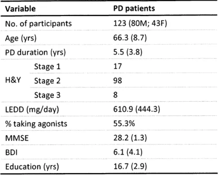
We recruited 123 patients with idiopathic PD from the Movement Disorders Units at the Massachusetts General Hospital and Brigham and Women's Hospital (Table 2.1). The inclusion criteria were: United Kingdom Parkinson's Disease Society Brain Bank diagnostic criteria,18 7 established by collaborating neurologists; mild to moderate disease indicated by Hoehn and Yahr (H&Y) stages 1-111; taking dopamine replacement therapy; no significant cognitive deficits indicated by Mini-Mental State Examination (MMSE)188 score 26; at least 12 years of schooling; and ability to give informed consent. The exclusion criteria were: history of a brain disorder other than PD; serious medical conditions (e.g., cancer, diabetes, heart disease); and severe depression indicated by a Beck Depression Inventory (BDI)1 89 score 18. All participants
gave written informed consent using procedures approved by the MIT Committee on the Use of Humans as Experimental Subjects and by the Partners Human Research Committee.
Participants were taking their normal dose of dopaminergic medications and were optimally medicated during testing. The self-identified racial and ethnic distribution of participants was: 122 White / not Hispanic or Latino and 1 Asian.
To compare dopaminergic medication among patients, each participant's dopaminergic drug regimen was converted to a levodopa equivalent daily dose (LEDD) according to a published146'190 formula: LEDD = levodopa/carbidopa regular (mg) + levodopa/carbidopa CR (mg) x 0.75 + [levodopa/carbidopa (mg) + levodopa/carbidopa CR (mg) x 0.75] x 0.33 if on
entacapone or tolcapone + [levodopa/carbidopa (mg) + levodopa/carbidopa CR (mg) x 0.75] x 1.2 if on 10 mg selegiline (x 1.1 if on 5 mg selegiline) + bromocriptine (mg) x 10 + pramipexole (mg) x 67 + requip (mg) x 20 + pergolide (mg) x 100.
2.2.2 Experimental design
On each trial, a left- or right-pointing green arrow appeared on a black computer screen (Figure 2.1). For Go trials, participants indicated the direction of this arrow by pressing the left or right arrow key on the keyboard as fast as possible, using their preferred index and middle fingers, respectively. The arrow stimulus remained on the screen until participants responded (max 2.5 sec). The next trial started after a 1.5 sec interval, during which the black screen remained blank.
On 25% of the trials, Stop trials, the arrow stimulus was replaced with a Stop signal (a red vertical bar) after a variable delay (Stop signal delay). We asked participants to inhibit their response when the Stop signal appeared. If they did so, the red bar remained on the screen for 2.5 sec. If participants erroneously pressed one of the arrow keys, the red bar disappeared immediately. The next trial started after a 1.5 sec interval.
The Stop signal delay started at 250 msec and was adjusted using an adaptive staircase method.191 If participants successfully inhibited their response on a Stop trial, the Stop signal delay was increased by 50 msec the next time a Stop signal appeared, thus making it harder to exert inhibitory control. If participants failed to inhibit themselves on a Stop trial, the Stop signal delay was decreased by 50 msec for the next Stop trial. This algorithm ensured that each
participant could inhibit roughly 50% of all Stop trials by the end of the experiment. This design allowed each participant to perform at his or her own inhibition threshold, equated the level of difficulty experienced by participants, and controlled for individual differences in speed of responding.64
We explained to the participants that they would not always be able to inhibit their response on Stop trials because the computer would adjust the difficulty of the task according to their performance level. We also asked them not to delay their response in anticipation of the Stop signal, but to inhibit their response when they saw the Stop signal. Participants completed 180 go and 60 Stop trials in 5 blocks with each block containing 36 Go and 12 Stop trials (240 trials total with an equal number of left- and right-pointing arrows in each block). Data analysis was limited to the fifth block to allow the staircase algorithm to converge on each participant's inhibitory threshold. Limiting the SSRT analysis to the fifth block ensured that all participants were performing at the same SSRT threshold-defined as the amount of advance warning a participant requires to be able to inhibit a habitual response 50% of the time-before they were compared with each other.
2.2.3 Genotyping
We extracted DNA from the venous blood of all participants using a QlAcube robotic workstation (Qiagen, Hilden, Germany). Aliquots of DNA were sent to Partners HealthCare Center for Personalized Genetic Medicine for genotyping. The DRD2 C957T (rs6277), DRD3 Ser9Gly (rs6280), and COMT Val158Met (rs4680) polymorphisms were genotyped using Sequenom hME chemistry, and DRD4 exon Ill VNTR was genotyped using a previously published
protocol.77 In our sample, 23, 69, and 31 patients carried the COMT Val/Val, Val/Met, and Met/Met genotypes, respectively. The DRD2 C957T break down was 21 C/C, 62 C/T, and 42 T/T. These distributions did not depart from the Hardy-Weinberg equilibrium (COMT: 2 =
1.975, df = 1, p = 0.160; DRD2: Xj = 0.132, df = 1, p = 0.716), indicating that allele frequencies
were in equilibrium in our cohort. Because only 8 and 12 participants fell in the D4 7+ and DRD3 C/C groups, respectively, we excluded DRD3 and DRD4 from further analyses.
2.2.4 Statistical analysis
The principal dependent variable was the SSRT, measured by subtracting the average Stop signal delay from the average correct Go reaction time in the final block.191 We also examined the participants' reaction times and error rates on Go trials. A univariate analysis of covariance (ANCOVA) compared each variable of interest among different genetic subgroups. We included age and sex in the ANCOVA as covariates because previous research uncovered age and sex differences in cognitive control ability92 19 3 and COMT enzyme activity.7 3 We also included LEDD, disease duration, and H&Y stage as covariates in the model to control for differences among participants in dopamine replacement dosage and the severity of motor symptoms.
To examine the impact of training on inhibitory ability, we compared SSRTs in the first and fifth blocks of the experiment. Because the staircase algorithm may not have converged to the 50% inhibitory threshold in the first block for all participants, we first corrected SSRTs for inhibition thresholds-defined as the number of successfully inhibited trials-in each block, and then carried a repeated measures ANCOVA on the adjusted SSRTs. We followed significant results
with post-hoc tests. All data were analyzed using MATLAB 2009a (MathWorks Inc., Natick, MA) and SPSS 11.5 (SPSS Inc., Chicago, IL).
2.3 Results
We characterized the participants in terms of age, sex, PD duration, H&Y stage, LEDD, number on agonists, MMSE, BDI, and education across COMT genotypes (Table 2.2). A significantly larger number of DRD2 C/C individuals were taking dopamine agonists as compared to C/T and T/T carriers (X2 = 6.915, df = 2, p = 0.032). Individuals with the C/T genotype of DRD2 were slightly, but significantly, older than C/C and T/T patients (C/C: M = 63.4, SD = 8.7; C/T: M =
68.6, SD = 8.4; T/T: M = 64.4, SD = 8.5; C/T vs. C/C : p = 0.048; C/T vs. T/T: p = 0.048). This age
difference was taken into account by including age a covariate in all analyses. Patients were well matched on all other characteristics across DRD2 genotypes (Table 2.3).
Because the green arrow in the Go trials was visible only for 2.5 seconds, we examined whether any participants missed this response window. Among the 123 participants, 117 (95.1%) never missed the window while 6 (4.9%; 2 COMT Val/Met & DRD2 C/C, 3 COMT Val/Met & DRD2 T/T,
1 COMT Met/Met & DRD2 T/T) participants missed the window on only a single Go trial. The
number of successfully inhibited trials did not differ statistically among DRD2 and COMT genotypes.
We used a univariate ANCOVA with SSRT as the dependent variable and genotype as the independent factor to examine effect of COMT variation on SSRT (Figure 2.2A). Age, sex, disease duration, total LEDD, and H&Y stage were covariates in the ANCOVA. The main effect
of COMT on SSRT was significant (F2,115 = 3.673, p = 0.028, i 2 = 6.0%). Planned post-hoc comparisons revealed that Val/Met and Met/Met participants had significantly higher SSRT thresholds than Val/Val individuals (Val/Met vs. Val/Val: p = 0.004 one-sided; Met/Met vs. Val/Val: p = 0.018 one-sided). The effect of COMT on accuracy (F2,115 = 0.518, p = 0.597) and reaction times (F2,115 = 1.852, p = 0.162) on Go trials was not significant (Figure 2.2B and C). The effect of DRD2 on SSRT (F2,115 = 0.336, p = 0.715), Go trial accuracy (F2,115 = 2.696, p = 0.072), and Go trial reaction times (F2,115 = 0.437, p = 0.647) was not significant.
To examine whether COMT variation interacted with training, we compared SSRTs in the first and fifth blocks. Because SSRT thresholds were significantly different between the two blocks
(p = 4.04 x 10¹²), we first corrected the SSRTs in the two blocks for this threshold difference: For each block, we ran a regression with the SSRT as the dependent variable and the percentage of successfully inhibited trials as the independent variable. Then we compared the standardized residuals from the two regressions. We ran a repeated measures ANCOVA with the standardized residuals as the dependent variables, and COMT genotype as the between-subjects factor. In both blocks, Val/Val carriers had lower standardized residuals (indicating better inhibitory ability) than those with at least one Met allele. Neither the main effect of the experimental block (F1,115 = 1.085, p = 0.300), nor the interaction between block and genotype (F2,115= 0.853, p = 0.429) were significant.
2.4
Discussion
This study examined whether polymorphisms in COMT, DRD2, DRD3, and DRD4 modulate inhibitory ability in PD. We addressed four specific questions: (1) Do COMT Met/Met and Val/Met carriers have longer SSRTs than Val/Val carriers?, (2) Do DRD2 T/T and C/T carriers have longer SSRTs than C/C carriers?, (3) Do DRD3 Gly/Gly carriers have longer SSRTs than Ser/Gly and Ser/Ser carriers?, and (4) Do D4.7- carriers have longer SSRTS than D4.7+ carriers?
We predicated that those individuals with variants that increase dopamine signaling would have reduced inhibitory ability due to a dopamine overdose in networks that are relatively preserved in the early stages of the PD. We found that patients who carried at least one Met allele of COMT, which confers increased dopamine levels in the PFC, had longer SSRTs than non-carriers. This reduction in inhibitory control was not accompanied by changes in accuracy or reaction times in trials without a Stop signal, indicating that increased SSRT was not due changes in performance or a general slowing of reaction times. Unlike COMT, DRD2 variation did not alter the SSRT. Due to sample sizes, we were unable to examine the influence of DRD3 and DRD4 on the SSRT.
The major finding of this experiment was that the Met allele of COMT resulted in a selective decrease in inhibitory ability in PD patients taking dopamine replacement therapy. Critically, this cognitive deficit was consistent with our prediction based on previous results showing an inverted-U relation between dopamine signaling and inhibitory ability. This finding highlights the future possibility of optimizing an individual's dopamine replacement therapy regimen based on their unique genetic profile.
Impact of COMT Val158Met polymorphism on SSRT
The observed effect of the COMTVal158Met polymorphism is consistent with the known neural substrates of response inhibition. Investigators have shown that normal function of the PFC, particularly right inferior frontal gyrus, is essential for successful response inhibition.1"1 Further, too much dopamine in the PFC results in a general reduction in neuronal activity.99-101 In an in vitro study in mice, investigators showed that application of high concentrations of dopamine to the PFC significantly reduced the number of action potentials produced by pyramidal neurons.100 Similarly, in well trained monkeys performing a spatial working memory tasks, high levels of D1 agonists significantly reduced the delay period activity of pyramidal neurons.99'101 Thus, it is likely that those with at least one Met allele of COMT had slowed SSRTs because of a reduction of neural activity in the PFC due to a dopamine overdose. We cannot rule out the possibility that the impact of COMT was due to its action at other nodes of the inhibitory network (e.g. pre-SMA or STN). Future functional imaging studies on the impact of the COMT Val158Met variation on response inhibition in PD patients may be able to localize specific nodes of interaction between dopamine replacement therapy and COMT variation.
No link between DRD2 C957T polymorphism and SSRT
The lack of a DRD2 effect on response inhibition was surprising. D2 receptors are densely
expressed in the basal ganglia (including caudate and putamen), which constitute one of the nodes of the response inhibition network. A previous study reported that healthy adults with variants of DRD2 that increased the expression of D2 receptors had better inhibitory control than those with reduced D2expression levels.194 Similarly, alcoholic carriers of the Al allele of
the ANKK1 Taq1A polymorphism, which is in linkage disequilibrium with DRD2 C957T and is associated with 30% to 40% reduced D2 receptor density in the striatum, had worse inhibitory
control than A2/A2 carriers.195 Further, PD patients who performed at the same level as controls in the Go/NoGo task had increased activity in the right caudate relative to controls, highlighting the importance of striatum for response inhibition in PD.196
No interaction between COMT and DRD2 on SSRT
Several investigators have shown a significant interaction between COMT and DRD2 polymorphisms in healthy adults. In a word serial position test of memory, those with COMT Val/Val and DRD2 C/C genotypes performed worse than those with Met/Met and T/T genotypes.197 Further, Met carries had significantly better working memory manipulation performance relative to Val/Val carriers, but only when they did not carry the Al allele of the ANKK1 TaqlA.1 98 Thus, to test for a possible interaction, we ran an ANCOVA with SSRT as the dependent variable, COMT and DRD2 as independent variables, and age, sex, disease duration, disease severity, and LEDD as covariates. The main effect of COMT remained significant in this model (p = 0.043), but as before, the main effect of DRD2 was not significant. We did not find an interaction between COMT and DRD2. Although this finding could be due to our small sample size (only 3 people were Val/Val and C/C carriers), the results suggest that the COMTCaIl158Met, and not DRD2 C957T, variation is the critical determinant of inhibitory ability in PD patients who take dopamine replacement therapy.