Publisher’s version / Version de l'éditeur:
Molecular nutrition & food research, 54, 5, pp. 1-11, 2010-01-01
READ THESE TERMS AND CONDITIONS CAREFULLY BEFORE USING THIS WEBSITE. https://nrc-publications.canada.ca/eng/copyright
Vous avez des questions? Nous pouvons vous aider. Pour communiquer directement avec un auteur, consultez la première page de la revue dans laquelle son article a été publié afin de trouver ses coordonnées. Si vous n’arrivez pas à les repérer, communiquez avec nous à PublicationsArchive-ArchivesPublications@nrc-cnrc.gc.ca.
Questions? Contact the NRC Publications Archive team at
PublicationsArchive-ArchivesPublications@nrc-cnrc.gc.ca. If you wish to email the authors directly, please see the first page of the publication for their contact information.
NRC Publications Archive
Archives des publications du CNRC
This publication could be one of several versions: author’s original, accepted manuscript or the publisher’s version. / La version de cette publication peut être l’une des suivantes : la version prépublication de l’auteur, la version acceptée du manuscrit ou la version de l’éditeur.
For the publisher’s version, please access the DOI link below./ Pour consulter la version de l’éditeur, utilisez le lien DOI ci-dessous.
https://doi.org/10.1002/mnfr.200900439
Access and use of this website and the material on it are subject to the Terms and Conditions set forth at
The location and behavior of α-tocopherol in membranes
Atkinson, Jeffrey; Harroun, Thad; Wassall, Stephen R.; Stillwell, William;
Katsaras, John
https://publications-cnrc.canada.ca/fra/droits
L’accès à ce site Web et l’utilisation de son contenu sont assujettis aux conditions présentées dans le site LISEZ CES CONDITIONS ATTENTIVEMENT AVANT D’UTILISER CE SITE WEB.
NRC Publications Record / Notice d'Archives des publications de CNRC:
https://nrc-publications.canada.ca/eng/view/object/?id=300a212d-ae14-4b24-b007-b3a12130672d
https://publications-cnrc.canada.ca/fra/voir/objet/?id=300a212d-ae14-4b24-b007-b3a12130672d
R
EVIEWThe location and behavior of a-tocopherol in membranes
Jeffrey Atkinson
1, Thad Harroun
2, Stephen R. Wassall
3, William Stillwell
4and John Katsaras
5 1Department of Chemistry, Brock University, St. Catharines, Ont., Canada2
Department of Physics, Brock University, St. Catharines, Ont., Canada
3
Department of Physics, Indiana University–Purdue University Indianapolis, Indianapolis, IN, USA
4
Department of Biology, Indiana University–Purdue University Indianapolis, Indianapolis, IN, USA
5
National Research Council, Chalk River, Ont., Canada
Received: September 8, 2009 Revised: October 27, 2009 Accepted: October 28, 2009
Vitamin E (a-tocopherol) has long been recognized as the major antioxidant in biological membranes, and yet many structurally related questions persist of how the vitamin functions. For example, the very low levels of a-tocopherol reported for whole cell extracts question how this molecule can successfully protect the comparatively enormous quantities of PUFA-containing phospholipids found in membranes that are highly susceptible to oxidative attack. The contemporary realization that membranes laterally segregate into regions of distinct lipid composition (domains), we propose, provides the answer. We hypothesize a-tocopherol partitions into domains that are enriched in polyunsaturated phospholipids, amplifying the concentration of the vitamin in the place where it is most needed. These highly disordered domains depleted in cholesterol are analogous, but organizationally antithetical, to the well-studied lipid rafts. We review here the ideas that led to our hypothesis. Experimental evidence in support of the formation of PUFA-rich domains in model membranes is presented, focusing upon docosahexaenoic acid that is the most unsaturated fatty acid commonly found. Physical methodologies are then described to elucidate the nature of the interaction of a-tocopherol with PUFA and to establish that the vitamin and PUFA-containing phospholi-pids co-localize in non-raft domains.
Keywords:
Lipid rafts / Lipid domains / Neutron scattering / NMR / PUFA
1
Introduction
The essential description of the antioxidant activity of toco-pherol has been well described and reviewed, and for this discussion we recommend references [1–7]. These and other studies pioneered the application of physical chemical principles to the action of phenolic antioxidants, establish-ing the rates of reaction for key steps in free radical-induced peroxidation of unsaturated fatty acids and phospholipids,
as well as the chain termination step of H-atom donation by such phenolic antioxidants as a-tocopherol. Much of this early work was performed in organic solution and, while forming the basis for the interpretation of tocopherol anti-oxidant chemistry, may not be entirely relevant for its action in phospholipid bilayers.
Of course, a considerable amount of work has focused on the biochemical aspects of a-tocopherol in model membranes of liposomes and vesicles [7–9]. Despite the complexities involved, to be able to explain and predict the antioxidant actions of tocopherol in membranes, we need to know how its orientation and dynamics within a bilayer [10–12] is affected by the nature of the bulk phospholipids. We should focus in particular on the role of polyunsaturated phospholipids since they are not only the chief substrates for peroxidative chemistry, but also they segregate into more fluid membrane domains where it would be advantageous Abbreviations: DHA, docosahexaenoic acid; DSC, differential
scanning calorimetry
Correspondence: Professor Jeffrey Atkinson, Departments of
Chemistry and Physics, Brock University, 500 Glenridge Ave, St. Catharines, Ont. L2S3A1, Canada
E-mail: jatkin@brocku.ca Fax: 11-905-682-9020
for tocopherol to preferentially reside. With the goal of providing a more detailed description of tocopherol’s action at the molecular level, work in the authors’ laboratories has focused on delineating the depth, orientation, and confor-mational dynamics of tocopherol in phospholipd bilayers; a reductionist approach to learning about a-tocopherol’s biochemistry.
From these experiments, a new thread of inquiry has emerged that tocopherol may fulfill its biological function by acting as both a lipid soluble antioxidant and as a critical structural component of membranes. We should expect that both roles would be dependent on the lipid composition that defines the local structure and dynamics of the membrane, an idea that is motivating our own research. In this article, we present a synthesis of these roles from a biophysical and chemical perspective, notably by drawing lessons from cholesterol research, and discuss the directions of future research that we would like to see.
1.1 a-Tocopherol beyond antioxidant chemistry
Several reviews have examined the complexities of anti-oxidant action of tocopherol in model systems such as liposomes and unilamellar vesicles [7, 13, 14]. These authors emphasize that any measure of the antioxidant action of tocopherol is a function of several different variables that may change between one assay and another and, by exten-sion, from one part of a cell membrane to another. For instance, there is a variability among the effectiveness of oxidation initiators, which in turn reveals that there are requirements for the presence of preformed lipid peroxyl radicals [7, 13, 14]. Dix and Aikens [15] have given a thor-ough critical perspective on the initiator problem, and Winterbourn [16] has provided an excellent overview of the relative reactivity and biological pertinence of reactive oxygen and nitrogen species.
Since tocopherol resides almost solely in phospholipid membranes in cells, the oxidizable substrates are predomi-nantly PUFA, but the concentration of tocopherol to PUFA is seemingly so low that recycling through reduction of the tocopheroxyl radical is required before the store of tocopherol is exhausted. Even higher membrane curvature, such as found in small unilamellar vesicles, supports faster lipid peroxidation, presumably due to ‘‘gaps’’ in head group packing that allow access of water-soluble radical initiators to the lipid core [17].
All of these factors conspire to confound the measured rates of lipid peroxidation. However, assaying lipid perox-idation in vitro can establish many of these conditions. Preparing known mixtures of pure phospholipids assures control of stoichiometry and the physical state of the lipids. In addition, specifically labeled tocopherol analogues can be used that can probe its dynamics and structure and perhaps even its oxidative state.
This opens up an approach to understand the nature of tocopherol’s complexity by reducing the surrounding
ques-tions to the basic interacques-tions of the constituent molecules. By doing so, we can look past the oxidation chemistry to the molecular associations of tocopherol with the variety of lipids it encounters in host cells, and answer a fundamental question: is a-tocopherol in the right place at the right time to be an antioxidant? We would also do well to draw parallels with cholesterol, which despite is minor chemical role in biology, its structural role cannot be underestimated. Where tocopherol prefers to spend its time may be the flip side of the cholesterol and lipid rafts question.
2
Membrane domains: A structure/
function role for a-tocopherol
2.1 Role of cholesterol in lipid rafts
It has never been easy to understand the reasons for the amazing diversity of biological lipids [18]. For example, it has been reported that there are some 30 000 different lipid molecular species just in the human meibum (tear gland) [19]! Cell membranes are composed, in part, of at least hundreds, perhaps thousands, of different lipids. It would be truly amazing if these lipids all behaved as inert mole-cules and did not display different affinities for one another. Yet in its original 1972 version, the Fluid Mosaic Model did not recognize the importance of lipid heterogeneity in membrane structure [20]. However, by 1974 a series of biophysical studies started to appear supporting the basic concept of lateral segregation of lipids into microdomains in membranes. Particularly noteworthy were cholesterol- and sphingolipid-enriched domains studied in model bilayer membranes in the 1970s by Biltonen and Thompson [21]. An early differential scanning calorimetry (DSC) study by Demel et al. [22] that established differential affinity for cholesterol was based on preferential obliteration by the sterol of melting transitions between two component monotectic lipids. This investigation showed that cholesterol association follows the sequence: sphingomyelin4 phosphatidylserine, phosphatidylglycerol4phosphatidyl-choline4phosphatidylethanolamine (SM4PS, PG4PC4 PE). Other similar reports followed, elaborating the nature of cholesterol–sphingolipid associations. The lipid micro-domain concept was formalized in a classic 1982 paper by Karnovsky et al. [23]. Therefore, the notion that cholester-ol–sphingolipid microdomains exist predated the concept of lipid rafts by more than a decade.
The discovery of what were coined ‘‘lipid rafts’’ in biological membranes [24] transformed thought in membrane research. They were first extracted from membranes at 41C by non-ionic detergents, particularly Triton X-100 [25, 26]. Isolated rafts were shown to contain about twice the amount of cholesterol and were also enri-ched in sphingolipids by about 50% when compared with the surrounding plasma membrane bilayer. The sterol was proposed to be the ‘‘glue’’ that holds them together since
rafts fall apart when cholesterol is extracted by cyclodextrin. Also, cholesterol is responsible for rafts existing in a liquid-ordered (lo) state [27] that is thicker than the surrounding
(non-raft) membrane that is in a liquid-disordered (ld), a.k.a.
liquid crystalline (La), state [25]. A characteristic of most
raft-lipids is that they have long saturated acyl chains. Using a wide variety of biophysical techniques, cholesterol has been shown to associate strongly with saturated acyl chains while avoiding polyunsaturated chains [28]. The lo state formed
when cholesterol mixes with saturated phospholipids and sphingolipids behaves as if it were halfway between the solid ordered (so), a.k.a. gel (Lb), and ldstates [29]. In the lostate
acyl chains are extended (have fewer gauche kinks) and so in this sense are gel like, but there is rapid axial rotation and lateral diffusion is almost as fast as in the ld state. What
makes rafts so influential is that they accumulate a family of important cell signaling proteins, giving function to a unique membrane structure [24].
2.2 Role of a-tocopherol in non-raft domains: De´ja` vu all over again?
2.2.1 a-Tocopherol and PUFA: complexation versus
association
A quick and admittedly superficial glance at the structure of a-tocopherol shows a remarkable homology to cholesterol. Both molecules are anchored at the aqueous interface by an OH group [11, 30, 31] have rigid rings at mid-structure and a branched chain protruding into the membrane interior. Admittedly, the amount of a-tocopherol in membranes is usually considerably less than cholesterol (such as in plasma membranes), but perhaps not in others, such as in the late endosome/lysosome [32, 33]. In lipid rafts, cholesterol has found a structurally influential niche. Perhaps a-tocopherol possesses an analogous role? Indeed, a-tocopherol was also being investigated for possible structural interactions with membrane lipids when cholesterol was first being shown to have a ‘‘structural’’ role through its association with sphin-gomyelin. The hope was to explain how vitamin E could fulfill its essential function as the major antioxidant in membranes where it has a special challenge in protecting highly oxidizable PUFA. a-Tocopherol is simply outnum-bered by the much more numerous potential oxidizable victims. Clearly, it would be advantageous if a-tocopherol and polyunsaturated lipids would preferentially partition into the same membrane locations, thus combining struc-tural and functional roles. But does this actually happen?
More than 30 years ago Lucy [34] proposed on the basis of molecular models that the conformation of the multiple double bonds in the PUFA chains of arachidonic acid-containing phospholipids creates pockets that can accom-modate the methyl groups at positions 40 and 80 on the isoprenoid side chain of a-tocopherol, resulting in the formation of complexes between the two molecules that are
ideally suited to preventing the oxidation of the highly vulnerable fatty acid. Experimental support was sparse and indirect, consisting of easier penetration of a-tocopherol into monolayers containing unsaturated phospholipid [35] and a reduction in permeability by a-tocopherol of membranes containing arachidonic acid [36]. Later it was shown using DSC that increasing amounts of a-tocopherol broadened and lowered the phase transition temperature of PUFA-containing PC samples, which was interpreted as being due to fluid phase immiscibilities and lateral phase separation of domains containing differing amounts of a-tocopherol [37]. In opposition was the failure of attempts to detect a direct manifestation of the predicted membrane stabilization as a change in molecular ordering and/or dynamics due to a-tocopherol that would occur preferentially in poly-unsaturated membranes. Especially troublesome was the observation that although a-tocopherol produced increases in ESR order parameters and correlation times of doxyl stearic acids probes, they are less in polyunsaturated than saturated PC membranes [38]. In addition, spin lattice relaxation times measured for methyl and methylene carbons on the phytyl side chain of a-tocopherol incorpo-rated into sonicated unilamellar vesicles were not signifi-cantly influenced by the double bond content of the phospholipids [39].
The concept of a-tocopherol-PUFA complexation was not confined to the Lucy model. Kagan proposed the formation of complexes between a-tocopherol and free unsaturated fatty acids largely on the basis of optical and NMR studies performed on mixtures in organic solution [40]. Involve-ment of the chromanol moiety, rather than the phytyl sidechain, was concluded. A model for the complex of linoleic acid with a-tocopherol was proposed in which a hydrogen bond is formed between the carboxyl of the fatty acid and the hydroxyl of the vitamin, while the 9,10- and 12,13- cis double bonds of the fatty acid adopt a conforma-tion that is complementary to the methyl groups on the chromanol moiety [41]. That a-tocopherol and free unsatu-rated fatty acids form complexes were similarly opined by Urano [42]. From 13C spin-lattice relaxation time and fluorescence quenching experiments performed in organic solution it was deduced that the methyl groups, but not the hydroxy group, on the chromanol group are responsible for complexation and that 3 or more double bonds in the fatty acid improve the interaction [41].
Evidence in support of the formation of a-tocopherol-free/fatty acid complexes in phospholipids bilayers, however, is indirect and less than convincing. On the one hand, greater reduction in fluidity due to incorporation of the vitamin into 1,2-dipalmitoylphosphatidylcholine (16:0-16:0PC (The numbers before and after the colon designate, respectively, the number of carbons and cis double bonds.)) bilayers containing free fatty acids with increasing number of double bonds was detected by fluorescence polarization of 1,6-diphenyl-1,3,5-hexatriene [43]. On the other hand, by2H
ordering produced by a-tocopherol between [2H62
]16:0-16:0PC membranes containing stearic (18:0) versus linoleic (18:2) free fatty acid [44].
Recent advances in our understanding of the conforma-tional dynamics of lipid chains containing multiple double bonds casts serious doubt, furthermore, on the ability of a-tocopherol to form complexes with PUFA. Complex formation is difficult to reconcile in terms of the rapid inter-conversion between torsional states, originating in the shallow energy barrier to rotation about the single bonds that separate the multiple double bonds, which occurs in PUFA [45]. Although long-lived a-tocopherol/polyunsaturated phospholi-pid complexes are unlikely, preferential association like cholesterol has for saturated sphingolipids are feasible.
2.2.2 PUFA’s aversion to cholesterol
Hypothesizing a structural role for a-tocopherol in a lipid domain analogous to that of cholesterol in lipid rafts should be based on a presumed preferential affinity of the vitamin for compounds most susceptible to oxidation, namely PUFA. With 22 carbons and 6 double bonds, docosahex-aenoic acid (DHA) is the most unsaturated PUFA commonly found in biological membranes [46] and so would be an ideal candidate for a-tocopherol association studies. We [47–49], as do others [50, 51], attribute the effi-cacy of DHA in preventing numerous human afflictions in part to the formation of DHA-rich, non-raft domains from which cholesterol is excluded. The tremendous disorder of PUFA chains is responsible for the aversion cholesterol has for DHA-containing phospholipids [48, 49]. Near approach of the steroid moiety is deterred by the wide variety of rapidly varying conformers that the DHA chain adopts.
There are a number of confirmatory experimental obser-vations. Closer contact with the 18:0 sn-1 chain in 18:0-22:6PC/[25,26,26,27,27,27-2H
7]cholesterol (1:1 mol) is revealed
by a higher rate of chain to sterol nuclear Overhauser enhancement spectroscopy (NOESY) in 1H magic angle spinning NMR experiments [50]. This result is reproduced in MD simulations on an 18:0-22:6PC/chol (3:1 mol) bilayer that corroborates the sterol favors solvation by saturated over polyunsaturated chains [52]. Partition coefficients for choles-terol are also smaller in unilamellar vesicles composed of PC with DHA than less unsaturated chains [53]. We have provi-ded unequivocal substantiation of poor affinity for the sterol by the greatly reduced solubility that was measured in PUFA-containing membranes by x-ray diffraction and solid-state2H
NMR [54]. In fact, we have shown that cholesterol sequesters to the bilayer center when forced to mix with PUFA chains in dipolyunsaturated lipids [55, 56]. Therefore, we can safely predict that polyunsaturated phospholipids should partition away from cholesterol, and hence lipid rafts, into different microdomains.
We have amassed proof from several experimental approaches that DHA-containing lipids segregate into
non-raft domains. Cold temperature detergent (Triton X-100) extraction of 16:0-22:6PE/egg SM/cholesterol (1:1:1 mol) membranes shows that egg SM and cholesterol phase sepa-rate almost exclusively (490%) into the detergent resistant membrane (DRM or raft) fraction, the hallmark of lipid rafts, whereas 16:0-22:6PE predominantly phase separates (70%) into the detergent soluble membrane (DSM, non-raft) frac-tion [57]. Separafrac-tion into SM-rich and PUFA-rich phases is further demonstrated by the presence of two endotherms in DSC scans for 16:0-22:6PE/egg SM (1:1 mol), and the intro-duction of cholesterol first affects the higher temperature endotherm attributed to the SM-rich phase [58]. This approach, of course, is the same methodology Demel et al. used to demonstrate cholesterol-SM affinity in the 1970s [22]. Solid-state 2H NMR spectra for [2H31]16:0-22:6PE/egg
SM (1:1 mol) [57] and 16:0-22:6PE/[2H
31]16:0SM (1:1 mol)
[59] establishes the presence of motionally distinct SM-rich (more ordered) and PUFA-rich (less ordered) domains that are o20 nm in size, an acceptable size for lipid rafts. Order in the SM-rich (raft) domain is increased more than the PUFA-rich (non-raft) domain when cholesterol (1:1:1 mol) is added. In a control system where monounsaturated oleic acid-containing 16:0-18:1PE substitutes for 16:0-22:6PE, the mutual exclusion of PE and cholesterol is much less.
2.2.3 a-Tocopherol in the membrane: where is it?
The next question we faced was to determine where a-tocopherol partitions in model membranes composed of DHA-containing PE- and SM-enriched regions. Our preli-minary detergent extraction and DSC work does offer persuasive support for the concept that the vitamin co-localizes with DHA [60]. The experiments indicate that although cholesterol associates primarily with the raft-lipid SM, a-tocopherol prefers the non-raft PUFA-containing phospholipid. We are currently extending these observations to include studies that will further demonstrate a-tocopherol partitions into domains enriched in DHA for which it has preferential affinity. Our strategy is to emulate earlier work that helped establish the opposite behavior for cholesterol whereby the sterol sequesters in rafts with SM for which it has strong affinity. Replacing a-tocopherol by cholesterol, the same SM/PE mixtures prepared from deuterated analogs that were employed in our previous 2H NMR investigations [57, 59] will be utilized to demonstrate that the vitamin differentially affects order within the PUFA-rich domains. Adapting an approach developed by Heerklotz and coworkers that uses isothermal titration calorimetry to measure the partitioning of cholesterol between cyclodextrin and lipid vesicles [61, 62], we shall substitute a-tocopherol for the sterol to assay its binding to DHA-containing phos-pholipid relative to less unsaturated phosphos-pholipids [63].
We have also begun a series of neutron diffraction experiments based on crystallography techniques and isotopic/isomorphic labeling of tocopherol. Neutron
diffraction does not result in a measure of electron density, typical of x-ray crystallography, although the same mathe-matics are involved. Instead, the interaction of neutrons and matter is via the nucleus, and is therefore isotope sensitive. Deuterium is also structurally isomorphic, and thus an ideal label for neutron diffraction. Here we used tocopherol labeled either by2H3 on the methyl attached to C5 of the
chromanol ring, or 2H2 on the C90 carbon along the
isoprenoid chain incorporated into 16:0-18:1PC or 18:1-18:1PC bilayers. Typically 5–6 Bragg diffraction peaks were recorded, meaning that the reconstructed unit cell has a crystallographic resolution of 8–10 A˚ . This is less than atomic resolution, since the bilayers were in the fluid phase. The difference between labeled (deuterated) and unla-beled (protonated) samples can be calculated by the measured structure factors with one important caveat: The structure factors for both experiments must be placed on the same relative scale. If they can, then the difference profile is simply the center of mass of the isotope label, with all other molecular components subtracted away. Furthermore, the distribution parameters can be shown to be of atomic resolution, even when the raw data isn’t. We have followed closely the discussion and methods of this topic from Han et al. [64] and our earlier work on deuterium labeled cholesterol [56].
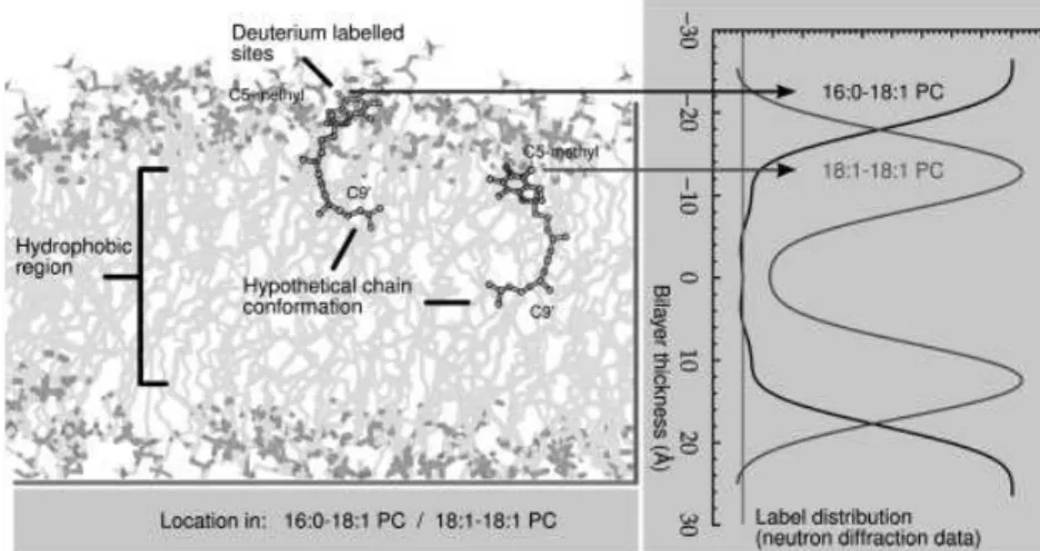
Figure 1 shows the mass distribution of a-tocopherol labeled with three2H on the methyl attached to C5 of the
chromanol ring, as measured in a 18:1-18:1PC membrane. The unit cell contains one bilayer, and the inter-lamellar water layers are located at the edges of the unit cell. The data place the reactive hydroxyl group at 13 A˚ from the bilayer center, as is also shown schematically in the figure. This position is at, or perhaps just below, the hydrophobic/ hydrophilic interface, nearly at the same depth as the phospholipid glycerol backbone ester. The location of the distal end (C90 of the isoprenoid side chain) of the toco-pherol is centered at the middle of the membrane, and with a broad distribution of nearly 1 A˚ (data not shown). Together these results confirm that tocopherol stands ‘‘upright’’ in
the bilayer, the hydroxyl group is just above the depth of the first carbon in the 18:1-18:1PC acyl chain. The chain of the tocopherol is highly disordered, since the C90 carbon, only 3/4 along the chain, is found in a broad range of locations about the center of the bilayer. If the chains were extended it would be interdigitated into the opposing bilayer leaflet, but the methyl branches make an all-trans configuration highly unlikely.
This unique and unambiguous insight into the location of tocopherol can be directly compared with atomic simu-lations. In future we plan to carry these experiments to other lipids, especially PUFA-containing lipids. The data included in Fig. 1 for [5 methyl-2H3] labeled a-tocopherol in
16:0-18:1PC, which reveals the vitamin molecule sits 10 A˚ higher than in 18:1:18:1PC, implies a profound sensitivity upon membrane unsaturation for the depth to which the vitamin penetrates. The planned neutron scattering experiments will be performed in conjunction with solid-state 2H NMR observation of deuterated analogs of both a-tocopherol and phospholipids and with MD simulations to provide a detailed view of dynamical organization.
3
Our hypothesis
We hypothesize that, in contrast to cholesterol, a-tocopherol preferentially incorporates into PUFA-rich domains and in doing so achieves ready access to the membrane component most at risk to oxidation. This co-localization of vitamin and PUFA would produce a concentration amplification that optimizes the protection of membranes from deleterious oxidation and eventual functional destruction. A simple calculation on the basis of molecular ratio illustrates, for instance, that as much as a 20-fold improvement in local concentration of a-tocopherol relative to PUFA would be achieved by this mechanism in membranes containing 5 mol% PUFA. How vitamin E that is present in small amounts within the plasma membrane can accomplish its accepted antioxidant function is, thus, answered.
Figure 1. Neutron scattering data showing
the position of the C5 methyl group on the chromanol and C90side chain methylene in
a-tocopherol (right panel) and a schematic illustration of the location of the vitamin (left panel) in 16:0-18:1PC and 18:1-18:1PC samples. Shown is a simple snapshot of just one possible conformation, illustrating a shorter effective length for the disordered sidechain, which places the C90group at the
a-Tocopherol may stabilize PUFA-rich, non-raft domains in a role parallel to that fulfilled by cholesterol in rafts.
A cartoon representation of our hypothesis is shown in Fig. 2. It shows the leaflet of a membrane in which a-tocopherol partitions into a DHA-rich domain while cholesterol and SM separate into a raft-like domain within a bulk lipid matrix. The smaller size of the chromanol group in a-tocopherol, which is approximately half the size of the steroid moiety in cholesterol, is the reason postulated for the difference. As the depth to which it penetrates the membrane precedes the sequence of double bonds that begins at position 4 in DHA [65], intimate proximity does not then necessitate adverse interactions between the rigid chromanol group and highly disordered double bonds while hydrogen bonding of the hydroxyl with the ester carbonyl on phospholipids to stabilize the domain can occur. The phenomenal flexibility of the PUFA chain [45], furthermore, produces a fluid membrane interior favorable to the segmental motion of the phytyl side chain with its methyl branches. In this arrangement that places the chromanol group close to the aqueous interface, a-tocopherol can effi-ciently trap peroxyl radicals created within PUFA chains and subsequently be restored. DHA chains isomerize so rapidly that they explore their entire conformational space within 50 ns [66]. The torsional states adopted include bent conformations that would bring even the lower portions of the PUFA chain up to the membrane surface [67–69] and into the vicinity of the chromanol group, which is also well situated for regeneration of the tocopheroxyl radical by water-soluble reducing agents including ascorbate.
4
Do we know where tocopherol does its
most important job?
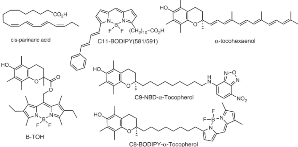
Other methodologies have the potential to contribute to testing the hypothesis presented here. Tools are available to assist in delineating the cellular location of peroxidative ‘‘hot
spots’’ in a cell as well as the location of tocopherol. For instance, several oxidatively sensitive fluorescence probes (Fig. 3) have been used to assess lipid peroxidation. Two often-used lipid-soluble probes are cis-parinaric acid [70–78] and C11-BODIPY(581/591) [77, 79–84]. cis-Parinaric acid oxidation products are non-fluorescent, thus it is the loss of parent fluorescence intensity that is monitored, whereas C11-BODIPY(581/591) undergoes a green-shift in its fluor-escence allowing ratio-based methods to track both the parent and oxidation product(s) [80, 85].
Recently one of us (J. A.) has completed the synthesis [86] of a fluorescent polyene analog of tocopherol called a-tocohexaenol (Fig. 3) which is exquisitely sensitive to oxidation and yet shares more structural features with a-tocopherol than any other available fluorophore. The hope is that a-tocohexaenol will be able to be used in a similar fashion as cis-parinaric acid, but be able to report more specifically on the location of tocopherol in a membrane. Other fluorescent analogues of tocopherol including C9-NBD-a-tocopherol [87] and C8-BODIPY-a-tocopherol (West, R., submitted) have been useful in determining the cellular location and transfer of tocopherol [88–92], but do not change fluorescence on oxidation.
A unique approach to fluorescence reporting of oxidative events in membranes has been reported by the group of Cosa [93–95]. They designed, synthesized and characterized a compound (B-TOH, Fig. 3) formed by conjugation of the chromanol carboxylic acid Trolox with a BODIPY fluor-ophore. B-TOH is not fluorescent as the free phenol due to self-quenching by a photoelectron transfer mechanism. However, once B-TOH has donated a phenolic hydrogen atom during a radical chain termination event, it becomes fluorescent and thus reports on the initiation and site of peroxidation. It remains to be seen whether B-TOH can report on peroxidative hot spots pertinent to the behavior of a-tocopherol, as its structure is considerably different. Combinations of the chromanol containing fluorophores may allow one to co-localize the steady-state presence of
Lipid raft
PUFA domain
Figure 2. A cartoon rendition of our
hypo-thesis where a-tocopherol partitions into a DHA-rich domain while cholesterol and SM separate into a raft-like domain within a bulk lipid matrix.
tocopherol with sites that are sensitive (change in fluores-cence intensity) during peroxidative events.
4.1 Is all of this relevant for understanding the biological activity of tocopherols?
The kinetic and mechanistic details that describe how tocopherol can act as a radical chain breaking antioxidant are now well established. Studies in model membrane systems have extended this work to phospholipid structures such as liposomes and vesicle bilayers. Progressing from this work to cellular biological membranes is a significant challenge. The biological function of a-tocopherol is made more interesting should we consider the possibility of mechanisms of action independent of antioxidant activity. Indeed, the effect of a-tocopherol on membrane resident proteins and enzymes, signaling cascades, and gene expression, may be related to its contribution to membrane structure and dynamics, and thus modulation of membrane-dependent signaling mechanisms such as protein and lipid kinases [96–98]. We have described here how our work using2H-NMR and neutron diffraction will enable a more detailed understanding of how tocopherol behaves when immersed in phospholipid bilayers of increasing poly-unsaturation.
How do we jump from model systems to the complexity of real biomembranes? It would seem that one of the key unknowns is the actual lipid composition of membranes in specific cellular locations. As we already know that toco-pherol is found in different amounts in different cell orga-nelles [32, 99] we should like to know whether the supporting phospholipid composition is changed as well. One possible approach to retrieving this information from cells is scanning imaging mass spectroscopy [100, 101].
This technique has already been used to image tocopherol location in cells [102] and tissues [103–105]. It would be fascinating if someone should now apply the tools of lipi-domics to collecting data not only on the location of
toco-pherol, but also the phospholipid profile at the same location. Lipidomic data analysis is sophisticated enough to discern the complete acyl chain populations of all of the common phos-pholipid headgroups. This would enable one to ask questions like: ‘‘What oxidizable lipid exists in those regions of highest (or lowest) tocopherol content?’’ ‘‘Are there regions of high PUFA content with relatively low tocopherol content?’’ Furthermore, scanning mass spectrometry could identify those products of lipid peroxidation that are known to be powerful signaling molecules [106, 107].
5
Concluding remarks
a-Tocopherol remains a most unusual character; it is a function in search of a home. Despite the fact that for decades vitamin E has been known as the major antioxidant in membranes and its molecular mode of action has been elucidated, how little is actually known about its physical location in biological membranes is remarkable. In this article we have reviewed the status of a-tocopherol’s orga-nization in membrane, including vertical location or depth, and have proposed lateral segregation of the vitamin into PUFA-rich domains. These domains are postulated in analogy with the very well studied lipid raft domain that currently plays an influential role in thought on membrane structure. While lipid rafts are SM-rich, lodomains that are
held together by cholesterol and have functions relating to cell signaling events, the non-raft domains proposed here are PUFA-rich, ld domains in which a-tocopherol
co-loca-lizes and serves its antioxidant role. Structural and func-tional roles for a-tocopherol, thus, would dovetail nicely in a mutually beneficial manner. By tracking after PUFA, the vitamin is placed ‘‘in the right place at the right time to be an antioxidant’’. A series of diverse physical methodologies are proposed to test the nature of the hypothesized a-toco-pherol/PUFA membrane domains.
The authors have declared no conflict of interest.
CO2H cis-parinaric acid N B N (CH2)10CO2H F F C11-BODIPY(581/591) N B N F F O O O HO B-TOH O HO α-tocohexaenol O HO H N N O N NO2 O HO N B N F F C9-NBD-α-Tocopherol C8-BODIPY-α-Tocopherol
Figure 3. Fluorescent probes used to
monitor lipid perocidation and the cellular location of a-tocopherol.
6
References
[1] Burton, G. W., Ingold, K. U., Vitamin E: applications of the principles of physical organic chemistry to the exploration of its structure and function. Acc. Chem. Res. 1986, 19, 194–201. [2] Niki, E., Antioxidants in relation to lipid peroxidation.
Chem. Phys. Lipids 1987, 44, 227–253.
[3] Burton, G. W., Ingold, K. U., Vitamin-E as an Invitro and Invivo Antioxidant. Ann. NY Acad. Sci. 1989, 570, 7–22. [4] Burton, G. W., Ingold, K. U., in: Miquel, J., Quintanilha,
A. T., Weber, H. (Eds.), Handbook of Free Radicals and
Antioxidants in Biomedicine, CRC Press, Inc, Boca Raton,
FL 1989, pp. 29–43.
[5] Burton, G. W., Vitamin E: molecular and biological func-tion. Proc. Nutr. Soc 1994, 53, 251–262.
[6] Niki, E., Noguchi, N., Dynamics of antioxidant action of vitamin E. Acc. Chem. Res. 2004, 37, 45–51.
[7] Fukuzawa, K., Dynamics of lipid peroxidation and anti-oxidion of alpha-tocopherol in membranes. J. Nutr. Sci.
Vitaminol. (Tokyo) 2008, 54, 273–285.
[8] Niki, E., Kawakami, A., Yamamoto, Y., Kamiya, Y., Oxidation of lipids. VIII. Synergistic inhibition of oxidation of phos-phatidylcholine liposome in aqueous dispersion by vitamin E and vitamin C. Bull. Chem. Soc. Jpn. 1985, 58, 1971–1975. [9] Niki, E., Takahashi, M., Komuro, E., Antioxidant activity of
vitamin-E in liposomal membranes. Chem. Lett. 1986, 1573–1576.
[10] Atkinson, J., Epand, R. F., Epand, R. M., Tocopherols and tocotrienols in membranes: a critical review. Free Rad.
Biol. Med. 2008, 44, 739–764.
[11] Quinn, P. J., Is the distribution of alpha-tocopherol in membranes consistent with its putative functions?
Biochemistry (Mosc.) 2004, 69, 58–66.
[12] Quinn, P. J., Molecular associations of vitamin E. Vitam.
Horm. 2007, 76, 67–98.
[13] Niki, E., Yoshida, Y., Saito, Y., Noguchi, N., Lipid per-oxidation: mechanisms, inhibition, and biological effects.
Biochem. Biophys. Res. Commun. 2005, 338, 668–676.
[14] Schnitzer, E., Pinchuk, I., Lichtenberg, D., Peroxidation of liposomal lipids. Eur. Biophys. J. 2007, 36, 499–515. [15] Dix, T. A., Aikens, J., Mechanisms and biological relevance
of lipid peroxidation initiation. Chem. Res. Toxicol. 1993, 6, 2–18.
[16] Winterbourn, C. C., Reconciling the chemistry and biology of reactive oxygen species. Nat. Chem. Biol. 2008, 4, 278–286. [17] Li, Q. T., Yeo, M. H., Tan, B. K., Lipid peroxidation in small
and large phospholipid unilamellar vesicles induced by water-soluble free radical sources. Biochem. Biophys. Res.
Commun. 2000, 273, 72–76.
[18] Dowhan, W., Molecular basis for membrane phospholipid diversity: why are there so many lipids? Annu. Rev.
Biochem. 1997, 66, 199–232.
[19] Nicolaides, N., Santos, E. C., The di- and triesters of the lipids of steer and human meibomian glands. Lipids 1985,
20, 454–467.
[20] Singer, S. J., Nicolson, G. L., The fluid mosaic model of the structure of cell membranes. Science 1972, 175, 720–731. [21] Estep, T. N., Mountcastle, D. B., Barenholz, Y., Biltonen, R.
L., Thompson, T. E., Thermal behavior of synthetic sphin-gomyelin-cholesterol dispersions. Biochemistry 1979, 18, 2112–2117.
[22] Demel, R. A., Jansen, J. W., van Dijck, P. W., van Deenen, L. L., The preferential interaction of cholesterol with different classes of phospholipids. Biochim. Biophys. Acta 1977, 465, 1–10.
[23] Karnovsky, M. J., Kleinfeld, A. M., Hoover, R. L., Klausner, R. D., The concept of lipid domains in membranes. J. Cell.
Biol. 1982, 94, 1–6.
[24] Simons, K., Ikonen, E., Functional rafts in cell membranes.
Nature 1997, 387, 569–572.
[25] Brown, D. A., London, E., Structure and function of sphingolipid- and cholesterol-rich membrane rafts. J. Biol.
Chem. 2000, 275, 17221–17224.
[26] Ahmed, S. N., Brown, D. A., London, E., On the origin of sphingolipid/cholesterol-rich detergent-insoluble cell membranes: physiological concentrations of cholesterol and sphingolipid induce formation of a detergent-inso-luble, liquid-ordered lipid phase in model membranes.
Biochemistry 1997, 36, 10944–10953.
[27] Brown, D. A., London, E., Structure and origin of ordered lipid domains in biological membranes. J. Membr. Biol. 1998, 164, 103–114.
[28] Wassall, S. R., Brzustowicz, M. R., Shaikh, S. R., Cherezov, V. et al., Order from disorder, corralling cholesterol with chaotic lipids. The role of polyunsaturated lipids in membrane raft formation. Chem. Phys. Lipids 2004, 132, 79–88.
[29] Polozov, I. V., Gawrisch, K., Characterization of the liquid-ordered state by proton MAS NMR. Biophys. J. 2006, 90, 2051–2061.
[30] Go´mez-Ferna´ndez, J. C., Villalain, J., Aranda, F. J., Ortiz, A.
et al., Localization of alpha-tocopherol in membranes. Ann. NY Acad. Sci. 1989, 570, 109–120.
[31] Salgado, J., Villalain, J., Gomez-Fernandez, J. C., Magic angle spinning C-13-NMR spin-lattice relaxation study of the location and effects of alpha-tocopherol, ubiquinone-10 and ubiquinol-ubiquinone-10 in unsonicated model membranes.
Eur. Biophys. J. 1993, 22, 151–155.
[32] Buttriss, J. L., Diplock, A. T., The relationship between alpha-tocopherol and phospholipid fatty acids in rat liver subcellular membrane fractions. Biochim. Biophys. Acta 1988, 962, 81–90.
[33] Rupar, C. A., Albo, S., Whitehall, J. D., Rat liver Iysosome membranes are enriched in alpha-tocopherol. Biochem.
Cell Biol. 1992, 70, 486–488.
[34] Diplock, A. T., Lucy, J. A., The biochemical modes of action of vitamin e and selenium: a hypothesis. FEBS Lett. 1973,
29, 205–210.
[35] Maggio, B., Diplock, A. T., Lucy, J. A., Interactions of tocopherols and ubiquinones with monolayers of phos-pholipids. Biochem. J. 1977, 161, 111–121.
[36] Diplock, A. T., Lucy, J. A., Verrinder, M., Zieleniewski, A., alpha-Tocopherol and the permeability to glucose and chro-mate of unsaturated liposomes. FEBS Lett. 1977, 82, 341–344. [37] Sanchez-Migallon, M. P., Aranda, F. J., Gomez-Fernandez, J. C., Interaction between alpha-tocopherol and heteroacid phosphatidylcholines with different amounts of unsatura-tion. Biochim. Biophys. Acta 1996, 1279, 251–258. [38] Wassall, S. R., Wang, L., McCabe, R. C., Ehringer, W. D.,
Stillwell, W., Electron spin resonance study of the inter-action of alpha-tocopherol with phospholipid model membranes. Chem. Phys. Lipids 1991, 60, 29–37. [39] Urano, S., Iida, M., Otani, I., Matsuo, M., Membrane
stabilization of vitamin E; interactions of alpha-tocopherol with phospholipids in bilayer liposomes. Biochem. Biophys. Res. Commun. 1987, 146, 1413–1418.
[40] Kagan, V. E., Tocopherol stabilizes membrane against phospholipase A, free fatty acids, and lysophospholipids.
Ann. NY Acad. Sci. 1989, 570, 121–135.
[41] Erin, A. N., Skrypin, V. V., Kagan, V. E., Formation of alpha-tocopherol complexes with fatty acids. Nature of complexes. Biochim. Biophys. Acta 1985, 815, 209–214. [42] Urano, S., Shichita, N., Matsuo, M., Interaction of vitamin-E and
its model compounds with unsaturated fatty-acids in homo-geneous solution. J. Nutr. Sci. Vitaminol. 1988, 34, 189–194. [43] Urano, S., Yano, K., Matsuo, M., Membrane-stabilizing
effect of vitamin E: effect of alpha-tocopherol and its model compounds on fluidity of lecithin liposomes.
Biochem. Biophys. Res. Commun. 1988, 150, 469–475.
[44] Wassall, S. R., Yang, R. C., Wang, L., Phelps, T. M. et al., Magnetic resonance studies of the structural role of vita-min E in phospholipid model membranes. Bull. Magn.
Reson. 1990, 12, 60–64.
[45] Feller, S. E., Gawrisch, K., MacKerell, A. D., Jr., Poly-unsaturated fatty acids in lipid bilayers: intrinsic and environmental contributions to their unique physical properties. J. Am. Chem. Soc. 2002, 124, 318–326. [46] Stillwell, W., Wassall, S. R., Docosahexaenoic acid:
membrane properties of a unique fatty acid. Chem. Phys.
Lipids 2003, 126, 1–27.
[47] Stillwell, W., Shaikh, S. R., Zerouga, M., Siddiqui, R., Wassall, S. R., Docosahexaenoic acid affects cell signaling by altering lipid rafts. Reprod. Nutr. Dev. 2005, 45, 559–579. [48] Wassall, S. R., Stillwell, W., Docosahexaenoic acid domains: the ultimate non-raft membrane domain. Chem.
Phys. Lipids 2008, 153, 57–63.
[49] Wassall, S. R., Stillwell, W., Polyunsaturated fatty acid-cholesterol interactions: domain formation in membranes.
Biochim. Biophys. Acta 2009, 1788, 24–32.
[50] Huster, D., Arnold, K., Gawrisch, K., Influence of docosa-hexaenoic acid and cholesterol on lateral lipid organization in phospholipid mixtures. Biochemistry 1998, 37, 17299–17308. [51] Mitchell, D. C., Litman, B. J., Molecular order and dynamics in bilayers consisting of highly polyunsaturated phospholipids. Biophys. J. 1998, 74, 879–891.
[52] Pitman, M. C., Suits, F., Mackerell, A. D., Jr., Feller, S. E., Molecular-level organization of saturated and
poly-unsaturated fatty acids in a phosphatidylcholine bilayer containing cholesterol. Biochemistry 2004, 43, 15318–15328. [53] Niu, S. L., Litman, B. J., Determination of membrane cholesterol partition coefficient using a lipid vesicle-cyclodextrin binary system: effect of phospholipid acyl chain unsaturation and headgroup composition. Biophys.
J. 2002, 83, 3408–3415.
[54] Shaikh, S. R., Cherezov, V., Caffrey, M., Soni, S. P. et al., Molecular organization of cholesterol in unsaturated phosphatidylethanolamines: X-ray diffraction and solid state 2H NMR reveal differences with phosphatidylcho-lines. J. Am. Chem. Soc. 2006, 128, 5375–5383.
[55] Harroun, T. A., Katsaras, J., Wassall, S. R., Cholesterol hydroxyl group is found to reside in the center of a polyunsaturated lipid membrane. Biochemistry 2006, 45, 1227–1233.
[56] Harroun, T. A., Katsaras, J., Wassall, S. R., Cholesterol is found to reside in the center of a polyunsaturated lipid membrane. Biochemistry 2008, 47, 7090–7096.
[57] Shaikh, S. R., Dumaual, A. C., Castillo, A., LoCascio, D.
et al., Oleic and docosahexaenoic acid differentially phase
separate from lipid raft molecules: a comparative NMR, DSC, AFM, and detergent extraction study. Biophys.
J. 2004, 87, 1752–1766.
[58] Shaikh, S. R., Brzustowicz, M. R., Gustafson, N., Stillwell, W., Wassall, S. R., Monounsaturated PE does not phase-separate from the lipid raft molecules sphingomyelin and cholesterol: role for polyunsaturation? Biochemistry 2002,
41, 10593–10602.
[59] Soni, S. P., LoCascio, D. S., Liu, Y., Williams, J. A. et al., Docosahexaenoic acid enhances segregation of lipids between: 2H-NMR study. Biophys. J. 2008, 95, 203–214. [60] Stillwell, W., LoCascio, D. S., T .urker Go¨rg .ulu, S., Wassall,
S. R., Is a-tocopherol the ‘‘glue’’ that holds PUFA non-raft domains together? Biophys. J. 2008, 94, 396.
[61] Tsamaloukas, A., Szadkowska, H., Slotte, P. J., Heerklotz, H., Interactions of cholesterol with lipid membranes and cyclodextrin characterized by calorimetry. Biophys. J. 2005, 89, 1109–1119.
[62] Tsamaloukas, A., Szadkowska, H., Heerklotz, H., Thermo-dynamic comparison of the interactions of cholesterol with unsaturated phospholipid and sphingomyelins. Biophys.
J. 2006, 90, 4479–4487.
[63] Williams, J. A., LoCascio, D. S., Tsamaloukas, A., Heerk-lotz, H. et al., Preferential interaction of a-tocopherol with PUFA-containing lipids characterized by isothermal titra-tion calorimetry. Biophys. J. 2009, 96, 608a.
[64] Han, X., Mihailescu, M., Hristova, K., Neutron diffraction studies of fluid bilayers with transmembrane proteins: structural consequences of the achondroplasia mutation.
Biophys. J. 2006, 91, 3736–3747.
[65] Stillwell, W., Dallman, T., Dumaual, A. C., Crump, F. T., Jenski, L. J., Cholesterol versus alpha tocopherol: effects on properties of bilayers made from heteroacid phospha-tidylcholines. Biochemistry 1996, 35, 13353–13362. [66] Soubias, O., Gawrisch, K., Docosahexaenoyl chains
isomerize on the sub-nanosecond time scale. J. Am.
[67] Huber, T., Rajamoorthi, K., Kurze, V. F., Beyer, K., Brown, M. F., Structure of docosahexaenoic acid-containing phos-pholipid bilayers as studied by H-2 NMR and molecular dynamics simulations. J. Am Chem. Soc. 2002, 124, 298–309. [68] Eldho, N. V., Feller, S. E., Tristram-Nagle, S., Polozov, I. V., Gawrisch, K., Polyunsaturated docosahexaenoic vs doco-sapentaenoic acid – differences in lipid matrix properties from the loss of one double bond. J. Am. Chem. Soc. 2003,
125, 6409–6421.
[69] Mihailescu, M., Gawrisch, K., The structure of poly-unsaturated lipid bilayers important for rhodopsin func-tion: a neutron diffraction study. Biophys. J. 2006, 90, L04–L06.
[70] Kuypers, F. A., van den Berg, J. J., Schalkwijk, C., Roelof-sen, B., Op den Kamp, J. A., Parinaric acid as a sensitive fluorescent probe for the determination of lipid peroxida-tion. Biochim. Biophys. Acta 1987, 921, 266–274.
[71] Van den Berg, J. J., Kuypers, F. A., Qju, J. H., Chiu, D. et al., The use of cis-parinaric acid to determine lipid peroxida-tion in human erythrocyte membranes. Comparison of normal and sickle erythrocyte membranes. Biochim.
Biophys. Acta 1988, 944, 29–39.
[72] van den Berg, J. J., Kuypers, F. A., Roelofsen, B., Op den Kamp, J. A., The cooperative action of vitamins E and C in the protection against peroxidation of parinaric acid in human erythrocyte membranes. Chem. Phys. Lipids 1990,
53, 309–320.
[73] Kagan, V. E., Tsuchiya, M., Serbinova, E., Packer, L., Sies, H., Interaction of the pyridoindole stobadine with peroxyl, superoxide and chromanoxyl radicals. Biochem.
Pharma-col. 1993, 45, 393–400.
[74] Tsuchiya, M., Kagan, V. E., Freisleben, H. J., Manabe, M., Packer, L., in: Packer, L. (Ed.), Oxygen Radicals in
Biolo-gical Systems, Pt D, Academic Press Inc, San Diego, CA
1994, pp. 371–383.
[75] McGuire, S. O., James-Kracke, M. R., Sun, G. Y., Fritsche, K. L., An esterification protocol for cis-parinaric acid-determined lipid peroxidation in immune cells. Lipids 1997, 32, 219–226.
[76] Fabisiak, J. P., Tyurina, Y. Y., Tyurin, V. A., Lazo, J. S., Kagan, V. E., Random versus selective membrane phos-pholipid oxidation in apoptosis: role of phosphatidylser-ine. Biochemistry 1998, 37, 13781–13790.
[77] Drummen, G. P., Makkinje, M., Verkleij, A. J., Op den Kamp, J. A., Post, J. A., Attenuation of lipid peroxidation by antioxidants in rat-1 fibroblasts: comparison of the lipid peroxidation reporter molecules cis-parinaric acid and C11-BODIPY(581/591) in a biological setting. Biochim.
Biophys. Acta 2004, 1636, 136–150.
[78] Serinkan, B. F., Tyurina, Y. Y., Babu, H., Djukic, M. et al., Vitamin E inhibits anti-Fas-induced phosphatidylserine oxidation but does not affect its externalization during apoptosis in Jurkat T cells and their phagocytosis by J774A.1 macrophages. Antioxid. Redox Signal. 2004, 6, 227–236. [79] Naguib, Y. M. A., A fluorometric method for measurement
of peroxyl radical scavenging activities of lipophilic anti-oxidants. Anal. Biochem. 1998, 265, 290–298.
[80] Pap, E. H. W., Drummen, G. P. C., Winter, V. J., Kooij, T. W. A. et al., Ratio-fluorescence microscopy of lipid oxidation in living cells using C11-BODIPY581/591. FEBS Lett. 1999,
453, 278–282.
[81] Ball, B. A., Vo, A., Detection of lipid peroxidation in equine spermatozoa based upon the lipophilic fluorescent dye C-11-BODIPY581/591. J. Androl. 2002, 23, 259–269.
[82] Drummen, G. P., van Liebergen, L. C., Op den Kamp, J. A., Post, J. A., C11-BODIPY(581/591), an oxidation-sensitive fluorescent lipid peroxidation probe: (micro)spectroscopic characterization and validation of methodology. Free
Radic. Biol. Med. 2002, 33, 473–490.
[83] Yoshida, Y., Shimakawa, S., Itoh, N., Niki, E., Action of DCFH and BODIPY as a probe for radical oxidation in hydrophilic and lipophilic domain. Free Radic. Res. 2003,
37, 861–872.
[84] Yeum, K. J., Beretta, G., Krinsky, N. I., Russell, R. M., Aldini, G., Synergistic interactions of antioxidant nutrients in a biological model system. Nutrition 2009, 25, 839–846. [85] Drummen, G. P., Gadella, B. M., Post, J. A., Brouwers, J. F.,
Mass spectrometric characterization of the oxidation of the fluorescent lipid peroxidation reporter molecule C11-BODIPY(581/591). Free Radic. Biol. Med. 2004, 36, 1635–1644.
[86] Wang, Y., Panagabko, C., Atkinson, J., Synthesis of alpha-tocohexaenol (alpha-T6) a fluorescent, oxidatively sensi-tive polyene analogue of alpha-tocopherol. Bioorg. Med.
Chem. 2010, 18, 777–786.
[87] Nava, P., Cecchini, M., Chirico, S., Gordon, H. et al., Preparation of fluorescent tocopherols for use in protein binding and localization with the alpha-tocopherol transfer protein. Bioorg. Med. Chem. 2006, 14, 3721–3736. [88] Qian, J. H., Morley, S., Wilson, K., Nava, P. et al., Intracellular
trafficking of vitamin E in hepatocytes: the role of tocopherol transfer protein. J. Lipid Res. 2005, 46, 2072–2082.
[89] Morley, S., Cross, V., Cecchini, M., Nava, P. et al., Utility of a fluorescent vitamin E analogue as a probe for toco-pherol transfer protein activity. Biochemistry 2006, 45, 1075–1081.
[90] Qian, J., Atkinson, J., Manor, D., Biochemical conse-quences of heritable mutations in the alpha-tocopherol transfer protein. Biochemistry 2006, 45, 8236–8242. [91] Morley, S., Cecchini, M., Zhang, W., Virgulti, A. et al.,
Mechanisms of ligand transfer by the hepatic tocopherol transfer protein. J. Biol. Chem. 2008, 283, 17797–17804. [92] Zhang, W. X., Frahm, G., Morley, S., Manor, D., Atkinson,
J., Effect of bilayer phospholipid composition and curva-ture on ligand transfer by the alpha-tocopherol transfer protein. Lipids 2009, 44, 631–641.
[93] Oleynik, P., Ishihara, Y., Cosa, G., Design and synthesis of a BODIPY-alpha-tocopherol adduct for use as an off/on fluorescent antioxidant indicator. J Am. Chem. Soc. 2007,
129, 1842–1843.
[94] Khatchadourian, A., Krumova, K., Boridy, S., Ngo, A. T.
et al., Molecular imaging of lipid peroxyl radicals in living
cells with a BODIPY-alpha-tocopherol adduct.
[95] Krumova, K., Oleynik, P., Karam, P., Cosa, G., Phenol-based lipophilic fluorescent antioxidant indicators: a rational approach. J. Org. Chem. 2009, 74, 3641–3651. [96] Brigelius-Flohe, R., Vitamin E: the shrew waiting to be
tamed. Free Radic. Biol. Med. 2009, 46, 543–554.
[97] Zingg, J. M., Modulation of signal transduction by vitamin E. Mol. Aspects Med. 2007, 28, 481.
[98] Zingg, J. M., Azzi, A., Non-antioxidant activities of vitamin E. Curr. Med. Chem. 2004, 11, 1113–1133.
[99] Buttriss, J. L., Diplock, A. T., The alpha-tocopherol and phospholipid fatty acid content of rat liver subcellular membranes in vitamin E and selenium deficiency.
Biochim. Biophys. Acta 1988, 963, 61–69.
[100] Mcdonnell, L., Heeren, R. M., Imaging mass spectrometry.
Mass Spectrom. Rev. 2007, 26, 606–643.
[101] Brunelle, A., Laprevote, O., Lipid imaging with cluster time-of-flight secondary ion mass spectrometry. Anal.
Bioanal. Chem. 2009, 393, 31–35.
[102] Monroe, E. B., Jurchen, J. C., Lee, J., Rubakhin, S. S., Sweedler, J. V., Vitamin E imaging and localization in the
neuronal membrane. J. Am. Chem. Soc. 2005, 127, 12152–12153.
[103] Touboul, D., Brunelle, A., Halgand, F., De La Porte, S., Lapre´vote, O., Lipid imaging by gold cluster time-of-flight secondary ion mass spectrometry: application to Duch-enne muscular dystrophy. J. Lipid Res. 2005, 46,
1388–1395.
[104] Mas, S., Touboul, D., Brunelle, A., Aragoncillo, P. et al., Lipid cartography of atherosclerotic plaque by cluster-TOF-SIMS imaging. Analyst 2007, 132, 24–26.
[105] Debois, D., Bralet, M., Le Naour, F., Brunelle, A., Lapre´vote, O., In situ lipidomic analysis of nonalcoholic fatty liver by cluster TOF-SIMS imaging. Anal. Chem. 2009, 81,
2823–2831.
[106] Berliner, J. A., Leitinger, N., Tsimikas, S., The role of oxidized phospholipids in atherosclerosis. J. Lipid. Res. 2009, 50, S207–S212.
[107] Subbanagounder, G., Watson, A. D., Berliner, J. A., Bioactive products of phospholipid oxidation: isolation, identification, measurement and activities. Free Radic.