HAL Id: tel-02961666
https://hal.archives-ouvertes.fr/tel-02961666
Submitted on 8 Oct 2020HAL is a multi-disciplinary open access archive for the deposit and dissemination of sci-entific research documents, whether they are pub-lished or not. The documents may come from teaching and research institutions in France or abroad, or from public or private research centers.
L’archive ouverte pluridisciplinaire HAL, est destinée au dépôt et à la diffusion de documents scientifiques de niveau recherche, publiés ou non, émanant des établissements d’enseignement et de recherche français ou étrangers, des laboratoires publics ou privés.
Virulence et spéciicité d’hôte de leptospires pathogènes
endémiques de Madagascar et ses îles voisines
Colette Cordonin
To cite this version:
Colette Cordonin. Virulence et spéciicité d’hôte de leptospires pathogènes endémiques de Madagascar et ses îles voisines. Bactériologie. Université de La Réunion, 2019. Français. �tel-02961666�
Université de La Réunion
Faculté des Sciences et TechnologiesEcole Doctorale (542) : Sciences Technologies et Santé UMR PIMIT (Processus Infecieux en Milieu Insulaire Tropical)
Thèse de Sciences
en vue de l’obtenion du grade de
Docteur en Biologie Moléculaire
Présentée par
Colete CORDONIN
Virulence et spéciicité d’hôte de leptospires pathogènes endémiques
de Madagascar et ses îles voisines
Virulence and host-speciicity of pathogenic Leptospira endemic to
Madagascar and surrounding islands
Thèse soutenue publiquement le 18 Mars 2019 devant le jury composé de
Didier Bouchon Professeur, Université de Poitiers Rapporteur
Serge Morand Directeur de recherche, CNRS, Montpellier Rapporteur Nathalie Charbonnel Directrice de recherche INRA-EFPA, Montpellier Examinatrice
Kathryn J. Allan PhD, University of Glasgow Examinatrice
Pablo Tortosa Maître de conférences, Université de la Réunion Directeur de thèse Patrick Mavingui Directeur de recherche, CNRS, la Réunion Co-directeur de thèse Marjolaine Roche Maître de conférence, Université de la Réunion Encadrante de thèse
Avant-propos
Cete thèse a été réalisée au sein de l’École Doctorale Sciences Technologies Santé de l’Université de la Réunion (ED542) et a été inancée par un contrat doctoral. Les travaux de cete thèse ont été menés au sein de l’UMR PIMIT (Processus Infecieux en Milieu Insulaire Tropical, Université de la Réunion, INSERM 1187, CNRS 9192, IRD 249) dirigée par le Dr Patrick Mavingui.
Les recherches ont été inancées par le programme de Fonds Européens de Développement Régional FEDER INTERREG V ECOSPIR (GURDTI/20170789-6875).
Les souches de leptospires uilisées lors de ces travaux ont été isolées lors de missions de terrains menées par le Centre de Recherche et de Veille sur les maladies émergentes de l’Océan Indien (CRVOI) à La Réunion, Mayote et Madagascar.
Remerciements
Je remercie tout d’abord mes directeurs, Pablo Tortosa et Patrick Mavingui, et mon encadrante, Marjolaine Roche, pour m’avoir encadrée durant cete thèse. Une équipe eicace et complémentaire.
Je remercie les membres de mon comité de suivi de thèse, Claire Valiente-Moro, Pierre Lefeuvre et Frédéric Pagès, pour leurs conseils judicieux et leur implicaion durant ces années de thèse.
Je remercie Serge Morand et Didier Bouchon d’avoir accepté d’évaluer cete thèse en tant que rapporteurs. Merci également à Kathryn J. Allan et à Nathalie Charbonnel d’avoir accepté de faire parie du jury de thèse en tant qu’examinatrices.
Un grand merci à Céline, David, Magali et Yann pour votre aide précieuse et sans qui cete thèse ne serait pas ce qu’elle est.
Merci à l’équipe de l’animalerie, Imade, Anaëlle J, Maéva et Fanny pour votre aide, vos conseils et votre bonne humeur.
Merci à tous les collègues de PIMIT et DéTROI qui ont contribué à l’abouissement de ce travail.
Une menion spéciale pour Élodie, Léa, Flora et (encore) Magali qui m’ont aidée à garder la tête hors de l’eau. À toutes les sardines qui commencent ou qui terminent leur thèse, je vous souhaite une bonne coninuaion et beaucoup de courage pour mener à bien vos travaux.
Enin, un grand merci à ma famille pour tous ces moments qui m’ont permis de me ressourcer. Et merci à toi, maman, de croire en moi et de m’avoir encouragée pendant toutes mes études.
Abstract
Leptospirosis is a zoonosis of main medical concern on several islands of southwestern Indian Ocean (SWIO), some of which recording among the highest human incidence worldwide. Over the last decade, molecular epidemiology invesigaions carried out under a One Health framework have revealed a wide variety of Leptospira lineages and disinct transmission chains throughout the islands of the region. These islands are home to pathogenic Leptospira lineages and animal reservoirs that are either introduced or endemic to the SWIO region. Interesingly, the regional distribuion of Leptospira diversity is associated with (i) a contrasted severity of human cases and (ii) disinct levels of speciicity of Leptospira towards their mammalian hosts. Speciically, endemic Leptospira appear less pathogenic in humans and display higher speciicity towards their animal reservoirs than their cosmopolitan counterparts.
To complete the dataset of Leptospira diversity in the SWIO region, we produced data from bats of eastern Africa. Results support the previously observed patern of host speciicity of Leptospira towards their bats hosts and, overlaid upon the biogeographic history of Malagasy bats, suggest that these volant mammals have colonized Madagascar from coninental Africa while hosing pathogenic Leptospira.
To beter understand the role of disinct Leptospira lineages in the contrasted epidemiology observed in the SWIO, we invesigated the pathogenicity of three Leptospira isolates from this region using a hamster model. Leptospira mayotensis and Leptospira borgpetersenii isolates were obtained from Tenrec ecaudatus (tenrec) on Mayote and Triaenops menamena (bat) in Madagascar, respecively, both mammals being endemic to the SWIO region. A Leptospira interrogans strain, which genotype has been reported in the majority of human acute cases on La Réunion, was isolated from the introduced Ratus ratus (rat). In keeping with a disinct severity of leptospirosis on Mayote and La Réunion, endemic bat-borne and tenrec-borne Leptospira were signiicantly less pathogenic than the control cosmopolitan rat-borne isolate.
The host speciicity of the isolates obtained from endemic hosts was addressed using experimental infecion of Ratus norvegicus, a known reservoir of pathogenic Leptospira. This animal model was challenged with all three isolates and mostly failed in supporing chronic infecion with bat-borne and tenrec-borne Leptospira. Hence, the strong host-speciicity of endemic Leptospira towards their hosts observed in the wild likely results from geneic determinants shaped by long-term co-evoluionary processes rather than from ecological constraints such as a lack of physical contact between introduced and endemic animal reservoirs.
Finally, we undertook full genome sequencing of regional strains in order to highlight genomic features that may be associated with virulence and host speciicity. Whole genome sequencing allowed the accurate classiicaion of Leptospira isolates obtained on SWIO islands. Comparaive genomics allowed to idenify genes speciic to a group or species of Leptospira but complex changes in Leptospira genome make diicult the ideniicaion of genomic elements responsible for speciic traits such as virulence and host speciicity.
Keywords: Leptospira, leptospirosis, southwestern Indian Ocean, rats, tenrecs, bats, hamsters, pathogenicity, host speciicity, whole genome sequencing
Résumé
La leptospirose est une zoonose d’importance médicale majeure dans les îles du Sud-Ouest de l’Océan Indien (SOOI) dont certaines enregistrent des incidences parmi les plus élevées au monde. Durant la dernière décennie, les données épidémiologiques moléculaires obtenues avec une approche « One Health » ont mis en évidence une grande diversité de lignées de leptospires ainsi que diférentes chaines de transmission sur les diférentes îles de la région. Les données moléculaires montrent la présence de diférents leptospires pathogènes et de réservoirs animaux introduits ou endémiques de cete région. La distribuion de ces diférentes lignées de leptospires est associée à (i) un contraste épidémiologique incluant des diférences dans la sévérité des cas humains et (ii) des niveaux de spéciicité d’hôtes diférents selon les leptospires considérés. Plus pariculièrement, les leptospires endémiques du SOOI semblent être moins pathogènes chez les humains et montrent une plus forte ainité pour leur réservoir que les leptospires cosmopolites.
Pour compléter nos connaissances sur l’histoire évoluive des leptospires du SOOI, nous avons produit des données provenant de chauves-souris de l’Afrique de l’Est. Ces données conirment la spéciicité de certaines lignées de leptospires envers leurs hôtes chiroptères et suggèrent que les chauves-souris d’Afrique ont colonisé Madagascar tout en étant infectées par leurs leptospires.
Ain de mieux comprendre le rôle des diférents leptospires dans l’épidémiologie régionale de la leptospirose, nous avons mesuré la pathogénicité de trois souches de leptospires retrouvées dans cete région à l’aide d’un modèle hamster. Des souches de Leptospira mayotensis et Leptospira borgpetersenii ont été isolés respecivement de Tenrec ecaudatus (tenrec) et Triaenops menamena (chauve-souris), deux mammifères endémiques du SOOI. Une souche de Leptospira interrogans, dont le génotype est retrouvé dans la majorité des cas humains graves à la Réunion, a été isolée de Ratus ratus (rat). En cohérence avec les données épidémiologiques humaines de Mayote et de La Réunion, les leptospires endémiques se sont révélées être signiicaivement moins pathogènes que la souche cosmopolite L. interrogans.
La spéciicité d’hôte des deux souches isolées de mammifères endémiques a été mise à l’épreuve par des infecions expérimentales de Ratus norvegicus, connu comme un réservoir important de leptospires. Les rats ont été infectés avec les trois isolats précédemment uilisé s. Les rats infectés par les souches endémiques n’ont pas développé d’infecion rénale chronique contrairement à la souche cosmopolite. Ces résultats montrent que la spéciicité d’hôte des leptospires endémiques observée in natura est probablement due à des facteurs généiques plutôt qu’à des facteurs écologiques, comme un manque de contacts physiques entre les réservoirs animaux endémiques et introduits.
Enin, le séquençage complet de souches de leptospires du SOOI a été réalisé ain d’ideniier des caractérisiques généiques pouvant être associées à la pathogénicité et la spéciicité d’hôte des leptospires pathogènes. Une classiicaion précise de souches de leptospires du SOOI a pu être réalisée sur la base des génomes complets. La comparaison de ces génomes a permis d’ideniier des gènes spéciiques à un groupe ou une espèce de leptospires. Cependant des modiicaions génomiques complexes rendent diiciles l’ideniicaion de caractérisiques génomiques responsables d’un phénotype pariculier tel que la virulence ou la spéciicité d’hôte.
Mots-clés : Leptospira, leptospirose, Sud-Ouest de l’Océan Indien, rats, tenrecs, chauves-souris, hamsters, séquençages de génomes complets
Table of Contents
Abstract...6
INTRODUCTION...13
I. General introducion on leptospirosis...14
II. The biogeography of Leptospira in SWIO is inimately linked to the evoluionary history of animal reservoirs...17
1. SWIO islands are home to a unique biodiversity shaped by colonizaion and radiaion events...17
2. Mammals from SWIO islands host several zoonoic pathogens including pathogenic Leptospira that are endemic to this insular ecosystem...20
3. The contrasted epidemiology of human leptospirosis in SWIO islands is associated with disinct transmission chains...26
III. Experimental animal models used in Leptospira research...29
1. Acute models of infecion...29
2. Chronic models of infecion...30
IV. Main conclusions and research hypotheses...31
CHAPTER 1: Invesigaion of the pathogenicity of three Leptospira isolates from southwestern Indian Ocean...33
I. Medical importance of Leptospira lineages in the SWIO region...34
II. Restoraion of virulence of three Leptospira isolates...36
III. Aricle 1: Three Leptospira strains from western Indian Ocean wildlife show highly disinct virulence phenotypes through hamster experimental infecion...37
IV. Main results...60
CHAPTER 2: Host speciicity of pathogenic Leptospira invesigated through experimental infecion ...61
I. Structure of Leptospira lineages in the SWIO region...62
II. Aricle 2: Pathogenic Leptospira and their animal reservoirs: tesing host speciicity through experimental infecion (in preparaion)...63
III. Main results...79
CHAPTER 3: Exploring Leptospira diversity through comparaive genomics...80
I. Leptospira whole-genome sequencing...81
II. Genome announcement: Complete genome sequences of three Leptospira mayotensis strains from tenrecs that are endemic to the Malagasy region...82
III. Comparaive genomics: methodology...85
1. Introductory statement...85
2. Methodology ...86
IV. Comparaive genomics: results and discussion...91
1. Taxonomic classiicaion...91
2. Pangenome characterisics...96
3. Genome-Wide Associaion Study (GWAS)...98
V. Conclusion...101
DISCUSSION and PERSPECTIVES...103
I. Virulence features of Leptospira isolates from the Malagasy region...105
II. Infecivity of endemic and host-speciic Leptospira isolates in new hosts...106
III. Genomic features of Leptospira...108
REFERENCES...110
List of figures
Figure 1. Leptospira phylogeny based on the concatenaion of 491 selected core genes...15
Figure 2. Worldwide incidence distribuion of leptospirosis cases...16
Figure 3. Timing of the colonizaion events involving terrestrial mammals from Africa to Madagascar ...18
Figure 4. Phylogeneic tree based on a 473 bp fragment of pathogenic Leptospira secY gene...23
Figure 5. Experimental procedure to restore the virulence of the Leptospira isolates...35
Figure 6. Experimental procedure to invesigate the virulence of the Leptospira isolates...36
Figure 7. Restoraion of virulence of Leptospira isolates...37
Figure 8. Experimental infecion of rats to invesigate the host speciicity of Leptospira isolates...63
Figure 9. Phylogeny of Leptospira interrogans...93
Figure 10. SNPs diversity among strains or within STs of L. interrogans, L. borgpetersenii and L. mayotensis...94
Figure 11. Phylogeny of Leptospira borgpetersenii...95
Figure 12. Phylogeny of Leptospira mayotensis...95
Figure 13. Pangenome matrix of Leptospira...97
Figure 14. Rarefacion curves of the pangenome and the core-genome of 46 Leptospira genomes. . .98
Figure 15. Analysis of discriminatory accessory genes of the diferent types of Leptospira...99
Figure 16. Analysis of discriminatory accessory genes of three Leptospira species...101
List of tables
Table 1. Analysis of molecular variance (AMOVA) based on Leptospira secY gene...22
Table 2. Potenial hosts of Leptospira spp. in SWIO islands recorded during the last decade...25
Table 3. Leptospira genomes publicly available used in comparaive genomics...89
Table 4. Leptospira genomes obtained in this study and used in comparaive genomics...90
List of abbreviations
5-Fu 5-FluorouracilAFAS Albumin faty acid supplement AMOVA Analysis of molecular variance COG Cluster of orthologous genes
DAPC Discriminant analysis in principal components EMJH Ellinghausen-McCullough-Johnson-Harris GWAS Genome wide associaion study
ICU Intensive care unit Ma Million years ago
MAT Microscopic aggluinaion test
Mb Megabase pair
MLST Mulilocus sequence typing
qPCR Quanitaive polymerase chain reacion SNP Single nucleoide polymorphism
ST Sequence type
SWIO Southwestern Indian Ocean WGS Whole genome sequencing
INTRODUCTION
I. General introducion on leptospirosis
Leptospirosis is one of the most widespread zoonosis worldwide. It is caused by a spirochete from the genus Leptospira and can be asymptomaic or cause a wide range of symptoms ranging from a febrile illness to the most severe form known as Weil’s disease, and characterized by high fever, jaundice and muli-organ failure. The disease can be transmited from animals to humans through direct or indirect contact with the issues or urine of infected animals. Wounded skin and mucosal membranes may facilitate the entry of the bacteria into a human host (Evangelista and Coburn, 2010; WHO, 2003), such infecions being recorded in a number of cases reporing occupaional (e.g. veterinarian, slaughterhouse workers) or recreaional aciviies (e.g. water sports, caving).
Leptospirosis can be diagnosed employing diferent tests including bacteriological (i.e. isolaion of leptospires from paients’ blood and their observaion under a dark-ield microscope), serological (e.g. microscopic aggluinaion test (MAT) and enzyme-linked immunosorbent assay (ELISA)) or molecular methods (Allan et al., 2015; Vijayachari et al., 2008). Anibioics are used to treat the disease in the early phase: doxycycline or amoxicillin are used to treat the mild forms of leptospirosis and penicillin or cetriaxone may be used in the most severe forms (Lau et al., 2018). However the treatment of late and severe forms with anibiotherapy may be not eicient and generate complicaions such as Jarisch-Herxheimer reacion in which high amounts of cytokines are released and induce an acute inlammatory response (Guerrier et al., 2017; Pappas and Cascio, 2006). Detecion, prevenion, clinical manifestaion and treatment of the disease in humans and domesic animals have been the subject of a number of comprehensive reviews and books (Adler, 2015; Bhari et al., 2003; Lau et al., 2018; Levet and Haake, 2015).
Although the global morbidity of the disease is unknown, a recent modeling study incorporaing informaion from several databases and publicaions suggests that the disease indeed afects more than one million people worldwide and causes 58,900 deaths annually (Costa et al., 2015). These igures place leptospirosis as a leading zoonoic cause of morbidity and mortality worldwide. However, because of the asymptomaic forms of the disease, self-resolving illness, non-speciic symptoms or the lack of accessibility to robust diagnosic faciliies, incidence of leptospirosis is currently largely underesimated (Lau et al., 2018; Picardeau, 2017). Higher morbidity is reported in studies exploring the disease in rural or tropical regions, in which climates and environments are conducive to survival of the bacterium (Cassadou et al., 2016; Costa et al., 2015; de Vries et al., 2014; WHO, 2003). The risk of contaminaion is thus expected to increase in the near future due to the growing popularity of water sports and the higher frequency of leisure travels to tropical countries
(Haake and Levet, 2015). For example, in Germany, the proporion of leptospirosis human cases associated to travel increased from 15.7% in 2003 to 40.7% in 2008 (Bandara et al., 2014). Climate is also an important factor for leptospirosis transmission. Several outbreaks have been reported ater heavy rainfalls or looding events (Amilasan et al., 2012; Balbuena et al., 2010; Dechet et al., 2012; Pagès et al., 2015b; Sanders et al., 1999; Sehgal et al., 2002; Smith et al., 2013) . Hence, global warming is also expected to induce an increase in the number of leptospirosis cases in Europe by creaing environmental condiions favorable for the development and/or the maintenance of the bacteria (Picardeau, 2017).
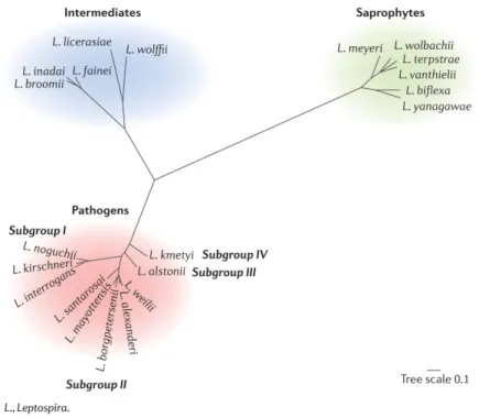
Leptospira are highly diversiied bacteria. A classiicaion based on the anigenic properies of the bacteria describes more than 250 pathogenic serovars grouped into 24 anigenically-related serogroups (Evangelista and Coburn, 2010). However, the rapid development of genotyping and genomics is acceleraing a switch to molecular classiicaions. The genus Leptospira currently comprises at least 21 species and is divided into three clusters: the pathogenic cluster includes at least ten Leptospira species, a second intermediate cluster is composed of species of unknown pathogenicity or causing mild illness while a third cluster includes free-living saprophyic species (Figure 1). The bacterium harbors a large genome size, ranging from 3.8 to 4.7 Mb, which is associated to its ability to survive in diverse environments (Bhari et al., 2003; Picardeau, 2017) and consising of two chromosomes in addiion to a variable number of smaller circular replicons.
Figure 1. Leptospira phylogeny based on the concatenaion of 491 selected core genes. (Picardeau, 2017)
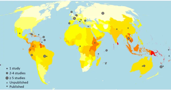
Although leptospirosis has a worldwide distribuion in both industrialized and developing countries (Bhari et al., 2003), the incidence of the disease is higher in tropical and subtropical areas with 73% of global reported cases occurring in tropical regions (Costa et al., 2015). As shown in Figure 2, the burden of the disease is paricularly high in the Caribbean islands, East sub-Saharan Africa, South-East Asia and Oceania (Costa et al., 2015; Evangelista and Coburn, 2010; Pappas et al., 2008; Yang, 2018).
Figure 2. Worldwide incidence distribuion of leptospirosis cases. 0-3 (white), 7-10 (yellow), 20-25 (orange), > 100 (red) cases per 100,000 people (Costa et al., 2015)
Noteworthily, disease incidence is maximal in tropical islands (Pappas et al., 2008) where the limited diversity of animal reservoirs typical of insular ecosystems may enhance Leptospira transmission (Derne et al., 2011).
This thesis will focus on southwestern Indian Ocean (SWIO) region, which records some of the highest incidence worldwide. In the last 20 years, a large body of serological and molecular data gathered on the diferent islands of the region has shown that these territories actually display disinct epidemiological paterns in terms of incidence and severity of the disease (Biscornet et al., 2017; Bourhy et al., 2010, 2012, 2014; Bovet et al., 1999; Desvars et al., 2011, 2012, 2013a; Gomard et al., 2014; Guernier et al., 2016; Lagadec et al., 2016; Yersin et al., 1998). In addiion, a wide diversity of pathogenic Leptospira lineages has been reported in both human acute cases and reservoir animals. These epidemiological paterns may result from the co-evoluion of Leptospira spp. and their animal hosts in a muli insular ecosystem considered as a leading biodiversity hotspot (Dietrich et al., 2018; Myers et al., 2000). Indeed, the unique biodiversity hosted by SWIO islands may
have led to the emergence of zoonoic pathogens that are unique to this region, including endemic Leptospira lineages and species, with consequences on human health that are key to this thesis. A large part of the following introducion synthesizes bibliographic together with original research data on Leptospira and leptospirosis in the SWIO. Leptospira diversity and evoluion are reviewed and their consequences on the contrasted epidemiology of human disease in the region are discussed.
II. The biogeography of Leptospira in SWIO is inimately linked to the evoluionary history of animal reservoirs.
1. SWIO islands are home to a unique biodiversity shaped by colonizaion and radiaion events.
The SWIO is considered one of the ive leading biodiversity hotspots in the world with Malagasy region hosing 3.2% of global endemic plants and 2.8% of global endemic vertebrates, all encompassed in 0.4% of Earth’s land surface (Myers et al., 2000). The region is composed of islands, including Madagascar, Mauriius, Mayote, La Réunion, the Seychelles, and the Union of the Comoros, with diferent surface areas, geographical isolaion and geological histories; these aspects have in turn contributed to the evoluion of the biodiversity sheltered by each island. True oceanic islands including the Comoros and Mascarene archipelagos, as well as coralline Seychelles, have emerged de novo from volcanic hot spots. They are disinctly younger than landmasses with a Gondwana origin, such as Madagascar and the graniic Seychelles. The composite structure of this muli-insular ecosystem is associated with disinct levels of species richness and endemism, as best exempliied by plant and animal species that have been invesigated in the region (Myers et al., 2000).
The diversity of microorganisms, including zoonoic pathogens, has been more recently explored. In animals, these invesigaions have shown that microbial diversity and distribuion result in part from the occurring reservoir species that include endemic and introduced taxa (Dietrich et al., 2014a, 2018, Wilkinson et al., 2014a, 2014b). Herein, we review the general drivers of species diversiicaion and structuraion with a special focus on Madagascar mammalian diversity. Indeed, the natural history of Malagasy mammals is complex but well established, and has directly inluenced the diversiicaion and distribuion of associated zoonoic microorganisms. Using leptospirosis as a model of zoonoic disease, we present epidemiological evidence highlighing the links exising between the extant mammalian diversity and associated pathogenic Leptospira, together with the consequences of these biogeographical paterns on human health at a regional level.
Origin of Madagascar’s unique biodiversity
The high bioic endemism on Madagascar results from geographical isolaion in deep geological ime associated with plate tectonics, and muliple colonizaion events that led to some of the more pronounced adapive radiaions in the world. Madagascar was part of the Gondwana superconinent unil it started to separate during the Mesozoic era. Indo-Madagascar, which included what is today the Indian subconinent, was isolated from Africa between 130 and 160 Ma. India separated 88 Ma from Indo-Madagascar and subsequently drited towards its current posiion (Bukontaite et al., 2015; Samonds et al., 2013; Wirta et al., 2008). While driting northwards, large porions splintered of the Indian landmass and today form the graniic islands of the Seychelles.
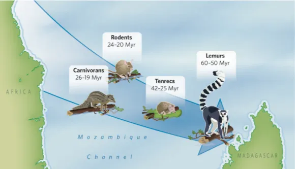
The vast majority of the extant fauna of Madagascar is not related to what is known in the Mesozoic fossil record, but to the Cenozoic period when the ancestors of extant animals colonized the island following its isolaion. Repiles and ishes were the predominant fauna on Madagascar before the mass exincion at the K-T boundary, ater which mammals and birds became more prevalent (Ganzhorn et al., 2013; Samonds et al., 2013). According to the most parsimonious hypothesis, the ancestors of all four extant groups of terrestrial mammals successfully colonized Madagascar from coninental Africa through four asynchronous arrival events taking place during the Cenozoic. Lemurs (Lemuridae) established irst (70-41 Ma), followed by tenrecs (Tenrecidae) (50-20 Ma), carnivores (Carnivorans) (33-14 Ma) and nesomyine rodents (Nesomyinae) (30-15 Ma) (Ali and Huber, 2010; Cox, 2000; Poux et al., 2005) (Figure 3).
Figure 3. Timing of the colonizaion events involving terrestrial mammals from Africa to Madagascar (Krause,
2010)
Two alternaive hypotheses have been proposed to explain these four successful ancestral colonizaion events. According to the land bridge hypothesis, Madagascar estrangement from Africa was followed by the emergence of small land areas located in the Mozambique Channel that may have represented rest areas and thus helped mammals to achieve and colonize Madagascar (McCall, 1997). However, such rest areas would have been located along the Davie Ridge, a 1200-km long North-South high rise located in the Mozambique Channel, and the diferent colonizaion events would have occurred in a limited area of Madagascar near to landmasses on the Davie Ridge, while mammalian colonizaion of Madagascar seems to have occurred at several randomly distributed sites (Ali and Huber, 2010). Moreover, it is recognized that these land areas would have been separated from each other from at least 230 km. Lastly, this stepping stone scenario should have led to a higher taxonomic diversity of Quaternary or extant mammals on Madagascar than currently recognized.
According to the second and alternaive sweepstakes hypothesis, mammals could have crossed the Mozambique Channel by rating on loaing vegetaion mats or large trees. The premise of this hypothesis was considered improbable because present oceanic current would not have allowed rating from Africa to Madagascar. In 2010, Ali and Huber demonstrated that oceanic currents in the Mozambique Channel were diferent from the present situaion. Indeed, during the Eocene, Africa, Madagascar and Australia were much further South, meaning that Madagascar was located within the convergence zone under the inluence of subtropical aniclockwise gyre, which resulted in oceanic currents passing from eastern Africa towards Madagascar. Hence, this would have allowed rats of vegetaion to drit across the Mozambique Channel from Africa to Madagascar in less than a month (Ali and Huber, 2010; Samonds et al., 2013). However, such colonizaion might have also been facilitated by special physiological traits of land vertebrates, such as torpor and/or the ability to lower metabolism that would have facilitated successful arrival on Madagascar. Madagascar and Africa slowly drited northwards unil the Early Miocene. At that ime, colonizaion of Madagascar via vegetaion mats became virtually impossible given the change in ocean currents from Madagascar to coninental Africa (Ali and Huber, 2010).
Unlike terrestrial mammals, bats, which ly, were only marginally inluenced by oceanic currents and could have colonized Madagascar from both African and Asian sources (Samonds et al., 2012, 2013). Recent invesigaions support that bats have arrived on Madagascar within the last 15 Ma (Samonds et al., 2013), and within the modern fauna there is evidence for at least 24 colonizaion events (Samonds et al., 2012), mostly of African origin.
Madagascar has sheltered intense biota radiaion.
Following these colonizaion events, Madagascar held considerable ecological diversity with numerous diferent biomes that facilitated an extraordinary series of adapive radiaions and gave rise to the modern taxa present on the island (Bukontaite et al., 2015; Eger and Mitchell, 1996). The mountainous landscapes of Madagascar provide diferent climates and three broad types of forest formaions: eastern humid, western dry-deciduous forests and spiny forests (Ganzhorn et al., 2013). The diversity of climates and vegetaion types, superimposed on broad elevaional and laitudinal clines, provides key aspects for animal adaptaion to each speciic environment and resuling in large-scale speciaion. For instance, tenrecs underwent a high level of adapive radiaion 29 Ma associated with changes in their morphology, physiology, behavior and ecology (Poux et al., 2008). The subsequent colonizaion of Madagascar by carnivores and rodents added more ecological pressure, requiring addiional adapive specializaion within Tenrecidae (Poux et al., 2008).
Madagascar possesses a notable diversity of bats, with around 46 species reported from the island, included in nine diferent families (Goodman and Ramasindrazana, 2018). A majority of bats species are endemic to Madagascar and/or neighboring islands, most of them originaing from Africa (Cardif and Jenkins, 2016). For instance, nine of the 11 Miniopterus spp. currently recognized on Madagascar are endemic to the island, while the two remaining species are also found in the Comoros archipelago. This genus underwent two main radiaion events circa 4-2 Ma and 1.25-0.75 Ma, probably related in part to climaic changes in mountainous areas of Madagascar (Chrisidis et al., 2014).
2. Mammals from SWIO islands host several zoonoic pathogens including pathogenic Leptospira that are endemic to this insular ecosystem.
Volant and terrestrial mammals are known reservoirs for a number of viral and bacterial zoonoic pathogens (Brook and Dobson, 2015; Calisher et al., 2006; Helmy et al., 2017; Heredia and García, 2018; Mostafavi et al., 2018; Pacheco et al., 2017). Recent studies carried out in Madagascar and surrounding islands have shown that the regional mammalian diversity is no excepion. Hantaviruses, responsible for hemorrhagic fever with renal syndrome and hantavirus cardiopulmonary syndrome, have been described in rodents on Mayote (Filippone et al., 2016). Astroviruses, responsible for gastroenteriis among children less than two years old, are found in several bat species from Mozambique and Madagascar (Hoarau et al., 2018; Lebarbenchon et al., 2017). A high diversity of paramyxoviruses responsible for respiratory tracts infecion are widespread on Indian Ocean islands and are hosted by bats, rodents, tenrecs as well as introduced terrestrial small mammals (Ghawar et al., 2017; Mélade et al., 2016; Wilkinson et al., 2012, 2014b). Among the documented bacterial zoonoses occurring on Indian Ocean islands, plague is both emblemaic and of
regional concern as Madagascar is considered as one of the leading foci of plague re-emergence. Further, ricketsioses and bartonelloses transmited through lea bites have been documented in Madagascar and La Réunion (Dieme et al., 2015; Kreppel et al., 2014), the former recently being associated with several human cases of murine typhus (Balleydier et al., 2015). Finally, leptospirosis, considered as the most prevalent bacterial zoonosis worldwide, is a major public health issue in the southwestern Indian Ocean (Pappas et al., 2008). Molecular data produced from mammals’ samples have recently highlighted the biogeography of Leptospira on some southwestern Indian Ocean islands, and certain islands have phylogeneically disinct Leptospira species/lineages, presumably associated with diferent land mammalian adapive radiaions in relaively deep geological ime (Dietrich et al., 2018). Altogether, data accumulated through mulidisciplinary research programs have shown that the biogeography of mammalian species actually shapes the diversity of occurring Leptospira (Dietrich et al., 2018; Gomard et al., 2016).
The natural history of pathogenic Leptospira on SWIO islands is ightly associated to the evoluionary history of mammalian reservoirs.
During the last decade, several studies have explored the diversity of pathogenic Leptospira in the wild endemic and introduced fauna of SWIO islands. These studies have revealed that small mammals endemic to Madagascar harbor a broad diversity of Leptospira lineages mostly unique to the island and grouping within 3 disinct major species, namely L. borgpetersenii, L. mayotensis and L. kirschneri. In addiion, these bacterial lineages express strong levels of speciicity towards their mammalian reservoir hosts, paricularly for bat-borne Leptospira. Indeed, Malagasy bats and their associated Leptospira are an interesing biological model to study the evoluion of host-pathogen interacions as the island’s bat fauna is composed of more than 40 disinct species, of which 80% are endemic. The invesigaion of Malagasy bats showed that each genus or family is reservoir of at least one and in certain cases a handful of Leptospira lineages (Dietrich et al., 2014b; Gomard et al., 2016; Lagadec et al., 2012).
At an evoluionary scale, the detected diversity of bat-borne Leptospira is best explained by (i) co-diversiicaion of Leptospira and bats and (ii) a few host-switches that may have been facilitated by the ecology and behavior of these animals, paricularly physical contact in day-roost sites between geneically disinct species such as Myois goudoi and Miniopterus spp. (Gomard et al., 2016). Such a scenario required further exploraion of bats from coninental Africa, considered as the source of colonizing lineages and including common ancestors with the majority of the extant Malagasy bat fauna (Cardif and Jenkins, 2016). For this, we recently invesigated pathogenic Leptospira sheltered by bats from Mozambique, revealing a wide diversity related to L. borgpetersenii, L. interrogans, L. kirschneri and L. noguchii, a bacterial species community also reported in Malagasy bats. Using the
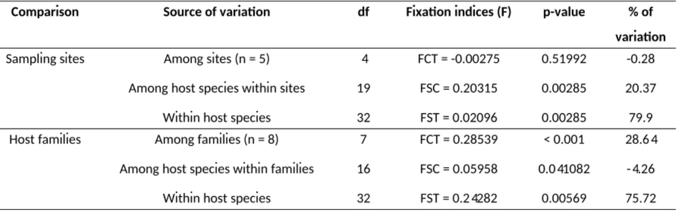
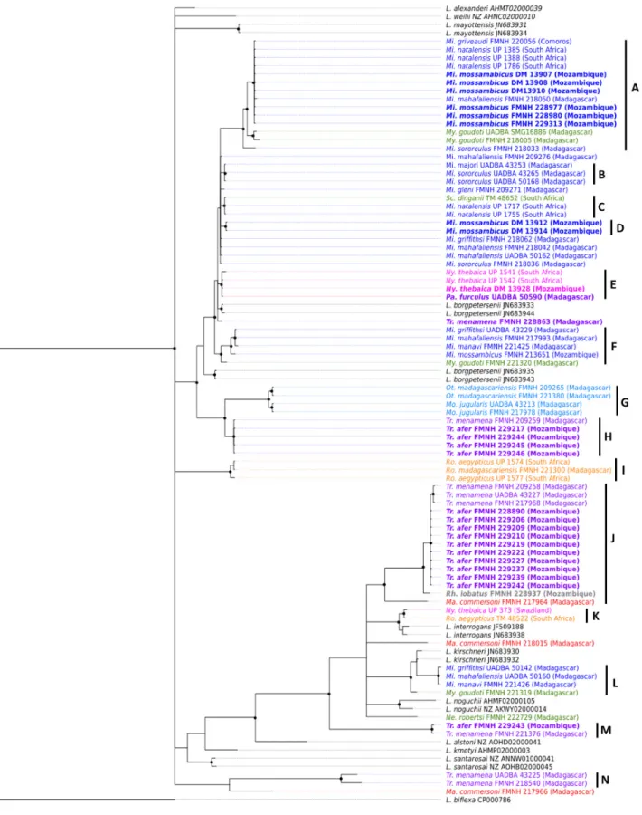
polymorphic secY gene marker, we constructed a phylogeny comprising samples from SWIO islands and coninental Africa. The phylogeneic tree depicted in Figure 4 shows that the topology is driven by mammalian host community rather than geography. Indeed, well-supported clades are composed of lineages shared by closely related bats species collected on Mozambique, Madagascar or Comoros. This patern is best exempliied by several monophyleic clades composed of Leptospira lineages shed by bat genera common to Mozambique and the Malagasy Region (Figure 4). For instance, clades A and F contain Leptospira genotypes found in the family Miniopteridae from Mozambique, Madagascar and/or Comoros, while clades H, I and J are composed of Leptospira lineages sheltered by Triaenops afer and Triaenops menamena, from Mozambique and Madagascar, respecively. There are some excepions to this patern, such as Scotophilus dinganii and Miniopterus natalensis carrying closely related Leptospira as well as Nycteris thebaica and Paratriaenops furculus shedding Leptospira with genotypes embedded in the same clade. However, the analysis of molecular variance (AMOVA) performed on molecular data of Leptospira in SWIO does not show any geographical structuraion of Leptospira lineages (p = 0.52) but rather suggests a structuraion according to their bat host families (p < 0.001) (Table 1). These results raise the hypothesis that certain lineages of ancestral bats colonizing Madagascar from eastern Africa were actually carrying Leptospira. Altogether, the natural history of Malagasy mammals and their associated microbial biomes suggest that the current diversity of pathogenic Leptospira on the island results from colonizaion of mammalian reservoirs from coninental Africa and subsequent co-diversiicaion processes. To our knowledge, this is the irst documented dataset supporing the existence of endemic zoonoic pathogens resuling from colonizaion and subsequent radiaion of their mammalian hosts.
Table 1. Analysis of molecular variance (AMOVA) based on Leptospira secY gene.
In each analysis, a populaion is deined as all Leptospira sequences detected in a single bat species at a single sampling site.
Comparison Source of variaion df Fixaion indices (F) p-value % of variaion
Sampling sites Among sites (n = 5) Among host species within sites
Within host species
4 19 32 FCT = -0.00275 FSC = 0.20315 FST = 0.02096 0.51992 0.00285 0.00285 -0.28 20.37 79.9 Host families Among families (n = 8)
Among host species within families Within host species
7 16 32 FCT = 0.28539 FSC = 0.05958 FST = 0.24282 < 0.001 0.041082 0.00569 28.64 -4.26 75.72 22
Figure 4. Phylogeneic tree based on a 473-bp-fragment of pathogenic Leptospira secY gene
Bayesian inference with HKY+I+G subsituion model was used for analysis. Nodes with a black dot correspond to posterior probabiliies ≥ 0.80. Leptospira haplotypes are colored according to host family (blue: Miniopteridae, green: Vesperilionidae, pink: Nycteridae, red: Hipposideridae, purple: Rhinonycteridae, light blue: Molossidae, orange: Pteropodidae). Mi: Miniopterus, My: Myois, Sc: Scotophilus, Ny: Nycteris, Pa:
Paratriaenops, Tr: Triaenops, Ot: Otomops, Mo: Mormopterus, Ro: Rousetus, Ma: Macronycteris.
Endemic and introduced Leptospira display disinct levels of host-speciicity.
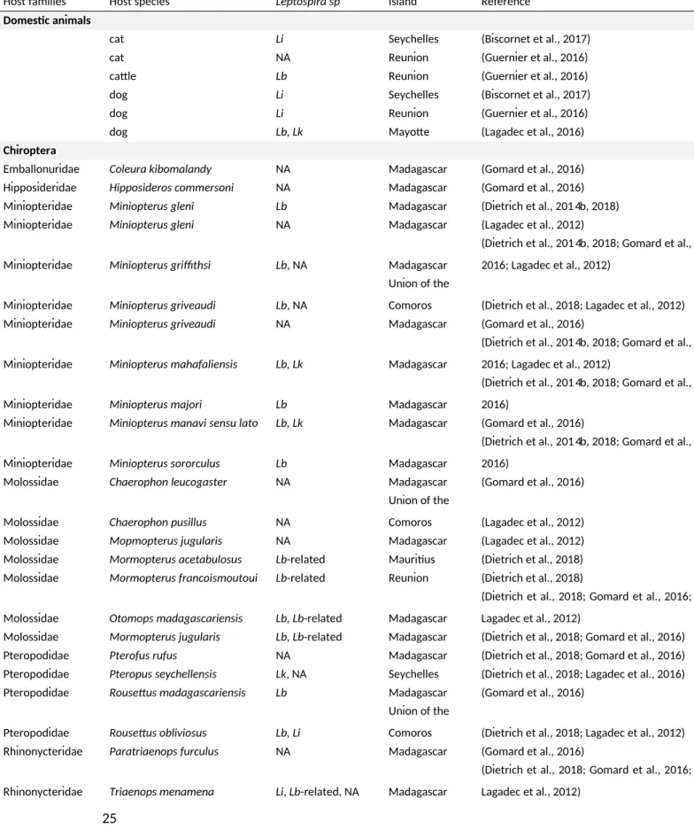
Although bat-borne Leptospira show ight host-speciicity, a detailed exploraion of terrestrial small mammals also brings forth interesing insights (Table 2). Indeed, the invesigaion of terrestrial mammals in diferent forests of Madagascar showed that tenrecs are the reservoirs of several disinct L. mayotensis lineages (Dietrich et al., 2015, 2018). Importantly, among the wild mammal fauna on Mayote (Lagadec et al., 2016), L. mayotensis was isolated only from Tenrec ecaudatus, a spiny tenrec introduced to the island and other neighboring islands from Madagascar and probably in the context of bush. Hence, co-diversiicaion appears as a major driver of Leptospira host-speciicity in volant and terrestrial mammals occurring on SWIO islands.
Synanthropic and wild introduced mammals have been invesigated on La Réunion and the Seychelles archipelago as well, showing that Ratus ratus and Ratus norvegicus harbor a limited diversity of Leptospira, mostly restricted to L. interrogans (Rahelinirina et al., 2010). Interesingly, the most prevalent Sequence Type (ST) found in rats from both islands has been reported in several other parts of the world (according to scheme 3 of MuliLocus Sequence Typing (MLST), see htp://www.pubmlst.org/leptospira/). The limited geneic diversity of Ratus-borne Leptospira and the cosmopolitan distribuion of the most prevalent ST are coherent with its recent introducion (Tortosa et al., 2017). Recently, a study invesigated the presence of Leptospira by sampling both introduced and endemic small mammals at the same forest sites on Madagascar. Leptospira co-infecions in introduced and endemic small rodents were highlighted following the implementaion of sophisicated detecion schemes focusing on possible co-infecions (Moseley et al., 2018). Although only L. interrogans had thus far been detected in introduced Ratus on Madagascar (Rahelinirina et al., 2010), these newly described co-infecions in introduced and endemic rodents also involved L. borgpetersenii and L. mayotensis (Moseley et al., 2018). It was suggested that introduced rodents may indeed support muliple infecions composed of introduced L. interrogans lineages and endemic Leptospira such as L. mayotensis shed by tenrecs. Rats may have acquired L. mayotensis infecion through direct or indirect contact with tenrecs (Moseley et al., 2018). Symmetrically, the presence of L. interrogans in co-infecions in endemic small nesomyine rodents from these same areas were reported although much more rarely (Moseley et al., 2018). A relaxed speciicity was also reported in R. Ratus from Mayote with L. interrogans and L. borgpetersenii being the most prevalent Leptospira spp. and L. mayotensis and L. kirschneri being present at a less extent (Desvars et al., 2012). These studies place Ratus spp. as potenial reservoirs of several Leptospira spp. but more detailed descripion of Leptospira strains and their hosts is necessary to assess host speciicity of these bacterial strains. Altogether, gathered data support that the geneic diversity of pathogenic Leptospira on SWIO islands is composed of endemic and introduced
Leptospira displaying disinct levels of host-speciicity ranging from strict speciicity for the former to more relaxed associaions for the later.
Table 2. Potenial hosts of Leptospira spp. in SWIO islands recorded during the last decade.
Host families Host species Leptospira sp Island Reference
Domesic animals
cat Li Seychelles (Biscornet et al., 2017) cat NA Reunion (Guernier et al., 2016) catle Lb Reunion (Guernier et al., 2016) dog Li Seychelles (Biscornet et al., 2017) dog Li Reunion (Guernier et al., 2016)
dog Lb, Lk Mayote (Lagadec et al., 2016)
Chiroptera
Emballonuridae Coleura kibomalandy NA Madagascar (Gomard et al., 2016) Hipposideridae Hipposideros commersoni NA Madagascar (Gomard et al., 2016) Miniopteridae Miniopterus gleni Lb Madagascar (Dietrich et al., 2014b, 2018) Miniopteridae Miniopterus gleni NA Madagascar (Lagadec et al., 2012)
Miniopteridae Miniopterus griithsi Lb, NA Madagascar
(Dietrich et al., 2014b, 2018; Gomard et al., 2016; Lagadec et al., 2012)
Miniopteridae Miniopterus griveaudi Lb, NA
Union of the
Comoros (Dietrich et al., 2018; Lagadec et al., 2012) Miniopteridae Miniopterus griveaudi NA Madagascar (Gomard et al., 2016)
Miniopteridae Miniopterus mahafaliensis Lb, Lk Madagascar
(Dietrich et al., 2014b, 2018; Gomard et al., 2016; Lagadec et al., 2012)
Miniopteridae Miniopterus majori Lb Madagascar
(Dietrich et al., 2014b, 2018; Gomard et al., 2016)
Miniopteridae Miniopterus manavi sensu lato Lb, Lk Madagascar (Gomard et al., 2016)
Miniopteridae Miniopterus sororculus Lb Madagascar
(Dietrich et al., 2014b, 2018; Gomard et al., 2016)
Molossidae Chaerophon leucogaster NA Madagascar (Gomard et al., 2016)
Molossidae Chaerophon pusillus NA
Union of the
Comoros (Lagadec et al., 2012) Molossidae Mopmopterus jugularis NA Madagascar (Lagadec et al., 2012) Molossidae Mormopterus acetabulosus Lb-related Mauriius (Dietrich et al., 2018) Molossidae Mormopterus francoismoutoui Lb-related Reunion (Dietrich et al., 2018)
Molossidae Otomops madagascariensis Lb, Lb-related Madagascar
(Dietrich et al., 2018; Gomard et al., 2016; Lagadec et al., 2012)
Molossidae Mormopterus jugularis Lb, Lb-related Madagascar (Dietrich et al., 2018; Gomard et al., 2016) Pteropodidae Pterofus rufus NA Madagascar (Dietrich et al., 2018; Gomard et al., 2016) Pteropodidae Pteropus seychellensis Lk, NA Seychelles (Dietrich et al., 2018; Lagadec et al., 2016) Pteropodidae Rousetus madagascariensis Lb Madagascar (Gomard et al., 2016)
Pteropodidae Rousetus obliviosus Lb, Li
Union of the
Comoros (Dietrich et al., 2018; Lagadec et al., 2012) Rhinonycteridae Paratriaenops furculus NA Madagascar (Gomard et al., 2016)
Rhinonycteridae Triaenops menamena Li, Lb-related, NA Madagascar
(Dietrich et al., 2018; Gomard et al., 2016; Lagadec et al., 2012)
Vesperilionidae Myois goudoi Lb, Lk, NA Madagascar
(Dietrich et al., 2014b, 2018; Gomard et al., 2016; Lagadec et al., 2012)
Vesperilionidae Neoromicia robertsi NA Madagascar (Gomard et al., 2016) Vesperilionidae Scotophilus marovaza NA Madagascar (Gomard et al., 2016)
Rodenia
Nesomyidae Eliurus minor Lb, Li + Lm or Lb * Madagascar
(Dietrich et al., 2014b, 2018; Moseley et al., 2018)
Nesomyidae Nesomys rufus Li + Lm or Lb * Madagascar (Moseley et al., 2018)
Muridae Mus musculus NA Madagascar
(Moseley et al., 2018; Rahelinirina et al., 2010)
Muridae Mus musculus Lb, Lk Reunion (Guernier et al., 2016)
Muridae Ratus spp. Lb, Li, Lk, Lm Mayote
(Desvars et al., 2012; Dietrich et al., 2018; Lagadec et al., 2016)
Muridae Ratus spp. Li Seychelles (Biscornet et al., 2017; Dietrich et al., 2018) Muridae Ratus spp. Li Reunion (Dietrich et al., 2018; Guernier et al., 2016)
Muridae Ratus spp. Lb, Li, Lm Madagascar
(Dietrich et al., 2018; Moseley et al., 2018*; Rahelinirina et al., 2010)
Soricomorpha
Soricidae Suncus murinus Li Reunion (Guernier et al., 2016)
Soricidae Suncus murinus NA Madagascar
(Moseley et al., 2018; Rahelinirina et al., 2010)
Afrosoricidae
Tenrecidae Hemicentetes nigriceps Lk Madagascar (Dietrich et al., 2014b, 2018)
Tenrecidae Hemicentetes semispinosus Lk, NA Madagascar
(Dietrich et al., 2014b, 2018; Moseley et al., 2018)
Tenrecidae Microgale cowani Lb, Lm Madagascar (Dietrich et al., 2014b, 2018) Tenrecidae Microgale dobsoni Lm Madagascar (Dietrich et al., 2014b, 2018) Tenrecidae Microgale longicaudata Lb Madagascar (Dietrich et al., 2014b, 2018) Tenrecidae Microgale majori Lb Madagascar (Dietrich et al., 2014b, 2018) Tenrecidae Microgale principula Lb Madagascar (Dietrich et al., 2014b, 2018) Tenrecidae Microgale spp NA Madagascar (Moseley et al., 2018)
Tenrecidae Tenrec ecaudatus Lm Mayote (Dietrich et al., 2018; Lagadec et al., 2016) NA = not available; Lb = Leptospira borgpetersenii; Li = Leptospira interrogans; Lk = Leptospira kirschneri; Lm =
Leptospira mayotensis
* = co-infecion
3. The contrasted epidemiology of human leptospirosis in SWIO islands is associated with disinct transmission chains.
Leptospirosis is of considerable medical concern in most if not all of the western Indian Ocean islands. For instance, the Seychelles Archipelago has been considered the country with the highest human incidence worldwide (Pappas et al., 2008). Similarly, human incidence is 10 to 50 imes higher on La Réunion and Mayote than in coninental France (Bourhy et al.; Centre naional de référence de la leptospirose, 2015). This is consistent with an incidence of leptospirosis overall higher in tropical areas, which humid climates are known drivers of bacterial persistence in the environment
(Costa et al., 2015). Interesingly, human incidence is even higher on small tropical islands possibly because limited species richness typical of insular ecosystems may facilitate transmission between wild mammalian reservoirs (Derne et al., 2011). As far as SWIO insular ecosystem is concerned, serological data obtained on diferent islands indicate diferent epidemiological paterns (Bourhy et al., 2012; Bovet et al., 1999; Desvars et al., 2011, 2013b; Yersin et al., 1998) . More recent invesigaions using molecular data and carried out through a One Health framework have illuminated the epidemiology of leptospirosis on several of these islands and highlighted the occurrence of sharply diferent transmission chains.
Madagascar has been previously considered as having very low levels of leptospirosis. In 2015, a human case of leptospirosis was reported from a traveler on La Réunion returning from Madagascar, probably infected with L. interrogans (Pagès et al., 2015a). The same year, a human seroprevalence of 2.9% was reported in the Moramanga District with Icterohaemorrhagiae ideniied as the most prevalent serogroup (Ratsitorahina et al., 2015). A 2018 study reported no case of leptospirosis from febrile paients based on PCR analysis of blood samples (Hagen et al., 2018). However, urine samples were not collected and these cases may have been misdiagnosed. This highlights the necessity of using correct protocols elaboraion to beter assess and understand the actual burden of human leptospirosis on Madagascar, which is sill largely unknown.
Human leptospirosis is also poorly described in the Union of the Comoros besides a few cases reported among travelers returning home (Subiros et al., 2017). In 2011, a serological invesigaion carried out using sera from human febrile cases highlighted the presence of leptospirosis on all three islands of the Union of the Comoros with seroprevalence extending from 3.4% on Grande Comore to 10.3% on Mohéli (Gomard et al., 2014). Again, the actual burden of the disease in this country remains an open quesion.
The epidemiology of leptospirosis on Mayote and La Réunion has been the subject of considerable studies. Further, the surveillance of leptospirosis is under the control of the same health system on both islands, Health Regional Agency (Agence Régionale de Santé, ARS). Hence, surveillance biases are expected to be limited, conferring to the epidemiological data produced on these French islands a good analysis power. Between 2010 and 2015, the annual mean incidence of human leptospirosis on Mayote was 100 cases per 100,000 habitants. Three per cent of the acute cases were admited to intensive care units (ICU) and 0.9% of total cases were lethal (Pagès et al., 2017a). Between 2015 and 2017, the incidence of leptospirosis on Mayote increased from 38.8 to 66.3 cases per 100,000 habitants. In 2016, 22% of human cases were hospitalized, 4.6% of the cases were admited to ICU and a mortality rate of 0.6% was reported (Subiros et al., 2017). In 2017, 19%
of leptospirosis human cases were hospitalized; among them, one third was admited to ICU and one paient died from the disease (mortality rate of 0.6%) (Cire océan Indien, 2018b). The serogroup Mini was the most prevalent one in human cases of leptospirosis, while Icterohaemorrhagiae has not been detected since 2008. This later serogroup was ideniied from human cases when more severe forms of leptospirosis were observed in paients and with a mortality rate reaching 23% (Subiros et al., 2017). Since 2008, Icterohaemorrhagiae serogroup has not been ideniied and the lethality rates dropped drasically to 0.6% in 2017 (Cire océan Indien, 2018b; Subiros et al., 2017). Molecular indings indicate that four species are involved in human leptospirosis on Mayote: L. borgpetersenii, L. interrogans, L. kirschneri and L. mayotensis. The three former species can be found in introduced mammals, such as rats and dogs. The fourth form of Leptospira on the island is restricted to T. ecaudatus, which is endemic to Madagascar and introduced to Mayote, and has been ideniied as the exclusive reservoir of L. mayotensis (Lagadec et al., 2016).
Between 2004 and 2015, a mean incidence of six cases of leptospirosis per 100,000 habitants was recorded on La Réunion, with hospitalizaion being signiicantly more frequent than on Mayote. Indeed, within the same period, 93% of total cases were hospitalized and among them, 33% were admited to ICU, while 3% of acute cases were lethal (Pagès et al., 2017b). Between 2014 and 2017, the annual incidence of leptospirosis varied between 4.7 and 7.2 cases per 100,000 habitants. The hospitalizaion rate was high (over 75%) and mortality rates averaged 5% except for the year 2017 when no person was reported to succumb to the disease. On La Réunion, Icterohaemorrhagiae is the most prevalent serovar and L. interrogans is largely dominant among human cases (Guernier et al., 2016; Pagès et al., 2017b). Both STs of L. interrogans detected in human cases on La Réunion (ST02 and ST34, MLST scheme 3) have a broad worldwide distribuion. Introduced Ratus appear as a major reservoir of Leptospira on the island, although only one ST of L. interrogans (ST02), idenical to one ST found in humans, is described in these animals. The absence of the second L. interrogans ST (ST34) in rats indicates that they are not the only source of L. interrogans infecing humans. Indeed, stray dogs may play a role in the transmission of the second haplotype of L. interrogans. Mus musculus and catle may also act as disease reservoirs as they shelter L. borgpetersenii with STs idenical to those characterized in a few human cases (Guernier et al., 2016).
Lastly, populaion-based studies carried out in the Seychelles since 1989 report high annual incidence rate of human leptospirosis, reaching 101 cases per 100,000 habitants between 1995 and 1996 (Yersin et al., 1998). In 2008, Seychelles was considered as having the highest incidence rate of human leptospirosis worldwide (Pappas et al., 2008). A longitudinal 12 months survey in 2014-2015 showed that this incidence decreased but remained high with a lethality rate of 11.8% (Biscornet et al., 2017). A large proporion of acute cases include severe manifestaions of the disease with
paients harboring one or several symptoms including jaundice, renal failure or pulmonary hemorrhage, the last one being the most important cause of death (Yersin et al., 1998). As on La Réunion, human cases of leptospirosis in the Seychelles are caused by a narrow diversity of Leptospira lineages. Most human cases involve the serogroup Icterohaemorrhagiae and only L. interrogans is detected among human acute cases and Ratus. Genotyping ideniied three STs in humans and animals, one corresponding to the most common ST found in rats and human cases from La Réunion (ST02) (Biscornet et al., 2017; Guernier et al., 2016). Importantly, in the Seychelles, Ratus does not appear as the main reservoir of human leptospirosis: ST02 is the only haplotype reported among rats and is involved in only one third of acute cases. Stray dogs may be involved in disease transmission to humans as one tested dog was found shedding the ST involved in the majority of human cases. However, complementary invesigaions are required to address the importance of dogs in the epidemiology of human leptospirosis (Biscornet et al., 2017).
The contrasted epidemiology of leptospirosis on SWIO islands indicates diferent transmission chains. The presence of diferent strains responsible for the disease depends on the local fauna (Table 2) and its biogeographic history (Dietrich et al., 2018). For instance, cosmopolitan and presumably introduced Leptospira (mainly L. interrogans) are responsible for most, if not all, human cases in the Seychelles and La Réunion, while original Leptospira lineages including endemic L. mayotensis are involved in most human cases on Mayote (Lagadec et al., 2016).
III. Experimental animal models used in Leptospira research
The invesigaion of leptospirosis through a One Health framework allowed highlighing the epidemiology of leptospirosis and the diferent transmission chains involved in human infecion. The use of experimental models helps understanding the mechanisms of infecion underlying Leptospira epidemiology as currently recognized in SWIO islands. Indeed, experimental approach can focus the invesigaions either on the acute phase or the chronic phase of the infecion, hence allowing disentangling the mechanisms at play in humans during acute infecion or in animal reservoirs during chronic infecion.
1. Acute models of infecion
Guinea pigs and hamsters mimic the symptoms of human leptospirosis and are used to study the acute phase of Leptospira infecion. Several Leptospira strains isolated around the world have been characterized in hamsters to describe the consequences of these infecions. These studies revealed several organs of interest as targets of Leptospira infecion. The mostly described damages include hemorrhage, congesion and iniltraion of inlammatory cells in the kidneys, lungs and liver
of infected animals (da Silva et al., 1995; Diniz et al., 2011; Matsui et al., 2015). Histopathological studies report the presence of urinary casts, nephriis, and glomerular and tubular damage in the kidneys of infected hamsters (Da Silva et al., 2010; Diniz et al., 2011; Forster et al., 2013; Matsui et al., 2015; Silva et al., 2008; Tomizawa et al., 2017; Villanueva et al., 2014). Petechial hemorrhage and alveolar collapse are oten observed in the lungs of hamsters infected with virulent strains of Leptospira (Da Silva et al., 2010; Silva et al., 2008; Tomizawa et al., 2017; Villanueva et al., 2014). Lastly, in the liver of infected animals, hepatocytes can be swollen and undergo degeneraion and necrosis while the loss of cohesion between hepatocytes can lead to jaundice (Alves et al., 1992; Diniz et al., 2011; Forster et al., 2013; Miyahara et al., 2014; Villanueva et al., 2014).
Rodent models of acute leptospirosis have also been used to describe the immune and inlammatory mechanisms underlying Leptospira infecion. Several studies highlighted that an overwhelming inlammatory response in hamsters following infecion with virulent strains of Leptospira could contribute to the severity of the disease, leading to renal lesions and a fatal outcome in most cases (Nakornpakdee et al., 2018; Nally et al., 2004; Vernel-Pauillac and Goarant, 2010; Vernel-Pauillac and Merien, 2006).
Experimental models of acute leptospirosis also allowed the ideniicaion of markers for early diagnosis of leptospirosis (Segawa et al., 2014; Vanaja et al., 2001) and have a crucial role in the development of efecive vaccines against the disease (Couinho et al., 2011; Murray et al., 2018; Oliveira et al., 2019). Models of acute infecion have also proven the eiciency of bioinformaics tools to beter understand leptospiral anigens structure and further improve the eiciency of vaccines (Garba et al., 2018; Hsieh et al., 2017; Murray et al., 2013).
2. Chronic models of infecion
Rats are renowned reservoirs for leptospirosis worldwide and thus provide a relevant model to study the mechanisms of renal colonizaion. Rats are not resistant to leptospirosis at their youngest age. As hamsters and guinea pigs, rat pups are suscepible to Leptospira infecion in the irst two weeks of their lives and undergo weight loss, jaundice and several issue damage, including pulmonary and cerebellum hemorrhage and hepatocytes disarrangement (Muslich et al., 2015). In these cases, leptospires can be detected in several organs, including the kidneys, liver, lung and cerebellum. Resistance to leptospirosis is expressed by rats aged of 23 days or more in which case leptospires are most oten detected in the kidneys only. Experimental Leptospira infecion in R. norvegicus mostly results in asymptomaic chronic renal infecion (Athanazio et al., 2008; Chagas-Junior et al., 2012; Nally et al., 2005, 2015; Tucunduva de Faria et al., 2007; Zilber et al., 2016). Histological studies on rats reveal no issue damage (Athanazio et al., 2008; Nally et al., 2005) except in cases where nephriis and iniltraion of lymphocytes and macrophages can be observed in the
kidneys (Nally et al., 2015; Tucunduva de Faria et al., 2007). Ratus norvegicus does not exhibit idenical ainiies to all Leptospira serovars. In 1981, Thierman et al have shown that the renal carriage of the serovar Icterohaemorrhagiae lasted longer than that of serovar Grippotyphosa. Thus, R. norvegicus appears as a relevant model to study and understand the host speciicity highlighted of leptospires by invesigaion of wild reservoirs in the ield.
IV. Main conclusions and research hypotheses.
Molecular and serological data gathered from SWIO islands in the last 20 years indicate that the local diversity of Leptospira lineages is related to the diversity of local mammalian reservoirs. Besides the heterogeneity of Leptospira lineages/species occurring on each island, it appears that Leptospira lineages unique to the region show a strong ainity for their endemic hosts in contrast with cosmopolitan lineages, which can be sheltered by a variety of mostly introduced mammalian hosts, including rodents, dogs and catle. The uniqueness of some Leptospira lineages/species shed by endemic mammals, such as tenrecs, likely results from long-term co-evoluion. These unique Leptospira can be considered as endemic to Madagascar and neighboring islands ; this is in contrast with the Leptospira lineages prevailing on La Réunion or the Seychelles, which have limited diversity and cosmopolitan distribuion likely associated with a recent introducion.
The paterns of Leptospira diversity on each island are in turn associated with disinct levels of severity in human cases. Indeed, cosmopolitan lineages such as L. interrogans ST02 commonly found in human cases on La Réunion and the Seychelles are associated with severe forms of leptospirosis. As far as endemic Leptospira are concerned, L. mayotensis is responsible for roughly 16% of human cases on Mayote (suppl. mat. Bourhy et al., 2012), where severe forms of human leptospirosis are less frequent than on La Réunion and the Seychelles (Biscornet et al., 2017; Cire océan Indien, 2018a, 2018b). To date, no bat-borne Leptospira has been detected in acute human cases from Madagascar, La Réunion, Mayote or the Seychelles, although high infecion prevalence have been reported in these volant mammals (Biscornet et al., 2017; Dietrich et al., 2015; Gomard et al., 2016; Guernier et al., 2016; Lagadec et al., 2016).
Altogether, the data summarized in this introductory chapter raise quesions about (i) a higher virulence of cosmopolitan Leptospira lineages as compared to lineages that are unique to the western Indian Ocean islands and (ii) a strong host speciicity of endemic Leptospira possibly resuling from co-diversiicaion of Leptospira and their hosts at evoluionary ime scales.
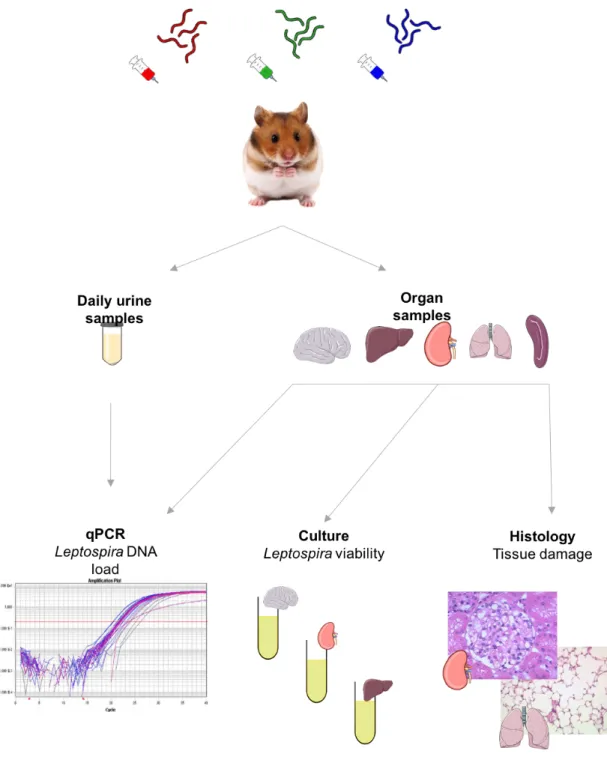
The virulence of cosmopolitan and endemic strains of Leptospira can be assessed through experimental infecion using diferent models to mimic and ascertain the impact of each lineage on human health. In the context of this thesis, we assessed the virulence of three strains of Leptospira
using a hamster model and strains isolated on regional islands: one cosmopolitan strain of L. interrogans isolated from R. Ratus on La Réunion, which genotype (ST02) is involved in the majority of human cases of leptospirosis on the island; one L. mayotensis isolate obtained from a tenrec and which genotype was previously found in human cases on Mayote (Bourhy et al., 2014; Lagadec et al., 2016); and one L. borgpetersenii endemic isolate obtained from a Malagasy bat and which genotype has not been reported in any human case thus far.
The ight host speciicity observed for endemic Leptospira lineages/species towards their mammalian reservoir can be challenged experimentally in order to determine whether this patern results from ecological or geneic determinants. Since rats are considered as a major reservoir of leptospirosis, they were used as animal models to invesigate the host speciicity of Leptospira and were challenged with all three Leptospira isolates. If experimentally infected rats fail to chronically maintain and shed L. mayotensis and/or L. borgpetersenii, the host speciicity observed in the ield likely involves geneic factors responsible for host-pathogen interacion. Symmetrically, if experimentally infected rats are found able to chronically excrete L. mayotensis and L. borgpetersenii, hence the host speciicity may actually result from ecological factors including the lack of physical contact between endemic and introduced reservoirs.
Finally, the exploraion and comparison of genomic features of Leptospira strains from SWIO islands and strains found elsewhere worldwide may shed light on the phenotypic contrasts observed among human acute cases. Altogether, this thesis aims at enlightening the contrasted epidemiology of leptospirosis observed in this region through experimental approaches that are complementary to the One Health approach implemented on several islands in the last decade.
The results produced in the frame of this thesis are presented according to three chapters that include the invesigaion of (i) the pathogenicity of three Leptospira strains isolated in SWIO islands, (ii) the speciicity of these strains towards their mammalian reservoirs and (iii) genomic components possibly associated to these features.