Biomechanical Structure-Function Relationships of Collagen
Tissues, B Cell Membranes, and Amyloid Fibers
by Carlos E. Castro
Submitted to the Department of Mechanical Engineering in Partial Fulfillment of the Requirements for the Degree of
ARCHIVES Doctor of Philosophy in Mechanical Engineering MASSACHUSEMS INSTrrUTE
at the OF TECHNOLOGY
Massachusetts Institute of Technology
February 2010 M
1
LIBRARIES
@ 2010 Masssachusetts Institute of Technology All rights reserved
Signature of Author... ...
Department of Mechanical Engineering
Certified by ... . ...
Matthe ang
Associate Professor of Biological and Mechanical Engineering Thesis Supervisor
Certified by ...
7I~yry
C. BoyceGail E. Kendall Professor of Mechanical Engineering Thesis Supervisor
Accepted by...
David E. Hardt Ralph E. and Eloise F. Cross Professor of Mechanical Engineering Graduate Program Committee Chair
This Doctoral Thesis has been examined by the following Thesis Committee:
Mary C. B'oyce, Ph.D. /
Thesis Supervisor and Committee Co-Chair
Gail E. Kendall Professor of Mechanical Engineering Massachusetts Institute of Technology
Roge D. Kamm, Ph.D.
Germeshausen Professor of Mechanical and Biological Engineering Massachusetts Institute of Technology
Matthew J. Lang.- ib.
Thesis Supervisor and Committee Co-Chair
Keck Assistant Professor of Biological and Mechanical Engineering Massachusetts Institute of Technology
Hidde Ploegh, Ph.D. Professor of Biology
Member, Whitehead Institute for Biomedical Research Massachusetts Institute of Technology
Biomechanical Structure-Function Relationships of Collagen
Tissues, B Cell Membranes, and Amyloid Fibers
by Carlos E. Castro
Submitted to the Department of Mechanical Engineering on October 19, 2009 in Partial Fulfillment of the
Requirements for the Degree of
Doctor of Philosophy in Mechanical Engineering ABSTRACT
Mechanical forces are critical to defining the physiological function of biological systems spanning length scales from 1 nm (single molecules) up to 1m (full mammalian systems). This work combines theoretical and experimental mechanics to gain insights into the physiological function of three biological systems at distinct length scales:
collagen tissues comprised of wavy fibers (- 1 mm), B lymphocyte membranes (- 1 gm),
and amyloid protein fibers (- 1 nm).
The initial portion of this thesis addresses the mechanics of fibrous collagen tissues such as ligaments, tendons, and pericardium that serve as load bearing components in biological systems. A novel micromechanical model describing the force-extension of wavy fibers comprising these tissues is integrated with bundle and network frameworks. The developed models accurately predict the mechanical behavior of bundled fiber tissues (i.e ligaments and tendon) and fibrous membranes (i.e. vessel walls and pericardium) and elucidate deformation mechanisms within these tissues.
Moving down in length scale, the second part of this thesis employs single cell experiments with optical tweezers to characterize the mechanical behavior of the B cell membrane, which is a critical component of its physiological functions including migration and antigen detection. Our results show that the mechanical properties of the membrane, specifically the effective viscosity of the membrane, evolve upon activation of B cell biochemical machinery. We further identify the presence of receptors in membrane nanotubes conferring B cells with the ability to sense antigen at remote locations.
Lastly, this thesis studies the aggregation and underlying structure of amyloid forming proteins by characterizing their physical properties at the fibril and single molecule level. Amyloid formation, which is associated with many diseases including Alzheimer's, results from the aggregation of misfolded proteins. This work combines optical trapping with fluorescence imaging to quantify the physical properties and molecular interactions of amyloid fibers formed from polymorphic variants of the yeast prion protein, Sup35. Our results show that Sup35 polymorphism leads to distinct physical properties of
amyloid aggregates. We further subject fibers to unfolding and rupture to elucidate structural details of misfolded Sup35.
Thesis Co-supervisor: Matthew J. Lang
Title: Associate Professor of Mechanical and Biological Engineering Thesis Co-supervisor: Mary C. Boyce
Acknowledgements
When I decided to pursue my PhD at MIT, I didn't know what I was getting into. Five years later I can look back at my experience with a great appreciation for how much I have grown both as a scientist and as a person. I was very blessed to be surrounded by caring people who made the last four years some of the best years of my life. I can't convey how grateful I am to them, but I would like to mention how they impacted my life. First I would like to thank DuPont, NIH, and SMART for funding my education and research throughout my PhD. The NIH Biomechanics Training Grant that funded two years of my PhD greatly contributed to my education in terms of linking me to other graduate students working in biomechanics to expand my knowledge of the field.
The impact of my thesis work was greatly enhanced due to the vision and forward thinking of my thesis committee. It was very valuable to have Roger Kamm, a leader in the field of biomechanics, supporting my work. His broad knowledge of the field was an
important asset to make my work as novel as possible. I also appreciate Roger taking extra time to help me with my future career by giving helpful advice and writing several reference letters. Hidde Ploegh was an invaluable addition to my committee. I was very lucky to take his Immunology class in the semester when I was writing my thesis proposal. Our B cell collaboration stemmed from a conversation I had with Hidde after class one day. I am grateful to Hidde's foresight, which enabled our collaboration to
make an important impact to the field of Immunology.
I could not have asked for more from my advisors, Mary Boyce and Matt Lang, both from an academic and a personal standpoint. The prospect of working with Mary, one of the foremost researchers in solid mechanics, was a big factor in my decision to attend MIT. I learned as much about mechanics through discussions in her office as I did from classes. Her guidance helped me develop into an independent thinker and effective researcher. Matt has been a great role model for me over the last four years. After four years of Matt's training I have learned how to build, troubleshoot, and optimize cutting edge biophysics instrumentation. Whether it was aligning a laser, setting up an experimental assay, or troubleshooting optics, Matt was always willing to spend time in the lab personally teaching me how to be an experimentalist. Both Matt and Mary also invested countless time into preparing me for a career in academia and gave me invaluable guidance during my job search process. Thanks to them, I feel well prepared to perform novel research in a biomechanics lab of my own.
Several other professors made important contributions to my life over my years at MIT. I was lucky to collaborate with Sue Lindquist on the amyloid project. After experiencing Sue's scientific guidance, it is easy to see how she has become a leader in the field of prion biology and protein conformation. Thanks to Gareth McKinley for convincing me to join the Program in Polymer Science and Technology. Gareth was a great teacher and mentor outside the classroom especially in my first year at MIT. I am grateful to Mark Bathe, Krystyn Van Vliet, and Jennifer McManus for spending many hours in advising
me on my career path and becoming good friends in the process. I also thank Alan Grodzinsky, Lalit Anand, John Hutchinson, and Bob Cohen for being positive influences on my academic career. I owe a big thanks to the MIT staff, Una Sheehan, Leslie Regan, Joan Kravit, Olga Parkin, Kristine Marzilli, Chris Connaire, Greg Sands, Tony Pulsone, and Annmarie Donovan, who were so helpful throughout my years here.
I am thankful to my collaborators who all also became good friends. Dr. Andrew Hu, a post-doc in the Ploegh Lab, was a huge help on the B cell project. He was a great mentor in the field of biology, and I always enjoyed our coffee breaks. I worked closely with Dr. Jijun Dong, a post-doc in the Lindquist lab, on the amyloid project. She taught me almost everything I know about prion biology. Countless hours at the trap were made much more enjoyable by having a great person like Jijun to get to know. I am also thankful to Dr. Alec Robertson and Dr. Sun Taek Kim who I was lucky to collaborate with on other projects and to Dave Quinn for his work on building the one of the trapping instruments. Thanks to Rajdeep, Nuo, Melis, Sai, Katia, Ethan, Meredith, Renaud, Tim, Juha, Lifeng, Chia-Ling, Damien, Brian, Ting-Ting, and Casey from the Boyce Lab and Mo, Yongdae, Hyungsuk, Marie Eve, Ricardo, Ted, Bill, Miriam, Mariya, Sangjin, Ben, Becky, Adrien, and Sergio from the Lang Lab who where a joy to work with. I owe a special thanks to Sharon Soong, Adam Mulliken, Mohit Garg, and Jeff Palmer from the Boyce lab and Jorge Ferrer, Ricardo Brau, Peter Tarsa, and David Appleyard from the Lang Lab. As senior members of the lab when I joined, they made me feel welcome and spent extra time helping me learn the ropes.
I would like to thank the lifelong friends I have made outside of lab who made an impact on my life as I hope I did on theirs. I am grateful to Rory, Frank, Barbara, Grinia, Sharon, Sungyon, Amit, Anjuli, Angela, Tony, Donny, Borjan, Andrej, and Dave who I shared so many good times with. I am especially grateful to Cathal, Conor, Al, and Bob, who were my roommates while at MIT, for always making me look forward to getting back to my apartment. I also am grateful to the unbelievables soccer team and MIT grad soccer club who were important parts of my time here. I owe a special thanks to my girlfriend, Gida Cotran, for helping me to stay sane through the mad rush to finish my thesis. She is one of the most amazing girls I have ever known. She has made me as happy as I have ever been, and I am very lucky to have her in my life.
Finally, and most importantly, I am thankful to my family: my parents Jose and Ana; my brother Jose; my two sisters Gina and Claudia; my sister-in-law Patty; my
brothers-in-law Todd and Jeff; my nieces and nephews Alexia, Gabriel, Isabela, Colin, Conor, and Brady; and my extended family, especially my aunt Ruth. The hardest part of my PhD has been being away from my family for several years. There are always tough times in any graduate program, but their love and support always kept me going. I can proudly say that my best friends throughout my life have been my family, and I can't wait to get back to Ohio to share my life with them again. I am especially thankful to my mom and dad. Thanks to my mom for all her prayers and always being there to do the work behind the scenes. Thanks to my dad for his words of wisdom (i.e. parent talks), and showing me how wonderful a life in academia could be. I would consider it my greatest success in life to raise a family like they have.
Table of Contents
List of Figures... 13
List of Tables... 17
Chapter 1: Introduction and Background... 19
1.1 Introduction... 19
1.1.1 Crimped Collagen Fibers... 21
1.1.2 Mechanics of B Lympocyte Membranes... 22
1.1.3 Physical Properties of Prion Amyloid Fibers... 25
1.2 Optical Trapping Overview... 27
1.2.1 Position Calibration of an Optical Trap... 31
1.2.2 Stiffness Calibration of an Optical Trap... 32
1.3 Optical Trapping Instrumentation... 36
1.4 References... 39
Chapter 2: An Elastica Approximate for Fibers and Fibrous Networks... 43
2.1 Introduction... 43
2.2 The Elastica Approximate Model... 46
2.2.1 Review of the Elastica Solution... 47
2.2.2 Limit of Small Extension... 53
2.2.3 Limit of Large Forces... 54
2.2.4 Composite Small Extension and Large Force Limits... 57
2.2.5 Incorporation of Axial Stretching... 60
2.2.6 Cross-Sectional Geometry and Microstructural Heirarchy... 62
2.2.7 Variation of Crimp Geometry... 64
2.3 Application of EAM to Fit Collagen Fascicle Data... 66
2.4 Incorporation Model into Networks... 70
2.5 Conclusions... 74
2.6 Appendix 2A: Extension to General Fiber Geometry... 79
2.7 References... 85
Chapter 3: Physiological Function of B Cell Membrane Mechanics... 89
3.1 Introduction... 89
3.2 Experimental Methods... 96
3.3 Review of Membrane Tethering Mechanics... 99
3.4 Membrane Tethering Experimental Results... 103
3.4.1 Cell-bead Adhesion Results... 103
3.4.2 Membrane Tethering Force-extension... 103
3.4.3 Membrane Mechanical Properties... 104
3.4.4 Tether Force Relaxation... 107
3.5 C onclusions... 116
3.6 Appendix 3A: Mechanical Equilibrium of Liquid Tubes... 120
3.7 Appendix 3B: Samples of Tether Force-extension Data... 126
3.8 R eferences... 146
Chapter 4: B cell Biochemical and Biomechanical Machinery... 149
4 .1 Introduction... 149
4.2 Experimental Methods... 153
4.2.1 Experimental Methods... 153
4.2.2 Combined Membrane Tethering and mIg Fluorescence... 153
4.3 Experimental Results... 155
4.3.1 XBP-1Ko Deficient B Cells... 155
4.3.2 mig Composition of B Cell Membrane Nanotubes... 158
4 .4 D iscussion... 164
4 .5 R eferences... 170
Chapter 5: Physical Properties of Yeast Prion Amyloid Fibers... 173
5 .1 Introduction... 173
5.2 Physical Properties of NM Fibers... 177
5.2.1 NM Amyloid Fiber Equilibrium Morphologies... 177
5.2.2 Fluorescence Imaging of Thermal Fluctuations... 179
5.2.3 Force-extension Behavior of Homogeneous NM fibers... 184
5.2.4 Force-extension Behavior of Kinked NM fibers... 190
5.3 Molecular Interaction of Yeast Prion Proteins... 193
5 .4 D iscussion... 205
5.4.1 Physical Properties of NM fibers... 205
5.4.2 Molecular Interactions of Yeast Prion Proteins... 210
5.5 Appendix 5A: Experimental Methods... 213
5.5.1 Protein Purification and Labeling... 213
5.5.2 Fluorescence Imaging of Morphology... 213
5.5.3 Fluorescence Imaging of Thermal Fluctuations... 214
5.5.4 Force-extension with Fluorescence Imaging... 215
5.5.5 Unfolding and Rupture of Prion Molecular Interactions... 217
5.5.6 Imaging of Fibers after Rupture Events... 219
5.5.7 Melting Curves of NM and RA2-5 Fibers... 219
5.6 Appendix 5B: Sample Amyloid Fiber Data Curves... 220
5.6.1 Force-extension Curves... 220
5.6.2 High Force Unfolding and Rupture Curves... 230
List of Figures
Figure 1.1 Mechanics at biology's length scales... 20
Figure 1.2 Stress-strain behavior of crimped collagen fibers... 22
Figure 1.3 Physical Interaction of B and T cells... 23
Figure 1.4 Optical trapping forces... 29
Figure 1.5 Optical traps behave like a linear spring... 31
Figure 1.6 Position sensing device voltage response... 32
Figure 1.7 Power spectrum of a trapped bead... 33
Figure 1.8 Stokes drag experiment... 35
Figure 1.9 Combined force-fluorescence trap layout... 37
Figure 1.10 Large force trap layout... 38
Figure 2.1 Fibers with wavy geometry... 46
Figure 2.2 Geometry used to approximate wavy fibers... 47
Figure 2.3 Numerical elastica solution for wavy fiber force-extension... 51
Figure 2.4 Linear elastic wavy fiber small extension solution... 54
Figure 2.5 Large force wavy fiber approximation... 57
Figure 2.6 Elastica approximate model for wavy fiber force-extension... 59
Figure 2.7 Elastica approximate model with axial extension included... 62
Figure 2.8 Effect of cross-sectional geometry and structural hierarchy... 64
Figure 2.9 Effect of random crimp geometry... 66
Figure 2.10 Force-extension behavior of collagen fascicle... 67
Figure 2.12 Simulation of simple shear in a 2D network... 74
Figure 2.A1 Schematic of generalized geometry... 79
Figure 2.A2 Small extension and large force limits for general geometry... 81
Figure 2.A3 Elastica approximate model for general geometry with axial extension included ... 82
Figure 2.A4 Effect of varying generalized geometry... 84
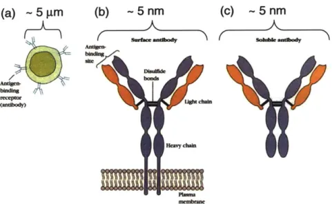
Figure 3.1 The B cell receptor... 90
Figure 3.2 B cells stimulated with lipopolysaccharide... 91
Figure 3.3 Leukocyte rolling and extravasation... 92
Figure 3.4 B cell spreading during antigen detection... 93
Figure 3.5 Biomechanical machinery of B cells... 94
Figure 3.6 Membrane tethering experimental background... 95
Figure 3.7 Membrane tethering experimental assay... 98
Figure 3.8 Schematic of energetic contributions to membrane tethering... 100
Figure 3.9 Cell-bead adhesion results... 103
Figure 3.10 Representative data of force versus extension and time... 104
Figure 3.11 Steady tether force results... 105
Figure 3.12 Steady tether force versus velocity... 106
Figure 3.13 Tether force relaxation data... 107
Figure 3.14 Results for force relaxation fit to double exponential model... 108
Figure 3.15 Static tether force versus radius... 109
Figure 3.16 Schematic of radial growth during force relaxation... 111
Figure 3.17 Experimental validation of radial growth... 112
Figure 3.19 Prediction of tether force relaxation with radial growth model... 116 Figure 3.A1 Mechanical equilibrium of membrane tubes... 120 Figure 4.1 B cell capping upon antigen detection... 150 Figure 4.2 XBP-1 deficiency leads to decreased mig expression and changes in lipid com position... 15 1 Figure 4.3 Membrane nanotubes physically connect T cells ... 153 Figure 4.4 Steady tether force results for XBP-1 KO cells... 156 Figure 4.5 XBP-1 KO Plasmablasts have lower effective viscosity... 157 Figure 4.6 Prediction of tether force relaxation behavior of XBP-1 KO B cells using radial
grow th m odel... 158 Figure 4.7 BCR is not present in membrane tether pulled after a 30 minute incubation
w ith fluorescent H EL... 159 Figure 4.8 BCR is present in membrane tethers pulled prior to a 5 minute incubation
w ith fluorescent H EL... 159 Figure 4.9 Dependence of BCR mobility on B cell activation by HEL... 161 Figure 4.10 Dependence of BCR mobility on B cell activation by HEL in XBP1-Ko B
c e lls ... 16 2 Figure 4.11 BCRs are pulled into membrane tethers during extension... 163 Figure 4.12 BCR occasionally appears as discrete spots along the tether with spatial pe riod icity... 164 Figure 5.1 Morphologies of NM fibers reconstituted at 4 0C and 37 0C... 179 Figure 5.2 Determining persistence length from thermal fluctuations... 182 Figure 5.3 Results of persistence length and kink torsional stiffness from NM fiber
therm al fluctuations... 183 Figure 5.4 Experimental tethered fiber assay... 185 Figure 5.5 Experimental results of combined force-extension and fluorescence
Figure 5.6 NM fiber mechanical properties from force-extension data... 189
Figure 5.7 Force-extension model for kinked fibers... 192
Figure 5.8 Loading and Unloading profiles for NM fibers with large force... 194
Figure 5.9 Examples of unfolding and rupture of NM fibers... 195
Figure 5.10 Multiple loading cycles on single NM fibers... 196
Figure 5.11 Imaging of surface-bound fiber fragment after rupture... 197
Figure 5.12 Examples of rare refolding events... 198
Figure 5.13 Determination of length of unfolding domains... 200
Figure 5.14 Distributions of unfolding lengths... 201
Figure 5.15 Melting curves for wild-type NM and RA2-5 fibers... 202
Figure 5.16 Bond kinetics determined from lifetime versus rupture force... 205
List of Tables
Table 1.1 Summary of amyloid related diseases... 26 Table 3.1 Membrane mechanical properties throughout differentiation... 106
Chapter 1
Introduction and Background
1.1 INTRODUCTION:
Mechanical forces play an important role in defining the physiological function of biological systems that span lengths scales from 1 nm (proteins and single molecules) up to 1 m (full mammalian systems). For example, conformational changes on the order of a few nanometers in ion channels which regulate intracellular concentration of ions such as Ca2+ can be driven by membrane stresses or strains [1-3]. At cellular length scales, endothelial cells (- 10-100 gm) which line blood vessel walls align and form actin stress fibers in the direction of shear flow [4-6]; and some cells can upregulate collagen production in response to mechanical strains [7]. Furthermore, an entire field of research exists to study the biomechanics of human movement. This thesis studies representative structures at the molecular (-1nm), cellular, and tissue (-1mm) length scales where mechanics plays a critical role in defining physiological function.
The approach taken to understand and model the mechanical behavior of biological systems must be tailored to the length scale of interest. In general biological systems in themselves can span several length scales. For example, B cells have a diameter of
have a diameter of - 10-100 nm, and the immunoglobulins expressed on the B cell membrane that initiate signaling and activation have dimensions of - 1 nm. Often times, describing the mechanical behavior of these complex systems requires a combination of different modeling approaches. Similarly, probing the mechanical behavior of biological systems requires experimentation tailored to the length scale of interest to guide and validate models. In this thesis we use a combination of applied experimental and theoretical mechanics to understand the role of forces and mechanical properties in the physiological function of systems at three distinct length scales: collagen tissues (~
mm), B lymphocytes (- pm), and prion amyloid fibers (- nm). Figure 1.1 shows how
these systems fit into a spectrum of some of the applied mechanics approaches used to describe the mechanical behavior of biological systems at different length scales.
B Cell Membrane
Collagen Fibers
Amyloid Fibers
Molecular Statistical Micromecha I Continuum
Dynamics \Mechanics ModelinVI Mechanics
10.d 104 10- 104 10-2 100 (m)
Figure 1.1 Mechanics at biological length scales. Biology spans length scales ranging from single proteins (-nm) to full organisms (-m). This thesis focuses on systems at three distinct length scales: (left ) amyloid fibers [Images Courtesy of Dr. Jijun Dong], (middle) B cells [8], and (right) collagen fibers [9]. An abbreviated list of mechanical modeling approaches tailored to the length scale of interest is shown in yellow. The approaches are often complimentary since biological systems often span several length scales in themselves.
20
Our general research approach combines system manipulation by molecular and cell biology techniques, measurement by force spectroscopy with optical tweezers, and mechanical modeling using a combination of the approaches listed in figure 1.1. The main focus of this thesis is applying novel modeling and measurement techniques to answer relevant questions in biology. The system manipulation aspect of this research was done in collaboration with the Ploegh Lab for the B cell work and the Lindquist lab for the amyloid fiber work, both at the Whitehead Institute for Biomedical Research.
1.1.1 Crimped Collagen Fibers. Starting with the largest length scale, we first address
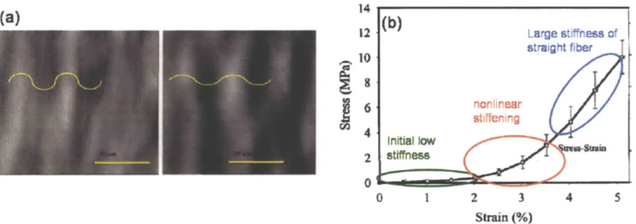
the mechanical behavior of collagen tissues comprised of networks of wavy collagen fibers in Chapter 2. This work focuses on the development of a micromechanical model to understand the stress-strain response of collagen tissues including ligaments, tendons, pericardium, and vessel walls whose primary load bearing constituent is a fibrous collagen network where the fibers generally have a wavy geometry [10-13]. The main physiological function of these soft tissues is to bear and transfer mechanical loads. Figure 1.2(a) shows an image of a bovine pericardium where the light and dark regions illustrate the highly aligned crimped fibers [12]. The wavy geometry of similar collagen fibers in rat tail tendons gives the nonlinear stress-strain response shown in figure 1.2(b) [11]. The wavy geometry initially deforms with little resistance by unbending of the crimped geometry, then experiences nonlinear stiffening while crimps straighten, and finally reaches a constant large stiffness when fibers become straight.
14 12 Large stiffness of straight fiber (0 6 nonlinear stiffening Initial low 2 stiffness 0 0 1 2 3 4 5 Strain (%)
Figure 1.2 Stress-strain behavior of crimped collagen fibers. (a) shows crimped fibers of a bovine pericardium all aligned in the same direction under no load (left) and after 15% strain (right) when crimps have begun to straighten (scale bars indicate 50 pm) [12]. (b) The crimped fiber gives a nonlinear stress-strain response with initially low stiffness
(unbending of crimps), nonlinear stiffening (straightening of crimps), and large stiffness response large strains (extension of straight fibers) [11].
During physiological processes, collagen tissues generally function in the nonlinear stiffening region of their stress-strain behavior [14]. Therefore, it is critical to accurately describe the nonlinear stiffening in order to describe the load bearing and transferring function of these tissues in physiological processes. In Chapter 2, we take a micromechanical modeling approach to develop a mechanical model for the force-extension behavior of wavy collagen fibers and bundles of fibers. This model accurately captures the full force-extension behavior of wavy fibers. Furthermore, our micromechanical model is useful to understand the deformation mechanisms of bundled fiber tissues such as tendons. We then integrate the wavy fiber model into a continuum mechanics model for fibrous networks to describe 2-dimensional tissues such as those found in vessel walls.
1.1.2 Mechanics of B lymphocyte Membranes. Working our way down in length
scale, Chapter 3 addresses the importance of the mechanical behavior of the B cell
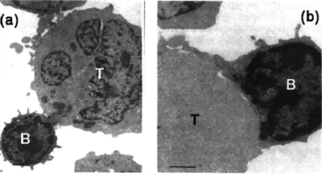
membrane during physiological functions. B cells are a critical component of the immune system that are responsible for detecting foreign antigens in mammals. During their lifetime, B lymphocytes must leave the bone marrow, circulate through the bloodstream, migrate into and through lymphoid organs where antigens are localized, probe lymphoid tissues for antigens, communicate with T lymphocytes, and migrate out of lymphoid tissues and re-enter the bloodstream. The lymphocyte membrane is a critical component for nearly all aspects of these functions, where its wrinkled topology facilitates easy expansion of the cell due to external stimuli and enables interaction and communication with surfaces, in particular the endothelial wall, lymphoid tissues, and T cells. For example, figures 1.3(a) and 1.3(b) shows a B cell interacting with a T cell. Contacts between B and T cells eventually form large areas of membrane contact. The excess B cell membrane that spreads out over the surface of the B cell is contained in the wrinkled geometry of the resting B cell membrane
Figure 1.3 Physical interactions of B and T cells. During their physiological function B cells interact with T cells. (a) shows an EM micrograph of initial contact between a B and T cell [Sanders et al, J Immunology 1986]. (b) Eventually these interactions lead to a large area of contact where the B cell membrane spreads out over the surface of the T cell (Scale bars are 1 gm) [15].
Here we perform single cell experiments with optical tweezers to quantify the mechanical properties of B cell membranes and track the evolution of those properties after B cell activation and throughout differentiation into antibody secreting Plasma cells. We further examine the dynamic stress relaxation behavior of membrane flow in the formation of membrane tethers (membrane nanotubes extracted from the surface of the cell), a process that occurs physiologically for example during cell rolling. A micro mechanical model is developed to describe the flow of membrane into tethers in order to relieve stress. All experiments are done on primary B cells that were extracted from B Cell Receptor (BCR) transgenic mice enabling us to probe the B cell in an antigen specific manner.
Chapter 4 addresses the inter-dependence of the biochemical and biomechanical machinery of the B cell. First, we investigate whether changes in the biochemical machinery have consequences for the biomechanical machinery of the B cell. Specifically, we quantify the membrane mechanical properties of X-box protein 1 (XBP-1) deficient B cells from BCR transgenic mice. XBP-1 is a transcription factor that regulates lipid synthesis and is critical for the differentiation of B cells into antibody secreting Plasma cells. We ask if XBP-1 deficiency results in changes in the mechanical behavior of the membrane that might impact B cell physiological function. Secondly, we investigate a potential biochemical role of membrane nanotube protrusions of B cells during antigen detection by employing a combined force and fluorescence approach with optical tweezers. Specifically, we ask if the BCR, the primary sensing agent of B cells, is present in membrane nanotubes extending from the
cell. BCR presence in nanotubes would confer cells with the ability to sense antigen at remote locations and possibly transfer antigens via nanotubes that form intercellular connections. Our results give insights into the immune function of the transcription factor XBP-1 in B cells as well as illustrate the cooperative nature of the B cell biochemical and biomechanical machinery. Furthermore, we shed new light on B cell function demonstrating the potential of B cells to sense antigens at remote locations via membrane nanotubes that protrude from their surface.
1.1.3. Physical Properties of Prion Amyloid Fibers. Taking one more step down in length scale, Chapter 5 examines the physical properties of amyloid-forming prion proteins. Amyloid fiber formation, which is associated with more than 30 diseases including disorders such as Alzheimer's, Creutzfeldt-Jacob and Parkinson's (see table 1.1), result from protein conformational changes leading to non-native structures that are subject to aggregation. Amyloids also serve important functions in diverse organisms, from bacteria to mammals that include biofilm formation, scaffolding, environmental adaptation and long term memory. In fact, it is now recognized that a capacity to accquire an amyloid conformation (albeit generally under nonphysiological conditions) is an inherent property of most peptides and proteins [16]. Furthermore, the amyloid self-assembly process has also been stimulated in synthetic preparations [17, 18]. Amyloid fibers exhibit mechanical properties comparable to those of spider silk [19]. Their impressive mechanical properties combined with the ease of assembly make amyloid fibers particularly suited for nanomaterials applications, including as templates
for conducting nanowire formation[20], as scaffolds for cell growth [21], and as functionalized biosensors [22].
Function Protoin Associated desaso
I ranni.4xon mrAhriula% Sinim RM101l1 rrotain A SAwonday 4;)Ktar imylomknis Apoproteln A4I Famikli amrylold polyneuropethy Type If Apollpoproteln A-I1 Familial amryloki polyneuropiathy Type 11
Transtyrotin Familial am'ylold polynauophy Type I Lkfi;uLftiin Ctaned- airt.idubib
Coagulation factors Fibrinogen Fibrinogen amytoidosis Erzymes Lysozy re Lyzozyme amyloiosis
Cytoskeletal proteins Keretn Ctareous smyloidosis
Taij Alzhoirytor's disease, frontotemporal dementia Hormonos Amylin Typo 11 diabetes
Caldioniin Mdullary carcinomna ot tho thyrcid Prolacti Aging pituit-y prolactfioras Inqtdin lnsuiin-rolarad amyboid Atrial matnuratic tarcter Atrial astyloidosis
Regulatory proteins Gelsclin FlnrlV hredluary amyloldosis
Protease rtilbitors CystanC lcelardc hedltarycrabralamyloldanglopathy
Imrmune tsystemn-related Iffmungtctuuin light ctlains (ic and X) Primary tiysteinic amnyl ks-, a r~rny c iwsatwd with minatlple mfyslorria lflulngIcui n leavy chain Primar-y systenuc omnylaiclosis
P2-Microglobmnin y remodialysis-relaed anyloidosis Cell-adhesion molecules Keratopthelir Carnea dystrophy
Unknown fuinction Lctodheruin M din) Aortic edial amyloldosis
Amyboid P ~ Alihoirnor's diseas, ccrobral arnlcici angiopethy Prion protein SpongA-ormoncophalopatA-rs
Apyloid B Aitsh Brtsh familial demntia Arnyloid Darnish Danish fawilii demntia
Lx-Synuclein Parkinsons disease
Table 1.1 A summary of amyloid-related diseases is given along with the corresponding amyloid forming protein and its natural biological function [23].
Prion proteins are a special class of amyloid fiber forming proteins -which are self-templating and thereby transmissible as disease vectors. Despite interest in amyloid fibers as novel materials and their importance in biology, little work has been done to quantify their physical and mechanical properties, and more specifically to relate these to protein structure and the self-assembly process. In this work, we employ a single molecule approach of combined force and fluorescence with optical trapping to quantify the physical properties of amyloid fibers formed from polymorphic variants (dissimilar structures from the same protein) of the yeast prion protein, Sup35. First, we employed fluorescence imaging to quantify the microstructure and thermal fluctuations of fibers in solution. The thermal fluctuations were used to identify the persistence length of two
subsets of fibers that are known to have different underlying prion structures [24]. We then developed a tethered fiber assay and performed single fiber force-extension experiments with optical tweezers to characterize the mechanical properties of prion fibers in bending and extension. Finally, we studied the molecular interactions that lead to misfolding and aggregation of prions by subjecting fibers to forces large enough to rupture those interactions. Our results have implications for the physical basis of strain diversity that results from polymorphic prion proteins. Furthermore, our unfolding and rupture experiments give insight into structural details of the prion conformation of Sup35.
1.2 OPTICAL TRAPPING OVERVIEW:
The primary experimental tool employed in this thesis is optical trapping. Optical traps function by the application of forces to micron scale particles through the application of laser light. The ability to apply forces to and trap particles using light was discovered by A. Ashkin 40 years ago [25]. Today optical force probes commonly referred to as optical traps or optical tweezers are widely applied in the fields of biophysics and biomechanics [26, 27]. Optical traps are capable of applying and measuring forces ranging from - 1-300 pN and measuring displacement with nm resolution making them ideal for the study of many biological systems. For example, the molecular motor kinesin, which transports intracellular cargo, is capable of applying forces of ~ 5 pN and achieve motion by systematic displacements ranging from -1-10 nm [28]. Optical tweezers have been applied to study the mechanics of a wide range of systems including molecular motors [29-31], biopolymers [32-35], filamentous biological
networks [36], F-actin and actin binding proteins [37, 38], and cell membranes [39, 40]. Additionally optical traps have been employed to manipulate bacteria, organelles, and entire cells [41, 42].
Optical traps use radiation pressure and momentum transfer of a tightly focused laser beam to manipulate and apply forces to particles ranging from -10 nm up to -10 pm. In general, spherical dielectric particles (i.e. glass or polystyrene beads) maximize the applied forces; however, optical traps can be used to manipulate any object on the appropriate length scale with some appreciable curvature such as a cell membrane [43]. Stable trapping is achieved using a high numerical aperture objective to focus a laser to a diffraction limited spot. Infrared lasers are often used to minimize the water absorption and hence sample heating [44]. Momentum is transferred to particles in the path of the laser due to reflection or refraction of the laser light. Figure 1.4 shows a schematic of a laser beam focused by a high numerical aperture objective passing through a spherical particle.
(a) (b)
F2 1n.
Figure 1.4 Optical trapping forces. (a) Optical traps utilize a high numerical aperture objective in order to focus an infrared laser to a diffraction limited spot (waist is ~ 500 nm). Forces are applied to particles (usually spherical beads) in the path of the laser through reflection (scattering forces) and refraction (momentum transfer). (b) Light bends when it enters the bead and when it leaves the bead due to the chance in index of refraction. In order to conserve momentum, a force is applied to the bead in the direction opposite the momentum change of light (F1 and F2). Some of the light reflects
directly of the bead, and results in a downstream force due to radiation pressure (Ftt). These force balance to provide an equilibrium position of the bead in the center of the laser just downstream of the focal point (i.e. waist).
Some of the light reflects off the bead resulting in a scattering force in the downstream direction, Ft. The light passing through the particle changes direction when enters and leaves bead due to the change in index of refraction resulting in a change in momentum. In order to conserve momentum, a force opposite to the momentum change of the light is applied to the bead, F, and F2. These forces create a
3-dimensional potential energy well for the bead with the equilibrium position just downstream of the focal point of the laser. If the bead is displaced from its equilibrium position the Gaussian intensity profile of the trapping laser results in a restoring force pushing the bead back towards its equilibrium position. The forces experienced by the trapped particle are proportional to the derivative of the gradient of the light. Thus, when
29
-the trapped particle is in -the center of -the trap, it experiences zero force on average, and when the bead is displaced from the center of the trap the restoring force increases linearly up to displacements of -100-150 nm. Therefore, an optically trapped bead can be approximated as a linear spring up to - 100-150 nm of displacement from its equilibrium position.
Optical traps can be characterized by a single parameter, the trap stiffness, ktrap. The displacement of the bead from its equilibrium position can then be used to determine
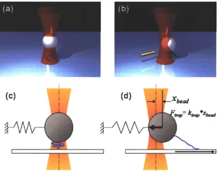
the force applied to the bead using the equation Ftrap = ktrap*xbead. If the bead is attached to a sample the trap can be used to apply a force to the sample. For example, figure 1.5(a) shows an optically trapped bead attached to a flexible polymer. The other end of the polymer is attached to a glass cover slip surface. If the sample is displaced relative to the trap as shown in figure 1.5(b), in this case by moving the coverslip, the bead will be pulled out of its equilibrium position. The restoring force, Fmp, is applied to the sample via the attachment to the bead. The deformation of the sample can be determined by the motion of the bead and the motion of the coverslip. Therefore, measuring the position of the bead can provide both the force applied to the sample, and the deformation of the sample. The resulting force versus deformation data can be used to determine relevant mechanical properties of the sample such as the bending stiffness in the case of polymers [28]. Alternatively, changes in force can be used to identify transitions of the sample such as folding or unfolding events of proteins [45-47].
(d)
Figure 1.5 Optical traps behave like a linear spring. An example tethered polymer assay is depicted in a 3D rendering (a-b) and in a 2D schematic illustrating the trap as a linear spring (c-d). (a,c) The bead is centered above the tether attachment point where it experiences essentially zero force. On average, the bead is located at the equilibrium position in the center of the laser. (b,d) The cover surface attachment point of the tether is displaced (this is usually done using a piezo electric microscope stage) and applies a force to the bead resulting in a restoring force pulling the bead back to the center of the trap, Ftap. The trap can accurately be modeled as a linear spring up to displacements of -100-150 nm.
1.2.1 Position Calibration of an Optical Trap. Quantifying optical trapping forces
relies on measuring the position of the bead, xtead, and knowledge of the trap stiffness,
ktiff. Xbead is generally measured either using video tracking or a position sensing device
(PSD) to monitor scatter from the trap laser or a secondary detection laser after passing through the bead. Video tracking utilizes visual measurement of the pixel location of the bead throughout the experiment [44]. The bead displacement is calculated using the pixel size and knowing the pixel location of the bead equilibrium position. PSDs read out a voltage based on the location where light hits the sensor. The 2 dimensional voltage response of the PSD as a function of bead position is calibrated by raster
scanning the bead in known increments through the detection region. This data is then fit to a fifth order polynomial which serves as a calibration curve to convert PSD
voltages into bead position. Figure 1.6 shows a typical voltage response for both axes of the PSD as a function of position. The position is shown in terms of the acousto-optic deflector (AOD) frequency. AODs steer the laser beam based on the frequency of an input signal. The AOD frequency can easily be converted to position in the sample plane using a calibration factor (has units of nm/MHz). Note that the AOD and PSD axes are oriented off axis by 45*. Our instruments employ PSD measurement of bead position. (a) (b) 4 4 . 0A OA 02 0.0 2 -o 2. .0.2 . -- 2
AOD Y Position [MH2] 4 -. AOD X Poastn Miz) ADD Y Positon Mz] 4A AOD X Posiion [MHz] Figure 1.6 Position sensing device voltage response. (a) X and (b) Y voltage signals from a position sensitive device were recorded as a trapped bead was raster scanned across the detection zone. For small displacements, the dependence of voltage signal on position is linear, but at larger displacements, the dependence becomes highly nonlinear. The position is shown in terms of AOD frequency which is converted to position in the sample plane using a calibration factor (nm/MHz).
1.2.2 Stiffness Calibration of an Optical Trap. Once the relation between bead
position and detector response is known, it can be used to calibrate the stiffness of the trap. In general the trap stiffness is a function of the laser power, the numerical aperture of the objective used, the diameter and index of refraction of the trapped bead, and the height of the bead of the coverslip surface [44]. Three methods are commonly
used to characterize the stiffness of the trap. The first and simplest method for stiffness calibration method relates the Brownian motion of the bead to the stiffness of the trap. Utilizing the linear spring approximation, the harmonic energy potential of the trap is
Utrp = I ktr x . This energy is related to the thermal energy of the medium, k8T, (kB is
2
Boltzmann's constant and T is absolute temperature) using the theorem of equipartition of energy [48].
kT = k, x2)
2 2t (1.1)
The trap stiffness can then be determined by measuring the positional variance of the bead,(x2). This method is very quick, but noise sources that increase the variance of bead position can lead to an underestimation of the trap stiffness. The second stiffness calibration method takes the one-sided power spectral density of a bead position trace and fits it to a Lorentzian function as shown in figure 1.7.
Figure 1.7 Power spectrum of a trapped bead [44]. The corner frequency can be related to the trap stiffness using equation 1.3.
The Lorentzian function gives the power spectrum as a function of frequency, S(f).
S (f )= 'BT12
67r3la(f 2 +f 2)(
The corner frequency, fo, is related to the trap stiffness using equation 1.3 [44].
fo = 127r2k,,ria
(1.3)
This method can reveal the presence of noise invisible to the other methods, and can be used to monitor local heating by the trap [49, 50].
The third method (Stokes drag) relies on applying a frictional drag force to the bead by fluid flow as shown in figure 1.8 and measuring the corresponding bead displacement, x. Fluid flow is generally accomplished by moving the sample relative to the stationary trap using a piezoelectric microscope stage.
Figure 1.8 Stokes drag experiment. Fluid flow relative to the bead applied a frictional drag force, Fd, which is balance by the trap force, Ft. The trap stiffness can be determined by dividing FE by the bead displacement, x.
The trap stiffness can then be determined using equation (1.4) where the drag force, Fd= 6,rraV, (Stokes drag equation for a sphere) is balanced by the trapping force,
F = k,,x , where q is the fluid viscosity, a is the bead radius, V is the fluid velocity, and
ktr is the trap stiffness.
6,r7raV = ka,,x (1.4)
Proximity to a surface, which is often the case in optical trapping experiments, alters the drag force according to Faxen's correction for flow parallel to the surface [44],
6,rraV, 5 (1.4)
1-
+45a
1a
16zb 8 b 256 IzbI 161 zb
35
where zb is the distance from the center of the bead to the surface. This method is the slowest, but it allows for characterization of any nonlinearities in the trap stiffness as a function of bead displacement and the maximum force capacity of the instrument. A detailed review of these calibrations methods is provided elsewhere [44, 51].
1.3 OPTICAL TRAPPING INSTRUMENTATION:
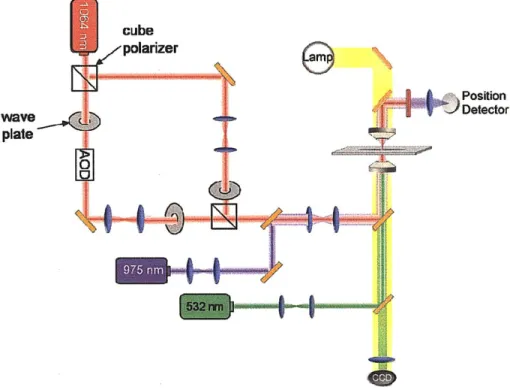
The experiments in this thesis were performed on two different optical trapping instruments in the Lang Laboratory. These instruments were built around inverted microscope platforms (Nikon, Melville, NY) that were heavily modified to improve functionality and stability. One of the instruments was designed for combined force and fluorescence applications at a single molecule level. This device combines separate lasers for optical trapping (1064 nm 4W capacity; Coherent, Santa Clara, CA), position detection (975 nm; Corning Lasertron, Bedford, MA), and fluorescence excitation (532 nm and 488 nm; World Star Tech, Toronto, ON) through a base that has improved mechanical stability. In addition, the setup includes a pair of computer controlled acousto-optic deflectors (AODs; IntraAction, Bellwood, IL), which permit precise steering of the trapping beam in two dimensions, and remote-controlled flipper mirrors and shutters, which facilitate rapid switching between bright-field imaging (CCD camera; DAGE-MTI, Michigan City, IN) and fluorescence imaging (EMCCD camera; Andor Technology, South Windsor, CT), and a piezoelectric stage (Physik Instrumente, Auburn, MA). This instrument was used for the membrane tethering with fluorescence (Chapter 4) and the fluorescent amyloid fiber force-extension (Chapter 5) experiments.
Figure 1.9 shows a schematic layout of the instrument. Further details of the instrument design are given elsewhere [52].
QM)
Position Detector AOD
EMCCD
Figure 1.9 Schematic layout of optical trap instrument used for combined force and fluorescence imaging experiments
The second instrument was optimized for the application of large forces for experiments at the cellular length scale. This device utilizes a similar setup to the previously described instrument with a few key differences. First, the trapping laser (1064 nm 1OW
capacity; IPG Photonics, Oxford, MA) is split into two branches of approximately equal power using a cube polarizer (ThorLabs; Newton, NJ). One branch (primary branch) passes through AODs and was generally used for position calibration and trapping at forces up to - 100 pN. The second branch that does not pass through the AODs, avoiding the -50% power loss, was used to achieve trapping forces up to -250 pN. When employing the second branch, it was initially aligned with the primary branch (position of the second branch was independently using picomotors to move one of the
telescope lenses). Position calibrations were performed using the primary branch with AODs. The bead was then transferred to the secondary branch prior to performing a stiffness calibration, and the experiment was carried out with the secondary branch. Secondly, while fluorescence excitation was available on this instrument (532 nm), it was not utilized since the instrument lacked an EMCCD camera for highly sensitive fluorescence imaging. This instrument was used for the membrane tethering experiments which did not require fluorescence imaging (Chapters 3 and 4) and for the amyloid unfolding and rupture experiments at large forces (Chapter 5). Figure 1.10 shows a schematic layout of this optical trapping instrument.
cube plarizer
Position
avDetector
plate
Figure 1.10 Schematic layout of the optical trap instrument used for large force applications.
1.4 REFERENCES
[1] Martinac, B., 2004. "Mechanosensitive ion channels: molecules of mechanotransduction". J Cell Sci. 117: p. 2449-2460.
[2] Blount, P. and Moe, P. C., 1999. "Bacterial mechanosensitive channels: integrating
physiology, structure and function". Trends Microbiol. 7: p. 420-424.
[3] Hamill, 0. P. and Martinac, B., 2001. "Molecular basis of mechanotransduction in living cells". Physiol Rev. 81: p. 685-740.
[4] Barbee, K. A., Davies, P. F. and Lal, R., 1994. "Shear Stress-Induced Reorganization of the Surface-Topography of Living Endothelial-Cells Imaged by Atomic-Force Microscopy". Circ Res. 74: p. 163-171.
[5] McCue, S., Noria, S. and Langille, B. L., 2004. "Shear-induced reorganization of endothelial cell cytoskeleton and adhesion complexes". Trends Cardiovas Med. 14: p.
143-151.
[6] Ohashi, T. and Sato, M., 2005. "Remodeling of vascular endothelial cells exposed to fluid shear stress: experimental and numerical approach". Fluid Dyn Res. 37: p. 40-59. [7] Ku, C. H., Johnson, P. H., Batten, P., Sarathchandra, P., Chambers, R. C., Taylor, P. M., Yacoub, M. H. and Chester, A. H., 2006. "Collagen synthesis by mesenchymal stem cells and aortic valve interstitial cells in response to mechanical stretch". Cardiovasc
Res. 71: p. 548-556.
[8] Fleire, S. J., Goldman, J. P., Carrasco, Y. R., Weber, M., Bray, D. and Batista, F. D., 2006. "B cell ligand discrimination through a spreading and contraction response". Science. 312: p. 738-741.
[9] Ottani, V., Raspanti, M. and Ruggeri, A., 2001. "Collagen structure and functional implications". Micron. 32: p. 251-260.
[10] Gasser, T. C., Ogden, R. W. and Holzapfel, G. A., 2006. "Hyperelastic modelling of arterial layers with distributed collagen fibre orientations". J Roy Soc Interface. 3: p. 15-35.
[11] Hansen, K. A., Weiss, J. A. and Barton, J. K., 2002. "Recruitment of tendon crimp with applied tensile strain". Journal of Biomechanical Engineering-Transactions of the Asme. 124: p. 72-77.
[12] Sacks, M. S., 2003. "Incorporation of experimentally-derived fiber orientation into a structural constitutive model for planar-collagenous tissues". Journal of Biomechanical Engineering-Transactions of the Asme. 125: p. 280-287.
[13] De Vita, R. and Slaughter, W. S., 2007. "A constitutive law for the failure behavior of medial collateral ligaments". Biomech Model Mechan. 6: p. 189-197.
[14] Viidik, A., Danielsen, C. C. and Oxlund, H., 1982. "On Fundamental and Phenomenological Models, Structure and Mechanical-Properties of Collagen, Elastin and Glycosaminoglycan Complexes". Biorheology. 19: p. 437-451.
[15] Sanders, V. M., Snyder, J. M., Uhr, J. W. and Vitetta, E. S., 1986. "Characterization of the Physical Interaction between Antigen-Specific B-Cells and T-Cells". J Immunol. 137: p. 2395-2404.
[16] Dobson, C. M., 2003. "Protein folding and misfolding". Nature. 426: p. 884-890. [17] Glover, J. R., Kowal, A. S., Schirmer, E. C., Patino, M. M., Liu, J. J. and Lindquist,
S., 1997. "Self-seeded fibers formed by Sup35, the protein determinant of [PSI+], a heritable prion-like factor of S-cerevisiae". Cell. 89: p. 811-819.
[18] MacPhee, C. E. and Dobson, C. M., 2000. "Formation of mixed fibrils demonstrates the generic nature and potential utility of amyloid nanostructures". Journal of the American Chemical Society. 122: p. 12707-12713.
[19] Smith, J. F., Knowles, T. P. J., Dobson, C. M., MacPhee, C. E. and Welland, M. E., 2006. "Characterization of the nanoscale properties of individual amyloid fibrils". Proceedings of the National Academy of Sciences of the United States of America. 103: p. 15806-15811.
[20] Scheibel, T., Parthasarathy, R., Sawicki, G., Lin, X. M., Jaeger, H. and Lindquist, S. L., 2003. "Conducting nanowires built by controlled self-assembly of amyloid fibers and selective metal deposition". Proceedings of the National Academy of Sciences of the United States of America. 100: p. 4527-4532.
[21] Gras, S. L., Tickler, A. K., Squires, A. M., Devlin, G. L., Horton, M. A., Dobson, C. M. and MacPhee, C. E., 2008. "Functionalised amyloid fibrils for roles in cell adhesion". Biomaterials. 29: p. 1553-1562.
[22] Baxa, U., Speransky, V., Steven, A. C. and Wickner, R. B., 2002. "Mechanism of inactivation on prion conversion of the Saccharomyces cerevisiae Ure2 protein". Proceedings of the National Academy of Sciences of the United States of America. 99: p. 5253-5260.
[23] Sigurdsson, E. M., Wisniewski, T. and Frangione, B., 2002. "Infectivity of amyloid diseases". Trends Mol Med. 8: p. 411-413.
[24] Krishnan, R. and Lindquist, S. L., 2005. "Structural insights into a yeast prion illuminate nucleation and strain diversity". Nature. 435: p. 765-772.
[25] Ashkin, A., 1970. "Acceleration and Trapping of Particles by Radiation Pressure". Phys Rev Lett. 24: p. 156-&.
[26] Moffitt, J. R., Chemla, Y. R., Smith, S. B. and Bustamante, C., 2008. "Recent advances in optical tweezers". Annu Rev Biochem. 77: p. 205-228.
[27] Neuman, K. C. and Nagy, A., 2008. "Single-molecule force spectroscopy: optical tweezers, magnetic tweezers and atomic force microscopy". Nat Methods. 5: p. 491-505.
[28] Khalil, A. S., Appleyard, D. C., Labno, A. K., Georges, A., Karplus, M., Belcher, A. M., Hwang, W. and Lang, M. J., 2008. "Kinesin's cover-neck bundle folds forward to generate force". P Natl Acad Sci USA. 105: p. 19247-19252.
[29] Mehta, A. D., Rief, M., Spudich, J. A., Smith, D. A. and Simmons, R. M., 1999. "Single-molecule biomechanics with optical methods". Science. 283: p. 1689-1695. [30] Abbondanzieri, E. A., Greenleaf, W. J., Shaevitz, J. W., Landick, R. and Block, S.
M., 2005. "Direct observation of base-pair stepping by RNA polymerase". Nature. 438: p. 460-465.
[31] Asbury, C. L., Fehr, A. N. and Block, S. M., 2003. "Kinesin moves by an asymmetric hand-over-hand mechanism". Science. 302: p. 2130-2134.
[32] Khalil, A. S., Ferrer, J. M., Brau, R. R., Kottmann, S. T., Noren, C. J., Lang, M. J. and Belcher, A. M., 2007. "Single M13 bacteriophage tethering and stretching".
Proceedings of the National Academy of Sciences of the United States of America. 104: p. 4892-4897.
[33] Wang, M. D., Yin, H., Landick, R., Gelles, J. and Block, S. M., 1996. "Stretching DNA with optical tweezers." Biophysical Journal. 70: p. Sup63-Sup63.
[34] Wen, J. D., Manosas, M., Li, P. T. X., Smith, S. B., Bustamante, C., Ritort, F. and Tinoco, I., 2007. "Force unfolding kinetics of RNA using optical tweezers. 1. Effects of experimental variables on measured results". Biophysical Journal. 92: p. 2996-3009.
[35] Baumann, C. G., Smith, S. B., Bloomfield, V. A. and Bustamante, C., 1997. "Ionic
effects on the elasticity of single DNA molecules". Proceedings of the National Academy of Sciences of the United States of America. 94: p. 6185-6190.
[36] Lee, H., Pelz, B., Ferrer, J. M., Kim, T., Lang, M. J. and Kamm, R. D., 2009.
"Cytoskeletal Deformation at High Strains and the Role of Cross-link Unfolding or Unbinding". Cellular and Molecular Bioengineering. 2: p. 28-38.
[37] Ferrer, J. M., Lee, H. S., Chen, J., Kamm, R. D. and Lang, M. J., 2007. "Mapping
the F-actin and actin binding proteins interactions: From macromechanics to single molecule biophysics". Biophys J: p. 303a-303a.
[38] Ferrer, J. M., Lee, H., Chen, J., Pelz, B., Nakamura, F., Kamm, R. D. and Lang, M. J., 2008. "Measuring molecular rupture forces between single actin filaments and
actin-binding proteins". P Natl Acad Sci USA. 105: p. 9221-9226.
[39] Raucher, D. and Sheetz, M. P., 1999. "Characteristics of a membrane reservoir
buffering membrane tension". Biophys J. 77: p. 1992-2002.
[40] Li, Z. W., Anvari, B., Takashima, M., Brecht, P., Torres, J. H. and Brownell, W. E., 2002. "Membrane tether formation from outer hair cells with optical tweezers". Biophys
J. 82: p. 1386-1395.
[41] Ashkin, A., Dziedzic, J. M. and Yamane, T., 1987. "Optical Trapping and Manipulation of Single Cells Using Infrared-Laser Beams". Nature. 330: p. 769-771. [42] Ashkin, A. and Dziedzic, J. M., 1987. "Optical Trapping and Manipulation of Viruses and Bacteria". Science. 235: p. 1517-1520.
[43] Holm, A., Sundqvist, T., Oberg, A. and Magnusson, K. E., 1999. "Mechanical manipulation of polymorphonuclear leukocyte plasma membranes with optical tweezers causes influx of extracellular calcium through membrane channels". Med Biol Eng Comput. 37: p. 410-412.
[44] Neuman, K. C. and Block, S. M., 2004. "Optical trapping". Rev Sci Instrum. 75: p.
2787-2809.
[45] Kellermayer, M. S. Z., Smith, S. B., Granzier, H. L. and Bustamante, C., 1997. "Folding-unfolding transitions in single titin molecules characterized with laser tweezers". Science. 276: p. 1112-1116.
[46] Onoa, B., Dumont, S., Liphardt, J., Smith, S. B., Tinoco, I. and Bustamante, C.,
2003. "Identifying kinetic barriers to mechanical unfolding of the T-thermophila
ribozyme". Science. 299: p. 1892-1895.
[47] Woodside, M. T., Anthony, P. C., Behnke-Parks, W. M., Larizadeh, K., Herschlag,
D. and Block, S. M., 2006. "Direct measurement of the full, sequence-dependent folding
landscape of a nucleic acid". Science. 314: p. 1001 -1004.
[48] Boltzmann, L., 1871. "Einige allgemeine Satze uber Warmegleichgewicht (Some general statements on thermal equilibrium)." Wiener Berichte. 63: p. 679-711.
[49] Peterman, E. J. G., Gittes, F. and Schmidt, C. F., 2003. "Laser-induced heating in optical traps". Biophysical Journal. 84: p. 1308-1316.
[50] Abbondanzieri, E. A., Shaevitz, J. W. and Block, S. M., 2005. "Picocalorimetry of
[51] Svoboda, K. and Block, S. M., 1994. "Biological applications of optical forces".
Annu Rev Biophys Biomol Struct. 23: p. 247-85.
[52] Brau, R. R., Tarsa, P. B., Ferrer, J. M., Lee, P. and Lang, M. J., 2006. "Interlaced
optical force-fluorescence measurements for single molecule biophysics". Biophys J.
91: p. 1069-1077.
Chapter 2
An Elastica Approximate for Fibers and Fibrous
Networks
2.1 INTRODUCTION:
Single fiber or chain mechanics play an important role in defining the mechanical response of biological molecules and networks, synthetic polymer networks, as well as woven and non-woven textiles. Single fiber mechanics has widely been approached by a consideration of the free energy of extension. In general, extension contributes to the free energy of the fiber in two ways: 1) a reduction in entropy due to a decrease in number of possible configurations a fiber may occupy with increasing extension, and 2) a change in enthalpy due to fiber deformation. The most commonly used models in molecular mechanics are the freely-jointed chain (FJC) model [53, 54] and the worm-like chain (WLC) model [55-57]; these models are based on entropic elasticity. Many biological and synthetic fibers have a limited configurational space and hence their mechanical behavior is dominated by changes in enthalpy; these fibers typically possess an initially wavy or crimped structure due to either growth or processing (figure 2.1). When force is applied to the fiber ends, the increase in fiber end-to-end distance is due to the unbending of the wavy structure. Hence, an Euler elastica model [58-65] is the physically appropriate representation of the force-extension behavior during fiber
![Table 1.1 A summary of amyloid-related diseases is given along with the corresponding amyloid forming protein and its natural biological function [23].](https://thumb-eu.123doks.com/thumbv2/123doknet/14165232.473765/26.918.125.804.190.541/summary-amyloid-related-diseases-corresponding-amyloid-biological-function.webp)
![Figure 1.7 Power spectrum of a trapped bead [44]. The corner frequency can be related to the trap stiffness using equation 1.3.](https://thumb-eu.123doks.com/thumbv2/123doknet/14165232.473765/33.918.255.657.753.1011/figure-power-spectrum-trapped-frequency-related-stiffness-equation.webp)
![Figure 1.9 shows a schematic layout of the instrument. Further details of the instrument design are given elsewhere [52].](https://thumb-eu.123doks.com/thumbv2/123doknet/14165232.473765/37.918.260.662.195.548/figure-shows-schematic-layout-instrument-details-instrument-design.webp)
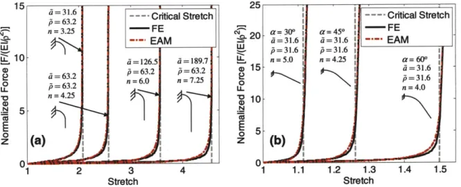
![Figure 2.10 (a) Shows results of fitting the model to force vs. stretch behavior obtained by Hansen et al [11] for a collagen fascicle with an average cross-sectional area of 0.078 mm 2](https://thumb-eu.123doks.com/thumbv2/123doknet/14165232.473765/67.918.154.726.334.576/figure-results-fitting-behavior-obtained-collagen-fascicle-sectional.webp)