2010/285
Changing nucleotide specificity of the DEAD-box helicase
Hera abrogates communication between the Q-motif
and the P-loop
Julian Strohmeier
1, Ines Hertel
2, Ulf Diederichsen
1,
Markus G. Rudolph
3and Dagmar Klostermeier
2,a,*
1
Institute for Organic and Biomolecular Chemistry,
University of Go¨ttingen, D-37077 Go¨ttingen, Germany
2Division of Biophysical Chemistry, Biozentrum,
University of Basel, CH-4056 Basel, Switzerland
3F. Hoffmann-La Roche, CH-4070 Basel, Switzerland
* Corresponding authore-mail: [email protected]
Abstract
DEAD-box proteins disrupt or remodel RNA and protein/
RNA complexes at the expense of ATP. The catalytic core
is composed of two flexibly connected RecA-like domains.
The N-terminal domain contains most of the motifs involved
in nucleotide binding and serves as a minimalistic model for
helicase/nucleotide interactions. A single conserved
gluta-mine in the so-called Q-motif has been suggested as a
con-formational sensor for the nucleotide state. To reprogram the
Thermus thermophilus RNA helicase Hera for use of
oxo-ATP instead of oxo-ATP and to investigate the sensor function
of the Q-motif, we analyzed helicase activity of Hera Q28E.
Crystal structures of the Hera N-terminal domain Q28E
mutant (TthDEAD_Q28E) in apo- and ligand-bound forms
show that Q28E does change specificity from adenine to
8-oxoadenine. However, significant structural changes
accom-pany the Q28E mutation, which prevent the P-loop from
adopting its catalytically active conformation and explain the
lack of helicase activity of Hera_Q28E with either ATP or
8-oxo-ATP as energy sources. 8-Oxo-adenosine, 8-oxo-AMP,
and 8-oxo-ADP weakly bind to TthDEAD_Q28E but in
non-canonical modes. These results indicate that the Q-motif not
only senses the nucleotide state of the helicase but could also
stabilize a catalytically competent conformation of the
P-loop and other helicase signature motifs.
Keywords: crystal structure; Hera; hydrogen bond
network; nucleotide specificity; 8-oxo-adenosine; ribosome
biogenesis.
Introduction
RNA helicases couple the chemical energy from ATP
hydroly-sis to structural rearrangements of RNA and RNA/protein
aPresent address: Institute for Physical Chemistry, University of
Muenster, Corrensstrasse 30, D-48149 Muenster, Germany.
complexes. As such they touch upon virtually all processes
of RNA metabolism including transcription, translation,
RNA editing, viral replication, and ribosome biogenesis
(Cordin et al., 2006). DEAD-box proteins form the largest
RNA helicase family (Hilbert et al., 2009). They are named
according to the characteristic sequence of their Walker B
motif, which is implicated in ATP binding. DEAD-box
pro-teins share a common modular architecture (Figure 1A): a
helicase core of two flexibly connected RecA-like domains
(RecA_N and RecA_C), which are sometimes flanked by
additional domains that confer substrate binding specificity,
mediate protein/protein interactions or may contribute to
duplex separation (Tsu et al., 2001; Kossen et al., 2002;
Grohman et al., 2007; Linden et al., 2008; Mohr et al., 2008;
Del Campo et al., 2009). In addition, DEAD-box helicases
can also form dimers (Klostermeier and Rudolph, 2009;
Rudolph and Klostermeier, 2009) via dedicated dimerization
domains, as has recently been found for the
Thermus
ther-mophilus
He
at resistant
R
NA-dependent
A
TPase, Hera
(Morlang et al., 1999), a DEAD-box RNA helicase
presum-ably involved in ribosome assembly and assembly of RNase P
(Linden et al., 2008). Upon cooperative binding of RNA and
ATP (or an ATP analog), the helicase core collapses to form
a composite RNA binding site traversing both RecA-like
domains, as exemplified by crystal structures of helicase/
RNA complexes (Andersen et al., 2006; Bono et al., 2006;
Sengoku et al., 2006; von Moeller et al., 2009). The RecA_N
domain of DEAD-box proteins contains most of the
con-served sequence motifs required for nucleotide binding, such
as the Q-motif and the P-loop (or Walker A motif), and the
DEAD-box which connects the P-loop to the SAT motif
(Figure 1A). RecA_N can thus serve as a minimalistic model
to study nucleotide binding to RNA helicases (Rudolph et
al., 2006; Hilbert et al., 2010).
Based on mutational analyses and sequence comparisons,
the Q-motif has been defined as a stretch of nine amino acids
with an almost invariant (99%) glutamine (Benz et al., 1999;
Tanner, 2003; Tanner et al., 2003). Crystal structures of
heli-case/nucleotide complexes showed that the glutamine side
chain directly binds to the adenine base of the nucleotide via
a double hydrogen bond (Figure 1B). Such a glutamine is
also present in reverse gyrases from extremophiles and in
several other ATP-binding P-loop containing proteins, such
as viral packaging motors and endonucleases, but also in
proteins involved in ATP synthesis and chromosome
segre-gation (Iyer et al., 2004; Draper and Rao, 2007; Tsay et al.,
2009). It was suggested that the Q-motif may be a
confor-mational sensor for nucleotide binding, relaying the signal
of a bound nucleotide to the P-loop (Cordin et al., 2004). A
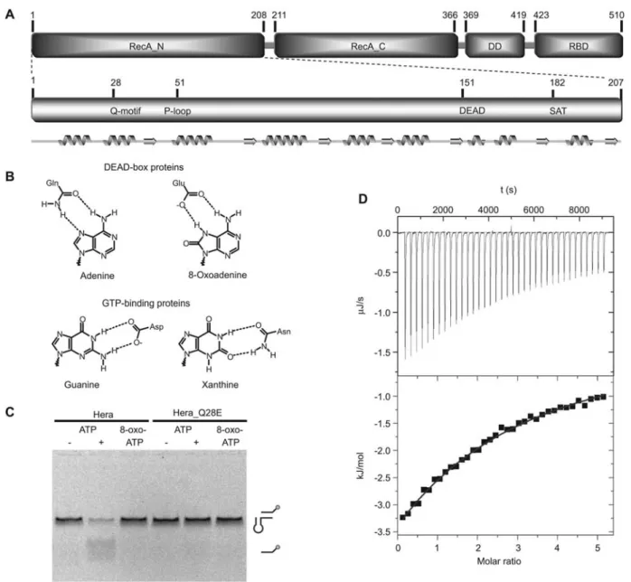
Figure 1 Overview of Hera and structure-sequence relationship of the N-terminal domain.
(A) The upper panel displays the Hera domain organization. The helicase core consists of two RecA-like domains and is followed by a dimerization and C-terminal RNA-binding domain. The lower panel highlights the conserved helicase motifs in the N-terminal RecA-like domain (RecA_N) with its secondary structure elements indicated below. The figure is drawn to scale. Numbers indicate domain boundaries and residue positions. (B) Comparison of the nucleotide specificity determinants in DEAD-box helicases (top) and several classes of GTPases (bottom). In these GTPases an aspartic acid contacts the exocyclic amino group and a ring nitrogen atom of guanine. Replacement by asparagine alters specificity to xanthine nucleotides. In DEAD-box proteins, the conserved glutamine residue selects adenine nucleotides via a bifurcated hydrogen bond donor of the side chain carboxamide to the N6 and N7 nitrogen atoms of the nucleobase. Mutation of this glutamine to glutamic acid removes the hydrogen bond donor. This hydrogen bond could be restored by use of 8-oxo-adenosine nucleotides, as these predominantly are in the oxo-tautomer in solution. (C) Unwinding of a 32/9mer RNA substrate derived from 23S rRNA by wild-type Hera and the Q28E mutant. The upper band represents the fluorescently labeled double-stranded substrate and the lower band is the fluorescent single strand after unwinding (cartoons on right). No helicase activity is detected with Hera/8-oxo-ATP, Hera_Q28E/ATP or Hera_Q28E/8-oxo-ATP, whereas Hera/ATP unwinds this substrate. (D) Determination of 8-oxo-adenosine affinity for TthDEAD_Q28E by ITC. Evaluation of the data based on a single binding site yieldsKas1728"41.7M(Kds579
m
M), DHs-26.3"0.4 kJ/mol and calculatedTDSs-7.8 kJ/mol for 8-oxo-adenosine. Measurements for 8-oxo-nucleotides were not performed due to scarcity of material and multiple binding modes for 8-oxo-AMP and 8-oxo-ADP for TthDEAD_Q28E as determined from the crystal structures.
possible way to probe the Q-motif for communication with
the P-loop would be to alter the nucleotide specificity of the
helicase while keeping spatial requirements and similar
hydrogen bonding potential around the nucleotide binding
site. For several classes of GTP-binding proteins, a strategy
was devised where an aspartate to asparagine mutation
shift-ed nucleotide specificity from guanine to xanthine (Figure
1B). Examples include H-Ras (Zhong et al., 1995), EF-Tu
(Hwang and Miller, 1987; Weijland et al., 1993), Ypt1 (Jones
et al., 1995), Rab-5 (Hoffenberg et al., 1995; Rybin et al.,
1996), FtsY (Powers and Walter, 1995), adenylosuccinate
synthetase (Kang et al., 1994) and Goa (although here a
second mutation was essential for shifting nucleotide
speci-ficity) (Yu et al., 1997). Xanthine nucleotides are only
tran-siently produced during purine metabolism and are not
populated as triphosphates
in vivo, allowing for separation
of the signaling effect of a specific GTPase in the
back-ground of other GTPases. A similar reasoning as for
GTP-binding proteins would allow, for instance, 8-oxo-adenine
nucleotides to bind to a helicase where the conserved
glu-tamine of the Q-motif is mutated to glutamate and also allow
for selectively activating the helicase with altered specificity
in vivo. Using the T. thermophilus RNA helicase Hera as a
model, the Q28E mutation was introduced in both authentic
Hera (Hera_Q28E) and the N-terminal RecA-like domain
(TthDEAD_Q28E). Although the 8-oxo-adenine base did
bind to TthDEAD_Q28E, the mutation in Hera rendered the
helicase functionally inactive. Crystal structures for several
nucleotide complexes revealed the expected hydrogen
bond-ing mode for the 8-oxo-adenine nucleobase but conveyed
interesting structural changes with respect to binding of the
ribose and phosphate parts compared to the TthDEAD/AMP
structure. The structures of TthDEAD_Q28E provide the
molecular basis for the role of the conserved glutamine in
the Q-motif in communication of the P-loop with the DEAD
and SAT motifs and explain the loss of helicase activity in
Hera_Q28E.
Results
8-Oxo-adenosine and the nucleotide triphosphate
(8-oxo-ATP) were synthesized and the expected tautomeric form
(Figure 1B) was confirmed by NMR analysis, in line with
previous results on 8-oxo-adenine derivatives (Chatgilialoglu
et al., 2006). 8-Oxo-ATP was tested as an energy source for
Hera helicase activity (Figure 1C). Although wild-type Hera
displayed ATP-dependent RNA unwinding activity on a 32/
9mer RNA substrate derived from 23S rRNA (Linden et al.,
2008), no unwinding of RNA in the presence of 8-oxo-ATP
was observed. By contrast, Hera_Q28E showed no
unwind-ing activity, neither with ATP nor with 8-oxo-ATP as the
energy source (Figure 1C). These results indicate that the
stereochemically rather conservative mutation Q28E strongly
impacts helicase activity. Isothermal titration calorimetry of
8-oxo-adenosine as a probe for base- and ribose-mediated
binding to TthDEAD_Q28E yielded a reaction enthalpy
DHs-26.3"0.4 kJ/mol and a dissociation constant
K
ds
579
m
M(Figure 1D). ATP affinities of DEAD-box proteins
typically range between 80
m
Mto )1 m
M(Cordin et al.,
2006). For Hera, nucleotide affinities for ATP analogs are
;200
m
M(M.H. Linden and D. Klostermeier,
unpublish-ed data). For the TthDEAD/AMP complex, we previously
determined a
K
ds
246
m
M. The AMP co-crystal structure
shows several interactions of the phosphate moiety with the
P-loop (Rudolph et al., 2006), indicating a substantial
ener-getic contribution to binding. Thus, a mere twofold reduction
(DDGs1.7 kJ/mol at 298 K) in affinity for the
Tth-DEAD_Q28E/8-oxo-adenosine complex points toward
spe-cific binding of the modified nucleobase 8-oxo-adenine to
the mutated Q-motif. Owing to lack of material, no detailed
binding
studies
were
possible
with
8-oxo-adenosine
nucleotides.
To rationalize the effect of the Q28E mutation on the RNA
helicase activity of Hera and to define the binding mode of
8-oxo-adenosine and derived nucleotides, we determined
crystal structures of unliganded TthDEAD_Q28E and its
complexes with adenosine, AMP and
8-oxo-ADP in several crystal forms. The Hera/nucleotide
comp-lexes were obtained
in situ by limited hydrolysis of
8-oxo-ATP during the crystallization process. The structures
were determined by molecular replacement using the
previ-ously determined dimeric TthDEAD structure as the search
model (PDB-ID 2gxs) (Rudolph et al., 2006). The models
were refined to resolutions of 1.4–2.6 A
˚ (Table 1).
Overall structure of TthDEAD
The structure of TthDEAD_Q28E displays all salient
fea-tures of a DEAD-box domain resembling a RecA-fold (Story
and Steitz, 1992; Story et al., 1992): a central seven-stranded
parallel b-sheet that is flanked by five and four a-helices on
either side (Figure 2A). Compared to the canonical RecA
fold, the DEAD domain possesses an N-terminal extension
of approximately 25 residues that contain the Q-motif (see
below) and cap the nucleotide binding domain. In all three
crystal forms discussed here, TthDEAD_Q28E forms dimers
of the same kind as the wild-type TthDEAD. The prominent
feature is a 14-stranded intermolecular b-sheet in a top-down
fashion (Figure 2B). Superposition of the first monomer of
the six dimeric TthDEAD_Q28E structures (two dimers are
present in triclinic form II) onto the first monomer of the
dimeric TthDEAD (PDB-ID 2gxs) structure yields a root
mean square deviation (rmsd) of -1.3 A
˚ over all Ca atoms.
Based on this superposition, structural plasticity for the
sec-ond monomer is apparent with displacements of up to 5.2 A
˚
and rotation up to 8.48. The variability within the dimers is
most prominent when the wild-type TthDEAD/AMP and
TthDEAD_Q28E/8-oxo-ADP structures are compared (see
below). Although there is no indication for dimerization of
either wild-type or TthDEAD_Q28E in solution, this dimer
has now been observed in four independent crystal settings
and appears to be a low energy state that is populated at least
at high concentrations of TthDEAD. Full-length Hera is
indeed a stable dimer but here dimerization is achieved by a
dedicated dimerization domain outside the helicase core that
is currently unique to Hera (Klostermeier and Rudolph,
2009).
Effect of the Q28E mutation on the structure of TthDEAD
In the structure of TthDEAD with orthophosphate bound (no
apo-structure is available for the wild-type) the carboxamide
side chain of Gln28 forms four hydrogen bonds: two with
T able 1 Data collection and refinement statistics. Dataset 3MWJ 3MWL 3MWK 3NBF 3NEJ apo, form I 8-oxo-adenosine 8-oxo-AMP 8-oxo-AMP and apo, form III 8-oxo-ADP , form II Data collection 38.3 – 1.40 46.9 – 1.60 46.6 – 1.45 46.5 – 1.9 43.4 – 2.6 Resolution range (A ˚) a (1.42 – 1.40) (1.63 – 1.60) (1.48 – 1.45) (1.93 – 1.90) (2.7 – 2.6) Space gr oup P4 1 21 2P 41 21 2P 41 21 2P 1 P 31 21 Cell dimensions (A ˚, 8)a s 94.5, c s 108.1 a s 92.2, c s 109.0 a s 91.9, c s 108.0 a s 60.6, b s 60.7, a s 59.7, c s 239.9 c s 74.1, a s 68.9, b s 77.3, g s 72.6 Unique reflections 90 708 (1895) 60 859 (1582) 82 310 (3261) 70 329 (2091) 15 772 (1515) Multiplicity 20.2 (20.5) 8.7 (3.6) 15.2 (14.5) 2.4 (2.4) 4.0 (2.7) Completeness (%) 100 (100) 97.3 (70.1) 99.9 (99.9) 95.7 (94.6) 94.2 (86.4) Rsym /R meas (%) b 7.6/8.4 7.8/8.6 8.6/9.1 6.2/7.1 21.7/27.8 A verage I/ s (I) 15.0 (1.9) 13.2 (1.1) 15.7 (1.4) 7.6 (1.2) 7.2 (1.2) Refinement 38.3 – 1.40 46.9 – 1.6 46.0 – 1.45 41.5 – 1.90 40.0 – 2.6 Resolution range (A ˚) (1.44 – 1.40) (1.63 – 1.60) (1.49 – 1.45) (1.93 – 1.90) (2.8 – 2.6) No. of reflections 83 072 (4352) 55 169 (1672) 75 950 (5165) 64 866 (2377) 15 080 (2267) Rcryst (%) c 14.1 (20.7) 19.6 (30.3) 15.9 (25.5) 18.4 (32.5) 21.2 (32.7) Rfr ee (%) c 17.6 (25.1) 23.6 (36.3) 20.9 (31.8) 22.2 (33.9) 26.9 (40.3) No. of residues/waters 424/279 424/387 424/323 842/266 406/3 Phase err or (8 ) d 16.9 25.2 20.3 27.1 26.9 rmsd Bonds/Angles (A ˚, 8) 0.018/1.72 0.006/1.1 0.022/2.0 0.007/1.1 0.004/0.9 Ramachandran plot (%) e 94.9/4.6/0.6 94.3/5.4/0.3 95.7/4.0/0.3 93.2/6.8/0 78.0/22.0/0 A verage B values (A ˚ 2) f 20.5 "10.3, 34.5 "12.3, 20.6 "9.3, 39.0 "18.2, 22.6 "8.3, 35.6 "11.9, 40.5 "17.7, 43.6 "17.0, 56.3 "24.2, 56.2 "21.7 32.8 "7.6, n.a. 34.5 "8.2, 43.0 "11.8 34.2 "5.9, 43.1 "12.7 43.0 "18.0, 50.8 "17.9, 44.3 "8.9, 52.0 "22.7 aV alues in par enthesis corr espond to the highest resolution shell. bCalculated with SCALA (Evans, 2006). cR cryst s 8 ±± Fo ±-±Fc ±± / 8 ±F o ±, wher e Fo and Fc ar e the structur e factor amplitudes fr om the data and the model, respectively . Rfr ee is Rcryst with 5% of test set structur e factors. dCalculated with PHENIX (Zwart et al., 2008). eCalculated using PROCHECK (Laskowski et al., 1993). Numbers reflect the per centage amino acid residues in the cor e: allowed and gener ous allowed regions, respectively . fEntries for pr otein monomers followed by the last two entries for water and ligands, respectively .
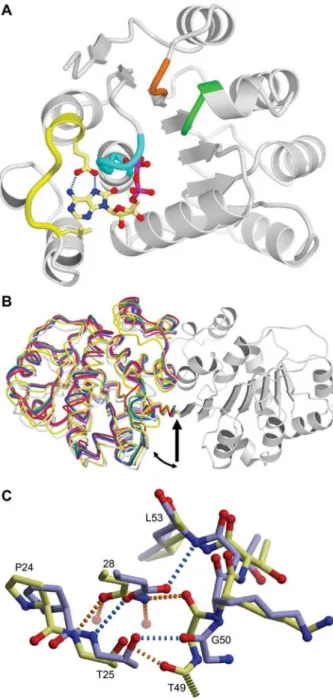
Figure 2 Salient features of the ThDEAD_Q28E structures. (A) Overview of the ThDEAD_Q28E monomer. The mutated Q-motif Q28E, the hydrophobic residue upstream of the Q-Q-motif (Leu21) and 8-oxo-ADP are shown as yellow stick models. The two key hydrogen bonds from Glu28 to 8-oxo-adenine are drawn as blue dashed lines. The P-loop is marked in cyan, the DEAD-box in green and the SAT motif in orange. (B) The TthDEAD dimer as present in the crystal structures is mediated by an intermolecular b-sheet. The dimers are superimposed on one monomer of the TthDEAD/ AMP complex (PDB-ID 2gxs, gray on right side). The second mon-omers are drawn as backbone traces and are colored as follows: TthDEAD/AMP in gray, TthDEAD_Q28E apo form I in green, TthDEAD_Q28E apo form III in red, TthDEAD_Q28E/8-oxo-aden-osine in blue, TthDEAD_Q28E/8-oxo-AMP in cyan and the two dimers of TthDEAD_Q28E/8-oxo-ADP of crystal form II are col-ored in yellow and magenta. Domain movements are apparent, hing-ing about the center of the b-sheet (arrow). (C) Effect of the Q28E mutation on the local structure of TthDEAD. Wild-type and mutant are colored yellow and blue, respectively.
water molecules and two with protein atoms (Figure 2C).
The Gln28 side chain carbonyl group hydrogen bonds to the
main chain amide of the Q-motif residue Thr25 and the NH
2-group of Gln28 contacts the main chain carbonyl of the
P-loop residue Gly50, thus effectively connecting the Q-motif
and the P-loop. The apo-TthDEAD_Q28E structure reveals
several consequences of the Gln28Glu mutation. First, no
water molecules are present. Second, the hydrogen bond to
Gly50 is disrupted due to opposition of two hydrogen bond
acceptors. Gly50 and adjoining residues of the P-loop move
away from their position in the TthDEAD structure by up to
2.6 A
˚ . This shift could be induced by electrostatic repulsion
from Glu28 that is likely deprotonated under the pH values
of crystallization (7.5–9.0). Third, the movement of the
P-loop and a shift of Glu28 relative to Gln28 enable a novel
hydrogen bond between the amide nitrogen of Leu53 and
Glu28. Interestingly, a hydrogen bond between the side chain
of Thr25 in the Q-motif and the P-loop is retained in both
cases. Although in the wild-type the carbonyl group of Thr49
accepts this hydrogen bond, this role is served by Gly50 due
to its rotation in the Gln28E mutation (Figure 2C). Thus,
already in the absence of nucleotides there are significant
changes of the apo-structures in wild-type and Q28E
Tth-DEAD, which impact on the communication between
heli-case motifs.
Unusual nucleotide binding to TthDEAD_Q28E
Apart from Hera, several crystal structures of DEAD-box
proteins in complex with AMP are now known including
DDX3X (Ho¨gbom et al., 2007), eIF4A (Schu¨tz et al., 2008),
DDX53 (PDB-ID 3iuy, unpublished) and DDX47 (PDB-ID
3ber, unpublished). Super-positions of these complexes
reveal virtually identical conformations for the AMP ligand
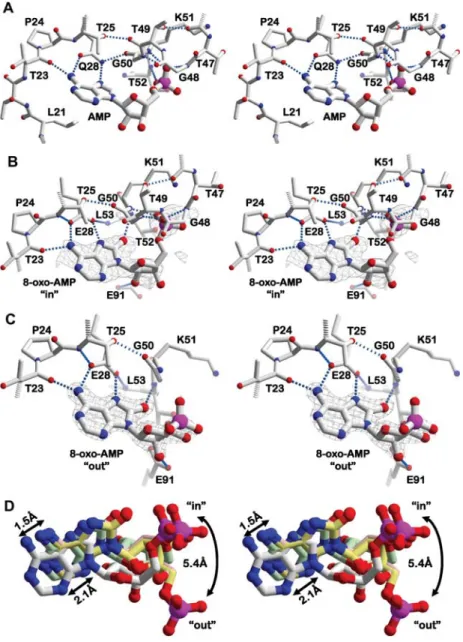
and the P-loop (binding site shown for Hera in Figure 3A).
In all structures, the adenine base binds to the conserved
glutamine side chain of the Q-motif (Q28 in Hera). In most
DEAD-box proteins, an aromatic side chain located
approx-imately seven residues upstream of the glutamine stacks onto
the nucleotide base. In 88% from a set 277 sequences, this
residue is a phenylalanine (Tanner et al., 2003). In other
cas-es, the aromatic side chain can be replaced by aliphatic
res-idues such as L21 in Hera or an isoleucine in DDX53. In
Hera, Q28 and T25 of the Q-motif form hydrogen bonds
with the main chain carbonyl groups of P-loop residues G50
and T49, respectively. The phosphate moiety of the
nucleo-tide is bound in a canonic fashion by main chain amide
nitro-gen atoms of T47, T49, and G50 of the P-loop (Figure 3A).
Interactions between the Q-motif, the nucleotide and the
P-loop establish a three-dimensional network that is primed for
sensing the type and phosphorylation state of the nucleotide.
Binding of 8-oxo-adenosine and 8-oxo-AMP
As anticipated, the nucleotide base of 8-oxo-adenine binds
to the side chain of Glu28 via a bidentate hydrogen bond.
Analogous to the wild-type situation, a third hydrogen bond
is formed by the exocyclic amino group with the carbonyl
group of Thr23 (Figure 3B). The oxo-group in 8-oxo-adenine
Figure 3 Details of nucleotide monophosphate binding to TthDEAD and the Q28E mutant.
(A) Stereo-view of the hydrogen bond network in the AMP structure (PDB-ID 2gxs) for comparison. (B) Stereo-view of the hydrogen bond network around 8-oxo-AMP with the monophosphate moiety bound to the P-loop (PDB-ID 3nbf). (C) Stereo-view of the hydrogen bond network in another 8-oxo-AMP complex (PDB-ID 3mwk). The phosphate part does not bind to the P-loop in this structure. Its flexibility is indicated by a deteriorating electron density compared to the nucleobase. Note the retention of the dual hydrogen bonds between the nucleobases and E28 in (B) and (C) when compared to the wild-type Q28 in (A) and the shifts of the P-loops. Omit map electron densities are drawn as a gray mesh and contoured at 3s. (D) Stereo-view of the protein-based superposition of AMP, adenosine and 8-oxo-AMP ligands. Carbon atoms of 8-oxo-AMP are colored in light gray, those of 8-oxo-adenosine are drawn in rose and carbon atoms of the in- and out-conformations of 8-oxo-AMP are shown in green and yellow, respectively. 8-Oxo-adenosine (hydrogen bond network not shown) superimposes very closely with 8-oxo-AMP. The nucleobases of the 8-oxo-compounds have shifted significantly compared to adenine. The ribose-phosphate part of 8-oxo-AMP has rotated about the glycosidic bond, placing the phosphate of 8-oxo-AMP 5.4 A˚ away from its position in the AMP structure.
forms an additional hydrogen bond to the backbone amide
group of Gly50, which is possible due to a rotation of the
Lys51-Thr52 peptide plane. This binding pattern for the
8-oxo-adenine base is universal for all 8-oxo-adenosine
nucle-otide complexes described here. In the 8-oxo-AMP complex,
there are two binding modes for the ribose-phosphate part of
8-oxo-AMP, termed in and out. The in-conformation (Figure
3B) corresponds to the classic localization of the phosphate
moiety in the P-loop where negative charge(s) are neutralized
by contacts with the side chain of Lys51, hydrogen bonds to
the main chain amides of P-loop residues and the macro
dipole of the a-helix C-terminal to the P-loop (Figure 1A).
By contrast, the out-conformation (Figure 3C) represents a
non-productive situation where the phosphate moiety does
not bind to the P-loop but is oriented towards bulk solvent
and appears rather flexible, as judged from weak electron
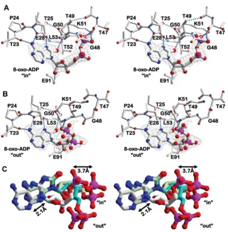
Figure 4 Details of 8-oxo-ADP binding to the TthDEAD_Q28E mutant.
(A) Stereo-view of the hydrogen bond network formed by 8-oxo-ADP in the P-loop bound in-conformation. (B) Corresponding view of the out-conformation not bound to the P-loop. (C) Protein-based superposition of AMP and 8-oxo-ADP. The two conformations of 8-oxo-ADP, shown in green and cyan, differ with respect to the position of their phosphate parts. The nucleobases are shifted to a similar magnitude as observed for 8-oxo-AMP (Figure 3). The terminal phosphates of AMP in wild-type TthDEAD and 8-oxo-ADP in the in-conformation are 3.7 A˚ apart, despite similar interactions with the P-loop.
density. The presence of such a conformation indicates that
binding of the phosphate part in 8-oxo-AMP to the P-loop
may not be as energetically favorable as for the adenine
nucleotides. Superposition of the proteins in the two
mono-phosphate complexes reveals that the out-conformation is
realized by rotations about the glycosidic and the sugar
C4-C5 bonds, in sum leading to a displacement of the phosphate
moiety by 5.4 A
˚ (Figure 3D). A possible explanation for the
presence of the out-conformation is apparent by comparing
the placement of the nucleobases within the active site: the
additional oxygen atom in 8-oxo-adenine seems to push and
tilt the nucleobase away from the P-loop by 1.5–2.1 A
˚ . A
concomitant pull on the sugar-phosphate moiety may explain
a loss in affinity for the P-loop, such that both conformations
can be trapped in crystal structures.
Binding of 8-oxo-ADP
The 8-oxo-adenine base in the 8-oxo-ADP complex
estab-lishes exactly the same interactions to TthDEAD_Q28E as
described above for 8-oxo-adenosine and 8-oxo-AMP
(Fig-ure 4). In addition, the two in- and out-conformations of the
ribose-phosphate moiety are also realized in the 8-oxo-ADP/
TthDEAD_Q28E complexes. Both conformations are well
defined by electron density and display different
conforma-tions of the ribose-phosphate parts when bound inside and
outside the P-loop, respectively. In the in-conformation, the
b-phosphate, which entertains four direct hydrogen bonds to
amide nitrogen atoms of the P-loop, is placed at
approxi-mately the same location as the a-phosphate in the
8-oxo-AMP complex (compare Figures 2B and 3A). Compared to
the wild-type TthDEAD/AMP complex, the b-phosphate of
8-oxo-ADP is located approximately 3.7 A
˚ from the position
of the a-phosphate in AMP (Figure 4C). Apart from crystal
contacts (not shown), the artificial out-conformation (Figure
4B) is stabilized by a single hydrogen bond of the
b-phos-phate to the amide nitrogen atom of the Thr49-Gly50 peptide
bond, which is flipped compared to the in-conformation.
Based on the comparisons between the 8-oxo-nucleotide/
TthDEAD_Q28E complexes, it can be concluded that the
P-loop exhibits considerable plasticity, both in the absence and
presence of non-natural nucleotides.
Communication of helicase motifs in the nucleotide-binding domain
Crystal structures of DEAD-box helicases in complex with
ADP are available from DDX5 (PDB-ID 3fe2), DDX10
Figure 5 Communication of helicase motifs within the N-terminal Rec_A-like domain.
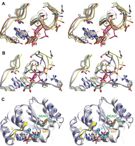
(A) Close-up stereo-view of the superposition of ADP-bound DEAD-box helicases DDX5 (PDB-ID 3fe2, blue), DDX10 (PDB-ID 2pl3, pink), DDX20 (PDB-ID 2oxc, green) and DDX52 (PDB-ID 3dkp, brown) with TthDEAD_Q28E in complex with 8-oxo-ADP (in-confor-mation, yellow). For the ADcomplexes, the conformations of the Q-motif (labeled Q) vary considerably but the conformations of the P-loops (labeled P) are quite similar. The conformations of these motifs in TthDEAD_Q28E are very different from those of the ADP-complexes. (B) Close-up stereo-view of the superposition of RNA/ADPNP complexes of Vasa (PDB-ID 2db3, light blue), DBP5 (PDB-ID 3fht, green), DDX19 (PDB-ID 3g0h, pink) with TthDEAD_Q28E in complex with 8-oxo-ADP (in-conformation, yellow). The conformations of the Q-motif and the P-loops in these RNA complexes are very similar but again differ from the conformation in Tth-DEAD_Q28E. (C) Stereo-view of a superposition of the RecA_N domains of TthDEAD_Q28E in complex with 8-oxo-ADP (in-conformation grey) and theDrosophila melanogaster Vasa/RNA/ADPNP complex (light blue). Although the main body of the domains remains constant, there are concerted movements of the Q-motif, the nucleotide, the P-loop, the DEAD-box and the SAT motif when the structures are compared to each other.
(PDB-ID 2pl3), DDX19 (PDB-ID 3ews), DDX20 (PDB-ID
2oxc) and DDX52 (PDB-ID 3dkp), which are all
unpublish-ed, except for DDX19 (Collins et al., 2009). Superposition
of these complexes with the TthDEAD_Q28E/8-oxo-ADP
structure reveals that the Q-motif exhibits structural
plastic-ity, with the Q28E-complex marking one extreme (Figure
5A). Interestingly, in all ADP-complexes the P-loops adopt
a very similar conformation that is again different from the
TthDEAD_Q28E/8-oxo-ADP structure. The b-phosphate of
8-oxo-ADP occupies a different position in the P-loop
com-pared to ADP, due to a different puckering of the ribose (see
below).
To estimate how strong the P-loop conformation in
8-oxo-nucleotide complexes deviates from the active conformation
in a closed RNA- and ATP-bound helicase, a structural
com-parison with the RNA complexes of Vasa (Sengoku et al.,
2006), DDX19 (Collins et al., 2009) and DBP5 (von Moeller
et al., 2009) was performed (Figure 5B). Both the P-loop
and the Q-motif in all three independent RNA/helicase
struc-tures have very similar conformations down to the level of
side chain conformations, which contrasts the situation in the
ADP-complexes. However, the Q-motif conformations
mark-edly differ from the TthDEAD_Q28E/8-oxo-ADP complex
(Figure 5B). Within the different 8-oxo-nucleotide
complex-es, there is also substantial P-loop plasticity (Figures 2 and
3). The Q28E mutation may increase the P-loop flexibility
independent of the bound nucleotide and lead to a
non-pro-ductive placement of the phosphate moiety in the P-loop. For
example, the conformation of the ribose at the C59-position
is different for 8-oxo-ADP compared to ADP and the ATP
analog ADPNP, placing the b-phosphate of 8-oxo-ADP at a
position between the b- and g-phosphate of ADPNP (Figure
5B). In addition, 8-oxo-adenine nucleotide binding to
TthDEAD_Q28E has structural consequences that extend
beyond the P-loop (Figure 5C). In nucleotide-free and
apo-structures of DEAD-box proteins, Lys51 (
T.th. numbering)
connects the P-loop to Glu152 of the DEAD-box via a salt
bridge, and DEAD-box residue Asp154 links to Ser182 and
Thr184 of the SAT motif. Binding of ADP/Mg
2qor ATP/
Mg
2qdisrupts the Lys51-Glu152 salt bridge and Mg
2qnow
connects the b-phosphate with Glu152 (Shi et al., 2004),
which keeps the interaction between the motifs intact. In the
8-oxo-ADP complex, the connection of the P-loop and the
DEAD-box is still in the ground state, with Lys51
electro-statically contacting Asp151 and Glu152 of the DEAD-box,
probably because no Mg
2qis bound in this structure.
Com-parison of the three closed helicase structures Vasa, DDX19
and DBP5 shows that their DEAD and SAT motifs share
virtually identical conformations, but again they differ from
the 8-oxo-ADP structure. Likewise, among the
8-oxo-nucle-otide complexes, the conformations of the DEAD and SAT
motifs are very similar. In the SAT motif, the main
differ-ences between the closed conformations and the Hera_Q28E/
8-oxo-nucleotide complexes are a shift of the conserved
threonine by 3.3 A
˚ (Ca-distance to Vasa) and a flip of the
Ala-Thr peptide plane. Taken together, these observations
indicate that the Q28E mutation and/or the bound
8-oxo-nucleotides prevent establishment of the hydrogen bonding
network between the P-loop and the SAT motif and thus
abolish the RNA unwinding activity of Hera_Q28E.
Discussion
In this study, the role of the conserved glutamine residue in
the Q-motif of the DEAD-box helicase Hera for nucleotide
specificity and communication with the P-loop was probed
by mutation to glutamic acid and adjustment of the
nucleo-base from adenine to 8-oxo-adenine. Upon changing the
hydrogen bonding pattern of both the Q-motif residue 28 and
the nucleobase, the double hydrogen bond between the
glu-tamine of the Q-motif and the nucleobase was retained. This
approach was successful with respect to maintaining
speci-ficity, but failed to keep a native interaction between the
Q-motif and the P-loop. Consequently, no helicase activity of
full-length Hera_Q28E with 8-oxo-ATP as energy source
was observed.
A similar approach of mutating a conserved asparagine
residue to aspartic acid in GTP-binding proteins has
suc-cessfully changed specificity from guanine to xanthine
nucle-otides in various examples. The question arises as to why
this approach was successful and, in addition to changing the
nucleotide specificity, the XTPase and effector binding
activ-ity was also retained. The situation in GTP-binding proteins
is rather simple: the asparagine/aspartate contacts only the
nucleotide but no other protein parts. The xanthine atoms
that differ from guanine point into the solvent, which can
explain why signaling functionality of the XTPases is
retained. By contrast, crystal structure analyses of
Tth-DEAD_Q28E/8-oxo-nucleotide complexes provide two main
explanations as to why helicase function is lost when the
analogous glutamine-to-glutamate mutation is introduced:
First, the Q-motif engages in interactions with the adenine
base and also with the P-loop contacting the phosphate
moi-ety of the bound nucleotide. The Q28E mutation opposes
two hydrogen bond acceptors, E28 (Q-motif) and G50
(P-loop), which leads to a shift of the P-loop that could prompt
all other conformational changes observed in the DEAD and
SAT motives. Second, the introduction of an oxygen atom
at the C8 position of adenine was essential to reverse the
hydrogen bonding potential of N7 from acceptor to donor.
This additional oxygen atom is small enough to retain the
anti-conformation of the nucleobase in nucleotides but
intro-duces another hydrogen bond acceptor that interacts with a
main chain amide nitrogen of the P-loop. Thus, two
addi-tional P-loop interactions, one from the Q-motif and another
from the nucleotide, are present in the Q28E/8-oxo system
that may be responsible for disrupting the communication
between helicase motifs.
Mutational analyses of the conserved glutamine residue in
the Q-motif of DEAD-box proteins has been performed
pre-viously with the yeast translation-initiation factors eIF4A
(Q48; Tanner et al., 2003) and Ded1 (Q169; Cordin et al.,
2004). Mutation of these glutamine residues to either
gluta-mate or alanine displayed lethal phenotypes in yeast.
Sur-prisingly, ATP and ADP binding to the mutants was still
observed in UV-crosslinking and ATPase competition
exper-iments (Tanner et al., 2003), but ATPase activities were
reduced significantly (Cordin et al., 2004), paralleled by little
or no RNA unwinding. From these studies, a differential
effect of the mutations on ADP and ATP binding was
inferred with different determinants for binding of the two
nucleotides, and the Q-motif was postulated as a molecular
‘on/off switch’. The growth phenotypes and lacking helicase
activities of the eIF4A and Ded1p Q-mutants are in line with
the central role of the Q-motif glutamine for sensing the
nucleotide state of the DEAD-box protein.
The structure of TthDEAD_Q28E demonstrates that
mutants with a glutamic acid instead of the essential
gluta-mine residue will not tolerate an adenine base, and we did
not detect binding of Hera_Q28E to mantADP (data not
shown). However, a secondary nucleotide binding site close
to the RNA-binding site is observed in the TthDEAD_Q28E/
8-oxo-ADP crystal structure (data not shown) and a
pyro-phosphate was observed in the same region of DDX52
(unpublished). Possibly, these secondary sites could
contrib-ute to the residual nucleotide binding as reported for eIF4A
and Ded1p. Taken together, the structural and mutational data
thus suggest that the conserved glutamine of the Q-motif
plays a crucial role in adenine recognition and signal relay
to the P-loop for unwinding activity. By contrast, structural
data do not support a role of the Q-motif as a ‘switch’ that
recognizes the phosphorylation state of bound nucleotide(s)
as it only interacts with the nucleobase.
A significant number of DEAD-box proteins have been
structurally characterized in complex with adenine
nucleo-tides, ranging from ATP analogs to ADP and AMP. Since
the determination of the TthDEAD/AMP complex structure,
three more AMP complexes have been determined with
DDX53, DDX47 (Schutz et al., 2010) and DDX3X (Ho¨gbom
et al., 2007). The binding of AMP to helicases is not
func-tionally relevant, but it is mechanistically interesting as it
points to the nucleobase as the key for nucleotide affinity.
By contrast, binding of the phosphates seems energetically
neutral. This notion is corroborated by equilibrium
dissoci-ation constants for the TthDEAD/AMP (Rudolph et al.,
2006) and TthDEAD_Q28E/8-oxo-adenosine complexes
(this work) in the high micromolar range, similar to those
reported for ADP and ATP analogs in a number of other
DEAD-box proteins (Cordin et al., 2006), including Hera
(M.H. Linden and D. Klostermeier, unpublished). As the
phosphate groups in nucleotides do not provide additional
binding energy, it could be concluded that the energy from
hydrogen bonds of the phosphate part to the P-loop is
trans-formed into conformational changes of the DEAD and SAT
motifs toward a closure of the helicase inter-domain cleft in
the ATP state (Yao et al., 1997; Kim et al., 1998; Johnson
and McKay, 1999; Caruthers et al., 2000; Linden et al.,
2008). In TthDEAD_Q28E, the hydrogen bonding network
connecting the Q-motif, the P-loop, the DEAD-box and the
SAT motif is maintained and these motifs have shifted in a
concerted fashion when compared to the structures of the
DEAD-box proteins Vasa, DDX19 and DBP5 in complex
with RNA and ADPNP (Sengoku et al., 2006; Collins et al.,
2009; von Moeller et al., 2009). However, the abnormal
P-loop conformation that is dictated by the mutated Q-motif
leads to a displacement of the phosphate moiety of bound
nucleotides, effectively abolishing the nucleotide-sensing
capability of the P-loop. As a consequence, the relay of
con-formational changes that eventually leads to a closure of the
cleft in the helicase core is blocked at the initial stage,
abro-gating unwinding activity.
Materials and methods
Synthesis of 8-oxo-adenosine and derived nucleotides
All reagents were of analytical grade and used without further puri-fication. Solvents were of the highest grade available. Dry solvents were stored over molecular sieves (4 A˚ ). Glass equipment utilized for reactions under inert atmosphere was flame dried before use.
1
H- and13
C-NMR spectra were recorded with a Varian Unity 300 spectrometer, a Varian Inova 500 spectrometer or a Varian Inova 600 spectrometer. Chemical shifts are quoted in parts per million (ppm) downfield of tetramethylsilane. Abbreviations for multiplic-ities are: s, singlet; d, doublet; t, triplet; m, multiplet; br, broad. Coupling constants are given in Hz. Mass spectrometry was carried out using Finnigan LQC or TSQ 7000 instruments. HRMS spectra were measured with a Bruker APEX-Q IV 7T instrument. HPLC analysis was performed using a Pharmacia A¨ kta basic instrument (pump type P-900, variable wavelength detector) with different sol-vent systems. Compounds were analyzed using a JASCO ReproSil column ODS-A, 5
mm, RP-C18, 250=4.6 mm, with a flow rate of
1 ml/min. Semi-preparative purification was performed using a JAS-CO ReproSil column ODS-A, RP-C18, 250=10 mm, with a flow rate of 3 ml/min. The synthesis of 8-oxo-adenosine and thenucle-oside-59-triphosphate was essentially performed as described (Scheme 1; Chatgilialoglu et al., 2006).
8-Bromo-adenosine (2)
An aqueous solution of sodium acetate (0.5M, 100 ml) was treated with acetic acid until pH 4.0 was reached. Adenosine (1) (1.50 g, 5.61 mmol) was added to the buffer and stirred under gentle heating until the solid was completely dissolved. The solution was allowed to cool to room temperature and a solution of bromine (2.69 g, 16.8 mmol) in water (50.0 ml) was added. The resulting reaction mixture was stirred at room temperature overnight. The solution was then decolorized by addition of solid sodium bisulfite and neutral-ized by addition of aqueous sodium hydroxide solution (50%). The suspension was allowed to stand at 58C overnight. The solid was then collected by filtration and the filter cake was washed with water (3=10.0 ml) and acetone (1=10.0 ml). The pure product was dried under vacuum to yield 1.41 g (4.08 mmol, 73%).1H-NMR
(300 MHz, DMSO-D6, 358C): ds3.48–3.57 (m, 1 H, H59), 3.68 (td, 2J H,Hs12.1 Hz, 3JH,Hs3.9 Hz, 1 H, H59), 3.99 (dd,3JH,Hs6.4 Hz, 3J H,Hs3.9 Hz, 1 H, H49), 4.18–4.23 (m, 1 H, H39), 5.06–5.13 (m, 1 H, H29), 5.18 (d,3J H,Hs4.2 Hz, 1 H, 39-OH), 5.40–5.47 (m, 2 H, 29-OH, 59-OH), 5.84 (d, 3J H,Hs6.8 Hz, 1 H, H19), 7.51 (sbr, 1 H, NH2), 8.12 (s, 1 H, H2) ppm. 13C-NMR (500 MHz, DMSO-D6, 358C): ds62.0 (C59), 70.8 (C49), 71.0 (C29), 86.6 (C39), 90.3 (C19), 119.5 (C5), 126.9 (C8), 149.7 (C4), 152.2 (C2), 154.9 (C6) ppm. ESI-MS m/z (rel.%): 346.0 (100) wMqHxq , 368.0 (54) wMqNaxq . HRMS (ESI): 346.0139 (calculated for C10H13BrN5O4: 346.0145),
367.9956 (calculated for C10H12BrN5O4Na: 367.9965).
8-Oxo-adenosine (3)
A solution of 8-bromo-adenosine (2) (346 mg, 1.00 mmol), 2-mer-captoethanol (234 mg, 210
ml, 3.00 mmol) and triethylamine
(1.01 g, 1.39 ml, 10.0 mmol) in water (100 ml) was stirred under reflux for 4 h. Afterwards, the solvent was removed under reduced pressure and the crude product was purified by semi-preparative HPLC. Yield: 254 mg, 898mmol, 90%.
1H-NMR (300 MHz, D 2O, 358C): ds3.84 (dd,2J H,Hs12.8 Hz,3JH,Hs3.9 Hz, 1 H, H59), 3.93 (dd, 2J H,Hs12.7 Hz, 3JH,Hs2.8 Hz, 1 H, H59), 4.25 (dd, 3J H,Hs6.6 Hz, 3JH,Hs3.2 Hz, 1 H, H49), 4.48 (dd, 3JH,Hs5.5 Hz, 3J H,Hs3.2 Hz, 1 H, H39), 4.99 (dd, 3JH,Hs5.6 Hz, 3JH,Hs6.3 Hz, 1 H, H29), 5.90 (d,3J H,Hs6.5 Hz, 1 H, H19), 8.04 (s, 1 H, H2) ppm. 13C-NMR (300 MHz, D 2O, 358C): ds62.1 (C59), 70.9 (C39), 71.3 (C29), 85.5 (C49), 86.2 (C19), 104.4 (C5), 146.2 (C4), 147.5 (C2), 151.0 (C8), 153.0 (C6) ppm. ESI-MS m/z (rel.%): 282.1 (100) wM-Hx-. HRMS (ESI): 282.0850 (calculated for C10H12N5O5: 282.0844).
Semi-preparative HPLC (RP-C18, 5–60% B wAsMilliQ-H2O,
BsMeCN:H2O 8:2x in 30 min): 11.21 min.
8-Oxo-adenosine-59-triphosphate (4)
A solution of 8-oxo-adenosine (3) (42.5 mg, 150
mmol) and proton
sponge (129 mg, 602mmol) in trimethyl phosphate (2.00 ml) was
stirred at room temperature for 0.5 h. The solution was cooled to 08C and phosphorous oxychloride (25.2 mg, 15.1ml, 165
mmol)
was added drop-wise. The clear solution was then stirred at 08C for 2 h. A mixture of tributylamine (111 mg, 143ml, 600
mmol) and
bis-tri-n-butylammonium pyrophosphate (535 mg, 975mmol) in dry
DMF (750ml) was added in one portion. The reaction was allowed
to stir at 08C for 15 min, and added drop-wise to cold TEAA buffer (0.2M, 25.0 ml, pHs7). The solution was stored at 48C overnight, washed with ethyl acetate (4=20.0 ml) and evaporated to dryness. The crude product was purified by analytical HPLC. Yield: 33.5 mg,Scheme 1 Reaction scheme for the synthesis of 8-oxo-ATP.
(i) Bromine, acetate buffer pH 4, room temperature (rt), overnight, 73%. (ii) 2-Mercaptoethanol, Et3N, H2O, reflux, 4 h, 90%. (iii) wax
Trimethyl phosphate, proton sponge, rt, 0.5 h. wbx POCl3, 08C, 2 h. wcx bis-tri-n-butylammonium pyrophosphate, Bu3N, DMF, 08C, 15 min.
wdx 0.2MTEAA buffer pH 7.4, 48C, 22 h. 64.1
mmol, 43%.
1H-NMR (600 MHz, D 2O, 358C): ds4.18–4.23 (m, 1 H, H59), 4.27–4.32 (m, 2 H, H49, H59), 4.62–4.64 (m, 1 H, H39), 5.25 (t,3J H,Hs5.6 Hz, 1 H, H29), 5.94 (d,3JH,Hs5.4 Hz, 1 H, H19), 8.21 (s, 1 H, H2) ppm. 13C-NMR (500 MHz, D 2O, 358C): ds61.5 (C59), 72.4 (C39), 72.7 (C29), 85.1 (C49), 88.4 (C19), 107.1 (C5), 149.4 (C4), 150.0 (C2), 153.9 (C8), 155.5 (C6) ppm.31P-NMR (300 MHz, D2O, 358C): ds-10.7 (d, Js9.7 Hz, g-P), -11.2 (d, Js19.5 Hz, a-P), -23.1 (t, Js19.5 Hz, b-P) ppm. HRMS (ESI): 521.98366 (calculated for C10H15N5O14P3: 521.98338). AnalyticalHPLC (RP-C18, 4% B wAs0.2MTEAA, Bs0.2MTEAA:MeCN
95:5x): 9.85 min.
8-Oxo-AMP was producedin situ during crystallization from 8-oxo-ATP by unspecific nuclease activity. Addition of trace amounts of shrimp alkaline phosphatase was used to generate 8-oxo-adeno-sine from 8-oxo-ATP during crystallization. For ITC measurements, HPLC purified 8-oxo-adenosine was used (see below). As reported previously (Chatgilialoglu et al., 2006), NMR spectra did not show the tautomeric 8-hydroxy-adenosine, which would have the same hydrogen bonding potential as adenine.
Protein purification and helicase assay
The genes coding for authenticT. thermophilus Hera and TthDEAD were cloned as previously described (Rudolph et al., 2006; Linden et al., 2008). The Q28E mutation was introduced using the Quik-change (Qiagen, Hilden, Germany) procedure and the sequences were verified. Hera_Q28E and TthDEAD_Q28E were purified as described previously (Rudolph et al., 2006; Linden et al., 2008). After size exclusion chromatography (S200, GE Healthcare, Munich, Germany) in 50 mM Tris/HCl pH 7.5, 200 mM NaCl,
TthDEAD_Q28E was concentrated to 2.23 mM by ultrafiltration
(10 kDa MW cut-off, Millipore, Schwalbach, Germany) and stored at -808C. Unwinding activity was measured using a 32/9mer RNA substrate derived from 23S rRNA as described (Linden et al., 2008) with 10
m
MHera or Hera_Q28E, 5m
MRNA, and 5 mMATP or 8-oxo-ATP in 50 mMTris/HCl pH7.5, 150 mMNaCl, 5 mMMgCl2at 258C for 30 min. Products were analyzed by native polyacryla-mide gel electrophoresis.
Crystallization, data collection, structure determination and refinement
TthDEAD_Q28E was crystallized under similar conditions to wild-type TthDEAD (Rudolph et al., 2006) but these crystals did not diffract X-rays. Ade novo search for crystallization conditions at 228C in the sitting drop vapor diffusion setup yielded several con-ditions from a sparse matrix screen (Index, Hampton Research, Ali-son Viejo, CA, USA) that after adjustment resulted in three diffracting crystal forms. Tetragonal form I was grown by 1:1 mix-ing of 0.65–0.8 mMTthDEAD_Q28E with 20–25% PEG3350 or
PEG2000-MME, 0.2 M Li2SO4, 0.1 M Tris/HCl pH 8.0–9.0.
Tri-clinic form II was obtained by 1:1 mixing of 0.6 mM Tth-DEAD_Q28E and 15% PEG3350, 0.2 M sodium citrate, 0.1 M HEPES/NaOH pH 7.5. A perfectly twinned hexagonal form III was obtained by 1:1 mixing of 0.6 mM TthDEAD_Q28E and 15% PEG3350, 0.2Msodium malate, 0.1MHEPES/NaOH pH 7.5. For
nucleotide complexes a twofold molar excess of 8-oxo-ATP and 10 mMMgCl2was added to the protein prior to crystallization. No
complex with 8-oxo-ATP but complexes of TthDEAD_Q28E with partially hydrolyzed nucleotides were obtained. In the case of 8-oxo-adenosine, hydrolysis was induced by adding trace amounts of alkaline phosphatase to crystallization trials containing 8-oxo-ATP, although 8-oxo-adenosine could have been used directly for co-crys-tallization. For the TthDEAD_Q28E complexes with 8-oxo-AMP and 8-oxo-ADP, the respective nucleotide was generated fortuitously in situ from 8-oxo-ATP without addition of phosphatase, incorpo-rated into TthDEAD_Q28E during crystallization and identified based on electron density. Crystals were hyper-quenched (Warkentin and Thorne, 2007) in liquid nitrogen after dragging through paraffin oil. Data were collected at 100 K at beamlines PX-II and PX-III of the Swiss Light Source, reduced with XDS (Kabsch, 1988), and scaled with SCALA (CCP4, 1994, apo-forms) or SADABS (Bruker, ligand complexes). Tetragonal crystals (form I) were of space group
P41212 with two molecules in the asymmetric unit. The triclinic
form II has four molecules per asymmetric unit and the twinned hexagonal crystal form III contains two molecules per asymmetric unit. All structures were determined by molecular replacement using the program PHASER (CCP4, 1994; McCoy et al., 2005) and PDB-ID 2gxs as search model (Rudolph et al., 2006). Models were built in COOT (Emsley et al., 2010) and refined with REFMAC5 (3MWJ and 3MWK) (CCP4, 1994) or PHENIX (all others) (Zwart et al., 2008) with 5% of reflections reserved for Rfree cross-validation
(Bru¨nger, 1992). The test set was kept consistent for data of the same Laue symmetry (3MWJ, 3MWL, 3MWK). For the twinned hexagonal data the test set was assigned in thin shells. The twin law and refined twin fraction are -h, -k, l and 0.49, respectively. The two-fold NCS axis for this structure is located at fractional coor-dinates (0.5, 0.47, z), close to a symmetry element of Laue group 6/mmm, thus offering an explanation for the twinning. Data collec-tion and refinement statistics are summarized in Table 1. Ligands were identified based on electron density and refined at full occu-pancy. No alternative conformations of the ligands were observed but parts of the ligands that are not contacted by protein atoms display elevated B-values. Figures were created with Bobscript (Esnouf, 1997) and Raster3D (Merritt and Murphy, 1994). Coordi-nates and structure factors were deposited in the Protein Data Bank with the accession number(s): 3MWJ, 3MWK, 3MWL, 3NBF and 3NEJ.
Isothermal titration calorimetry (ITC)
Binding of 8-oxo-adenosine to TthDEAD_Q28E was determined at 298 K in 50 mMTris/HCl pH 7.5, 200 mMNaCl using an isothermal titration calorimeter (VP-ITC, MicroCal, Inc., Freiburg, Germany). 83.7
m
MTthDEAD_Q28E in the cell was titrated with 8ml
injec-tions of a 1.907 mM8-oxo-adenosine solution. The concentrationof 8-oxo-adenosine was determined by UV-spectroscopy using a molar extinction coefficient at 269 nm and pH 7.1 of ´269s
15 000M-1 cm-1(Cho and Evans, 1991). ITC data were analyzed
using the manufacturer’s software based on a 1:1 stoichiometry as defined by the crystal structure.
Acknowledgments
We thank the staff at SLS beamlines PX-II and PX-III and Manuel Hilbert for support during data collection and Caroline Loew for help with the ITC measurement. This work was supported by grants from the Swiss National Science Foundation (to D.K.).
References
Andersen, C.B., Ballut, L., Johansen, J.S., Chamieh, H., Nielsen, K.H., Oliveira, C.L., Pedersen, J.S., Seraphin, B., Le Hir, H., and Andersen, G.R. (2006). Structure of the exon junction core complex with a trapped DEAD-box ATPase bound to RNA. Science313, 1968–1972.
Benz, J., Trachsel, H., and Baumann, U. (1999). Crystal structure of the ATPase domain of translation initiation factor 4A from Saccharomyces cerevisiae – the prototype of the DEAD box protein family. Structure7, 671–679.
Bono, F., Ebert, J., Lorentzen, E., and Conti, E. (2006). The crystal structure of the exon junction complex reveals how it maintains a stable grip on mRNA. Cell126, 713–725.
Bru¨nger, A.T. (1992). Free R value: a novel statistical quantity for assessing the accuracy of crystal structures. Nature355, 472– 475.
Caruthers, J.M., Johnson, E.R., and McKay, D.B. (2000). Crystal structure of yeast initiation factor 4A, a DEAD-box RNA heli-case. Proc. Natl. Acad. Sci. USA97, 13080–13085.
CCP4 (1994). The Collaborative Computational Project Number 4, suite programs for protein crystallography. Acta Cryst. D50, 760–763.
Chatgilialoglu, C., Navacchia, M.L., and Postigo, A. (2006). A fac-ile one-pot synthesis of 8-oxo-7,8-dihydro-(2’-deoxy)adenosine in water. Tetrahedron47, 711–714.
Cho, B.P. and Evans, F.E. (1991). Structure of oxidatively damaged nucleic acid adducts. 3. Tautomerism, ionization and protonation of 8-hydroxyadenosine studied by 15
N NMR spectroscopy. Nucleic Acids Res.19, 1041–1047.
Collins, R., Karlberg, T., Lehtio, L., Schutz, P., van den Berg, S., Dahlgren, L.G., Hammarstrom, M., Weigelt, J., and Schuler, H. (2009). The DEXD/H-box RNA helicase DDX19 is regulated by an a-helical switch. J. Biol. Chem.284, 10296–10300. Cordin, O., Banroques, J., Tanner, N.K., and Linder, P. (2006). The
DEAD-box protein family of RNA helicases. Gene367, 17–37. Cordin, O., Tanner, N.K., Doere, M., Linder, P., and Banroques, J. (2004). The newly discovered Q motif of DEAD-box RNA heli-cases regulates RNA-binding and helicase activity. EMBO J.23, 2478–2487.
Del Campo, M., Mohr, S., Jiang, Y., Jia, H., Jankowsky, E., and Lambowitz, A.M. (2009). Unwinding by local strand separation is critical for the function of DEAD-box proteins as RNA chap-erones. J. Mol. Biol.389, 674–693.
Draper, B. and Rao, V.B. (2007). An ATP hydrolysis sensor in the DNA packaging motor from bacteriophage T4 suggests an inch-worm-type translocation mechanism. J. Mol. Biol.369, 79–94. Emsley, P., Lohkamp, B., Scott, W.G., and Cowtan, K. (2010).
Fea-tures and development of Coot. Acta Cryst.D66, 486–501. Esnouf, R.M. (1997). An extensively modified version of
MOLS-CRIPT that includes greatly enhanced coloring capabilities. J. Mol. Graph.15, 132–134.
Evans, P. (2006). Scaling and assessment of data quality. Acta Cryst. 62, 72–82.
Grohman, J.K., Del Campo, M., Bhaskaran, H., Tijerina, P., Lam-bowitz, A.M., and Russell, R. (2007). Probing the mechanisms of DEAD-box proteins as general RNA chaperones: the C-ter-minal domain of CYT-19 mediates general recognition of RNA. Biochemistry46, 3013–3022.
Hilbert, M., Karow, A.R., and Klostermeier, D. (2009). The mech-anism of ATP-dependent RNA unwinding by DEAD box pro-teins. Biol. Chem.390, 1237–1250.
Hilbert, M., Kebbel, F., Gubaev, A., and Klostermeier, D. (2010). eIF4G stimulates the activity of the DEAD box protein eIF4A by a conformational guidance mechanism. Nucleic Acids Res., in revision. In press, PMID 21062831
Hoffenberg, S., Nikolova, L., Pan, J.Y., Daniel, D.S., Wessling-Res-nick, M., Knoll, B.J., and Dickey, B.F. (1995). Functional and structural interactions of the Rab5 D136N mutant with xanthine nucleotides. Biochem. Biophys. Res. Commun.215, 241–249. Ho¨gbom, M., Collins, R., van den Berg, S., Jenvert, R.M., Karlberg,
T., Kotenyova, T., Flores, A., Karlsson Hedestam, G.B., and Schiavone, L.H. (2007). Crystal structure of conserved domains 1 and 2 of the human DEAD-box helicase DDX3X in complex with the mononucleotide AMP. J. Mol. Biol.372, 150–159. Hwang, Y.W. and Miller, D.L. (1987). A mutation that alters the
nucleotide specificity of elongation factor Tu, a GTP regulatory protein. J. Biol. Chem.262, 13081–13085.
Iyer, L.M., Makarova, K.S., Koonin, E.V., and Aravind, L. (2004). Comparative genomics of the FtsK-HerA superfamily of pump-ing ATPases: implications for the origins of chromosome seg-regation, cell division and viral capsid packaging. Nucleic Acids Res.32, 5260–5279.
Johnson, E.R. and McKay, D.B. (1999). Crystallographic structure of the amino terminal domain of yeast initiation factor 4A, a representative DEAD-box RNA helicase. RNA5, 1526–1534. Jones, S., Litt, R.J., Richardson, C.J., and Segev, N. (1995).
Require-ment of nucleotide exchange factor for Ypt1 GTPase mediated protein transport. J. Cell Biol.130, 1051–1061.
Kabsch, W. (1988). Evaluation of single crystal x-ray diffraction data from a position sensitive detector. J. Appl. Cryst.21, 916– 924.
Kang, C., Sun, N., Honzatko, R.B., and Fromm, H.J. (1994). Replacement of Asp333 with Asn by site-directed mutagenesis changes the substrate specificity of Escherichia coli adenylo-succinate synthetase from guanosine 5’-triphosphate to xantho-sine 5’-triphosphate. J. Biol. Chem.269, 24046–24049. Kim, J.L., Morgenstern, K.A., Griffith, J.P., Dwyer, M.D., Thomson,
J.A., Murcko, M.A., Lin, C., and Caron, P.R. (1998). Hepatitis C virus NS3 RNA helicase domain with a bound oligonucleo-tide: the crystal structure provides insights into the mode of unwinding. Structure6, 89–100.
Klostermeier, D. and Rudolph, M.G. (2009). A novel dimerization motif in the C-terminal domain of the Thermus thermophilus DEAD box helicase Hera confers substantial flexibility. Nucleic Acids Res.37, 421–430.
Kossen, K., Karginov, F.V., and Uhlenbeck, O.C. (2002). The car-boxy-terminal domain of the DExDH protein YxiN is sufficient to confer specificity for 23S rRNA. J. Mol. Biol.324, 625–636. Laskowski, R.A., MacArthur, M.W., Moss, D.S., and Thornton, J.M. (1993). PROCHECK: a program to check the stereochemical quality of protein structures. J. Appl. Cryst.26, 283–291. Linden, M.H., Hartmann, R.K., and Klostermeier, D. (2008). The
putative RNase P motif in the DEAD box helicase Hera is dis-pensable for efficient interaction with RNA and helicase activity. Nucleic Acids Res.36, 5800–5811.
McCoy, A.J., Grosse-Kunstleve, R.W., Storoni, L.C., and Read, R.J. (2005). Likelihood-enhanced fast translation functions. Acta Cryst.D61, 458–464.
Merritt, E.A. and Murphy, M.E.P. (1994). Raster3D Version 2.0 – a program for photorealistic molecular graphics. Acta Cryst. D50, 869–873.
Mohr, G., Del Campo, M., Mohr, S., Yang, Q., Jia, H., Jankowsky, E., and Lambowitz, A.M. (2008). Function of the C-terminal domain of the DEAD-box protein Mss116p analyzedin vivo and in vitro. J. Mol. Biol. 375, 1344–1364.
Morlang, S., Weglohner, W., and Franceschi, F. (1999). Hera from Thermus thermophilus: the first thermostable DEAD-box helicase with an RNase P protein motif. J. Mol. Biol.294, 795– 805.
Powers, T. and Walter, P. (1995). Reciprocal stimulation of GTP hydrolysis by two directly interacting GTPases. Science 269, 1422–1424.
Rudolph, M.G., Heissmann, R., Wittmann, J.G., and Klostermeier, D. (2006). Crystal structure and nucleotide binding of the Ther-mus thermophilus RNA helicase Hera N-terminal domain. J. Mol. Biol.361, 731–743.
Rudolph, M.G. and Klostermeier, D. (2009). The Thermus ther-mophilus DEAD box helicase Hera contains a modified RNA recognition motif domain loosely connected to the helicase core. RNA15, 1993–2001.
Rybin, V., Ullrich, O., Rubino, M., Alexandrov, K., Simon, I., Sea-bra, M.C., Goody, R., and Zerial, M. (1996). GTPase activity of Rab5 acts as a timer for endocytic membrane fusion. Nature383, 266–269.
Schu¨tz, P., Bumann, M., Oberholzer, A.E., Bieniossek, C., Trachsel, H., Altmann, M., and Baumann, U. (2008). Crystal structure of the yeast eIF4A-eIF4G complex: an RNA-helicase controlled by protein-protein interactions. Proc. Natl. Acad. Sci. USA 105, 9564–9569.
Schutz, P., Karlberg, T., van den Berg, S., Collins, R., Lehtio, L., Hogbom, M., Holmberg-Schiavone, L., Tempel, W., Park, H.W., Hammarstrom, M., et al. (2010). Comparative structural analysis of human DEAD-box RNA helicases. PLoS One5.
Sengoku, T., Nureki, O., Nakamura, A., Kobayashi, S., and Yokoya-ma, S. (2006). Structural basis for RNA unwinding by the DEAD-Box proteinDrosophila Vasa. Cell 125, 287–300. Shi, H., Cordin, O., Minder, C.M., Linder, P., and Xu, R.M. (2004).
Crystal structure of the human ATP-dependent splicing and export factor UAP56. Proc. Natl. Acad. Sci. USA101, 17628– 17633.
Story, R.M. and Steitz, T.A. (1992). Structure of the recA protein-ADP complex. Nature355, 374–376.
Story, R.M., Weber, I.T., and Steitz, T.A. (1992). The structure of theE. coli recA protein monomer and polymer. Nature 355, 318–325.
Tanner, N.K. (2003). The newly identified Q motif of DEAD box helicases is involved in adenine recognition. Cell Cycle 2, 18–19.
Tanner, N.K., Cordin, O., Banroques, J., Doere, M., and Linder, P. (2003). The Q motif: a newly identified motif in DEAD box helicases may regulate ATP binding and hydrolysis. Mol. Cell 11, 127–138.
Tsay, J.M., Sippy, J., Feiss, M., and Smith, D.E. (2009). The Q motif of a viral packaging motor governs its force generation and com-municates ATP recognition to DNA interaction. Proc. Natl. Acad. Sci. USA106, 14355–14360.
Tsu, C.A., Kossen, K., and Uhlenbeck, O.C. (2001). The Escheri-chia coli DEAD protein DbpA recognizes a small RNA hairpin in 23S rRNA. RNA7, 702–709.
von Moeller, H., Basquin, C., and Conti, E. (2009). The mRNA export protein DBP5 binds RNA and the cytoplasmic nucleo-porin NUP214 in a mutually exclusive manner. Nat. Struct. Mol. Biol.16, 247–254.
Warkentin, M. and Thorne, R.E. (2007). A general method for hyperquenching protein crystals. J. Struct. Funct. Genomics8, 141–144.
Weijland, A., Sarfati, R., Barzu, O., and Parmeggiani, A. (1993). Asparagine-135 of elongation factor Tu is a crucial residue for the folding of the guanine nucleotide binding pocket. FEBS Lett. 330, 334–338.
Yao, N., Hesson, T., Cable, M., Hong, Z., Kwong, A.D., Le, H.V., and Weber, P.C. (1997). Structure of the hepatitis C virus RNA helicase domain. Nat. Struct. Biol.4, 463–467.
Yu, B., Slepak, V.Z., and Simon, M.I. (1997). Characterization of a Goa mutant that binds xanthine nucleotides. J. Biol. Chem.272,
18015–18019.
Zhong, J.M., Chen-Hwang, M.C., and Hwang, Y.W. (1995). Switch-ing nucleotide specificity of Ha-Ras p21 by a sSwitch-ingle amino acid substitution at aspartate 119. J. Biol. Chem.270, 10002–10007. Zwart, P.H., Afonine, P.V., Grosse-Kunstleve, R.W., Hung, L.W., Ioerger, T.R., McCoy, A.J., McKee, E., Moriarty, N.W., Read, R.J., Sacchettini, J.C., et al. (2008). Automated structure solution with the PHENIX suite. Methods Mol. Biol.426, 419–435. Received September 21, 2010; accepted November 30, 2010