THE JOURNAL OF INFECTIOUS DISEASES • VOL. 160, NO.5. NOVEMBER 1989 © 1989 by The University of Chicago. All rights reserved. 0022-1899/89/6005-0037$01.00
Myospherulosis: An Iatrogenic Complication of
Antisyphilitic 1i'eatment?
COLLEAGUES - A 42-y-old Swiss male gardener was referred to the dermatology outpatient clinic in July 1988 with a history of two painful masses located symmetrically in the buttocks. The right mass had been partly excised at another institution a few weeks previously and was described as a whitish nodule. Histologic ex-amination revealed a cystic structure filled with rounded bodies containing small spheres, compatible with coccidioidomycosis. In view of the patient's origin, the fact that he had not traveled abroad, and his excellent physical condition, the diagnosis was reconsidered. Serology for coccidioidomycosis was negative, as were all cultures from the tissue specimen including that on Sabouraud's medium.
Further interrogation revealed a history of syphilis in 1974. The dermatology outpatient clinic files showed that at that time the patient had been treated for meningovascular syphilis with a 21-d course of intramuscular procaine penicillin.
Physical examination was unremarkable except for a firm, ten-der 4 x 2-cm nodule located deep in the left buttock. The over-lying skin was unremarkable. There was no evidence of recur-rence on the right buttock and the excision scar was normal. The erythrocyte sedimentation rate and leukocyte count were normal and serology for human immunodeficiency virus and venereal disease research laboratory test were negative. No abnormalities were observed on chest radiographs.
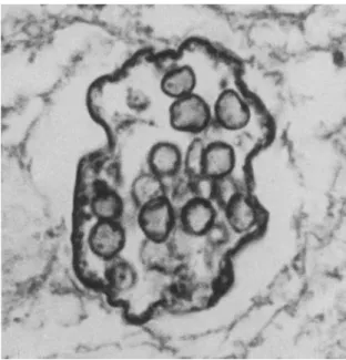
Tissue blocks were obtained from the other institution; sec-tions were cut and stained with hematoxylin and eosin, van Gie-son-Elastin, periodic acid-Schiff, Giemsa, Masson's trichrome, Pearl's stain, Grocott, and Lepehne benzioline for hemoglobin. The thick outer fibrohyalin capsule contained granulation tissue in places with occasional foreign body giant cells and groups of foam cells near cholesterol crystals in certain areas. The cavity contained a fine granular material in which were numerous thin-walled spherules of various sizes. Most of these contained 2-20 smaller spherules (4-6 J.lm). The histologic picture and staining characteristics were consistent with myospherulosis (figureI).
The first description, by McClatchie et al. [1] in 1969, of myo-spherulosis in Africa concerned cases found in subcutaneous tis-sue. In 1977 in the USA, Kyriakos [2] described 16 cases, all iden-tified as ear, nose, and throat (ENT) pathology. Only 6 cases have been documented in Europe, all in France. Of the 67 cases de-scribed worldwide, 36 had an ENT localization, 28 were in pe-ripheral soft tissues, and 3 in unusual locations: breast, ovary, and central nervous system. Only one documented case involv-ing subcutaneous tissues has been clearly linked to an antisyphi-litic treatment consisting of intramuscular injections of benzathine penicillin [3]. In the present case, the first to our knowledge to be reported from Switzerland, we think the development of myospherulosis can be directly linked to the intramuscular injec-tions of procaine penicillin administered 14 y earlier.
Theories regarding the pathogenesis of myospherulosis have evolved from early hypotheses implicating fungi, algae, and pollens to current points of view that the lesions are iatrogenic. Experimen-tally it has been shown that contact between oil-based substances
Please address requests for reprints to Dr. Martine Bouvier, Unite de Sante communautaire et Medecine tropicale, Hopital Cantonal, 1211 Geneve 4, Switzerland.
913
Figure1. Sac containing numerous spherules 4-6 J.lm in diameter (hematoxylin-eosin stain; x 560).
and red blood cells can produce a lesion typical of myospherulo-sis. The substances may be exogenous, such as hemostatic gauze impregnated with petrolatum or antibiotics, as seen in the ENT cases, or oily preparations of injectable drugs as seen in the sub-cutaneous cases. Endogenous substances such as debris of trau-matic or necrotic origin have also been implicated in the patho-genesis. The coating of erythrocytes by lipids produces the typical spherules ("bag of marbles") seen on histologic examination [4-6]. The incidence of myospherulosis is probably greatly underes-timated considering the wide use of intramuscular injections, in particular of antibiotics. In the present case, the long delay be-fore the appearance of symptoms, the unspecific character of the lesions, and the difficulty in identifying the unusual histologic lesion all delayed the correct diagnosis. This case reminds us that myospherulosis must be included in the differential diagnosis of subcutaneous nodules of undetermined origin to prevent patients from undergoing unnecessary investigations and treatment.
SABINE KINlOCH, MARTINE BOUVIER, SEBASTIAN LUCAS, N. JEREMIAH Cox Dermatology and Medical Outpatient Clinics and Tropical Medicine Unit, H6pital Cantonal Universitaire, and Department of Clinical Pathology, Centre Medical Universitaire, Geneva, Switzerland; and Department of Histopathology, University College and Middlesex School of Medicine, London, UK
References
1. McClatchie S, Warambo MW, Bremner AD. Myospherulosis: a previously unreported disease? Am J Clin Pathol 1969; 51:699-704
914
and middle ear. A possible iatrogenic disease. Am J Clin Pathol1977;67:118-130
3. Beurlet J, Groussard0,Ravisse P, Berger G, Destombes P. La myospherulose: une enigme posee par de curieux "sacs de billes." Arch Anat Cytol Pathol1979;27:5-13 4. Rosai J. The nature of myospherulosis of the upper
respira-tory tract. Am J Clin Pathol1978;69:475-481
Correspondence
5. Wheeler TM, McGavran MH. Myospherulosis-further ob-servations. Am J Clin Pathol1980;73:685-686. 6. Vuong PN, BavieraE,Houissa-Vuong S, Fombeur JP, Oriol
R, Mesnil C. La myospherulose: une maladie iatrogene. A propos de deux observations avec etude immunomor-phologique et experimentale. Semin Hop Paris1985;61:9-15
THE JOURNAL OF INFECTIOUS DISEASES • VOL. 160, NO.5. NOVEMBER 1989 ©1989 by The University of Chicago. All rights reserved. 0022-1899/89/6005-0038$01.00
Registry on Mucormycosis in Dialysis Patients
COLLEAGUES - Sane et at.[1]reported a patient with myelofibro-sis and transfusional iron overload, treated with deferoxamine, who died from disseminated mucormycosis caused byRhizopus oryzae.An analogous case was reported recently [2], and deferox-amine therapy is proposed to be the risk factor for this zygomy-cete infection.Sane et at. [1] noted that mucormycosis has been reported in hemodialysis patients worldwide. As of1February1989, 25cases had been reported among nondiabetic dialysis patients[3-9],and we are aware of several more unpublished cases.
Several authors have linked this infectious complication to the use of deferoxamine. Of the25reported dialysis patients,22were receiving deferoxamine for the treatment of either aluminum over-load (18 cases) or iron overover-load (4 cases). The infection was fatal in22 (88070)of25patients. In most instances, the diagnosis was made postmortem. When the fungus was cultured, it was always genusRhizopus.The mechanism underlying this fulminant in-fection in dialysis patients treated with deferoxamine is unknown. To identify predisposing factors, we are establishing a registry of dialysis patients who have developed mucormycosis. Thus, we request physicians who have observed this mycosis in dialysis pa-tients to contact us. We will respond with a detailed question-naire in the hope of identifying risk factors for this infection. (To respond, see footnotes.)
To report patients to the registry, questionnaires may be obtained from the following: Dr. Johan Boelaert, Algemeen Ziekenhuis St Jan, B-8000 Brugge, Belgium; Dr. Andrew Fenves, Baylor University Medical Center, 3550 Gaston Ave., Dallas, TX 75246; and Dr. Jack Coburn, Nephrology Section (WlllL), West Los Angeles V.A. Medical Center, Los Angeles, CA 90073.
Reprints are available 'from Dr. Boelaert at the address shown above.
JORAN R. BOELAERT, ANDREWZ.FENVES, JACK W. COBURN Algemeen Ziekenhuis St Jan, Brugge, Belgium; Baylor University Medical Center, Houston, Texas; West Los Angeles (California) Veterans Administration Hospital
References
1. Sane A, Manzi S, Perfect J, Herzberg AJ, Moore JO. Deferox-amine treatment as a risk factor for zygomycete infection [letter]. J Infect Dis1989;159:151-152
2. Rex JH, Ginsberg AM, Fries LF, Pass HI, Kwong-Chung KJ. Cunninghamella bertholletiae infection associated with deferoxamine therapy. Rev Infect Dis1988;10:1187-1194 3. Boelaert JR, van Roost GF, Vergauwe PL, Verbanck 11, de
Vroey C, Segaert ME The role of desferrioxamine in dialysis-associated mucormycosis: report of three cases and review of the literature. Clin Nephrol1988;29:261-266 4. Windus OW, Stokes TJ, Julian BA, Fenves AZ. Fatal
rhizo-pus infections in hemodialysis patients receiving deferoxa-mine. Ann Intern Med1987;107:678-680
5. Segal R, Zoller KA, Sherrard OJ, Coburn JW. Mucormyco-sis: a life-threatening complication of deferoxamine ther-apy in long-term dialysis patients [abstract]. Kidney Int 1988;33:238
6. Sombolos K, Kalekou H, Barboutis K, Tzarou V. Fatal phycomycosis in a hemodialyzed patient receiving deferox-amine [letter]. Nephron1988;49:169-170
7. Kerr PG, Turner H, Davidson A, Bennett C, Maslen M. Zygomycosis requiring amputation of the hand: an isolated case in a patient receiving haemodialysis. Med J Aust 1988;148:258-259
8. Morris OJ, Altus P. Rhinocerebral mucormycosis in an anephric patient. South Med J 1988;81:400-403
9. Hisanaga S, Fujimoto S, Wakisaka 0, Yamamoto Y, Tamaka K, Koita H, Kono T. Cardiac mucormycosis associated with deferoxamine use in a hemodialysis patient [in Japanese]. Kidney and Dialysis1988;24:143-148