Revised ESTS guidelines for preoperative mediastinal lymph node
staging for non-small-cell lung cancer
†
Paul De Leyn
a,*, Christophe Dooms
b, Jaroslaw Kuzdzal
c, Didier Lardinois
d, Bernward Passlick
e,
Ramon Rami-Porta
f, Akif Turna
g, Paul Van Schil
h, Frederico Venuta
i, David Waller
j,
Walter Weder
kand Marcin Zielinski
l aDepartment of Thoracic Surgery, University Hospitals Leuven, Leuven, Belgium
b Department of Pneumology, University Hospitals Leuven, Leuven, Belgium c
Department of Thoracic Surgery, Jagiellonian University Collegium Medicum Krakow, Krakow, Poland
d Department of Thoracic Surgery, University Hospital Basel, Basel, Switzerland e
Department of Thoracic Surgery, Albert-Ludwigs-University Freiburg, Freiburg, Germany
f Department of Thoracic Surgery, University Hospital Mutua de Terrassa and CIBERES Lung Cancer Group, Terrassa, Barcelona, Spain g
Department of Thoracic Surgery, University Hospital Istanbul, Istanbul, Turkey
h Department of Thoracic and Vascular Surgery, Antwerp University Hospital, Antwerp, Belgium i
Department of Thoracic Surgery, University Hospital, Rome, Italy
j Department of Thoracic Surgery, Glenfield Hospital Leicester, Leicester, UK k
Department of Thoracic Surgery, University Hospital Zurich, Zurich, Switzerland
l Department of Thoracic Surgery, Pulmonary Hospital Zakopane, Zakopane, Poland
* Corresponding author. Department of Thoracic Surgery, University Hospitals Leuven, Herestraat 49, 3000 Leuven, Belgium. Tel: +32-16346820; fax: +32-16346821; e-mail: [email protected] (P. De Leyn).
Received 3 October 2013; received in revised form 16 December 2013; accepted 20 December 2013
Abstract
Accurate preoperative staging and restaging of mediastinal lymph nodes in patients with potentially resectable non-small-cell lung cancer (NSCLC) is of paramount importance. In 2007, the European Society of Thoracic Surgeons (ESTS) published an algorithm on preoperative mediastinal staging integrating imaging, endoscopic and surgical techniques. In 2009, the International Association for the Study of Lung Cancer (IASLC) introduced a new lymph node map. Some changes in this map have an important impact on mediastinal staging. Moreover, more evidence of the different mediastinal staging technique has become available. Therefore, a revision of the ESTS guidelines was needed. In case of computed tomography (CT)-enlarged or positron emission tomography (PET)-positive mediastinal lymph nodes, tissue confirmation is indicated. Endosonography [endobronchial ultrasonography (EBUS)/esophageal ultrasonography (EUS)] withfine-needle aspiration (FNA) is thefirst choice (when available), since it is minimally invasive and has a high sensitivity to rule in mediastinal nodal disease. If negative, surgical staging with nodal dissection or biopsy is indicated. Video-assisted mediastinoscopy is preferred to mediastinoscopy. The combined use of endoscopic staging and surgical staging results in the highest accuracy. When there are no enlarged lymph nodes on CT and when there is no uptake in lymph nodes on PET or PET–CT, direct surgical resection with systematic nodal dissection is indicated for tumours ≤3 cm located in the outer third of the lung. In central tumours or N1 nodes, preoperative mediastinal staging is indicated. The choice between endoscopic staging with EBUS/EUS and FNA or video-assisted mediastinoscopy depends on local expertise to adhere to minimal requirements for staging. For tumours >3 cm, preoperative mediastinal staging is advised, mainly in adenocarcinoma with high standardized uptake value. For restaging, invasive techniques providing histological information are advisable. Both endoscopic techniques and surgical procedures are available, but their negative predictive value is lower compared with the results obtained in baseline staging. An integrated strategy using endoscopic staging techniques to prove mediastinal nodal disease and mediastinoscopy to assess nodal response after induction therapy needs further study. Keywords:Lung cancer• Preoperative staging • Surgical staging • Endoscopic staging • Restaging
INTRODUCTION
For patients with non-small-cell lung cancer (NSCLC) and no systemic metastasis, mediastinal staging is very important as it pro-vides accurate information on the extent of the disease, guides the choice of treatment and determines the patient’s prognosis.
In 2007, the European Society of Thoracic Surgeons (ESTS) pub-lished an algorithm on preoperative mediastinal staging based on the current available literature [1]. These guidelines integrated imaging, endoscopic and surgical techniques. They were widely used and have been prospectively validated. Their negative pre-dictive value (NPV) is 0.94 [2].
However, since 2007, there have been substantially more infor-mation and evidence on mediastinal staging techniques. In 2009, the International Association for the Study of Lung Cancer (IASLC) †Presented at the 21st European Conference on General Thoracic Surgery,
Birmingham, UK, 26–29 May 2013.
© The Author 2014. Published by Oxford University Press on behalf of the European Association for Cardio-Thoracic Surgery. All rights reserved.
G
U
IDELIN
E
introduced a new lymph node map of the lungs and mediastinum that resulted from an international and multidisciplinary consen-sus [3]. Some new changes in this map have an important impact on mediastinal staging. Moreover, new insights into the import-ance of restaging and techniques for mediastinal restaging have become available. Therefore, the ESTS Council approved the ini-tiative by the working group to revise and update the previous guidelines on mediastinal staging.
METHODOLOGY
There were several meetings of the working group. The project was discussed in the Council at the ESTS meeting in Essen ( June 2012). There were several meetings (Essen, Zürich, Brussels and Birmingham) where the participants presented their experience and discussed the relevant literature published since 2007. Initial findings were presented and discussed at the ESTS meeting in Birmingham (May 2013). Thefinal paper was put on the website for discussion by all ESTS members. Their remarks were discussed and included in thefinal manuscript.
For recommendations, a level of evidence and grading of rec-ommendation is given. This was adapted from the Infectious Diseases Society of America–United States Public Health Service grading system (Table1) [4].
It is evident that both in primary staging and in restaging, not every technique is available in every centre. Therefore, staging and restaging techniques can differ between different countries and centres.
IMPACT OF NEW IASLC LYMPH NODE MAP
There are several modifications compared with the previous Naruke and Mountain and Dresler maps [5,6], but probably the most important modification from the clinical point of view is the shift of the anatomical mediastinal midline to the left paratracheal margin, the so-called mediastinal oncological midline [3]. This change is important to be understood by radiologists, bronchos-copists, nuclear medicine specialists and surgeons because they have to locate the nodes correctly. The clinical implications of this new definition of the mediastinal midline, that affect exclusively nodal stations 2R, 2L, 4R and 4L (for the rest of the nodal stations,
the mediastinal midline remains unchanged) is that involved pre-tracheal lymph nodes and lymph nodes on the left of the anatom-ical midline but on right side of the oncologanatom-ical midline are classified as N2 in case of right-lung tumours but as N3 in case of left-lung tumours.
Another important modification for mediastinal staging is that the anatomical borders of the lymph node (LN) stations are clearly defined. This is especially relevant for the lower border of stations 4R and 4L. For the right lower paratracheal lymph nodes (station 4R), the lower border is the lower margin of the azygos vein. On the left side, the lower border of the left paratracheal lymph nodes (station 4L) is the upper rim of the left pulmonary artery. By cervical mediastinoscopy (and by endoscopic techniques), the lymph nodes below the azygos vein and below the upper rim of the pulmonary artery can be biopsied and they should be labelled, respectively, as 10R and 10L (Fig.1and Table2with permission from IASLC).
Definition of nodal zone and nodal station
A nodal zone is an anatomical area that includes one or several neighbouring nodal stations. The supraclavicular and the subcar-inal zones include one nodal station each, station 1 and station 7, respectively. However, the limits of both nodal stations 1 and 7 are wider than they used to be in the previous maps. The other nodal zones include two, three or six nodal stations. It is import-ant to realize that, in theory, a single N2 zone may have from one to multiple nodes involved in one or several nodal stations, and the nodes may be small or large. The concept of nodal zones is of especial value for those patients who will not undergo surgi-cal treatment. For those receiving chemotherapy, radiotherapy or their combination, the precise anatomical location of the nodes involved is not so important. So, the nodal zones help locate nodal involvement without having to define the exact ana-tomical location of the nodes. However, nodal stations are im-portant for those patients in whom surgical treatment is required. Precise nodal location is important preoperatively to guide surgi-cal treatment, and also intra- and postoperatively to indicate further treatment. This is especially relevant in the upper medi-astinal zone. Whether the right or the left paratracheal nodes are involved or not is important to confirm or rule out N2 or N3 disease and to select patients for surgical (multimodality) treatment.
Table 1: Level of evidence and grading of recommendation
Level of evidence
I Evidence from at least one large randomized controlled trial of good methodological quality (low potential for bias) or meta-analyses of well-conducted randomized trials without heterogeneity
II Small randomized trials or large randomized trials with a suspicion of bias (lower methodological quality) or meta-analyses of such trials or of trials with demonstrated heterogeneity
III Prospective cohort studies
IV Retrospective cohort studies or case–control studies
V Studies without control group, case reports and experts opinions Grading of recommendation
A Strong evidence for efficacy with a substantial clinical benefit, strongly recommended
B Strong or moderate evidence for efficacy but with a limited clinical benefit, generally recommended
C Insufficient evidence for efficacy or benefit does not outweigh the risk or the disadvantages (adverse events, costs,…), optional D Moderate evidence against efficacy or for adverse outcomes, generally not recommended
RATIONALE FOR PREOPERATIVE MEDIASTINAL
NODAL STAGING
The current guidelines for the treatment of lung cancer are deter-mined by the clinical status of the mediastinal nodes. The aim of
mediastinal staging is to exclude with the highest certainty and the lowest morbidity patients with mediastinal nodal disease since these patients will not benefit from upfront surgery [7,8].
There is controversy regarding the best treatment of N2 disease because of the heterogeneity of nodal involvement. Also, patient
Figure 1:The IASLC lymph node map including the proposed grouping of lymph node stations into‘zones’ for the purposes of prognostic analysis (Rusch et al. [3] with permission).
G
U
IDELIN
and tumour characteristics and extent of resection play a role in the selection of treatment modality for these patients. In the IASLC paper [9], 4277 of 11 619 patients clinically staged as cN2cM0 underwent resection and had information on pN
category. Only a subgroup of 2876 patients underwent complete (R0) resection without any induction therapy and had information on nodal location and pN category based on pathological staging from lymphadenectomy. An exploratory analysis of the impact of Table 2: Anatomical definitions for each lymph node station and station grouping by nodal zones
Lymph node station Anatomical limits Supraclavicular zone
#1: Low cervical, supraclavicular and sternal notch nodes
Upper border: lower margin of cricoid cartilage.
Lower border: clavicles bilaterally and, in the midline, the upper border of the manubrium. 1R designates right-sided nodes, and 1L left-sided nodes in this region.
For lymph node station 1, the midline of the trachea serves as the border between 1R and 1L. Upper zone
#2: Upper paratracheal nodes 2R: Upper border: apex of the right lung and pleural space and, in the midline, the upper border of the manubrium. Lower border: intersection of caudal margin of innominate vein with the trachea.
As for lymph node station 4R, 2R includes nodes extending to the left lateral border of the trachea.
2L: Upper border: apex of the lung and pleural space and, in the midline, the upper border of the manubrium. Lower border: superior border of the aortic arch.
#3: Prevascular and retrotracheal nodes
3a: Prevascular.
On the right: Upper border: apex of the chest. Lower border: level of carina. Anterior border: posterior aspect of the sternum. Posterior border: anterior border of the superior vena cava.
On the left: Upper border: apex of the chest. Lower border: level of carina. Anterior border: posterior aspect of the sternum. Posterior border: left carotid artery.
3p: Retrotracheal.
Upper border: apex of the chest. Lower border: carina.
#4: Lower paratracheal nodes 4R: includes right paratracheal nodes, and pretracheal nodes extending to the left lateral border of the tracheal. Upper border: intersection of caudal margin of innominate vein with the trachea.
Lower border: lower border of the azygos vein.
4L: includes nodes to the left of the left lateral border of the trachea, medial to the ligamentum arteriosum. Upper border: upper margin of the aortic arch.
Lower border: upper rim of the left main pulmonary artery. Aorto-pulmonary zone
#5: Subaortic (aorto-pulmonary window)
Subaortic lymph nodes lateral to the ligamentum arteriosum. Upper border: the lower border of the aortic arch. Lower border: upper rim of the left main pulmonary artery. #6: Para-aortic nodes (ascending
aorta or phrenic)
Lymph nodes anterior and lateral to the ascending aorta and aortic arch. Upper border: a line tangential to the upper border of the aortic arch. Lower border: the lower border of the aortic arch.
Subcarinal zone
#7: Subcarinal nodes Upper border: the carina of the trachea.
Lower border: the upper border of the lower lobe bronchus on the left; the lower border of the bronchus intermedius on the right.
Lower zone
#8: Para-oesophageal nodes (below carina)
Nodes lying adjacent to the wall of the oesophagus and to the right or the left of the midline, excluding subcarinal nodes.
Upper border: the upper border of the lower lobe bronchus on the left; the lower border of the bronchus intermedius on the right.
Lower border: the diaphragm.
#9: Pulmonary ligament nodes Nodes lying within the pulmonary ligament. Upper border: the inferior pulmonary vein. Lower border: the diaphragm.
Hilar/interlobar zone
#10: Hilar nodes Includes nodes immediately adjacent to the mainstem bronchus and hilar vessels including the proximal portions of the pulmonary veins and main pulmonary artery.
Upper border: the lower rim of the azygos vein in the right; upper rim of the pulmonary artery on the left. Lower border: interlobar region bilaterally.
#11: Interlobar nodes Between the origin of the lobar bronchi.
a#11s: between the upper lobe bronchus and bronchus intermedius on the right. a
#11i: between the middle and lower bronchi on the right. Peripheral zone
#12: Lobar nodes Adjacent to the lobar bronchi. #13: Segmental nodes Adjacent to the segmental bronchi. #14: Subsegmental nodes Adjacent to the subsegmental bronchi.
The International Association for the Study of Lung Cancer lymph node map. Adapted with permission from ref. [3]. #: nodal station number.
a
lymph node zones on survival was performed in a subgroup (N = 1992 patients) from this cohort, finding that pathological single N1 zone ( pN1a) had better prognosis than multiple logical N1 zones ( pN1b), that the prognosis of multiple patho-logical N1 zones was the same as that of single pathopatho-logical N2 zone ( pN2a) andfinally, that the prognosis of multiple pathologic-al N2 zones ( pN2b) was significantly worse. Five-year survival rates for pN1a, pN1b, pN2a and pN2b were 48, 35, 34 and 20, re-spectively. These survival data should be interpreted with caution and have already been misinterpreted. It is important to have in mind that these survival analyses were performed in resected patients with pathologically staged tumours and thus based on results from lymphadenectomy: information on nodal status was available from station 2 and from stations 4 to 9 in all contributing institutions. Additionally, all centres but one provided documenta-tion on stadocumenta-tions 11 and 12, and most had informadocumenta-tion on stadocumenta-tions 1, 13 and 14, while half of them provided documentation on station 3. Therefore, the results from this highly selected pop-ulation of patients used for this specific analysis cannot be extra-polated to the clinical staging setting. This is why these data cannot be invoked to propose upfront surgical treatment for patients with presumed clinical single N2 zone determined with the current clinical staging guidelines. No pretreatment test [computed tomography (CT), positron emission tomography (PET), endobronchial ultrasonography (EBUS), esophageal ultra-sonography (EUS) and mediastinoscopy] can be compared with lymphadenectomy, except the lymphadenectomies performed through the transcervical approach (video-assisted mediastinal lymphadenectomy—VAMLA and transcervical extended medias-tinal lymphadenectomy— TEMLA). Therefore, there is room for a well-designed prospective study to evaluate the possible role of primary surgery in preoperatively proven single zone or single station N2 disease.
There is a subgroup of patients with pretreatment histologically proven N2 disease who are candidates for surgical multimodality treatment. These patients are treated with induction chemotherapy or induction chemoradiotherapy. In case of downstaging of the mediastinal lymph nodes or major response in those lymph nodes and in the tumour, resection with systematic nodal dissection can be performed with acceptable morbidity and mortality and reward-ing 5-year survival. There are several prognostic indicators, some of which are related to the primary tumour and others are related to the extent of nodal disease. To include patients for surgical multi-modality treatment, the disease should be initially technically resectable. Excluded for surgical multimodality are patients with unresectable disease such as extracapsular disease (can be clearly visualized by mediastinoscopy) or bulky N2 disease based on CT. Fit patients with extracapsular disease and/or bulky N2 disease should be treated with definitive chemoradiotherapy.
Bulky N2 disease is not well defined but it correlates with the radiographic group A, as described in the American College of Chest Physicians (ACCP) Evidence-based Clinical Practice Guidelines [10]. This group is defined as mediastinal infiltration, where the discrete lymph nodes cannot be distinguished or mea-sured. Bulky is not strictly related to the size of the lymph nodes, but it is considered by this committee that lymph nodes larger than 25 mm short axis will also be defined as bulky disease (Level V). Bulky disease can be restricted to a single station, but usually represents multistation or multiple zonal involvement. Since this paper deals with preoperative lymph node staging, techniques to obtain histology in bulky mediastinal nodal disease are beyond the scope of this article.
PRIMARY MEDIASTINAL LYMPH NODE STAGING
Several techniques are available and their use depends on local availability and local expertise.
These techniques include: (i) imaging techniques, (ii) endoscopic techniques and (iii) surgical techniques.
Although we should aim for the test with the highest sensitivity and NPV, the working group considers a rate of unforeseen pN2 disease of 10% as acceptable. After thorough mediastinal staging, this unforeseen pN2 is mostly single station resectable nodal disease.
Imaging techniques
Chest CT scan.
CT remains important in lung cancer imaging. However, due to its low sensitivity (55%) and specificity (81%), it is impossible to solely rely on CT scan [10]. A CT scan may help us in selecting the appropriate procedure for tissue sampling due to the anatomical images it provides.PET
–CT scan.
The addition of PET to CT results in more accurate lymph node staging than CT alone with an overall sensitivity of 80–90% and specificity of 85–95%. PET–CT has a high NPV for detecting mediastinal nodal disease in peripherally located NSCLC. Exceptions include:(i) suspected N1 nodes, (ii) tumour >3 cm and
(iii) centrally located tumour without suspected nodes on CT or PET scan.
(a) In a study from Japan [11], 30% of 143 patients with N1 disease on CT scan (lymph node short axis of >1 cm) were found to have pathological N2–N3.
(b) A recent meta-analysis [12] has shown that the NPV of PET–CT for tumours ≤3 cm was 94% (649 patients) com-pared with 89% for tumours >3 cm (130 patients) staged as T2 (6th edition of TNM). Thisfinding was confirmed in a recent prospective study from Spain [13]. For peripheral tumours≤3 cm, the NPV of PET–CT was 92% while it was 85% for tumours >3 cm. Based on these studies, we now recommend that for peripheral tumours (outer third of the lung)≤3 cm without enlarged (hilar and/or medias-tinal) lymph nodes on CT and with PET-negative nodes, further mediastinal staging can be omitted. There was a substantial difference in the rate of mediastinal nodal dis-ease between adenocarcinoma and other tumour hist-ology (risk ratio 2.72). Also, high 18F-Fluorodeoxyglucose (FDG) uptake in the primary lesion was associated with a greater risk of occult nodal metastasis. For tumours >3 cm (mainly adenocarcinoma with high FDG uptake), further mediastinal staging techniques providing histology should be considered (Fig.2).
(c) Leeet al. [14] examined the prevalence of pathological N2 disease in patients with clinical Stage I NSCLC (6th edition of TNM version) with negative mediastinum on PET and CT. In 2.9% of peripheral tumours (outer third of lung), N2 disease was found, while the prevalence of N2 disease was 21.6% in central tumours.
G
U
IDELIN
Diffusion-weighted magnetic resonance imaging.
Advances in magnetic resonance imaging (MRI) technology have allowed acquisition of diffusion-weighted MRI (DWI), which provides excellent tissue contrast because of the difference in the diffusion of water molecules among tissues. The technique yields qualitative and quantitative information that reflects changes at cellular level and provides unique insights into tumour cellularity and the integrity of cell membranes. In a recent meta-analysis [15], the accuracy of DWI and 18F-FDG-PET–CT was evaluated. The pooled sensitivity for DWI was 0.95 (95% CI 0.85–0.98) and significantly better than for FDG-PET–CT 0.89 (95% CI 0.85–0.91). However, at this moment, there are no large prospective studies comparing the value of DWI and FDG-PET and it is too early to determine the true value of DWI in nodal staging in patients with NSCLC.Endoscopic techniques
Conventional transbronchial needle aspiration.
Although the conventional transbronchial needle aspiration (TBNA) technique has been available for almost three decades, its use in routine clinical practice has been adopted only by a minority (10–15%) ofpulmonologists for mediastinal nodal staging of patients with potentially resectable Stage I–III lung cancer. Major reasons for its underuse are its dependency on nodal size (>15–20 mm short axis on CT scan) and operator skills. Meta-analyses reported a sensitivity of 78% and a false-negative rate of 28% for conventional TBNA in clinical N2 disease with a high disease prevalence of 81% [16,17]. A conventional blind TBNA is useful if it leads to proof of N3 disease, but too often does not exclude N3 disease in cases of proven N2 disease.
Endoscopic ultrasonography: EUS-
fine-needle aspiration
and EBUS-TBNA
Practical aspects.
Although E(B)US-TBNA is performed in some centres under general anaesthesia, EBUS and EUS are more often performed in an outpatient setting under local anaesthesia with moderate sedation.EBUS is able to visualize superior and inferior mediastinal LNs at stations 2R/2L, 4R/4L and 7, as well as hilar LNs at stations 10, 11 and even 12, as described on the new LN map [3]. EUS particularly visualizes superior mediastinal lymph nodes in station 4L and inferior mediastinal nodes in stations 7, 8 and 9, as described on the new LN map [3]. Thus, EUS-fine-needle aspiration (FNA)
complements other techniques, as several of these LNs (stations 8 and 9) are not accessible by EBUS-TBNA or mediastinoscopy. Although some expert centres considered EUS-FNA of lymph nodes in stations 5 or 6, currently available data are limited, and therefore, we do not recommend routine use of this procedure for this indication [18].
It is possible to visualize and sample lymph nodes with a short axis of >5 mm and the optimal number of aspirations per station has been reported to be 3 [19]. When mediastinal nodal staging is required, systematic nodal sampling is feasible by endosonogra-phy. Indeed, several endosonography series have shown a mean or median number of sampled mediastinal nodal stations of 3–4 per patient [20–26]. Nodal stations 4R, 4L and 7 should always be sought during the endosonographic examination and described in the medical report. In addition, the largest node measuring >5 mm on ultrasonography within each of these stations as well as FDG-avid nodes within each of these nodal stations should be sampled for pathological analysis. On indication, nodal stations 10R and 10L can be biopsied. To avoid contamination while using one single needle for an EBUS or EUS procedure, the order of nodal sampling should begin at the level of N3 nodes followed by N2 nodes before ending with N1 nodes.
Performance characteristics.
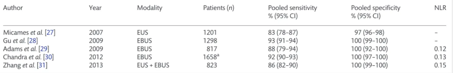
Several meta-analyses of EUS-FNA alone, EBUS-TBNA alone and combined EUS + EBUS reported a pooled sensitivity of 83–94% for mediastinal staging of lung cancer (Table2) [27–31]. Only one randomized controlled trial (Aster trial, 20) has been performed, comparing the two staging strategies proposed in the ESTS 2007 guidelines (either mediastinoscopy or alternatively endosonography followed by mediastinoscopy) [1]. There was no difference in sensitivity or NPV when mediastinoscpy was compared with endoscopic staging. However, the staging strategy starting with combined endosonography and if negative combining it with surgical staging has proved to detect significantly more mediastinal nodal N2/3 disease compared with media-stinoscopy alone [20]. Another consequence is that the implementation of endosonography for baseline mediastinal nodal staging clearly reduces the need for mediastinoscopy [32]. On the other hand, the negative likelihood ratio reported by three of the meta-analyses is 0.13–0.15 (Table3) [29–31]. This implies that the probability of having mediastinal nodal involvement for any individual patient with a negative endosonography result is 13–15%. This probability based on endosonography alone is in our opinion not low enough to directly proceed to a surgical resection. Therefore, in the routine practice, we still recommenda preoperative surgical staging procedure [i.e. video-assisted mediastinoscopy (VAM)] in case of a negative endosonography. However, there is evidence coming from prospective studies performed in experienced endosonography centres that mediastinoscopy may not improve sensitivity after a well-performed negative endosonography with needle aspiration of at least three mediastinal nodal stations in patients with low (<35%) prevalence of mediastinal disease [21,26,33]. EBUS-TBNA and EUS-FNA are safe procedures with reported minor complications in <1% of cases [27,
28,34]. With the rapidly increasing number of procedures, occasional
reports of moderate-to-severe complications have been published, such as pneumothorax requiring chest tube drainage, infection of bronchogenic cyst, empyema, lung and/or mediastinal abscess and haemopneumomediastinum. So far, only one death has been reported related to an EBUS-TBNA procedure [35].
Surgical staging techniques
Cervical mediastinoscopy.
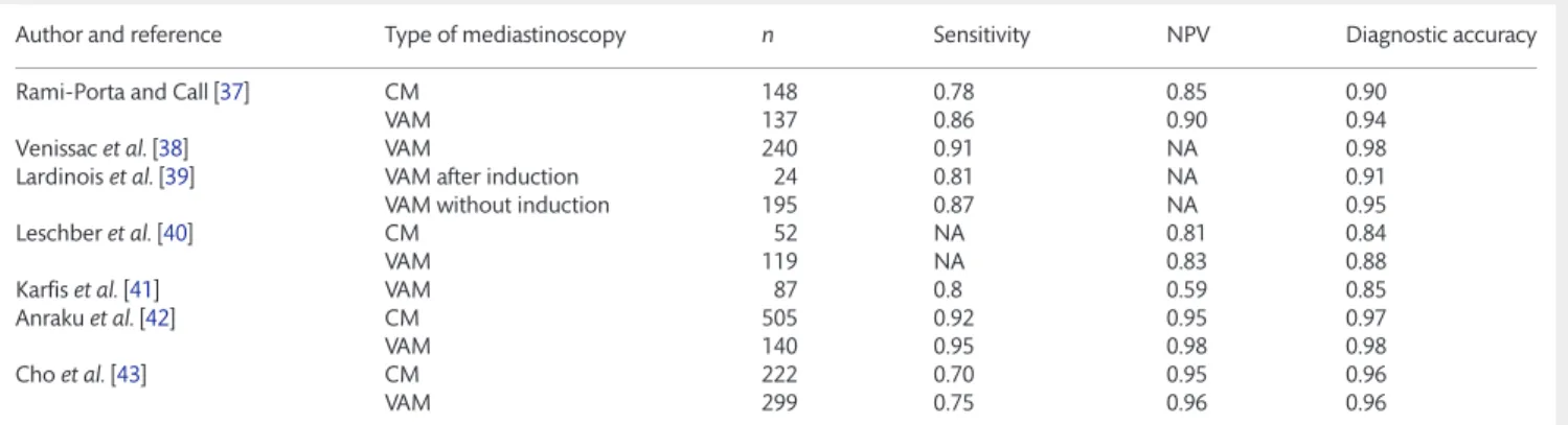
Cervical mediastinoscopy through a pretracheal suprasternal incision was introduced by Carlens in 1959 and further popularized by Pearson in North America. It allows a full mapping of the ipsilateral and contralateral superior mediastinal lymph nodes. Cervical mediastinoscopy is performed under general anaesthesia and can be safely done as an outpatient procedure. For many years, it was the gold standard for invasive staging of patients with potentially operable lung cancer. Since 1995, use of video techniques has been introduced leading to VAM. VAM clearly improved visualization and teaching [36] since both the trainer and the trainee can share the magnified image on the monitor. For more details on the technique of cervical mediastinoscopy, we refer to a recent publication on this topic [37].There are only retrospective studies comparing the safety and accuracy of conventional mediastinoscopy with VAM (Table 4). Although some authors [40, 42, 43] found an increase in the number of LN or LN stations biopsied, no difference in sensitivity or NPV was found (Table4). In some of these studies, a reduction in the complication rate (mainly of recurrent nerve palsy) was observed. Very recently [44], a best evidence topic has been published on the safety and accuracy of VAM compared with con-ventional mediastinoscopy (Table 5). The authors analysed 108 papers published between 1989 and 2011. There were 5156 conventional mediastinoscopies and 956 VAMs. Both procedures
Table 3: Published meta-analyses on endobronchial and oesophageal endosonography with FNA for mediastinal nodal staging of lung cancer
Author Year Modality Patients (n) Pooled sensitivity % (95% CI)
Pooled specificity % (95% CI)
NLR
Micameset al. [27] 2007 EUS 1201 83 (78–87) 97 (96–98) – Guet al. [28] 2009 EBUS 1298 93 (91–94) 100 (99–100) – Adamset al. [29] 2009 EBUS 817 88 (79–94) 100 (92–100) 0.12 Chandraet al. [30] 2012 EBUS 1658a 92 (90–93) 100 (97–100) 0.13
Zhanget al. [31] 2013 EUS + EBUS 823 86 (82–90) 100 (99–100) 0.15
a
Some small series also included sarcoidosis.
n: number; EUS: esophageal ultrasonography; EBUS: endobronchial ultrasonography; NLR: negative likelihood ratio.
G
U
IDELIN
are safe with no mortality in that time frame and a low morbidity. Although by VAM more lymph node stations are sampled, the NPV and accuracy were identical.
Although the videomediastinoscope is not strictly necessary to achieve a thorough, clinically acceptable mediastinoscopy, it has many advantages over the conventional one: larger and clearer images, the possibility to simultaneously share the procedure with trainees and all personnel in the operative theatre, the possibility to record the operation for future educational uses and discussion and the possibility to improve its teaching without compromising the safety or accuracy of the procedure. Moreover, it allows bi-manual dissection with possibilities to perform nodal dissection and removal rather than sampling or biopsy. This is especially im-portant and technically feasible for the subcarinal LN station. After removal of station 7 LNs, the oesophagus can be clearly visualized. The ESTS working group recommends performing VAM.
Video-assisted thoracoscopic surgery.
Although video-assisted thoracoscopic surgery (VATS) can reach almost every mediastinal lymph node station, it is more invasive than cervical mediastinoscopy (it needs double lumen intubation), it is limited by pleural adhesions and it can evaluate only ipsilateral nodal disease. For the para-aortic lymph nodes (station 6) and thesubaortic lymph nodes (station 5), left VATS is a surgical technique that allows obtaining large tissue samples. It is indicated when enlarged PET-positive lymph nodes are visualized at Level 5 or 6. These lymph node stations cannot be biopsied by routine mediastinoscopy, E(B)US-FNA. An alternative to VATS is the left anterior mediastinotomy. In some experienced centres, extended mediastinoscopy is performed for these lymph node stations and it gives good NPVs: 0.89–0.97 [37]. Extended cervical mediastinoscopy is performed from the mediastinoscopy incision [45].
VAMLA and TEMLA.
During the last decade, two new invasive staging techniques representing more radical methods of mediastinal exploration have been introduced: VAMLA [46] and TEMLA [47]. These two techniques aim for a complete removal of all mediastinal nodes with the surrounding adipose tissue to improve the accuracy of staging. VAMLA is completely performed with the use of a videomediastinoscope, whereas TEMLA uses a 5- to 8-cm collar incision in the neck and elevates the sternum with a hook. The dissection is performed in an open way and with the use of a videomediastinoscope. By VAMLA, the lymph nodes which are usually accessible through mediastinoscopy are removed. By TEMLA, more lymph node stations are accessibleTable 4: Staging values of conventional mediastinoscopy and videomediastinoscopy
Author and reference Type of mediastinoscopy n Sensitivity NPV Diagnostic accuracy Rami-Porta and Call [37] CM 148 0.78 0.85 0.90
VAM 137 0.86 0.90 0.94
Venissacet al. [38] VAM 240 0.91 NA 0.98 Lardinoiset al. [39] VAM after induction 24 0.81 NA 0.91 VAM without induction 195 0.87 NA 0.95 Leschberet al. [40] CM 52 NA 0.81 0.84
VAM 119 NA 0.83 0.88
Karfiset al. [41] VAM 87 0.8 0.59 0.85 Anrakuet al. [42] CM 505 0.92 0.95 0.97
VAM 140 0.95 0.98 0.98
Choet al. [43] CM 222 0.70 0.95 0.96
VAM 299 0.75 0.96 0.96
Adapted from Rami-Porta and Call [37].
CM: conventional mediastinoscopy;n: number of patients; NA: not available; NPV: negative predictive value; PPV: positive predictive value; VAM: video-assisted mediastinoscopy.
Table 5: Overall comparison VAM vs CM (studies 1989–2011)
VAM (n = 956) CM (n = 5156) P-value
Mortality 0 0
Morbidity 0.83–2.9% 0–5.3% NS
No. of LN biopsied 6–8.5 5–7.13 NS
No. of LN stations sampled 1.9–3.6 2.6–2.98 NS
Accuracy 87.9–98.9% 83.8–97.2% NS
NPV 83.0–98.6% 81.0–98.7% NS
Adapted from Zakkaret al. [44].
such as the prevascular, the para-aortic, the subaortic and the para-oesophageal lymph node stations. The NPV is very high and approaches 98.7% for TEMLA. The results of VAMLA and TEMLA regarding sensitivity and side effects are shown in Table4. Although there is no doubt that the accuracy of mediastinal staging increases when lymphadenectomy is performed compared with nodal biopsy, these techniques have a higher morbidity and mortality. The complications after VAMLA and TEMLA are well recorded (Table6) and are probably more studied in detail than after CM or VAM. These procedures are performed in very experienced centres. For VAMLA, mainly problems with recurrent nerve palsy and important scarring with an impact on subsequent resection are reported. The published data for TEMLA are mainly from one very experienced centre and there are concerns on morbidity and mortality.
For TEMLA and VAMLA, we conclude that currently available data regarding its use are limited and, therefore, we do not recommend its use except of clinical trials. We encourage other centres to publish their data with these new staging techniques.
MINIMAL REQUIREMENTS FOR MEDIASTINAL
NODAL STAGING
The ESTS clinical practice guidelines 2013 for preoperative medi-astinal nodal staging recommend that at least the following nodal stations should be explored and biopsied:
(i) right and left lower paratracheal lymph nodes (stations 4R and 4L) and
(ii) subcarinal lymph nodes (station 7).
If present, the right and left upper paratracheal stations 2R and 2L should also be biopsied.
When required to determine subsequent treatment strategy, lymph node stations 10R (below the azygos vein) and 10L (below the upper rim of left pulmonary artery) should be biop-sied. In case of left-sided tumours, stations 5 and 6 should be biopsied if it changes the treatment strategy. The same applies
to the lower mediastinal lymph nodes (stations 8 and 9). Nodal biopsy of these stations could be indicated in case extracapsular (non-resectable) nodal disease is expected from imaging studies.
ALGORITHM FOR PRIMARY MEDIASTINAL
STAGING
The algorithm for preoperative mediastinal staging is shown in
Fig.2. For NSCLC, both for mediastinal and for distant staging, PET
or PET–CT is indicated.
(i) Direct surgery can be performed if all of these three criteria apply: no suspect lymph node detected by CT or PET, a tumour of≤3 cm (Stage IA), located in the outer third of the lung (Level IIA).
(ii) In case of enlarged mediastinal lymph nodes on CT or PET-positive lymph nodes, tissue confirmation is indicated. In this case, endosonography (EBUS/EUS) with FNA is thefirst choice (when available) since it is minimally invasive and has a high sensitivity to rule in mediastinal nodal disease (Level IA). If negative, video-assisted mediastinoscopy is indicated (Level IB). The combined use of endoscopic staging and sur-gical staging results in the highest accuracy. For patients with a left upper lobe tumour, surgical staging of the aorto-pulmonary window nodes (if enlarged on CT and/or PET– CT-positive) can be performed (by anterior mediastinotomy, VATS or extended cervical mediastinoscopy) if involvement changes treatment strategy (Level V).
(iii) Invasive staging by E(B)US/mediastinoscopy is indicated if at least one of these criteria applies: central lesion, suspect N1 nodes (Level IIB). In case of tumours >3 cm (mainly in adeno-carcinoma with high FDG uptake), the NPV for mediastinal nodal disease is <90% and invasive staging may be considered (Level IIB). Although a high FDG update in the primary tumour is a predictor of N2 disease, the ideal cut-off of stan-dardized uptake value (SUV) has not yet been determined above which invasive mediastinal nodal staging is required. In addition, the SUV measurement is not yet standardized from Table 6: Results of VAMLA and TEMLA
Author Procedure n NPV Sensitivity Side effect
Hürtgenet al. [46] VAMLA 46 100% 100% Recurrent laryngeal nerve palsy: 2.2%
Scarring with impact on subsequent resection: 25% Leschberet al. [48] VAMLA 23 100% 100% Blood loss >100 ml : 12%
Witteet al. [49] VAMLA 144 NA 100% Recurrent laryngeal nerve palsy: 3.4% Vascular lesions: 2.1%
Mediastinitis: 0.7% Marked scarring: 19% Zielinskiet al. [50] TEMLA 256 97.4% 94% Mortality: 0.3%
Recurrent laryngeal nerve palsy: 2.5% Pneumothorax: 0.7%
Pleural effusion: 1.1%
Yooet al. [51] VAMLA 108 NA NA Recurrent laryngeal nerve palsy: 3.4% n: number of patients; NPV: negative predictive value; VAMLA: video-assisted mediastinal lymphadenectomy; NA: not available; TEMLA: transcervical extended mediastinal lymphadenectomy.
G
U
IDELIN
one centre to another, and therefore, a visual interpretation of the FDG uptake on PET is to be preferred [52]. In all of the above-mentioned cases, there is the choice between VAM with biopsy or lymph node dissection or endoscopic staging by EBUS/EUS with FNA. The choice depends on local expert-ise to adhere to minimal requirements for staging (Level V). If video-assisted mediastinoscopy is negative, these patients can undergo surgical treatment. They also can undergo surgi-cal treatment after negative EBUS/EUS if the number of nodes explored and the number of needle passes in each node meet the established requirements. Otherwise, surgical ex-ploration is recommended after negative EBUS/EUS.
(iv) If only CT is available, we refer to the algorithm of the 2007 edition of the ESTS guidelines [1].
MEDIASTINAL RESTAGING AFTER INDUCTION
THERAPY
Mediastinal downstaging after induction therapy for locally advanced Stage III NSCLC is an important prognostic factor for long-term survival. Patients with persisting mediastinal involve-ment have a worse prognosis compared with those with proven mediastinal downstaging.
The same techniques used in primary staging can be used for mediastinal restaging. Non-invasive imaging techniques are not accurate enough for mediastinal restaging. PET provides additional metabolic information, but there are conflicting data regarding its use. PET has been shown to be more accurate in predicting the T
component than the N status [52]. Although experience is rather limited, integrated PET–CT combining precise anatomical and functional information seems to be more accurate for restaging [53]. In a prospective study of 93 patients who were restaged by chest CT and integrated PET–CT after induction chemoradiother-apy, repeat PET–CT was found to be more accurate than CT alone for all pathological stages [54]. However, there were 20% false-negative and 25% false-positive cases. So, in case of suspicion of residual mediastinal disease, nodal biopsies are still required [54].
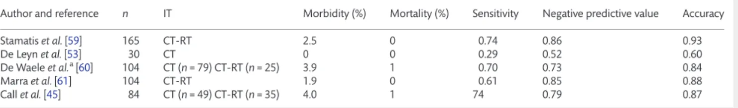
Different techniques providing histology can be used for re-staging (Tables7and8). Endoscopic techniques can be used, but EBUS-TBNA reported a variable NPV of 20 [56] and 78% [58]. The difference in the NPV may be explained by the prevalence of ypN2 after induction therapy, which was 94% in the study of Herth and 44% in the study of Szlubowski. These results empha-size that a negative EBUS for restaging should be confirmed by in-vasive surgical mediastinal restaging.
Remediastinoscopy (reMS) was found to be technically feasible, also after induction therapy [59,62]. However, reMS is used only in very selected experienced centres and is not widely adoptable due to severe fibrosis. Although feasible, the accuracy is lower than that of mediastinoscopy for primary staging and this ques-tions the timing of mediastinoscopy (baseline or at restaging, timing of further radiotherapy after induction which should not be delayed).
In experienced hands, TEMLA is also an accurate restaging tech-nique. In a series of 63 patients, induction chemotherapy (n = 54) or chemoradiotherapy (n = 9) was administered for N2 or N3 NSCLC. Initial mediastinoscopy was performed in 7 patients. Sensitivity, specificity and accuracy of TEMLA were 95.5, 100 and
Table 7: Restaging with EUS and EBUS after induction therapy
Technique Author n Sensitivity Specificity Accuracy EBUS Krasniket al. [55] 83 0.70 1.0 0.75 EBUS Herthet al. [56] 124 0.76a 1.0 0.77
EUS Stigtet al. [57] 25 0.92 1.0 0.92
EBUS Szlubowskiet al. [58] 61 0.67 0.86 0.80 EUS von Bartheldet al. [18] 58 0.44 NR 0.60
aNegative predictive value was only 20%.
EUS: endoscopic (oesophageal) ultrasound; EBUS: endobronchial ultrasound;n: number of patients; NR: not reported.
Table 8: Restaging with repeat mediastinoscopy after induction therapy
Author and reference n IT Morbidity (%) Mortality (%) Sensitivity Negative predictive value Accuracy Stamatiset al. [59] 165 CT-RT 2.5 0 0.74 0.86 0.93 De Leynet al. [53] 30 CT 0 0 0.29 0.52 0.60 De Waeleet al.a[60] 104 CT (n = 79) CT-RT (n = 25) 3.9 1 0.70 0.73 0.84 Marraet al. [61] 104 CT-RT 1.9 0 0.61 0.85 0.88 Callet al. [45] 84 CT (n = 49) CT-RT (n = 35) 4.0 1 74 0.79 0.87 a
Combined, updated series.
98.3%, respectively [63]. In a recent retrospective analysis from the same institution, EBUS/EUS and TEMLA performed for restaging after neoadjuvant treatment were compared in 78 patients. Sensitivity, specificity and NPV of TEMLA were 97, 100 and 99%, respectively [50].
Owing to severefibrosis, reMS should not be attempted after VAMLA or TEMLA.
Only one study reported the results of VATS for restaging after induction therapy [64]. In this Cancer and Leukemia Group B 39 803 trial, a negative result of VATS was defined as negative lymph node biopsies from at least three lymph node stations, whereas a positive result consisted of a pathological proof of per-sisting N2 disease or the demonstration of pleural carcinomatosis. Sensitivity, specificity and NPV of VATS for restaging were 67, 100 and 73%, respectively. Restaging by VATS is feasible, but requires single-lung ventilation and is limited to one hemithorax only.
An alternative approach that needs prospective validation is to rely on endosonography for baseline mediastinal nodal staging and afirst mediastinoscopy for restaging after induction therapy. In this‘restaging’ setting, the NPV of ‘a first and more easy and safe mediastinoscopy’ was 90% (with a prevalence of ypN2 of 46%) [39].
We conclude that optimal mediastinal lymph node staging is a truly multidisciplinary process, with a variety of possible techni-ques, to be performed by experienced hands.
Conflict of interest: none declared.
REFERENCES
[1] De Leyn P, Lardinois D, Van Schil P, Rami-Porta R, Passlick B, Zielinski M et al. ESTS guidelines for preoperative lymph node staging for non-small cell lung cancer. Eur J Cardiothorac Surg 2007;32:1–8.
[2] Gunluoglu MZ, Melek H, Medetoglu B, Demir A, Kara H, Dincer S. The validity of preoperative lymph node staging guidelines of the European Society of Thoracic Surgeons in non-small cell lung cancer. Eur J Cardiothorac Surg 2011;40:287–90.
[3] Rusch VW, Asamura H, Watanabe H, Giroux DJ, Rami-Porta R, Goldstraw P. The IASLC lung cancer staging project. A proposal for a new international lymph node map in the forthcoming seventh edition of the TNM classifi-cation for lung cancer. J Thorac Oncol 2009;4:568–77.
[4] Dykewicz CA. Summary of guidelines for preventing opportunistic infections among hematopoietic stem cell transplant recipients. Clin Infect Dis 2001;33:139–44.
[5] Naruke T, Suemasu K, Ishikawa S. Lymph node mapping and curability at various levels of metastasis in resected lung cancer. J Thorac Cardiovasc Surg 1978;76:833–9.
[6] Mountain C, Dresler C. Regional lymph node classification for lung cancer staging. Chest 1997;111:1718–23.
[7] Pearson FG, Delarue NC, Ilves R, Todd TR, Cooper JD. Significance of posi-tive superior mediastinal nodes identified at mediastinoscopy in patients with resectable cancer of the lung. J Thorac Cardiovasc Surg 1982;83:1–11. [8] Funatsu T, Matsubaru Y, Hatakenaka R, Kosaba S, Yasuda Y, Ikeda S. The role of mediastinoscopic biopsy in preoperative assessment of lung cancer. J Thorac Cardiovasc Surg 1992;104:1688–95.
[9] Rusch VW, Crowley J, Giroux DJ, Goldstraw P, Im JG, Tsuboi Met al.. International Staging Committee; Cancer Research and Biostatistics; Observers to the Committee; Participating Institutions. The IASLC lung cancer staging project: proposals for the revision of the N descriptors in the forthcoming seventh edition of the TNM classifications for lung cancer. J Thorac Oncol 2007;2:603–12.
[10] Silvestri GA, Gonzalez AV, Jantz MA, Margolis ML, Gould MK, Tanoue LT et al. Methods for staging non-small cell lung cancer. Diagnosis and man-agement of lung cancer, 3rd ed: American College of Chest Physicians evidence-based clinical practice guidelines. Chest 2013;143(Suppl): e211s–50s.
[11] Hishida T, Yoshida J, Nishimura M, Nishiwaki Y, Nagai K. Problems in the current diagnostic standards of clinical N1 non-small cell lung cancer. Thorax 2008;63:526–31.
[12] Wang J, Welch K, Wang L, Kong F-M. Negative predictive value of positron emission tomography for stage T1-2N0 non-small-cell lung cancer: a meta-analysis. Clin Lung Cancer 2012;13:81–9.
[13] Gómez-Caro A, Boada M, Cabañas M, Sanchez M, Arguis P, Lomeña F et al. False-negative rate after positron emission tomography/computer tomography scan for mediastinal staging in cI stage non-small-cell lung cancer. Eur J Cardiothorac Surg 2012;42:93–100.
[14] Lee PC, Port JL, Korst RJ, Liss Y, Meherally DN, Altorki NK. Risk factors for occult mediastinal metastases in clinical stage I non-small cell lung cancer. Ann Thorac Surg 2007;84:177–81.
[15] Wu LM, Xu JR, Gu HY, Hua J, Chen J, Zhang Wet al. Preoperative medias-tinal and hilar nodal staging with diffusion-weighted magnetic resonance imaging and fluorodeoxyglucose positron emission tomography/com-puted tomography in patients with non-small-cell lung cancer: which is better? J Surg Res 2012;178:304–14.
[16] Holty J, Kuschner W, Gould M. Accuracy of transbronchial needle aspir-ation for mediastinal staging of non-small cell lung cancer: a meta-analysis. Thorax 2005;60:949–55.
[17] Detterbeck FC, Jantz M, Wallace M, Vansteenkiste J, Silvestri GA. American College of Chest Physicians. Invasive mediastinal staging of lung cancer: ACCP evidence-based clinical practice guidelines (2nd edition). Chest 2007;132(Suppl 3):202S–20S.
[18] von Bartheld MB, Versteegh MI, Braun J, Willems LN, Rabe KF, Annema JT. Transesophageal ultrasound-guidedfine-needle aspiration for the mediastinal restaging of non-small cell lung cancer. J Thorac Oncol 2011; 6:1510–5.
[19] Lee HS, Lee GK, Lee HS, Kim MS, Lee JM, Kim HYet al. Real-time endo-bronchial ultrasound-guided transendo-bronchial needle aspiration in medias-tinal staging of non-small cell lung cancer: how many aspirations per target lymph node station? Chest 2008;134:368–74.
[20] Annema J, van Meerbeeck J, Rintoul R, Dooms C, Deschepper E, Dekkers OMet al. Mediastinoscopy versus endosonography for mediastinal nodal staging of lung cancer: a randomized trial. JAMA 2010;304:2245–52. [21] Szlubowski A, Zieliński M, Soja J, Kołodziej M, Figura J, Cmiel A et al. A
combined approach of endobronchial and endoscopic ultrasound-guided needle aspiration in the radiologically normal mediastinum in non-small-cell lung cancer staging -a prospective trial. Eur J Cardiothorac Surg 2010; 37:1175–9.
[22] Herth FJF, Krasnik M, Kahn N, Eberhardt R, Ernst A. Combined endoscopic endobronchial ultrasound-guidedfine-needle aspiration of mediastinal lymph nodes through a single bronchoscope in 150 patients with sus-pected lung cancer. Chest 2010;138:790–4.
[23] Hwangbo B, Lee GK, Lee HS, Lim KY, Lee SH, Kim HYet al. Transbronchial and transesophagealfine-needle aspiration using an ultrasound broncho-scope in mediastinal staging of potentially operable lung cancer. Chest 2010;138:795–802.
[24] Block M. Endobronchial ultrasound for lung cancer staging: how many sta-tions should be sampled? Ann Thorac Surg 2010;89:1582–7.
[25] Ohnishi R, Yasuda I, Kato T, Tanaka T, Kaneko Y, Suzuki Tet al. Combined endobronchial and endoscopic ultrasound-guidedfine needle aspiration for mediastinal nodal staging of lung cancer. Endoscopy 2011;43: 1082–9.
[26] Yasufuku K, Pierre A, Darling G, de Perrot M, Waddell T, Johnston Met al. A prospective controlled trial of endobrochial ultrasound-guided trans-bronchial needle aspiration compared with mediastinoscopy for medias-tinal lymph node staging of lung cancer. J Thorac Cardiovasc Surg 2011; 142:1393–400.
[27] Micames CG, McCrory DC, Pavey DA, Jowell PS, Gress FG. Endoscopic ultrasound-guidedfine-needle aspiration for non-small cell lung cancer staging: a systematic review and meta-analysis. Chest 2007;131:539–48. [28] Gu P, Zhao YZ, Jiang LY, Zhang W, Xin Y, Han BH. Endobronchial
ultrasound-guided transbronchial needle aspiration for staging of lung cancer: a systematic review and meta-analysis. Eur J Cancer 2009;45: 1389–96.
[29] Adams K, Shah PL, Edmonds L, Lim E. Test performance of endobronchial ultrasound and transbronchial needle aspiration biopsy for mediastinal staging in patients with lung cancer: systematic review and meta-analysis. Thorax 2009;64:757–62.
[30] Chandra S, Nehra M, Agarwal D, Mohan A. Diagnostic accuracy of endo-bronchial ultrasound-guided transendo-bronchial needle biopsy in mediastinal lymphadenopathy: a systematic review and meta-analysis. Respir Care 2012;57:384–91.
G
U
IDELIN
[31] Zhang R, Ying K, Shi L, Zhang L, Zhou L. Combined endobronchial and endoscopic ultrasound-guidedfine needle aspiration for mediastinal lymph node staging of lung cancer: a meta-analysis. Eur J Cancer 2013;49:1860–7. [32] Tournoy KG, De Ryck F, Vanwalleghem LR, Vermassen F, Praet M, Aerts JG
et al. Endoscopic ultrasound reduces surgical mediastinal staging in lung cancer: a randomized trial. Am J Respir Crit Care Med 2008;177:531–5. [33] Herth FJ, Eberhardt R, Krasnik M, Ernst A. Endobronchial
ultrasound-guided transbronchial needle aspiration of lymph nodes in the radiologic-ally and PET normal mediastinum in patients with lung cancer. Chest 2008;133:887–91.
[34] Varela-Lema L, Fernández-Villar A, Ruano-Ravina A. Effectiveness and safety of endobronchial ultrasound-transbronchial needle aspiration: a systematic review. Eur Respir J 2009;33:1156–64.
[35] Navani N, Brown JM, Nankivell M, Woolhouse I, Harrison RN, Jeebun V et al. Suitability of endobronchial ultrasound-guided transbronchial needle aspiration specimens for subtyping and genotyping of non-small cell lung cancer. A Multicenter Study of 774 Patients. Am J Respir Crit Care Med 2012;185:1316–22.
[36] Martin-Ucar AE, Chetty GK, Vaughan R, Waller DA. A prospective audit evaluating the role of video-assisted cervical mediastinoscopy (VAM) as a training tool. Eur J Cardiothorac Surg 2001;26:393–5.
[37] Rami-Porta R, Call S. Invasive staging of mediastinal lymph nodes: medias-tinoscopy and remediasmedias-tinoscopy. Thorac Surg Clin 2012;22:177–89. [38] Venissac N, Alifano M, Mouroux J. Video-assisted mediastinoscopy:
experience from 240 consecutive cases. Ann Thorac Surg 2003;76:208–12. [39] Lardinois D, Schallberger A, Betticher D, Ris HB. Postinduction
video-mediastinoscopy is as accurate and safe as video-video-mediastinoscopy in patients without pretreatment for potentially operable non-small cell lung cancer. Ann Thorac Surg 2003;75:1102–6.
[40] Leschber G, Sperling D, Klemm W, Merk J. Does video-mediastinoscopy improve the results of conventional mediastinoscopy? Eur J Cardiothorac Surg 2008;33:289–93.
[41] Karfis EA, Roustanis E, Beis J, Kakadellis J. Video-assisted cervical mediasti-noscopy: our seven-year experience. Interact CardioVasc Thorac Surg 2008;7:1015–8.
[42] Anraku M, Miyata R, Compeau C, Shargall Y. Video-assisted mediastino-scopy compared with conventional mediastinomediastino-scopy: are we doing better? Ann Thorac Surg 2010;89:1577–81.
[43] Cho JH, Kim J, Kim K, Choi YS, Kim HK, Shim YM. A comparative analysis of video-assisted mediastinoscopy and conventional mediastinoscopy. Ann Thorac Surg 2011;92:1007–11.
[44] Zakkar M, Tan C, Hunt I. Is video mediastinoscopy a safer and more effect-ive procedure than conventional mediastinoscopy? Interact CardioVasc Thorac Surg 2012;14:81–4.
[45] Call S, Rami-Porta R, Obiols C, Mitjans MS, Gonzalez-Pont G, Bastús-Piulats Ret al. Repeat mediastinoscopy in all its indications: experi-ence with 96 patients and 101 procedures. Eur J Cardiothorac Surg 2011; 39:1022–7.
[46] Hürtgen M, Friedel G, Toomes H, Fritz P. Radical video-assisted mediasti-noscopic lymphadenectomy (VAMLA)—technique and first results. Eur J Cardiothorac Surg 2002;21:348–51.
[47] Kuzdzal J, Zielinski M, Papla B, Szlubowski A, Hauer L, Nabialek Tet al. Transcervical extended mediastinal lymphadenectomy—the new opera-tive technique and early results in lung cancer staging. Eur J Cardiothorac Surg 2005;27:384–90.
[48] Leschber G, Holinka G, Linder A. Video-assisted mediastinoscopic lym-phadenectomy (VAMLA)—a method for systematic mediastinal lymph node dissection. Eur J Cardiothorac Surg 2003;24:192–5.
[49] Witte B, Wolf M, Huertgen M, Toomes H. Video-assisted mediastinoscopic surgery: clinical feasibility and accuracy of mediastinal lymph node staging. Ann Thorac Surg 2006;82:1821–7.
[50] Zielinski M, Szlubowski A, Kołodziej M, Orzechowski S, Laczynska E, Pankowski J et al. Comparison of endobronchial ultrasound and/or endoesophageal ultrasound with transcervical extended mediastinal lymphadenectomy for staging and restaging of non-small-cell lung cancer. J Thorac Oncol 2013;8:630–6.
[51] Yoo DG, Kim YH, Kim DK, Kim HR, Park S. Clinical feasibility and surgical benefits of video-assisted mediastinoscopic lymphadenectomy in the treatment of resectable lung cancer. Eur J Cardiothorac Surg 2011;40: 1483–6.
[52] Dooms C, Vansteenkiste J. Prognostic value offluorodeoxyglucose uptake in non-small cell lung cancer: time for standardization and validation. J Thorac Oncol 2010;5:583–4.
[53] De Leyn P, Stroobants S, De Wever W, Lerut T, Coosemans W, Decker G et al. Prospective comparative study of integrated PET-CT compared with remediastinoscopy in the assessment of residual mediastinal lymph node disease after induction chemotherapy for mediastinoscopy-proven stage IIIA-N2 non-small cell lung cancer: a Leuven Lung Cancer Group Study. J Clin Oncol 2006;24:333–9.
[54] Cerfolio RJ, Bryant AS, Ojha B. Restaging patients with N2 (stage IIIa) non-small cell lung cancer after neoadjuvant chemoradiotherapy: a prospect-ive study. J Thorac Cardiovasc Surg 2006;131:1229–35.
[55] Krasnik M, Ernst A, Eberhardt R, Yasufuku K, Herth F. EBUS-TNA for medi-astinal restaging. Eur Resp J 2006;28(Suppl 50):601s–2s.
[56] Herth FJ, Annema JT, Eberhardt R, Yasufuku K, Ernst A, Krasnik Met al. Endobronchial ultrasound with transbronchial needle aspiration for restaging the mediastinum in lung cancer. J Clin Oncol 2008;26:3346–50. [57] Stigt JA, Oostdijk AH, Timmer PR, Shahin GM, Boers JE, Groen HJ.
Comparison of EUS-guidedfine needle aspiration and integrated PET-CT in restaging after treatment for locally advanced non-small cell lung cancer. Lung Cancer 2009;66:198–204.
[58] Szlubowski A, Herth FJ, Soja J, Kołodziej M, Figura J, Cmiel A et al. Endobronchial ultrasound-guided needle aspiration in non-small-cell lung cancer restaging verified by the transcervical bilateral extended mediastinal lymphadenectomy—a prospective study. Eur J Cardiothorac Surg 2010;37:1180–4.
[59] Stamatis G, Fechner S, Hillejan L, Hinterthaner M, Krbek T. Repeat medias-tinoscopy as a restaging procedure. Pneumologie 2005;59:862–6. [60] De Waele M, Serra-Mitjans M, Hendriks J, Lauwers P, Belda-Sanchis J, Van
Schil Pet al. Accuracy and survival of repeat mediastinoscopy after induc-tion therapy for non-small cell lung cancer in a combined series of 104 patients. Eur J Cardiothorac Surg 2008;33:824–8.
[61] Marra A, Hillejan L, Fechner S, Stamatis G. Remediastinoscopy in restaging of lung cancer after induction therapy. J Thorac Cardiovasc Surg 2008;135: 843–9.
[62] Pauwels M, Van Schil P, De Backer W, Van den Brande F, Eyskens E. Repeat mediastinoscopy in the staging of lung cancer. Eur J Cardiothorac Surg 1998;14:271–3.
[63] Zieliński M, Hauer L, Hauer J, Nabiałek T, Szlubowski A, Pankowski J. Non-small-cell lung cancer restaging with transcervical extended medias-tinal lymphadenectomy. Eur J Cardiothorac Surg 2010;37:776–80. [64] Jaklitsch MT, Gu L, Demmy T, Harpole DH, D’AMico TA, McKenna RJ et al.
Prospective phase II trial of preresection thoracoscopic mediastinal restaging after neoadjuvant therapy for IIIA (N2) non-small cell lung cancer: results of CALGB protocol 39803. J Thorac Cardiovasc Surg 2013; 146:9–16.