Expression of the synthetic gene of an artificial DDT-binding
polypeptide in Escherichia coli
Rudolf Moser, Stefan Frey, Karl Miinger,
Thomas Hehlgans, Stephan KJauser, Hanno Langen, Ernst-L.Winnacker1, Ronald Mertz1 and Bernd Gutte Biochemisches Institut der Universitat Zurich, Winterthurerstrassc 190, CH-8057 Zurich, Switzerland, and ' Laboratorium fur Molekulare Biologie (Genzentrum), D-8033 Martinsried, FRG
This paper reports the expression of an artificial functional polypeptide in bacteria. The gene of a designed 24-residue DDT-binding polypeptide (DBP) was inserted between the BamYQ and PsA cleavage sites of plasmid pUR291. The hybrid plasmid, pUR291-DBP, was cloned in Escherichia coli JM109. After induction by isopropyl-/3-D-thiogalactopyranoside a fu-sion protein was expressed in which DBP was linked to the COOH-tenninus of (3-galactosidase. DBP, which is stable to trypsin, was obtained by tryptic digestion of the fusion pro-tein and subsequent fractionation of the tryptic peptides by reversed-phase h.p.l.c. Recombinant and chemically syn-thesized DBP showed identical chromatographic properties, amino add composition, and chymotryptic digestion patterns. Both the /3-galactosidase-DBP fusion and isolated recombi-nant DBP bound DDT. The fusion protein was 25 times as potent as the designed 24-residue DBP in activating a cytochrome P-450 model system using equimolar catalytic amounts of the two proteins.
Key words: protein design/DDT-binding polypeptide/pUR291
hybrid plasmid/fusion protein/recombinant DDT-binding poly-peptide
could be efficiently expressed in a suitable organism, and (ii) if there was expression, the physicochemical and biological proper-ties of the primary translation product (inevitably a fusion pro-tein containing the DBP sequence) could be studied, and DDT binding and metabolism by a DBP+ and a DBP~ strain of
E. coli could be compared.
Materials and methods
Construction of the hybrid plasmid pUC8-DBP
The eight DNA fragments constituting the double-stranded DNA of the DBP gene (Figure 2, nucleotide sequences in boxes) for insertion between the BamHl and Pstl cleavage sites of die polylinker of plasmid pUC8 (Vieira and Messing, 1982) were synthesized by the solid phase phosphoramidite method (Beaucage and Caruthers, 1981) using protected deoxyribonucleotide-morpholino-methoxyphosphines (Dorper and Winnacker, 1983). After synthesis and complete deprotection they were purified by electrophoresis on preparative denaturing 16% polyacrylamide gels. The bands containing the target oligodeoxynucleotides were
Introduction
Artificial, man-made proteins may be useful for research, biotechnology and medicine, yet our limited understanding of the relationship between amino acid sequence and folding of a polypeptide chain is, at present, an unsurmountable obstacle to the design of novel biologically active proteins. Artificial func-tional peptides, however, have been prepared by several authors (reviewed by Gutte et al., 1984; Moser et al., 1985).
We have designed a 24-residue polypeptide (Moser et al., 1983) (Figure 1) capable of binding the insecticide DDT ~ 1000 times more strongly than bovine serum albumin, a well-known carrier of hydrophobic molecules (kD of the peptide-DDT
com-plex in 55% aqueous ethanol = 0.9 x 10~6 M). The tools for the design were model building and secondary structure predic-tion (Chou and Fasman, 1978; Levitt, 1978). The artificial DDT-binding polypeptide (DBP) was synthesized by the solid phase method (Merrifield, 1963; Barany and Merrifield, 1979) and was shown by c.d. measurements in 55% aqueous ethanol to contain 42% /3-sheet (Moser, 1986) which agreed well with the /3-sheet content of the model (Moser et al., 1983). The purified synthetic product was homogeneous by various analytical criteria and could be crystallized; the crystals, however, were not suitable for X-ray structural analysis.
The present work was attempted mainly for two reasons: (i) it was useful to demonstrate that genes of non-natural polypeptides
19
Fig. 1. Structural formula of 4,4'-DDT [l,l,l-trichloro-2,2-bis(4-chloro-phenyl)-ethane] (top) and sequence and proposed secondary structure of the designed 24-residue DDT-binding polypeptide (bottom). Dotted lines indicate hydrogen bonds between NH and CO groups of the backbone and side chains.
-12 -11 -10 - 9 - 8 - 7 - 6 - 5 - 4 - 3 - 2 - 1 1 2 3 4 5 6 7 8 9 10 11 Met Thr Met H e Thr Asn Ser Arg Gly Ser Lys Arg Met Thr Phe H e Arg Pro Asn Val Gly Ala Met
5 ' - ATG ACC ATG A n ACG AAT TCC CGG GpA TCC AAG CGT ATG ACC TTT ATC|CGT CCT AAC GTT GGC GCC ATG 31 - TAC TGG TAC TAA TGC TTA AGG GCC CCT AGfc TTC GCA TAC TGG AAA TAG GCA G G A I T T G CAA CCG CGG TAC
Bantil b d
12 13 14 15 16 17 18 19 20 21 22 23 24
Ser Asn Phe Tyr His Tyr Pro Asn l i e H e H e Thr Phe Stop Stop Leu Gin Pro Ser Leu Ala Leu
Table I. DDT degradation by hemin— cysteine
/3-galactosidase-DBP fusion protein Protein (jiM) No protein added /3-Galactosidase-DBP DBP /3-Galactosidase 0.24b 2.45 120 6 in 24 DDT 51 59.5 54.5 86 51
h in the presence of the
degradation (%)"
TC6|AAC TTC TAC CAC TAT CCG AAC ATC A i p A H ACC TTT TAA TAG CTG CAfc CCA AGC TTG GCA CTG - 31
AGC TTG AAG /frG GTG ATA G6C TTG TAG TAG TAA TGfc AAA ATT ATC A c GTC GGT TCG AAC CGT GAC - 51
* R Pstl
Fig. 2. Structure of the gene of the artificial 24-residue peptide inserted between the BamHl and Pstl cleavage sites of the polylinkcr of plasmid pUC8
(Vieira and Messing, 1982). The synthetic fragments constituting the double-stranded DNA of the gene are lettered a - h . Codons - 1 2 to - 6 encode the NH2-terminal residues of /3-galactosidase, codons - 5 to —3 are part of the polylinker, and codons - 2 and - 1 encode the basic residues Lys and Arg. The
codons for the artificial 24-residue peptide are numbered 1 - 2 4 and are followed by two stop codons. Expression would yield a 36-residue 'precursor' comprising residues —12 to 24. The residues of the designed DDT-binding polypeptide are underlined.
extracted with water, and urea and salts were removed by reverse-phase h.p.l.c. on a C-18 column (Brownlee) using a gradient of acetonitrile in water buffered with 100 mM triethylammonium acetate (pH 7.0). Assembly of the DBP gene followed a previous-ly published procedure (Rommens et al., 1983). The mixture of the oligodeoxynucleotides b to g (50 pmol each in 40 /il) was incubated at pH 7.4 and 37°C in the presence of 1 mM ATP and T4 polynucleotide kinase for 45 min. It was then heated to 95 °C for 3 min and, after the addition of oligodeoxynucleotides a and h, allowed to cool to 4°C overnight. Ligation was carried out at 16°C for 16 h using fresh ATP (1 mM) and T4 polynucleo-tide ligase (50 /il, pH 7.4). The ligation mixture was separated on a 12% polyacrylamide gel and stained with ethidium bromide. The band of correct length (90 bp) was extracted (Maxam and Gilbert, 1980) and the synthetic DBP gene was obtained from the solution by two-fold precipitation with ethanol. In order to construct the hybrid plasmid pUC8-DBP, pUC8 DNA (5 /tg) was digested with BamHl and Pstl and the large fragment isolated by electrophoresis on a 1 % agarose gel. One /tg of the lineariz-ed vector was ligatlineariz-ed with the synthetic gene present in a ten-fold molar excess (0.34 /tg). The hybrid plasmid formed was transfected to competent E. coli JM83 cells which were then in-cubated on agar plates at 37 °C for 24 h in the presence of am-picillin (50 /ig/ml) and Xgal (5-bromo-4-chloro-indolyl-/3-D-galactopyranoside, 0.9 mg per plate). White clones were mainly formed. In contrast with blue clones which are obtained after transformation of strain JM83 by pUC8 (Ullmann et al., 1967; Langley et al., 1975), white clones have no /3-galactosidase activity indicating that they harbour the hybrid plasmid. To check size and sequence of the synthetic insert, the hybrid plasmid of 5-ml cultures of 18 white clones was isolated (Birnboim and Doly, 1979; Marko et al., 1982) and digested with BamHl and Pstl. In all cases the small fragment of these cleavages contained the expected 90 bp as shown by polyacrylamide gel electrophoresis. Six of the small fragments (or synthetic inserts) were sequenced by the method of Maxam and Gilbert (Maxam and Gilbert, 1980) and were found to have the correct sequence.
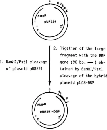
Construction of the hybrid plasmid pUR291-DBP (Figure 3)
Plasmid pUR291 (Ruther and Mflller-Hill, 1983) was treated with restriction endonucleases BamHl and Pstl and the digestion mix-ture separated by electrophoresis on a 1 % agarose gel. The large fragment of pUR291 was ligated to the DBP gene isolated from
T h e concentration of cysteine is rate-limiting. •The low solubility of/3-galactosidase-DBP in 0.05 M NH4HCO3-dirnethylformanTide (5:6, v/v) did not allow protein
concentration-degradation rate studies.
a BamHUPstl digest of pUC8-DBP. The pUR291-DBP hybrid plasmid was transfected into competent E. coli JM109 cells. Ampicillin-resistant clones containing the hybrid plasmid were identified by digestion of the extractable DNA of quick lysates with BamHUPstl and EcoKUPstl, respectively, and gel elec-trophoretic analysis of the restriction patterns.
Expression and isolation of the $-galactosidase-DBP fusion protein
Electrophoresis of cell extracts of pUR291- and pUR291-DBP-transformed E. coli JM109 on 7.5% SDS-polyacrylamide gels showed that LPTG induced strongly both lacZ' and the lacZ'-DBP fusion. Maximum yields were obtained by addition of IPTG (1—2 mM) to transformed E. coli JM109 during logarithmic growth (Esoo, - 0 . 8 ) and incubation at 25 °C for - 2 0 h. Cells were centrifuged, sonicated and extracted with a Tris buffer, pH 7.4 (20 mM Tris-HCl, 10 mM MgCl2, 10 mM /3-mercapto-ethanol) to remove soluble proteins. The insoluble portions were extracted further with buffer containing 8 M urea, 0.1 M NH,-HCO3, 1 mM MgCl2, 1 mM dithiothreitol. After centrifugation, aliquots of the supernatants and the pellets were analyzed by elec-trophoresis (Figure 4). The supernatants were dialyzed against water and lyophilized. All steps of the isolation procedure were performed at 4°C. The lyophilized products were purified fur-ther by gel filtration on Sephacryl S-300 in a Tris-urea buffer, pH 7.4 (8 M urea, 20 mM T r i s - H C l , 10 mM 0-mercaptoethanol). Fractions containing the putative
j3-AMPR V
pUR291 I
1. BamHI/PstI cleavage of plasmid pUR291
2. ligation of the large fragment with the DBP gene (90 bp, ^ ) ob-tained by BamHI/Pstl cleavage of the hybrid plasmid pUC8-DBP
Fig. 3. Construction of the hybrid plasmid pUR291-DBP by insertion of the DBP-coding portion of pUC8-DBP (Figure 2) into BamHI/P^rl-treated pUR291 (Ruther and MQller-Hill, 1983). Expression of pUR291-DBP in
E. coli yields a 1050-residue fusion protein of the following sequence:
/3-galactosidase (l-1021)-Arg-Gly-Ser-Lys-Arg-DBP. Abbreviations of restriction sites: E, EcoRl; B, BamHl; S, Sail; P, Psll; H, HindJU.
m0 ttfc< W
12 3 4 5 6 7
Fig. 4. SDS-PAGE of wild-type E. coli /3-gaJactosidase (lanes 1, 4 and 7), /3-gaJactosidase of pUR291 -transformed E. coli JM109 (lanes 2 and 3), and 0-galactosidase-DBP fusion protein of pUR291 -DBP-transformed E. coli JM109 (lanes 5 and 6). Bands move from top to bottom; only part of the 300 x 180 mm gel is shown. In lanes 2 and 5, aliquots of the urea extracts of disrupted cells were applied. The pellets of these extracts were boiled in 0.125 M Tris-HCl, pH 6.8, containing 10% glycerol, 5% /3-mercaptoethanol, and 2% SDS and the supernatants of the centrifuged mixtures were applied to lanes 3 and 6.
galactosidase —DBP fusion protein or the /3-galactosidase of the control were identified by SDS-PAGE, pooled, dialyzed against water at 4°C and lyophilized.
Isolation and characterization of recombinant DBP
The putative /3-galactosidase —DBP fusion protein of pUR291-DBP-transformed E. coli JM109 and the /3-galactosidase
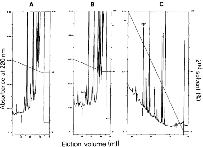
of the control were carboxymethylated (Craven et al., 1965) and then cleaved by trypsin (Steers et al., 1965). The mixtures of the tryptic peptides of control and sample were partially frac-tionated by reversed-phase h.p.l.c. (Figure 5A and B, respec-tively) on a C-8 column (250 X 4 mm, Macherey and Nagel) using 0.1% TFA (trifluoroacetic acid) as the first solvent and 60% acetonitrile in 0.1 % TFA as the second solvent. The recom-binant DBP (Figure 5B, arrow) was rechromatographed and then cleaved by chymotrypsin. Figure 5C shows the h.p.l.c. chro-matogram of the chymotryptic peptides of recombinant DBP. Chymotryptic hydrolysis of chemically synthesized DBP followed by h.p.l.c. gave an almost identical digestion pattern.
DDT degradation by cytochrome P-450 models (Sakurai, 1980)
Mixtures of DDT (0.12 mM), hemin (0.05 mM), cysteine (12 mM) and various proteins (concentrations given in Table I) were incubated in 0.05 M NH4HCO3-dimethylformamide (5:6, v/v) at 37 °C for 24 h. DDT degradation and product formation (Moser et al., 1985) were determined quantitatively (Table I) by h.p.l.c. on an RP-300-5C8-column using a gradient of acetonitrile in 0.1 % TFA.
Results and discussion
The pUC8-DBP hybrid plasmid
To derive the sequence of the DBP gene the codon usage of highly expressed E. coli proteins (Grosjean and Fiers, 1982) was chosen except for those positions at which restriction sites had to be created or abolished. The DBP gene was synthesized (Beaucage and Caruthers, 1981) and inserted between the BamHl and PstI cleavage sites of the polylinker region of plasmid pUC8 (Figure 2). The hybrid plasmid formed was cloned in E. coli JM83 (Yanisch-Perron et al., 1985). Gel electrophoresis and DNA se-quence analysis (Maxam and Gilbert, 1980) showed that the clon-ed DBP gene, isolatclon-ed from the BamHUPstl digest of the hybrid plasmid of transformed E. coli JM83 cells, had the expected size and sequence. Expression would yield a 36-residue fusion pro-tein (Figure 2) from which the desired 24-residue DDT-binding polypeptide could be obtained by tryptic cleavage at the pair of basic residues preceding the NH2-terminal methionine of DBP. The internal Arg-Pro bond is not susceptible to tryptic hydrolysis. Antibodies against DBP cross-reacted strongly with a chemical-ly synthesized reference sample of the 36-residue fusion protein (Figure 2). However, radioimmunoassays of samples of the cytosol, detergent extracts of the membranes, and culture medium taken at various stages of the logarithmic and stationary growth of the transformed E. coli JM83, were all negative. The lack of measurable amounts of translation product seemed to be caused mainly by the instability of the putative 36-residue fusion pro-tein towards endogenous proteases, because neither exogenous-ly added 24-residue nor 36-residue poexogenous-lypeptide could be detected immunologically in cell extracts even in the presence of protease inhibitors. Similar observations on analogous fusions and nonsense fragments of/3-galactosidase have been made previously (Goldschmidt, 1970; Itakura et al., 1977).
The pUR291-DBP hybrid plasmid
We surmized that the fusion of DBP with the COOH-terminus of /3-galactosidase might yield a more stable product. Using this scheme, Itakura et al. (1977) had been able to demonstrate the expression in E. coli of the synthetic gene of somatostatin, a 14-residue peptide hormone. Fusions employing /3-galactosidase as a protective 'carrier' can be easily analyzed by gel elec-trophoresis and assays of the enzymatic activity. We chose
B
Q_
CD
Elution volume (ml)
Fig. 5. Isolation of recombinant DBP from the tryptic digest of the carboxymethylated /3-galactosidase - DBP fusion protein by reverse-phase h.p.l.c. on a C-l
column. (A,B) Partial resolution of the tryptic digests of /3-galactosidase of the control and /3-galactosidase—DBP fusion protein, respectively;
(C) chymotryptic peptide map of recombinant DBP isolated from the DBP peak in B. The peaks marked (v) appeared in the chymotryptic digest of both recombinant and chemically synthesized DBP and had almost the same relative sizes. The arrow in A, B and C indicates the position of chemically synthesized DBP chromatographed under identical conditions.
plasmid pUR291 (Riither and Miiller-Hill, 1983) (Figure 3) as the expression vector. The lacZ' portion of the /3-galactosidase gene of pUR291 can be induced by inducers of the lac system and is extended at the 3'-end by a polylinker region. The hybrid plasmid pUR291-DBP was constructed as depicted in Figure 3 and was cloned in E. coli JM109 (Yanisch-Perron et al., 1985). After the expected structure of the DBP gene in the hybrid plasmid of transformed cells had been confirmed by gel elec-trophoretic analysis of restriction patterns, the expression of the putative 1050-residue /3-galactosidase-DBP fusion protein could be demonstrated and was compared with the expression of lacL' by pUR291-transformed E. coli JM109. Cells were incubated with IPTG, sonicated, extracted with 20 mM Tris-HCl buffer, pH 7.4, and centrifuged. Extracts and resuspended pellets were assayed for /3-galactosidase activity- using o-nitrophenyl-/3-D-galactopyranoside as substrate; the product formed (o-nitrophenol) was determined quantitatively at 420 nm (Steers et
al., 1971). In pUR291-DBP-transforrned cells 59% of the
measurable activity was found in the extract, the remainder was in the pellet. In contrast, 95% of the measurable /3-galactosidase activity of pUR291-transformed cells was soluble in the Tris buf-fer indicating that the putative fusion protein had reduced solu-bility. The enzyme assays also revealed that the specific activity of the presumptive /3-galactosidase-DBP fusion protein was — 20% lower than that of /3-galactosidase isolated from pUR291-transformed E. coli JM109. This correlated with dif-ferences in the structural stability of the two products. Whereas the putative /3-galactosidase—DBP fusion was partially
suscep-tible to digestion by cellular proteases, the /3-galactosidase of the control was stable. Digestion was increased considerably when pUR291-DBP-transformed E. coli cells were grown after IPTG induction at 37°C.
Isolation and characterization of recombinant DBP
Transformed E. coli cells were sonicated and extracted with 20 mM Tris-HCl, pH 7.4, to remove soluble proteins. The pellets were treated with 0.1 M NH4HCO3 containing 8 M urea and 1 mM dithiothreitol. After centrifugation the supernatants were dialyzed against water, lyophilized, and purified further by gel filtration on Sephacryl S-300. Gel electrophoresis (Figure 4) showed that, as expected, both the presumptive /3-galac-tosidase-DBP fusion protein and /3-galactosidase of the control displayed a bigger relative molecular mass than wild type /3-galac-tosidase from E. coli (1023 residues). This scheme of isolation of the /3-galactosidase —DBP fusion protein was more effective than affinity chromatography on Sepharose-4B-linked p-2smno-phenyl-/3-D-thiogalactopyranoside (Cuatrecasas, 1970) because the fusion protein was only poorly soluble in buffers not con-taining urea and bound only weakly to the immobilized substrate analogue.
The putative galactosidase —DBP fusion protein and the /3-galactosidase of the control were then carboxymethylated and cleaved by trypsin. Solid phase radioimmunoassays, using rab-bit antibodies against chemically synthesized DBP and I25 I-labelled protein A,- were positive for the mixture of the tryptic peptides of the sample derived from the
pUR291-DBP-transformed strain but were negative for the tryptic digests of wild-type /3-galactosidase and /3-galactosidase of the pUR291-transformed strain (results not shown). H.p.l.c. analysis of the tryptic peptides revealed that the digest of the putative /3-galactosidase—DBP fusion protein contained a peptide that eluted at the same position as chemically synthesized DBP (Figure 5B, arrow). This peptide was not present in the digest of the /3-galactosidase from the control (Figure 5A, arrow). The material under the marked peak of Figure 5B was rechromatographed for further analytical characterization. Amino acid analysis after acid hydrolysis of the recombinant product showed excellent agree-ment with the values obtained for the synthetic 24-residue DDT-binding polypeptide, and resolution by h.p.l.c. of the chymotryp-tic digests of recombinant product and chemically synthesized DBP gave almost identical peptide maps (Figure 5C).
Based on the enzymatic activity of the fusion protein and quan-titative amino acid analysis, the yield of recombinant DBP was 41% after Sephacryl S-300 chromatography of the urea-extractable material and 28 % after tryptic digestion of the purified fusion protein and resolution of the digest by h.p.l.c. Recombi-nant and chemically synthesized DBP were indistinguishable in their DDT-binding properties.
Characterization of the (3-galactosidase—DBP fusion protein
The dissociation constant of the DBP-DDT complex in 55% aqueous ethanol was 0.9 X 10~6 M as determined by the method of Hummel and Dreyer (1962). In a preliminary t.l.c. assay using 20% dimethylsulfoxide in 0.05 M NH4HCO3 as sol-vent, the DDT binding of the /3-galactosidase —DBP fusion was of the same order of magnitude. Wild-type /3-galactosidase did not have measurable DDT-binding activity. Moreover, the /3-galactosidase—DBP fusion protein was 25 times as potent as an equimolar amount of free DBP in stimulating the degradation of DDT by a cytochrome P^50 model system (Table I). The model system alone (hemin and excess cysteine; Sakurai, 1980) was able to degrade 51 % of the added DDT in 24 h. Wild-type /3-galactosidase had no effect. Preliminary studies showed that the increase of the DDT degradation rate depended on the con-centration of DBP (Table I). The nature of this observation will be investigated further.
This paper describes for the first time the cloning and expres-sion in E. coli of a designed, artificial polypeptide based on the construction of the pUR291-DBP gene hybrid plasmid. The DBP moiety of the isolated /3-galactosidase — DBP translation product was more active in DDT degradation than free DBP and the bacterial strain transformed by the hybrid plasmid can now be used to study the biological activity of the /3-galactosidase-DBP fusion protein in vivo. The system also lends itself to investiga-tions of the relainvestiga-tionship between amino acid sequence and DDT binding of the designed 24-residue polypeptide by exchange of residues in selected positions.
Acknowledgements
We thank the Schweizerische Nationalfonds for support of this work.
References
Barany.G. and Merrifidd.R.B. (1979) In Gross.E. and MeienhoferJ. (eds), The
Peptides. Vol. 2, Academic Press, NY, pp. 1-284.
Beaucage.S.L. and Caruthers.M.H. (1981) Tetrahedron Lett., 22, 1859-1862. Birnboim.H.C. and DolyJ. (1979) Nucleic Acids Res., 7, 1513-1523. Chou.P.Y. and Fasman.G.D. (1978) Adv. Enzymol., 47, 45-148.
Craven.G.R., Steers.E.Jr and Anfinsen.C.B. (1965) J. Biol. Oiem., 240, 2468-2477.
Cuatrecasas.P. (1970) J. Biol. Oiem.. 245, 3059-3065.
Dorper.T. and Winnacker,E.-L. (1983) Nucleic Acids Res., 11, 2572-2584. Goldschmidt.R. (1970) Nature, 228, 1151-1154.
Grosjean.H. and Fiers.W. (1982) Gene, 18, 199-209.
Gutte,B., Moser.R., Klauscr.S. and Weilenmann.M. (1984) In Ricard.J. and Comish-Bowden,A. (eds), Dynamics of Biochemical Systems. Plenum Publishing Corporation, NY, pp. 259-272.
HummcU.P. and Dreyer.W.J. (1962) Biochim. Biophys. Acta, 63, 530-532. Itakura.K., Hirose.T., Crea.R., Riggs.A.D., Heyneker.H.L., Bolivar.F. and
Boyer.H.W. (1977) Science, 198, 1056-1063.
Langfey.K.E., Fowler,A.V. andZabin,I. (1975)/. BioL Oiem., 250, 2587-2592. Levht.M. (1978) Biochemistry, 17, 4277-4285.
Marko.M.A., Chipperfield.R. andBimboim.H.C. (1982) Anal. Biochem., 121, 382-387.
Maxam,A.M. and Gilbert.W. (1980) Methods Enzymol., 65, 499-560. Merrifield.R.B. (1963) /. Am. Oiem, Soc., 85, 2149-2154.
Moser.R. (1986) Ph.D. Thesis, Universital Zurich.
Moser,R., Thomas.R.M. and Gutte.B. (1983) FEBS Lett., 157, 247-251. Moser.R., Klauser,S., Lcist.T., Langen.H., Epprecht.T. and Gutte.B. (1985)
Angew. Chem. (Engl. Ed), 24, 719-727.
RommensJ., MacKnight,D., Pomeroy-Cloney.L. and Jay.E. (1983) Nucleic Acids
Res., 11, 5921-5940.
RQther,U. and MQller-Hill.B. (1983) EMBO ]., 2, 1791-1794. Sakurai.H. (1980) Oiem. Pharm. Bull., 28, 3437-3439.
Steers.E.Jr, Craven.G.R, Anfinsen.C.B. and BethuneJ.L. (1965)7. Biol. Chem.,
240, 2478-2484.
Steers,E.Jr, Cuatrecasas.P. and PoUard.H.B. (1971) J. Biol. Oiem., 246, 196-200.
Ullmann.A., Jacob.F. and MonodJ. (1967) J. Mol. Biol., 24, 339-343. VieiraJ. and MessingJ. (1982) Gene, 19, 259-268.
Yanisch-Perron.C, VieiraJ. and MessingJ. (1985) Gene, 33, 103-111.
![Fig. 1. Structural formula of 4,4'-DDT [l,l,l-trichloro-2,2-bis(4-chloro- [l,l,l-trichloro-2,2-bis(4-chloro-phenyl)-ethane] (top) and sequence and proposed secondary structure of the designed 24-residue DDT-binding polypeptide (bottom)](https://thumb-eu.123doks.com/thumbv2/123doknet/14904515.655351/1.903.472.842.566.1028/structural-trichloro-trichloro-sequence-proposed-secondary-structure-polypeptide.webp)