Advance Access publication on June 23, 2010
Glycomimicry: Display of the GM3 sugar epitope on
Escherichia coli and Salmonella enterica sv Typhimurium
Karin Ilg2,3,Elif Yavuz2,4,Carola Maffioli3,
Bernard Priem1,4,and Markus Aebi1,2
3Institute of Microbiology, ETH Zurich, Zurich, Switzerland, and4CERMAV,
Grenoble, France
Received on April 9, 2010; revised on June 11, 2010; accepted on June 13, 2010
Oligosaccharides present on the surface of pathogenic bacteria play an important role in their interaction with their host. Bacteria with altered cell surface structures can be used to study these interactions, and glycoengi-neering represents a tool to display a glycoepitope on a different bacterium. Here, we present non-pathogenic Escherichia coli and Salmonella enterica serovar Typhi-murium expressing the sialyllactose oligosaccharide epitope of the ganglioside GM3. By expression of the ga-lactosyltransferase LgtE and the sialic acid transferase Lst as well as the CMP-sialic acid synthetase SiaB from Neisseria gonorrhoeae and Neisseria meningitidis in engi-neered strains devoid of the sialic acid catabolism, the GM3 sugar epitope was displayed on these bacteria as demonstrated by live cell immunostaining and a detailed analysis of their lipooligosaccharides. These strains offer the possibility to investigate the role of sialic acid in the recognition of bacteria by the immune system in a non-pathogenic background.
Keywords: bacterial glycosylation / chimeric LPS / gangliosides / glycoengineering
Introduction
Bacteria produce many oligo- and polysaccharide structures. A high percentage of these is found in structures of the cell wall, e.g., in peptidoglycan, lipopolysaccharides (LPS) and lipooli-gosaccharides (LOS). Some human enteropathogens, like Campylobacter jejuni and Neisseria meningitidis, produce sac-charide structures on their surface that resemble human carbohydrate structures (Tsai 2001; Yuki et al. 2004). This mechanism is termed molecular mimicry and might help the bacteria to avoid recognition by the human immune system. C. jejuni is one of the few bacteria able to synthesize sialic acid
(N-acetyl neuraminic acid; NeuNAc), and many strains include this sugar in their LOS, resulting in human ganglioside sugar mimicry (Aspinall, Fujimoto et al. 1994;Aspinall, McDonald et al. 1994;Ang et al. 2004). Gangliosides are glycosphingoli-pids containing at least one sialic acid residue. They are present in all mammalian cells but enriched in nerve cells and in brain tissue. The molecular mimicry of ganglioside sugar epitopes can lead to the autoimmune paralysis Guillain–Barré Syn-drome (GBS) in approximately 1 in 1000 humans infected with C. jejuni (Ang et al. 2004;Yuki et al. 2004). Patients pro-duce anti-ganglioside antibodies that do not only bind the ganglioside sugar epitopes on the surface of the pathogen but also the host's own gangliosides, mostly in the nodes of Ranvier and the motor nerve termini (Yuki et al. 1990; Hafer-Macko et al. 1996).
Recent studies used genetically engineered Escherichia coli to produce pure oligosaccharides in large quantities. These E. coli strains heterologously express glycosyltransferases from other bacteria, e.g., N. meningitidis, Neisseria gonorrhoeae or C. jejuni or even eukaryotes. The oligosaccharides produced include the carbohydrate moiety of nonsulfated HNK-1, Lewis X tetrasaccharides, globotriose and globotetraose, human milk oligosaccharides and the carbohydrate moieties of the gang-liosides GM1 and GM2 (Priem et al. 2002; Antoine et al. 2003,2005; Dumon et al. 2006;Yavuz et al. 2008). The as-sembly of these oligosaccharide structures takes advantage of the internalization of lactose by the genetically engineered bac-teria. After internalization, lactose is used in the cytoplasm as a platform for the addition of other saccharides by heterologously expressed glycosyltransferases. Most of the oligosaccharides produced are secreted into the medium and can be purified easily (Bettler et al. 1999; Priem et al. 2002). This approach of using E. coli as a living factory replaces the time-consuming and laborious chemical or chemo-enzymatic synthesis and also allows the large-scale production of oligosaccharides (Bettler et al. 1999).
The synthesis of oligosaccharides in vivo can be combined with an approach utilizing the bacterial cell surface as a scaf-fold for different saccharide structures. In Gram-negative bacteria, the outer membrane is an asymmetric bilayer with glycerophospholipids in the inner leaflet and LPS in the outer leaflet. LPS consists of the lipid A moiety, an inner and an outer core to which in some organisms a polymeric O-antigen region is attached (Raetz and Whitfield 2002). The lipid A moiety com-prises two glucosamine residues connected through a β1,6 linkage. Acyl chains attached to these glucosamines anchor the molecule in the outer membrane. Proximal to the lipid A is
1
To whom correspondence should be addressed: Markus Aebi, e-mail: [email protected]; Bernard Priem, e-mail: [email protected]
2
the inner core region. In most Gram-negative bacteria, this region is composed of two 3-deoxy-D-manno-oct-2-ulosonic acid (Kdo) and severalL-glycero-D-mannoheptose (Hep) resi-dues which can be substituted by phosphoethanolamine or hexoses. The distal outer core consists of hexoses and N-acetyl-hexoses (seeFigure 1) (Schnaitman et al. 1991;Parker et al. 1992;Pradel et al. 1992;Schnaitman and Klena 1993;Heinrichs et al. 1998). To date,five core types have been found for E. coli (R1–R4 and K-12), whereas for Salmonella enterica serovar Typhimurium (S. Typhimurium) only two have been described (Lüderitz et al. 1982; Heinrichs et al. 1998; Olsthoorn et al. 1998). In S. Typhimurium, the polymeric O-antigen region is attached to the last glucose residue of the outer core (Hellerqvist et al. 1969). The outer core structure in E. coli K-12 strains ter-minates on the heptose residue HepIV to which the O-antigen region is attached (Holst et al. 1991; Heinrichs et al. 1998). E. coli K-12 strains used in the laboratory, however, do not produce the polymeric O16 O-antigen due to an insertion in wbbL. This gene encodes the rhamnosyltransferase that is in-volved in the addition of rhamnose to the second position of the O16 backbone (Liu and Reeves 1994; Stevenson et al. 1994; Yao and Valvano 1994).
Bacterial cell surfaces can be used to display engineered oligosaccharide structures in their lipopolysaccharide layer by two different pathways. On the one hand, the O-antigen ligase WaaL dependent transfer of oligo- and polysaccharides to E. coli or S. Typhimurium lipid A core can be exploited. The structures transferred include the Haemophilus influenzae lipooligosaccharide structure, the O-antigens from Shigella dysenteriae or E. coli O111 as well as the C. jejuni heptasac-charide normally linked to protein (Spinola et al. 1990;Falt et al. 1996;Szymanski et al. 1999;Wang et al. 1999;Phillips et al. 2000;Xu de et al. 2007). This approach requires the assem-bly of the engineered oligosaccharides on the lipid carrier undecaprenylpyrophosphate. On the other hand, truncated lip-id A core structures as acceptors of foreign saccharlip-ide structures can be used. This approach has been employed by Paton and co-workers for the production of probiotics (Paton et al. 2001,2005). The E. coli R1 strain CWG308 with a trun-cation of the lipid A core at the glucose I residue is used in these
studies to heterologously express glycosyltransferases from C. jejuni and N. meningitidis. These then modify the truncated lipid A core to produce a chimeric LOS structure on E. coli. This system is similar to the“living factory” described above that employs the glucose moiety of internalized lactose as a plat-form for the addition of other saccharides.
We extended the approach by Paton and co-workers from E. coli R1 to E. coli K-12 and S. Typhimurium by constructing strains that lack the gene encoding the sialic acid aldolase NanA. This allowed the incorporation of sialic acid into oligo-saccharides within the cell (Priem et al. 2002). Additionally, the strains were deleted in glycosyltransferase genes which led to the truncation of the lipid A core terminating with the glucose I residue. This was then used to attach the GM3 epi-tope by the heterologous expression of glycosyltransferases from N. meningitidis and N. gonorrhoeae.
Results
Truncation of lipid A core in S. Typhimurium and E. coli K-12 and display of the GM3 sugar on the truncated lipid A core To produce a terminal Glc in E. coli K-12 lipid A core, the genes encoding the glycosyltranferases WaaO and WaaB were deleted. The resulting strain E. coli K-12ΔnanAΔwaaOΔwaaB was termed LPS-1. In S. Typhimurium, the same truncation of the lipid A core was achieved by deleting the genes encoding the galactosyltransferases WaaI and WaaB, and the resulting strain was termed SKI22. Both strains used were also deleted in the gene encoding the sialic acid aldolase NanA to prevent the degradation of sialic acid added externally (see below). In or-der to produce E. coli and S. Typhimurium with a GM3 sugar epitope on the cell surface, the strains with the truncated lipid A core, LPS-1 and SKI22, were transformed with a plasmid encoding theβ-1,4-galactosyltransferase LgtE from N. gonor-rhoeae (pBBRlgtE) and with a plasmid encoding the CMP-sialic acid synthetase SiaB as well as the CMP-sialic acid transferase Lst from N. meningitidis (pKI3*). The strains containing the corresponding vectors served as negative control. Strains were grown in Luria–Bertani (LB) medium supplemented with 2% sialic acid, and proteinase-K-treated whole cell extracts were
A
B
C
GlcNAc(α1-2) GalII(α1-6) HepIII(α1-7) KdoII(α2-4) O-PS-GlcII(α1-2)GalI(α1-3)GlcI(α1-3)HepII(α1-3)HepI(α1-5)KdoI(α2-6)LipidA
PEtN/P P
WaaB WaaI
O-PS-HepIV(α1-6) Gal(α1-6) HepIII(α1-7) KdoII(α2-4) GlcIII(α1-2)GlcII(α1-3)GlcI(α1-3)HepII(α1-3)HepI(α1-5)KdoI(α2-6)LipidA PEtN/P P WaaB WaaO LgtE Lst
NeuNAc(α2-3) HepIII(α1-7) KdoII(α2-4) Gal(α1-4)GlcI(α1-3)HepII(α1-3)HepI(α1-5)KdoI(α2-6)LipidA
PEtN/P P
Fig. 1. Lipopolysaccharide structures in S. Typhimurium (A), E. coli K-12 (B) and the engineered strains for GM3 display (C). Glc, glucose; Gal, galactose; Kdo, 3-deoxy-D-manno-oct-2-ulosonic acid; Hep,L-glycero-D-mannoheptose; GlcNAc, N-acetylglucosamine; NeuNAc, N-acetyl neuraminic acid; P, phosphate; PEtN, phosphoethanolamine. Adapted fromYethon, Gunn et al. (2000),Yethon, Vinogradov et al. (2000),Frirdich et al. (2003)andKaniuk et al. (2004).
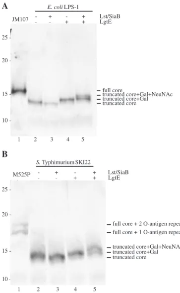
prepared. The display of the GM3 structure was judged by silver staining of a Tris-Tricine polyacrylamide gel electrophoresis (PAGE). Tofirst demonstrate the truncation of the lipid A core, proteinase-K-treated whole cell extracts of the parental strains can be seen in lane 1 ofFigure 2A for the LPS-1 parent JM107 and in lane 1 ofFigure 2B for M525P, the parental strain of SKI22. These strains show bands of higher apparent molecular mass than the strains with the truncated core, corresponding to the full core for JM107 or the full core with additional one or two O-antigen subunits for M525P. In lane 4 ofFigure 2A and B, the addition of the galactose residue by LgtE was detected for LPS-1 and SKI22, respectively, by a shift to a higher appar-ent molecular mass (seeFigure 2). The expression of SiaB and Lst led to the transfer of a sialic acid moiety and a further shift to a higher apparent molecular mass. This demonstrated that the GM3 epitope was produced in an E. coli K-12 derivative and a S. Typhimurium strain and that it was transferred to the
trun-cated lipid A core in both strains. In the strains containing the plasmids expressing LgtE and Lst/SiaB (lane 5 inFigure 2A and B), it was observed that the transfer of the sialic acid residue by Lst was not complete and that some truncated lipid A core with only galactose added was still present in the preparation.
Live cell microscopy demonstrated surface localization of GM3 sugar epitope on LPS-1 and SKI22
For immunological studies, it was important that the GM3 sugar epitope was present on the cell surface. As cell surface localization cannot be judged with whole cell extracts, immu-nofluorescence analysis of live cells was performed. Cells containing the plasmids encoding the galactosyltransferase LgtE, the CMP-sialic acid synthetase SiaB and the sialic acid transferase Lst grown in medium containing sialic acid (see above) were incubated with a mouse monoclonal anti-GM3 antibody, stained with 4, 6-diamino-2-phenylindole (DAPI) and an anti-mouse-IgM-Alexa647 conjugate. It is evident fromFigure 3that the antibody did not bind to cells contain-ing the truncated core (upper panel) or the truncated core with the galactose residue attached by LgtE (middle panel). When the genes encoding Lst and SiaB were expressed, cell surface staining was visible for E. coli LPS-1 and S. Typhi-murium SKI22 (lower panel in Figure 3A and B). This demonstrated that the GM3 sugar epitope was present on the surface of the bacterial cells, although it was perceivable again that not all cells carry this epitope.
Matrix-assisted laser desorption/ionization time-of-flight analysis revealed presence but low abundance of GM3 sugar epitope on truncated lipid A core
Mass spectrometry was performed for further analysis of the structures present on the truncated lipid A core of E. coli LPS-1 and S. Typhimurium SKI22. LOS of the strains display-ing the GM3 epitope and grown in medium containdisplay-ing sialic acid were isolated by the method developed byGalanos et al. (1969). The acyl chains attached at the hydroxyl groups of the glucosamine residues of the lipid A were cleaved by mild hydrazine treatment (Haishima et al. 1992). As can be seen inFigure 4B, the matrix-assisted laser desorption/ionization time-of-flight (MALDI-TOF) analysis demonstrated that neither the addition of galactose nor of sialic acid to the lipid A core was complete in the E. coli LPS-1 strain. In S. Typhimurium SKI22, the galactose residue was added to nearly all lipid A core mo-lecules, but sialic acid was not transferred to all galactosylated lipid A core molecules, similar to the situation in the E. coli strain (Figure 4D and B). As indicated in Figure 4, the mo-lecular composition of the truncated lipid A core structure deducted from the detected mass is a P3–Glc–Hep2– Kdo2–lipid A structure instead of the reported P4–Glc–Hep3– Kdo2–lipid A structure (Müller-Loennies et al. 2003). This is most probably due to the substrate specificity of the lipid A core glycosyl- and phosphotransferases and will be discussed in more detail below.
Quantification of display showed higher display levels in E. coli As means of quantification of the display of the GM3 epitope on the E. coli LPS-1 and the S. Typhimurium SKI22 strains, we carried out a colorimetric antibody binding assay. For this, live truncated core truncated core+Gal truncated core+Gal+NeuNAc full core - + Lst/SiaB - - +- ++ LgtE - + Lst/SiaB - - +- ++ LgtE E. coli LPS-1 JM107 1 2 3 4 5 10 -S. Typhimurium SKI22 M525P truncated core truncated core+Gal truncated core+Gal+NeuNAc full core + 1 O-antigen repeat full core + 2 O-antigen repeats
1 2 3 4 5
A
B
25 20 15 10 25 20 15-Fig. 2. The truncated lipid A core in E. coli LPS-1 (A) and S. Typhimurium SKI22 (B) can be modified with a GM3 epitope. Tris-Tricine PAGE of whole cell extracts from cells which were grown in the presence of external sialic acid and which expressed the indicated genes from plasmids followed by silver staining. Whole cell extracts from the parental strains were used as controls.
A
B
Vector controls
LgtE+vector control
LgtE+Lst/SiaB
DNA anti GM3 Merge DNA anti GM3 Merge
Fig. 3. Surface localization of the GM3 epitope on E. coli LPS-1 (A) and S. Typhimurium SKI22 (B). Immunofluorescence analysis of live cells that were grown in the presence of external sialic acid and which expressed the indicated genes from plasmids. Scale bar 5 µm.
1722.0 1989.2 2256.4 2523.6 2790.8 3058.0 70.3 0 20 40 60 80 100 Mass (m/z) 2180: GalGlcHep2Kdo2GlcNAcyl2P3 1722.0 1989.2 2256.4 2523.6 2790.8 3058.0 0 20 40 60 80 100 16.5 Mass (m/z) 2180: GalGlcHep2Kdo2GlcNAcyl2P3 1722.0 1989.2 2256.4 2523.6 2790.8 3058.0 0 20 40 60 80 100 Mass (m/z) 2180: GalGlcHep2Kdo2GlcNAcyl2P3 2472: NeuNAcGalGlcHep2Kdo2GlcNAcyl2P3 23.5 1722.0 1989.2 2256.4 2523.6 2790.8 3058.0 0 20 40 60 80 100 Mass (m/z) 2018: GlcHep2Kdo2GlcNAcyl2P3 2180: GalGlcHep2Kdo2GlcNAcyl2P3 2472: NeuNAcGalGlcHep2Kdo2GlcNAcyl2P3 46.8
A
B
C
D
Fig. 4. MALDI-TOF analysis of purified, O-deacylated LOS from E. coli LPS-1 and S. Typhimurium SKI22 demonstrates the addition of NeuNAc in the presence of Lst/SiaB. (A) E. coli LPS-1 expressing the gene encoding for LgtE and containing the vector control for the sialyltransferase. (B) E. coli LPS-1 expressing the genes encoding for LgtE and Lst/SiaB. (C) S. Typhimurium SKI22 expressing the gene encoding for LgtE and containing the vector control for the sialyltransferase. (D) S. Typhimurium SKI22 expressing the genes encoding for LgtE and Lst/SiaB.
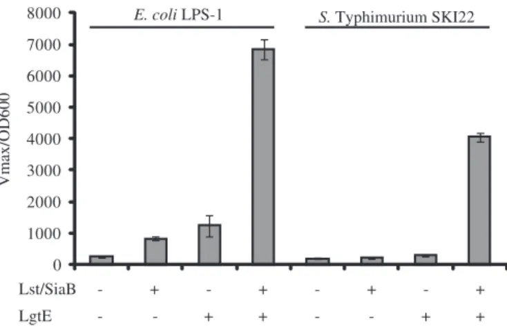
cells grown in medium containing sialic acid were incubated with the anti-GM3 antibody EM5 followed by an incubation with an anti-mouse-IgM-HRP conjugate. The assay was developed with the colorimetric substrate 2,2′-azino-bis(3-ethylbenzthiazoline-6-sulphonic acid) (ABTS) (see Materials and methods), and the kinetics of the color development were recorded. This assay demonstrated a 27-fold increase in signal intensity in the E. coli LPS-1 strain containing LgtE and Lst/ SiaB as compared to the LPS-1 strain harboring the vector con-trols (Figure 5, left). In S. Typhimurium SKI22, the increase in signal intensity was 20-fold between SKI22 strain containing LgtE and Lst/SiaB in comparison to the SKI22 strain with the vector controls (Figure 5, right). This showed that E. coli LPS-1 cells bound more anti-GM3 antibody EM5 than the S. Typhimurium SKI22 cells and thereby indicated higher dis-play levels of the GM3 sugar epitope on the E. coli strain.
Discussion
The connection between the autoimmune disease GBS and sia-lylated structures in bacterial LOS is still not clear. The usual infectious agents associated with subsequent GBS include Epstein–Barr virus, Mycoplasma pneumoniae, C. jejuni and cytomegalovirus (Hughes and Cornblath 2005). Of these, C. jejuni is known to express sialylated LOS that mimics human gangliosides (Aspinall, Fujimoto et al. 1994;Ang et al. 2004; Yuki et al. 2004). However, not every structure mimicking gang-liosides leads to GBS. N. meningitidis and H. influenzae strains have been demonstrated to produce LOS with a sialyl-LacNAc structure, which differs from the GM3 epitope only in the N-acetylgroup present at C2 of the Gal residue (Mandrell et al. 1992;Yamasaki et al. 1993). Nevertheless, none of these strains has been reported to be a statistically significant antecedent in-fection in GBS patients.
With the aim of providing well-defined model organisms in order to study the connection between ganglioside mimicry in the LOS and evocation of an autoimmune disease, we con-structed E. coli K-12 and S. Typhimurium that display the
GM3 sugar epitope on the cell surface as demonstrated by var-ious methods. Mass spectrometry analysis of the O-deacylated isolated LOS proved the addition of galactose and sialic acid re-sidues to the lipid A core. The molecular mass of the truncated lipid A core in the E. coli LPS-1 strain (2018 Da) was increased by 162 and 292 Da, respectively (Figure 5). However, the mo-lecular mass of 2018 Da for the truncated lipid A core did notfit with the calculated mass of 2291.95 Da for the chemical com-position lipid A–Kdo2–Hep3–Hex–P4of the three main glycoforms I–II–III present in E. coli K-12 (Müller-Loennies et al. 2003). The difference between the calculated and the ob-served molecular mass could be accounted for by the disappearance of one heptose residue and one phosphate group. In E. coli R1, it has been shown that the mutation of waaY yielded a strain with a core oligosaccharide devoid of phosphate on the HepII residue. Furthermore, a mutation of waaQ resulted in the loss of the branched HepIII residue on HepII, and it im-peded the activity of waaY (Yethon et al. 1998). It is also known that a mutation of waaG which encodes the glucosyltransferase acting on HepI not only leads to the truncation of the lipid A core immediately behind the inner core heptose residues but also to a lack of phosphate groups on HepII (Yethon, Vinogradov et al. 2000). It is likely that a similar co-dependency between the monosaccharides present in the lipid A core and the phosphor-ylation of saccharides exists in E. coli K-12. Most probably, the deletion of waaO and waaB yields a suboptimal substrate for the gene product of waaQ, thereby inhibiting the addition of the He-pIII residues. This thenfinally results in a lipid A–Kdo2–Hep2– Hex–P3structure because the gene product of waaY relies on the presence of the HepIII residue (Yethon et al. 1998). Yethon and co-workers also demonstrated that the genetic determinants for modifications of the lipid A core are conserved between E. coli and S. Typhimurium (Yethon, Gunn et al. 2000). Therefore, the structure lipid A–Kdo2–Hep2–Hex2–P3that we observed for the galactosylated truncated lipid A core in SKI22 can also be explained to stem from the same substrate specificities as in E. coli K-12. In a S. Typhimurium SKI22 strain containing the vec-tor controls for LgtE and Lst/SiaB, a product at 2018 Da was visible, which demonstrates that in S. Typhimurium SKI22 the truncated lipid A core also possesses the structure lipid A–Kdo2–Hep2–Hex–P3 (data not shown).
While mass spectrometry analysis and lipooligosaccharide separation by Tris-Tricine polyacrylamide gels demonstrated the presence of sialic acid on the truncated lipid A core struc-tures of E. coli LPS-1 and S. Typhimurium SKI22, antibody binding proved the α-2,3 linkage of the attached sialic acid residue. A competition assay with purified sialyllactose, but not with lactose, demonstrated the accuracy of the antibody binding (data not shown).
The E. coli and S. Typhimurium strain displaying the GM3 sugar epitope will be helpful tools when the role of sialic acid containing LOS structures in the recognition and the interaction of these structures with the immune sys-tem is being examined.
Materials and methods
Bacterial strains and growth conditions
A summary of bacterial strains used in this study can be found inTable I. Bacteria were grown in LB medium (10 g/L Bacto
0 1000 2000 3000 4000 5000 6000 7000 8000 Vmax/OD600 Lst/SiaB - + - + - + - + + + - - + + LgtE - -
E. coli LPS-1 S. Typhimurium SKI22
Fig. 5. Quantification of anti-GM3 binding to whole cells demonstrates higher display levels in E. coli LPS-1 when compared to S. Typhimurium SKI22. Live cells expressing the indicated genes from plasmids were incubated with the anti-GM3 antibody, and the amounts of bound antibody were quantified by the colorimetric assay described in the Materials and methodssection.
tryptone, 5 g/L Bacto yeast extracts, 5 g/L NaCl). For sialyla-tion experiments, LB was supplemented with 2% sialic acid (Jülich chiral solutions, Jülich, Germany). The pH of the LB with sialic acid was adjusted to 7.5 by addition of NaOH and sterilized by filtering. LB agar plates were supplemented with 1.5% (w/v) agar. Antibiotics were used at the following final concentrations: tetracyclin (tet) 10 µg/mL, ampicillin (amp) 100 µg/mL, kanamycin 50 µg/mL, chloramphenicol (cam) 25 µg/mL.
DNA manipulations
N. meningitidis siaB and lst were amplified from N. meningi-tidis MC58 (ATCC# BAA 335 D5) genomic DNA, while H. influenzae lst was amplified from H. influenzae RM118 genomic DNA (Derek Hood, Oxford). N. gonorrhoeae lgtE was amplified from N. gonorrhoeae genomic DNA. Restric-tion enzymes were purchased from MBI/Fermentas, whereas amplification of DNA was carried out with Pfu DNA poly-merase isolated in the Institute of Microbiology, ETH Zurich, by standard protocols. All cloning steps were carried
out in E. coli DH5α, and all constructs were confirmed by sequencing (Synergene Biotech, Schlieren, Switzerland). Construction of the E. coli K-12ΔnanAΔwcaJΔwaaOΔwaaB strain LPS-1
Inactivation of waaB and waaO was carried out by homolo-gous recombination using the suicide plasmid pKO3. Primers (l) and (m) were used for polymerase chain reaction (PCR) am-plification of 0.67 kb of DNA flanking the 5′ end of waaB from JM107, whereas primers (n) and (o) were used for PCR amplification of 0.69 kb of DNA flanking the 3′ end of waaO. The 665-bp fragment was digested with BamHI and SalI, while the 688-bp fragment was digested with BamH1 and SmaI. Both digested fragments were mixed and cloned into the suicide plasmid pKO3 digested with SmaI and SalI. The resulting re-combinant integrative plasmid pKO3-BO was transformed into TA1ΔnanA cells; positive clones were screened for correct in-tegration of the plasmid DNA by PCR with primers (l) and (o) (for the sequence, seeTable I). The truncation of wcaJ is de-scribed elsewhere (Priem et al.).
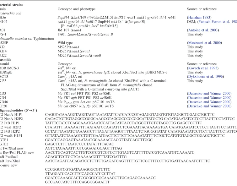
Table I.Bacterial strains, plasmids and oligonucleotides used in this study Bacterial strains
Strain Genotype and phenotype Source or reference Escherichia coli
DH5α SupE44ΔlacU169 (Φ80lacZΔM15) hsdR17 recA1 endA1 gyrA96 thi-1 relA1 (Hanahan 1983)
JM107 endA1 gyrA96 thi hsdR17 SupE44 relA1λ−Δ(lac-proAB)
[F′ traD36 proAB+ lacIqlacZΔM15] DSM; (Yanisch-Perron et al. 1985)
TA01 JM 107ΔnanA (Antoine et al. 2003)
LPS-1 TA01ΔnanAΔwcaJΔwaaOΔwaa B This study Salmonella enterica sv. Typhimurium
M525P Wild type (Mastroeni et al. 2000)
SKI2 M525PΔnanA This study
SKI14 M525PΔnanAΔwaaI This study SKI22 M525PΔnanAΔwaaIΔwaaB This study Plasmids
Plasmid Genotype Source or reference pBBR1MCS-3 TetR, bhr ori (Kovach et al. 1995)
pBBRlgtE TetR, bhr ori, N. gonorrhoeae lgtE cloned XbaI/SacI into pBBR1MCS-3 This study
pACT3 CamR, p15A ori (Dykxhoorn et al. 1996)
pKI3* CamR, p15A ori, N. meningitidis lst cloned XbaI/PstI with a C-terminal
FLAG-tag downstream of SiaB from N. meningitidis cloned SacI/XbaI with a C-terminal c-myc-tag into pACT3
This study
pKD3 bla FRT cat FRT PS1 PS2 oriR6K (Datsenko and Wanner 2000)
pKD4 bla FRT aph FRT PS1 PS2 oriR6K (Datsenko and Wanner 2000)
pKD46 bla PBADgam bet exo pSC101 oriTS (Datsenko and Wanner 2000)
pCP20 bla cat cI857λPRflp pSC101 oriTS (Datsenko and Wanner 2000)
Oligonucleotides (5′→3′)
LT2 NanA H1P1 CAGGTATAAAGGTAGGTAGTTAATATATTCATCATCCGTAGAGGTAGGTGTGTAGGCTGGAGCTGCTTC
LT2 NanA H2P2 CACACTGTTGTAGGCCGGGCAAGCGTAGCGCCCCCGGCATTATGCTG CATATGAATATCCTCCTTAGTTCCTATTCC LT2 I+B H1P1 TATTTCTATCTCAGGAAATGAATCCATTACATCACCTATGGGTTGTGTAGGCTG GAGCTGCTTC
LT2 waaI H2P2 CTATATTTTAAAAATTTTAATAATGCAATATTCTCGAAATTACAAAAGTGA CATATGAATATCCTCCTTAGTTCCTATTCC LT2 I+B H2P2 GCTATTTAATATCTAAAGTCTTTAGATTAAGGTTTTAACTCTGGGGTATAT CATATGAATATCCTCCTTAGTTCCTATTCC LT2 waaB H1P1 GTTATAATCTAAAGTCTGTTGAATGACTTCTTCTTCAAAATATTTTCTGCTCATGTGTAGGCTGGAGCTGCTTC LGTE1 GGATCCAGGAGTAAATAATGCAAAACCACGTTATCAGCTTGGC
LGTE2 GAGCTCTTTTAATCCCCTATATTTTACAC Lst Fw/XbaI new AGTCTAGAAATTTGTCGGAATGGAGTTTTAG
Lst Rev/PstI FLAG new AACCTGCAGTCACTTGTCGTCGTCGTCCTTGTAGTCATTTTTATCGTCAAATGTCAAAATC SiaB Fw/SacI AGAGCTCCTGCTCAAAAACGTTTTATCGATTTG
SiaB Rev/XbaI c-myc new AATCTAGATCACAGATCCTCTTCTGAGATGAGTTTTTGTTCGCTTTCCTTGTGATTAAGAATGTTTTC l CCCGGGTCGTGATAAAGGGCGTCTTC m TTAGGATCCACCTTCCAGCCATCCCTTAT n GGATCCAAAGCACTCGCGGCCGCAAAGCTTGCAGAGCAAAACC o GTCGACCATCTTTCCAGGGGGAATTT
Construction of the M525PΔnanAΔwaaIΔwaaB strain SKI22 For the step-by-step deletion of the nanA, waaI and waaB genes, the method introduced by Datsenko and Wanner was used (Datsenko and Wanner 2000). Primers and plasmids employed in this approach are listed inTable I. First, the gene encoding the sialic acid aldolase NanA was deleted. DNA sequences from template plasmid pKD4 which carries a kanamycin resistance cassette flanked by FLP recognition target (FRT) sites were amplified using primers LT2 NanA H1P1 and LT2 NanA H2P2. The PCR product obtained was electropo-rated into strain M525P carrying the λ red recombinase encoding plasmid pKD46. Transformants in which the nanA gene had been exchanged with the kanamycin resistance cas-sette were selected on LB plates containing 50 µg/mL final concentration kanamycin. Integration of the antibiotic cassette into the correct genomic region was verified by PCR. Loss of the antibiotic cassette from the chromosome was achieved by using pCP20 which encodes the FLP recombinase and recog-nizes the FRT sites. The resulting ΔnanA strain was termed SKI2 after verification of the genomic deletion of nanA by PCR. In SKI2, the waaI gene was deleted using the same approach and primers LT2 I+B H1P1 and LT2 waaI H2P2. The resulting ΔnanAΔwaaI strain was termed SKI14 after verification of the genomic deletion of waaI by PCR. In SKI14, the waaB gene was deleted using the same approach again and primers LT2 waaB H1P1and LT2 I+B H2P2. The resultingΔnanAΔwaaIΔwaaB strain was termed SKI22 after verification of the genomic deletion of waaB by PCR.
Analysis of lipid A core
Detection of lipid A core and its alteration was performed by silver staining. To achieve this, proteinase-K-treated whole cell extracts were prepared as follows: The equivalent of 1 OD600 of induced overnight LPS-1 or SKI22 cells harboring pBBRlgtE and pKI3* (or the corresponding vector controls) or the parental strains JM107 or M525P were resuspended in 500 µL 0.065 M Tris-HCl pH 6.8, 2% sodium dodecyl sulfate (w/v), 5% β-mercaptoethanol (v/v), 10% glycerin (v/v) and 0.05% bromo-phenol blue (w/v) and lysed for 5 min at 95°C. Proteins were digested by the addition of proteinase K (Roche,final concen-tration 0.4 mg/mL) for 1 h at 60°C. Equal volumes of each sample were then separated on a 17% Tris-Tricine-PAGE and stained with silver as described (Tsai and Frasch 1982).
Live cell microscopy
For live cell microscopy, the equivalent of 0.2 OD600 of in-duced overnight LPS-1 or SKI22 cells harboring pBBRlgtE and pKI3* (or the corresponding vector controls) were pelleted by centrifugation (8 min, 4°C, 8000 × g) and washed once with 1 mL phosphate-buffered saline (PBS). The samples were then treated with 5% bovine serum albumin (BSA) in PBS on a ro-tary wheel for 30 min at 4°C. Incubation with the primary GM3 recognizing antibody EM5 (Hugh Willison, Glasgow) in PBS/5% BSA followed for 45 min at 4°C on a rotary wheel. Unbound antibody was washed away in two washing steps us-ing 1 mL PBS. Cells were then stained with DAPI (10μg/mL final concentration), and bound antibody was visualized with a goat-anti-mouse-IgM-AlexaFluor647 conjugate (Invitrogen) in a 45-min incubation step on ice in the dark. Unbound DAPI and
secondary antibody were removed by washing the cells three times with PBS. Finally, cells were resuspended in 60 μL PBS for analysis under the microscope (Zeiss Axioplan 2), em-bedded in low melting agarose. Data acquisition and analysis for microscopy was carried out with Zeiss Axiovision 4.7, while images were combined using Adobe Photoshop CS3. Colorimetric antibody binding quantification to whole cells The procedure used for quantification of anti-GM3 antibody EM5 binding to whole cells was the same as for live cell micros-copy up to the incubation step with the secondary antibody. As secondary antibody, a goat-anti-mouse-IgM-horse radish pero-xidase conjugate (Santa Cruz) was used in PBS/5% BSA in an incubation step of 45 min at 4°C on a rotary wheel. Unbound horse radish peroxidase conjugate was washed away in two washing steps; cells were resuspended in 500μL 70 mM phos-phocitrate buffer pH 4.2 and distributed in triplicates in a 96-well plate. Afirst measurement for OD600was carried out before adding the substrate ABTS (final concentration 1 mM in 70 mM phosphocitrate buffer pH 4.2 with addition of 0.03% H2O2) to the wells and recording the color development at 405 nm for 5 min in a SpectraMaxPlus (Molecular Devices). For analysis, vmaxwas normalized to OD600.
Isolation of lipooligosaccharides
LOS of overnight induced LPS-1 or SKI22 cells harboring pBBRlgtE and pKI3* (or the corresponding vector controls) was extracted using the phenol–chloroform–light petroleum method (Galanos et al. 1969). In brief, cells equivalent to ap-proximately 4000 OD600were harvested and washed with 1× PBS. The bacteria were then resuspended in phenol:chloro-form:light petroleum (1:2.5:4) at a final concentration of 2 g per 50 mL and disrupted in a potter homogenizer. Samples were spun for 10 min at 4°C at 3800 × g, and the supernatant was incubated at 55°C in a water bath until complete evapora-tion of the chloroform-ether phase had taken place. LPS was precipitated by the addition of 1 volume of deionized H2O and pelleted for 10 min at 4°C at 3800 × g. At this stage, three separate layers were visible in the suspension, H2O in the up-per phase, LOS in the middle phase and phenol in the lower phase. The phenol phase was removed with a Pasteur pipette, and a second precipitation of LOS from the phenol phase was performed by the addition of 1 volume of deionized H2O, and the centrifugation step was repeated. Both LOS phases were combined and spun at 4°C for 15 min at 11,000 × g. Residual phenol was removed with a Pasteur pipette, and the centrifuga-tion step was repeated. The pellet containing the LOS was washed with 20 mL methanol, spun for 20 min at 17,500 × g, and the supernatant was discarded. Residual methanol was re-moved by heating the sample in a water bath to 55°C. The LOS pellet was resuspended in 10 mL of 0.1 mM MgCl2and centrifuged at 15°C, 100,000 × g for 16 h. Thefinal pellet was resuspended in 2 mL of ddH2O and dried down in a vacuum.
O-deacylation of isolated lipooligosaccharides
O-deacylation of LPS was performed by mild hydrazine treat-ment (Haishima et al. 1992). LPS was dissolved in hydrazine hydrate to afinal concentration of 20 mg/mL and incubated for 2 h at 37°C with constant shaking. To precipitate LPS after the
cleavage of the O-linked acyl chains, 15 volumes of ice-cold ac-etone were added. The sample was spun for 15 min at room temperature at 16,000 × g, and the pellet containing the O-deacylated LPS was washed with acetone and subjected to another centrifugation step for 15 min at room temperature at 10,000 × g. The supernatant was removed and the pellet air dried.
Mass spectrometry analysis
For MALDI-mass spectrometry profiling, samples dissolved in water at a final concentration of 5–10 mg/mL were mixed 1:1 with the 6-aza-2-thio-thymine (ATT) matrix (20 mg/mL in 70% MeOH with 10 mM ammonium citrate). Data acquisi-tion was performed on 4800 Peoteomic Analyzer, (Applied Biosystem, Framingham, MA) using linear negative ion mode, with a total of 20 sub-spectra of 125 laser shots and each with laser energy set at 7000.
Acknowledgements
We acknowledge all members from the labs of M.A. and B.P. for fruitful discussions. We thank Dr. Hugh Willison (Glasgow) for the EM5 antibody recognizing the GM3 structure and Yao-Yun Fan for performing the MALDI-TOF analysis. This work was supported by a Schweizerischer Nationalfonds grant (31003A_127098/1) to M.A. and a Marie Curie Early Stage Research Training Fellowship of the European Community's Sixth Framework Programme (MEST-CT-2004-5033) to E.Y.
Abbreviations
ABTS, 2,2′-azino-bis(3-ethylbenzthiazoline-6-sulphonic acid); BSA, bovine serum albumin; DAPI, 4, 6-diamino-2-phenylindole; FRT, FLP recognition target; GBS, Guillain–Barré Syndrome; Hep,L-glycero-D-mannoheptose; Kdo, 3-deoxy-D-manno-oct-2-ulosonic acid; LOS, lipooligosaccharide; LPS, lipopolysaccharide; MALDI-TOF, matrix-assisted laser desorption/ionization time-of-flight; NeuNAc, N-acetyl neuraminic acid; PBS, phosphate-buffered saline; PCR, polymerase chain reaction.
References
Ang CW, Jacobs BC, Laman JD. 2004. The Guillain–Barre syndrome: a true case of molecular mimicry. Trends Immunol. 25:61–66.
Antoine T, Bosso C, Heyraud A, Samain E. 2005. Large scale in vivo synthesis of globotriose and globotetraose by high cell density culture of metabolical-ly engineered Escherichia coli. Biochimie. 87:197–203.
Antoine T, Priem B, Heyraud A, Greffe L, Gilbert M, Wakarchuk WW, Lam JS, Samain E. 2003. Large-scale in vivo synthesis of the carbohydrate moi-eties of gangliosides GM1 and GM2 by metabolically engineered Escherichia coli. Chembiochem. 4:406–412.
Aspinall GO, Fujimoto S, McDonald AG, Pang H, Kurjanczyk LA, Penner JL. 1994. Lipopolysaccharides from Campylobacter jejuni associated with Guil-lain–Barre syndrome patients mimic human gangliosides in structure. Infect Immun. 62:2122–2125.
Aspinall GO, McDonald AG, Pang H, Kurjanczyk LA, Penner JL. 1994. Lipopolysaccharides of Campylobacter jejuni serotype O:19: structures of core oligosaccharide regions from the serostrain and two bacterial isolates from patients with the Guillain–Barre syndrome. Biochemistry. 33:241–249. Bettler E, Samain E, Chazalet V, Bosso C, Heyraud A, Joziasse DH, Wakarchuk WW, Imberty A, Geremia AR. 1999. The living factory: in vivo production of
N-acetyllactosamine containing carbohydrates in E. coli. Glycoconj J. 16:205–212.
Datsenko KA, Wanner BL. 2000. One-step inactivation of chromosomal genes in Escherichia coli K-12 using PCR products. Proc Natl Acad Sci USA. 97:6640–6645.
Dumon C, Bosso C, Utille JP, Heyraud A, Samain E. 2006. Production of Lew-is X tetrasaccharides by metabolically engineered Escherichia coli. Chembiochem 7:359–365.
Dykxhoorn DM, St Pierre R, Linn T. 1996. A set of compatible tac promoter expression vectors. Gene. 177:133–136.
Falt IC, Mills D, Schweda EK, Timmis KN, Lindberg AA. 1996. Construction of recombinant aroA salmonellae stably producing the Shigella dysenteriae serotype 1 O-antigen and structural characterization of the Salmonella/Shi-gella hybrid LPS. Microb Pathog. 20:11–30.
Frirdich E, Lindner B, Holst O, Whitfield C. 2003. Overexpression of the waaZ gene leads to modification of the structure of the inner core region of Escherichia coli lipopolysaccharide, truncation of the outer core, and reduction of the amount of O polysaccharide on the cell surface. J Bacteriol. 185:1659–1671.
Galanos C, Lüderitz O, Westphal O. 1969. A new method for the extraction of R lipopolysaccharides. Eur J Biochem. 9:245–249.
Hafer-Macko C, Hsieh ST, Li CY, Ho TW, Sheikh K, Cornblath DR, McKhann GM, Asbury AK, Griffin JW. 1996. Acute motor axonal neu-ropathy: an antibody-mediated attack on axolemma. Ann Neurol. 40:635–644.
Haishima Y, Holst O, Brade H. 1992. Structural investigation on the lipopoly-saccharide of Escherichia coli rough mutant F653 representing the R3 core type. Eur J Biochem. 203:127–134.
Hanahan D. 1983. Studies on transformation of Escherichia coli with plasmids. J Mol Biol. 166:557–580.
Heinrichs DE, Yethon JA, Whitfield C. 1998. Molecular basis for structural diversity in the core regions of the lipopolysaccharides of Escherichia coli and Salmonella enterica. Mol Microbiol. 30:221–232.
Hellerqvist CG, Lindberg B, Svensson S, Holme T, Lindberg AA. 1969. Structural studies on the O-specific side chains of the cell wall lipopolysac-charides from Salmonella typhi and S. enteritidis. Acta Chem Scand. 23:1588–1596.
Holst O, Zahringer U, Brade H, Zamojski A. 1991. Structural analysis of the heptose/hexose region of the lipopolysaccharide from Escherichia coli K-12 strain W3100. Carbohydr Res. 215:323–335.
Hughes RA, Cornblath DR. 2005. Guillain–Barre syndrome. Lancet. 366: 1653–1666.
Kaniuk NA, Vinogradov E, Whitfield C. 2004. Investigation of the structural requirements in the lipopolysaccharide core acceptor for ligation of O anti-gens in the genus Salmonella: WaaL“ligase” is not the sole determinant of acceptor specificity. J Biol Chem. 279:36470–36480.
Kovach ME, Elzer PH, Hill DS, Robertson GT, Farris MA, Roop RM 2nd, Peterson KM. 1995. Four new derivatives of the broad-host-range cloning vector pBBR1MCS, carrying different antibiotic-resistance cassettes. Gene. 166:175–176.
Liu D, Reeves PR. 1994. Escherichia coli K-12 regains its O antigen. Microbiology. 140(Pt 1):49–57 .
Lüderitz O, Freudenberg MA, Galanos C, Lehmann V, Rietschel ET, Shaw DH. 1982. Lipopolysaccharides of Gram-negative bacteria. Current Topics in Membranes and Transport. 17:79–115.
Mandrell RE, McLaughlin R, Aba Kwaik Y, Lesse A, Yamasaki R, Gibson B, Spinola SM, Apicella MA. 1992. Lipooligosaccharides (LOS) of some Hae-mophilus species mimic human glycosphingolipids, and some LOS are sialylated. Infect Immun. 60:1322–1328.
Mastroeni P, Vazquez-Torres A, Fang FC, Xu Y, Khan S, Hormaeche CE, Dougan G. 2000. Antimicrobial actions of the NADPH phagocyte oxi-dase and inducible nitric oxide synthase in experimental salmonellosis. II. Effects on microbial proliferation and host survival in vivo. J Exp Med. 192:237–248.
Müller-Loennies S, Lindner B, Brade H. 2003. Structural analysis of oligosac-charides from lipopolysaccharide (LPS) of Escherichia coli K-12 strain W3100 reveals a link between inner and outer core LPS biosynthesis. J Biol Chem. 278:34090–34101.
Olsthoorn MM, Petersen BO, Schlecht S, Haverkamp J, Bock K, Thomas-Oates JE, Holst O. 1998. Identification of a novel core type in Salmonella lipopolysaccharide. Complete structural analysis of the core region of the lipopolysaccharide from Salmonella enterica sv. Arizonae O62. J Biol Chem. 273:3817–3829.
Parker CT, Pradel E, Schnaitman CA. 1992. Identification and sequences of the lipopolysaccharide core biosynthetic genes rfaQ, rfaP, and rfaG of Escher-ichia coli K-12. J Bacteriol. 174:930–934.
Paton AW, Jennings MP, Morona R, Wang H, Focareta A, , Roddam LF, Paton JC. 2005. Recombinant probiotics for treatment and prevention of entero-toxigenic Escherichia coli diarrhea. Gastroenterology. 128:1219–1228. Paton AW, Morona R, Paton JC. 2001. Neutralization of Shiga toxins Stx1,
Stx2c, and Stx2e by recombinant bacteria expressing mimics of globotriose and globotetraose. Infect Immun. 69:1967–1970.
Phillips NJ, Miller TJ, Engstrom JJ, Melaugh W, McLaughlin R, Apicella MA, Gibson BW. 2000. Characterization of chimeric lipopolysaccharides from Escherichia coli strain JM109 transformed with lipooligosaccharide synthesis genes (lsg) from Haemophilus influenzae. J Biol Chem. 275:4747–4758.
Pradel E, Parker CT, Schnaitman CA. 1992. Structures of the rfaB, rfaI, rfaJ, and rfaS genes of Escherichia coli K-12 and their roles in assembly of the lipopolysaccharide core. J Bacteriol. 174:4736–4745.
Priem B, Gilbert M, Wakarchuk WW, Heyraud A, Samain E. 2002. A new fermentation process allows large-scale production of human milk oligosac-charides by metabolically engineered bacteria. Glycobiology. 12:235–240. Raetz CR, Whitfield C. 2002. Lipopolysaccharide endotoxins. Annu Rev
Bio-chem. 71:635–700.
Schnaitman CA, Klena JD. 1993. Genetics of lipopolysaccharide biosynthesis in enteric bacteria. Microbiol Rev. 57:655–682.
Schnaitman CA, Parker CT, Klena JD, Pradel EL, Pearson NB, Sanderson KE, MacClachlan PR. 1991. Physical maps of the rfa loci of Escherichia coli K-12 and Salmonella typhimurium. J Bacteriol. 173:7410–7411. Spinola SM, Kwaik YA, Lesse AJ, Campagnari AA, Apicella MA. 1990.
Clon-ing and expression in Escherichia coli of a Haemophilus influenzae type b lipooligosaccharide synthesis gene(s) that encodes a 2-keto-3-deoxyoctulo-sonic acid epitope. Infect Immun. 58:1558–1564.
Stevenson G, Neal B, Liu D, Hobbs M, Packer NH, Batley M, Redmond JW, Lindquist L, Reeves P. 1994. Structure of the O antigen of Escherichia coli K-12 and the sequence of its rfb gene cluster. J Bacteriol. 176:4144–4156.
Szymanski CM, Yao R, Ewing CP, Trust TJ, Guerry P. 1999. Evidence for a system of general protein glycosylation in Campylobacter jejuni. Mol Microbiol. 32:1022–1030.
Tsai CM. 2001. Molecular mimicry of host structures by lipooligosaccharides of Neisseria meningitidis: characterization of sialylated and nonsialylated lacto-N-neotetraose (Galbeta1-4GlcNAcbeta1-3Galbeta1-4Glc) structures in lipooligosaccharides using monoclonal antibodies and specific lectins. Adv Exp Med Biol. 491:525–542.
Tsai CM, Frasch CE. 1982. A sensitive silver stain for detecting lipopolysac-charides in polyacrylamide gels. Anal Biochem. 119:115–119.
Wang L, Curd H, Reeves PR. 1999. Immunization of mice with live oral vaccine based on a Salmonella enterica (sv Typhimurium) aroA strain expressing the Escherichia coli O111 O antigen. Microb Pathog. 27:55–59.
Xu de Q, Cisar JO, Osorio M, Wai TT, Kopecko DJ. 2007. Core-linked LPS expression of Shigella dysenteriae serotype 1 O-antigen in live Salmonella Typhi vaccine vector Ty21a: preclinical evidence of immunogenicity and protection. Vaccine. 25:6167–6175.
Yamasaki R, Griffiss JM, Quinn KP, Mandrell RE. 1993. Neuraminic acid is alpha 2–>3 linked in the lipooligosaccharide of Neisseria meningitidis serogroup B strain 6275. J Bacteriol. 175:4565–4568.
Yanisch-Perron C, Vieira J, Messing J. 1985. Improved M13 phage cloning vectors and host strains: nucleotide sequences of the M13mp18 and pUC19 vectors. Gene. 33:103–119.
Yao Z, Valvano MA. 1994. Genetic analysis of the O-specific lipopolysaccha-ride biosynthesis region (rfb) of Escherichia coli K-12 W3110: identification of genes that confer group 6 specificity to Shigella flexneri serotypes Y and 4a. J Bacteriol. 176:4133–4143.
Yavuz E, Drouillard S, Samain E, Roberts I, Priem B. 2008. Glucuronylation in Escherichia coli for the bacterial synthesis of the carbohydrate moiety of nonsulfated HNK-1. Glycobiology. 18:152–157.
Yethon JA, Gunn JS, Ernst RK, Miller SI, Laroche L, Malo D, Whitfield C. 2000. Salmonella enterica serovar typhimurium waaP mutants show in-creased susceptibility to polymyxin and loss of virulence in vivo. Infect Immun. 68:4485–4491.
Yethon JA, Heinrichs DE, Monteiro MA, Perry MB, Whitfield C. 1998. Involvement of waaY, waaQ, and waaP in the modification of Escheri-chia coli lipopolysaccharide and their role in the formation of a stable outer membrane. J Biol Chem. 273:26310–26316.
Yethon JA, Vinogradov E, Perry MB, Whitfield C. 2000. Mutation of the lipo-polysaccharide core glycosyltransferase encoded by waaG destabilizes the outer membrane of Escherichia coli by interfering with core phosphoryla-tion. J Bacteriol. 182:5620–5623.
Yuki N, Susuki K, Koga M, Nishimoto Y, Odaka M, Hirata K, Taguchi K, Miyatake T, Furukawa K, Kobata T, et al. 2004. Carbohydrate mimicry between human ganglioside GM1 and Campylobacter jejuni lipooligo-saccharide causes Guillain–Barre syndrome. Proc Natl Acad Sci USA. 101:11404–11409.
Yuki N, Yoshino H, Sato S, Miyatake T. 1990. Acute axonal polyneuropathy associated with anti-GM1 antibodies following Campylobacter enteritis. Neurology. 40:1900–1902.