Cite this article as: Most H, Reinhard B, Gahl B, Englberger L, Kadner A, Weber Aet al. Is surgery in acute aortic dissection type A still contraindicated in the presence of preoperative neurological symptoms? Eur J Cardiothorac Surg 2015;48:945–50.
Is surgery in acute aortic dissection type A still contraindicated in the
presence of preoperative neurological symptoms?
†
Henriette Most*
‡, Brigitta Reinhard
‡, Brigitta Gahl, Lars Englberger, Alexander Kadner, Alberto Weber,
Jürg Schmidli, Thierry P. Carrel and Christoph Huber
Department of Cardiac and Vascular Surgery, Inselspital University Hospital Berne, Berne, Switzerland
* Corresponding author. Department of Cardiovascular Surgery, Inselspital University Hospital Berne, 3010 Berne, Switzerland. Tel: +41-316-320845; fax: +41-316-329766; e-mail: [email protected] (H. Most).
Received 7 October 2014; received in revised form 3 December 2014; accepted 11 December 2014
Abstract
OBJECTIVES: Severe neurological deficit (ND) due to acute aortic dissection type A (AADA) was considered a contraindication for surgery because of poor prognosis. Recently, more aggressive indication for surgery despite neurological symptoms has shown acceptable postoperative clinical results. The aim of this study was to evaluate early and mid-term outcomes of patients with AADA presenting with acute ND.
METHODS: Data from 53 patients with new-onset ND who received surgical repair for AADA between 2005 and 2012 at our institution were retrospectively reviewed. ND was defined as focal motor or sensory deficit, hemiplegia, paraplegia, convulsions or coma. Neurological symp-toms were evaluated preoperatively using the Glasgow Coma Scale (GCS) and modified Rankin Scale (mRS), and at discharge as well as 3–6 months postoperatively using the mRS and National Institutes of Health Stroke Scale. Involvement of carotid arteries was assessed in the pre-and postoperative computed tomography. Logistic regression analysis was performed to detect predictive factors for recovery of ND. RESULTS: Of the 53 patients, 29 (54.7%) showed complete recovery from focal ND at follow-up. Neurological symptoms persisted in 24 (45.3%) patients, of which 8 (33%) died without neurological assessment at follow-up. Between the two groups ( patients with recovery and those with persisting ND), there was no significant difference regarding the duration of hypothermic circulatory arrest (28 ± 14 vs 36 ± 20 min) or severely reduced consciousness (GCS <8). Multivariate analysis showed significant differences for the preoperative mRS between the two groups (P < 0.007). A high preoperative mRS was associated with persistence of neurological symptoms (P < 0.02). Cardiovascular risk factors, age or involvement of supra-aortic branches were not predictive for persistence of ND.
CONCLUSION: More than half of our patients recovered completely from ND due to AADA after surgery. Severity of clinical symptoms had a predictive value. Patients suffering from AADA and presenting with ND before surgery should not be excluded from emergency surgery. Keywords:Aortic dissection type A• Neurological deficit • Coma • Recovery • Malperfusion
INTRODUCTION
Outcome of surgical repair for acute aortic dissection type A (AADA) complicated by neurological deficit (ND) has improved significantly over the past decade, but surgeons remain hesitant to operate in view of generally poorer prognosis and difficult pre-dictability [1–4], and some still advocate medical management or delayed surgery.
AADA is complicated in about 16–33% by malperfusion [2,5,6], which implies poorer prognosis [7, 8]. Neurological symptoms are observed in 17–44% [9,10], and arise from either occlusion of the supra-aortic vessels by the dissection flap, from hypoxic
encephalopathy secondary to shock or tamponade, or thrombo-embolism originating from the false lumen [4]. Surgery itself, requiring anticoagulation and hypothermic circulatory arrest (HCA), additionally increases the risk of a pre-existing neurological dysfunction.
Cerebral malperfusion in patients with AADA was demonstrated to be a predictor for detrimental outcome, and thus long consid-ered as a contraindication for emergent surgery [11,12]. Recently, more liberal indication for surgery has been adopted, and accept-able clinical results have been reported. Several newer studies and case reports put the evidence for adverse outcome in patients with AADA and preoperative ND in question [1,13]. Even in patients pre-senting in a comatose state, fortunate outcomes have been observed [13,14]. Early diagnosis and therapy of AADA are crucial, since lack of improvement has been associated with the time to †Presented at the 28th Annual Meeting of the European Association for
Cardio-Thoracic Surgery, Milan, Italy, 11–15 October 2014. ‡Thefirst two authors contributed equally to this study.
© The Author 2015. Published by Oxford University Press on behalf of the European Association for Cardio-Thoracic Surgery. All rights reserved.
A
O
RTIC
SURGER
Y
surgery >9 h [15], and treatment within 5 h after the onset of symp-toms has been reported to result in favourable outcome [13].
The aim of our study was to evaluate early and mid-term out-comes of patients with AADA and preoperative new-onset ND and tofind predictive factors, which would allow a better pre-operative assessment of risk and benefit of surgical repair in these patients.
METHODS
Patients and data collection
We reviewed our institutional database identifying all patients who underwent surgical repair for AADA, diagnosed according to the Stanford Classification, between January 2005 and December 2012. All clinical data were obtained by retrospective review of hospital records. This study was approved by the Institutional Review Board of the Inselspital, University Hospital Berne. Of 300 patients treated because of AADA, 53 (17.6%) showed new-onset ND due to AADA. Patient demographics, clinical history and
pres-entation, imaging findings, surgical management and
post-operative course were available. We were not able to precisely identify the time between the onset of symptoms and surgery in the majority of the patients, but surgery was never intentionally delayed because of ND at our institution.
Neurological assessment
Neurological symptoms were assessed by the emergency medi-cine team, anaesthesiologist and surgeon performing the oper-ation (Table 1), before strong sedative or narcotic medication was initiated. Neurological symptoms were evaluated using the Glasgow Coma Scale (GCS) [16] and modified Rankin Scale (mRS) [17] preoperatively (Table1). Additionally, the initial neurological symptoms were qualitatively classified.
Postoperatively, neurological evaluation contained GCS, mRS and the National Institutes of Health Stroke Scale (NIHSS) [18] at discharge, and again at follow-up (mRS and NIHSS). The symp-toms were compared with the ND described preoperatively, and a correlation between pre- and postoperative symptoms was pos-sible in all but the 6 patients who died. Follow-up was achieved in 92.5% of discharged patients (2 died not disease-related within
the next 8 months and 2 were transferred back to their home country after discharge). Follow-up consisted of consultations at our institution including CT scan at 3 and 12 months and regularly thereafter.
Surgical management
All patients underwent median sternotomy and cardiopulmonary bypass (CPB) was initiated via right axillary artery and right atrium. Femoral artery cannulation was used in 8 patients.
The extent of aortic arch repair was adapted to the site of the intimal tear and the size of the aortic arch. An open anastomosis with replacement of the small curvature of the arch was performed in the majority of patients (Table3). Surgical repair was performed in moderate or deep HCA in all but 2 patients. Thiopental was administered before initiation of circulatory arrest at a bladder tem-perature of 26°C. Acid–base balance was managed following ‘alpha-stat’ strategy.
During circulatory arrest, bilateral selective antegrade cerebral perfusion (ACP) was performed in all patients at a flow rate of 6–9 ml/kg/min, and cerebral oxygenation was monitored using near-infrared spectroscopy throughout the surgery since 2009.
Statistical analysis
Continuous variables are presented as mean ± standard; categories asn and %. Pre- and intraoperative variables were compared with theχ2test for categorical and unpaired Student’s t-test for
continu-ous variables.P-values were validated with non-parametric Mann– Whitney U-test. Logistic regression analysis was performed with persisting neurological symptoms as a dependent variable. We included at most two independent variables into the logistic regres-sions with respect to the small number of patients: on the basis of the descriptive statistics, we included preoperative mRS together with age, cardiovascular risk factors, duration of HCA, involvement of the carotid arteries (dissection or occlusion) and the clinical neurological presentation preoperatively evaluated by GCS into a logistic regression analysis.
AP-value of <0.05 was considered significant; all P-values and 95% confidence intervals were two-sided. All analyses were per-formed using the Stata software (version 12, StataCorp., College Station, TX, USA).
RESULTS
Based on the postoperative neurological assessment, we divided our cohort into two groups. All patients without focal ND at the time of follow-up (NIHSS = 0) were considered fully recovered N0 (n = 29) and compared with patients with persistent ND at
follow-up (NIHSS ≥1), termed group N1 (n = 24). Group N1 included
patients who died with signs of severe cerebral damage/oedema as assessed by CT scan or electroencephalogram but without possibility for clinical neurological assessment postoperatively.
Neurological outcome
In our cohort of patients with new preoperative ND, more than half of the patients (N0, 54%) recovered with no motor or sensory Table 1: Neurological assessment
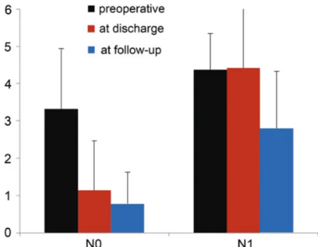
N0 (n = 29) N1 (n = 24) P-value GCS preoperative 12.5 (±3.9) 10.9 (±4.9) 0.17 GCS≤8 6 (20%) 9 (37%) 0.17 GCS postoperative 15.0 (±0.0) 10.9 (±5.7) <0.001 mRS preoperative 3.3 (±1.6) 4.4 (±1.0) 0.007 mRS postoperative 1.1 (±1.3) 4.4 (±1.7) <0.001 mRS follow-up 0.8 (±0.8) 2.8 (±1.5) <0.001 NIHSS postoperative 0.5 (±0.8) 17.8 (±17.8) <0.001 NIHSS follow-up 0.0 (±0.0) 3.6 (±4.4) <0.001 GCS: Glasgow coma scale; mRS: modified Rankin Scale; NIHSS: National Institutes of Health Stroke Scale.
deficit according to the NIHSS at 3 months postoperatively. With respect to the predictive value of neurological status, there was no difference between the groups regarding preoperative GCS. The severity of impairment with respect to the ability to perform daily routine actions independently, assessed by mRS, was predictive of neurological recovery (P < 0.007) as depicted in Table4. Patients who recovered had a significantly lower preoperative mRS score of 3.3 ± 1.6 compared with those in Group N1, who were severely impaired, exemplified by a score of 4.4 ± 1.0. We identified an mRS of >3 as a cut-off value for the lack of neurological recovery (odds ratio [OR]: 5.3,P = 0.01, 95% CI 1.46–19.61).
In all patients receiving surgery for AADA within the 8-year period (n = 300), we observed a new postoperative stroke rate of 12%. These permanent strokes were diagnosed by postoperative CT scan and neurological assessment.
Neurological recovery
In both groups, clinical impairment improved significantly until discharge and further at follow-up (Fig.1). Although the NIHSS in Group N0 was 0 by definition at the time of follow-up, the mRS showed minimal residual impairment in some patients due to cognitive deficit such as poor concentration leading to partial dis-ability. In the group lacking full recovery (N1), 41% of patients showed improved symptoms at follow-up, and 25% remained similarly impaired after surgery, whereas 33% had devastating outcome and died. Of the 15 patients completing follow-up in this group, 10 had an mRS of≤3, which classifies moderate disabil-ity in the sense of‘requires some help, but able to walk unassisted’. Thus, we could assume that of all surviving patients presenting with ND, 71% were living with an acceptable neurological outcome and quality of life.
Regarding the outcome of 14 patients, who presented with a se-verely reduced level of consciousness (Table 1), we did not find GCS level predictive for neurological outcome (Table4), but cannot conclude that it is not important. There was no correlation between the time from the onset of symptoms to the beginning of surgery and the severity of ND (mRS) in N1 patients either. Time to surgery was not statistically different or shorter in the comatose patients who fully recovered (N0: 3.1 ± 1.3 h vs N1: 5.8 ± 3.0 h, P = 0.07),
although there was a trend and the 2 patients with time to surgery of more than 8 h died with devastating cerebral damage.
Patient- and surgery-related parameters
Age and gender were not different between the two groups (Table2). Concerning the cardiovascular risk, there was a signi ficant-ly higher incidence of hypertension, smoking and famificant-ly history of cardiovascular disease in Group N0. Obesity and diabetes appeared more frequently in Group N1. Cardiovascular risk factors did not predict outcome in our cohort. There was a trend towards longer operation and CPB time (Table3), as well as the duration of HCA in patients of Group N1 (P = 0.095). These factors may be confounding factors, and might be explained by the fact that surgical repair could have been more complicated in these cases as a consequence of larger extent of the dissection. When mRS was adjusted for the duration of HCA, it was still a significant predictor of outcome (Table4).
Involvement of the supra-aortic vessels
One significant difference between both groups was a higher inci-dence of occlusion of the right carotid artery by the dissection in the
Table 2: Patient-related parameters
N0 (n = 29) N1 (n = 24) P-value
Age 58.6 (±13.0) 61.0 (±10.5) 0.46
Gender 0.50
Male 18 (62%) 17 (71%) 0.50
Cardiovascular risk factors
Hypertension 22 (76%) 16 (67%) 0.07 Obesity 4 (14%) 5 (21%) 0.04 Dyslipidaemia 8 (28%) 5 (21%) 0.07 Smoking 13 (45%) 5 (21%) 0.03 Diabetes mellitus 1 (3%) 1 (4%) 0.07 Family history 8 (28%) 3 (13%) 0.05
Table 3: Surgery-related parameters
N0 (n = 29) N1 (n = 24) P-value
Ascending aorta repair 1 1
Hemi-arch repair 24 (83%) 19 (79%)
Total arch repair 4 4
Operation time (min) 263.8 (±74.2) 295.8 (±120.3) 0.25 CPB (min) 152.9 (±52.0) 173.1 (±85.6) 0.29 Cross-clamping time (min) 94.5 (±35.4) 102.1 (±51.6) 0.52 Reperfusion (min) 36.1 (±17.5) 41.4 (±25.8) 0.38 HCA (min) 28.4 (±14.6) 36.6 (±20.1) 0.09 With ACP (min) 23.3 (±13.6) 30.6 (±17.3) 0.09 Without ACP (min) 6.2 (±5.8) 7.6 (±6.8) 0.42 Hypothermia (°C) 21.3 (±3.0) 20.8 (±2.8) 0.51 Cannulation site
axillary 25 (86%) 19 (79%) 0.47
femoral 4 (14%) 3 (13%)
axillary + femoral 0 (0%) 1 (4%) CPB: cardiopulmonary bypass; HCA: hypothermic circulatory arrest; ACP: antegrade cerebral perfusion.
Figure 1:Development of neurological symptoms resulting in clinical impair-ment of patients with neurological deficit due to acute aortic dissection type A. N0: group with postoperative NIHSS = 0; N1: patients with NIHSS≥1 at follow-up; mRS: modified Rankin Scale; NIHSS: National Institutes of Health Stroke Scale. A O RTIC SURGER Y
group showing persistent ND (P < 0.05), but it had no predictive value (Table4). There was also a trend when taking into account dis-section of the right carotid artery, but neither disdis-section nor occlu-sion of the left carotid artery was different (Table5). We did notfind a correlation between reconstitution of blood flow in the right carotid artery postoperatively and at follow-up to a favourable neurological outcome. Although symptoms of a right-sided stroke were more frequently found in the whole cohort, there was no dif-ference between groups regarding this parameter (34% in N0 vs 46% in N1). About 51% (15 in Group N0 vs 12 in Group N1) out of all patients had preoperative cranial CT or MRI with or without angio-gram, ruling out haemorrhage or demarcation of territorial stroke.
Mortality
In the complete cohort of 300 patients with AADA who received surgical repair between 2005 and 2012 at our institution, the 30-day mortality rate was 10% (32 patients). In patients with pre-operative new onset of neurological symptoms, this rate increased to 15% (Table6). At the time of follow-up, two additional deaths were recorded, and both patients died from non-disease-related causes, 1 of them residually impaired (N1).
Of the 8 patients who died in-hospital (Table6), 6 had disease-related, neurological detrimental outcome and life-support was withdrawn. Two patients with preoperative visceral malperfusion died from disease-related multiorgan failure, and the cerebral imaging showed severe cerebral lesions as well. Thus, all deceased
patients showed devastating neurological status postoperatively, resulting from cerebral oedema and/or haemorrhage in conjunc-tion with stroke.
DISCUSSION
Neurological symptoms are frequently found in patients with AADA, and the potential for recovery is difficult to predict: similar to thefindings of larger registries and other single-centre studies [6,19], everyfifth patient presented with new onset of neurological dysfunction in our cohort. Repeated observations of neurological recovery after surgery have been made in about 50% of these patients [3,15]. Therefore, surgeons are more likely to perform the extensive surgery necessary to repair the ascending aorta and
re-establish antegrade blood flow. But, how can we distinguish
patients who will recover their ND from those with detrimental outcome?
In view of the very poor prognosis of medical therapy with in-hospital mortality of more than 60% [20,21], surgery was recently deemed appropriate even in high-risk patients such as octogenar-ians [22] and in those with more complex AADA (with coronary is-chaemia or visceral malperfusion for instance). On the other hand, preoperative ND seemed to predict in-hospital death with an OR of 7.7 [23]. Other studies did notfind preoperative ND alone to be a predictor of mortality or even of postoperative ND [6].
At our institution, the decision whether to operate on a patient presenting with ND is made by the surgeon’s clinical judgement on a case-to-case basis, fairly liberal but not overly aggressive as indicated by the average GCS of 11.8 (±4.4). This strategy resulted in an overall favourable outcome with the rate of persistent ND and mortality comparable with or even exceeding previously pub-lished results [3,6,8].
We identified an mRS of >3 as an independent predictor of poor neurological improvement. Similarly, Morimoto et al. [15] also defined an NIHSS score of >11 to be associated with an un-favourable outcome. In their study, patients in the group without recovery even deteriorated from an average NIHSS score of 17 ± 7.2–22 ± 14.7. In contrast, the majority of patients in our study improved significantly over the course of the follow-up (Fig.1). It has been suggested, in a recent publication of the German Registry for Acute Aortic Dissection Type A (GERAADA), that pre-operative ND alone does not predict postpre-operative ND, and that malperfusion of three organ systems was predictive of persistent postoperative ND [6].
In accordance with the study by Morimoto et al. [15], the
present analysis confirmed that cerebral malperfusion supposedly resulting from either dissection or occlusion of the carotid arteries is not predictive of persistent ND and neither is recovery asso-ciated with a higher degree of postoperative regression of the Table 5: Involvement of the carotid arteries
N0 (n = 29) N1 (n = 24) P-value
Occlusion RICA 1 (4%) 5 (23%) 0.049
Dissection RICA 11 (42%) 15 (68%) 0.073
Occlusion LICA 1 (4%) 1 (5%) 0.90
Dissection LICA 9 (35%) 8 (36%) 0.90 RICA: right internal carotid artery; LICA: left internal carotid artery.
Table 6: Postoperative course and mortality
N0 (n = 29) N1 (n = 24) P-value Intubation (days) 1.1 (±1.2) 2.7 (±2.2) 0.003 Hospitalization (days) 12.6 (±5.1) 16.3 (±12.1) 0.15 Mortality In-hospital 0 (0%) 8 (33.3%) 0.002 At follow-up 1 (3.4%) 1 (4.2%)
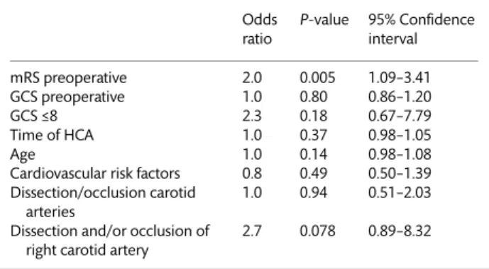
Table 4: Logistic regression analysis for predictive factors of persistent neurological deficit
Odds ratio P-value 95% Confidence interval mRS preoperative 2.0 0.005 1.09–3.41 GCS preoperative 1.0 0.80 0.86–1.20 GCS≤8 2.3 0.18 0.67–7.79 Time of HCA 1.0 0.37 0.98–1.05 Age 1.0 0.14 0.98–1.08
Cardiovascular risk factors 0.8 0.49 0.50–1.39 Dissection/occlusion carotid
arteries
1.0 0.94 0.51–2.03 Dissection and/or occlusion of
right carotid artery
2.7 0.078 0.89–8.32
mRS: modified Rankin Scale; GCS: Glasgow coma scale; HCA: hypothermic circulatory arrest.
involvement of the supra-aortic vessels. This speaks in favour of clinical assessment rather than putting too much emphasis on the radiological evidence. But, in many previous reports, cerebral mal-perfusion was defined as a significant risk factor for detrimental neurological outcome and mortality [1,6]. More detailed informa-tion on intracranial cerebral vessel perfusion may provide an add-itional value as intracranial Doppler may become more available in the future.
As to how the immediacy of surgery influenced our results
cannot be derived from our data, because we did not have suf fi-cient data to correlate outcome with the time to surgery in all patients. It is generally accepted now that an intentional delay of surgery, which results in 15–25% mortality, cannot be advocated anymore [24].
We observed a trend towards longer operating time, including cross-clamping time, time of circulatory arrest and ACP in the N1 group, but none of these parameters were predictive for recovery. Operative procedures of longer duration suggest more extensive repair and/or a more demanding intraoperative situs. However, there was no difference between both groups when the need for total arch repair was analysed. The majority of patients received an open distal anastomosis, consisting of an excision of the concave portion of the aortic arch and a slanting suture line towards proximal of the brachiocephalic trunk.
A further important aspect is the underlying pathophysiological mechanism leading to the ND. It seems important to distinguish between focal and generalized symptoms, such as coma. In patients with a generally reduced level of consciousness, this can be due to stroke, a prolonged post-ictal phase after epileptic seizure induced by general hypoxia, but also an effect of cardiac tamponade with consecutive deterioration of the cardiac function. Comatose patients represent an interesting cohort, for which an acceptable outcome has recently been published as well [13]. In our study, surgery was performed on a minority of patients pre-senting in a comatose state. Of these 14 patients, 6 recovered to various extent and were in Group N1, 6 of them recovered fully (N0). Although our findings in this cohort are limited by small numbers, we did not identify a correlation between the severely reduced level of consciousness with neurological outcome, but saw a trend towards shorter time to surgery and thus may con-clude, that surgery might prove beneficial even in these patients, when carried out immediately.
Cerebral protection strategies, including the arterial cannulation strategy, ACP, hypothermia and transcranial oxygen saturation moni-toring, have been considered to play a major role in the neurological outcome following surgery for AADA. Cannulation of the right sub-clavian arteryfirst is a strategy that was implemented at our institu-tion throughout the whole period of observainstitu-tion, and was only quit after frustrane trial of perfusion via this access. A report from the GERAADA registry stated that the type of cerebral protection did not impact neurological complication rate [25], which is in contrast to earlier reports, which found that ACP led to superior outcome [4].
Limitations
This study has certain limitations, mainly the limited sample size and the method of retrospective data analysis. Neurological as-sessment was limited to GCS and mRS, both rather crude assess-ment scores that are not validated in this specific situation.
For obvious reasons, the performance of randomized trials is highly unlikely in these patients, so the community will rely on
data from registries and case series such as this one. We believe that the highly defined and standardized approach at our institu-tion constitutes an advantage over more heterogeneous data acquired in registries and therefore single-centre studies should complement them.
We unfortunately do not have reliable data for all patients on the time between onset of symptoms and surgery. The diagnosis of AADA was frequently made by CT scan in an outside hospital, and the patient was transferred, resulting in 2–3 h delay between imaging and skin incision. Furthermore, some patients delayed admission to a hospital themselves and whether this contributed to neurological outcome is difficult to evaluate, since it is known that the dissection can progress over time.
CONCLUSION
We report an 8-year experience of treating AADA patients, of which roughly 20% showed new ND of a large variation from comatose to focal motor deficit only. A favourable outcome with either full re-covery or limited incapacitation (mRS <3) was achieved in more than 70% of this cohort. Crude assessment of neurological impair-ment via mRS was predictive of detriimpair-mental outcome, but the ma-jority even of comatose patients showed recovery to some extent, were discharged from our hospital and concluded thefirst follow-up. We conclude that even severely incapacitated patients should notper se be excluded from life-saving surgery.
ACKNOWLEDGEMENTS
We thank Florian Schoenhoff for critical reading and excellent comments on this manuscript.
Conflict of interest: Christoph Huber receives proctoring and lec-turing fees from Edwards Lifesciences and Symetis SA, and is a consultant for Medtronic, Inc.
REFERENCES
[1] Tanaka H, Okada K, Yamashita T, Morimoto Y, Kawanishi Y, Okita Y. Surgical results of acute aortic dissection complicated with cerebral mal-perfusion. Ann Thorac Surg 2005;80:72–6.
[2] Fann JI, Sarris GE, Miller DC, Mitchell RS, Oyer PE, Stinson EBet al. Surgical management of acute aortic dissection complicated by stroke. Circulation 1989;80(3 Pt 1):I257–63.
[3] Estrera AL, Garami Z, Miller CC, Porat EE, Achouh PE, Dhareshwar Jet al. Acute type A aortic dissection complicated by stroke: can immediate repair be performed safely? J Thorac Cardiovasc Surg 2006;132:1404–8. [4] Bonser RS, Ranasinghe AM, Loubani M, Evans JD, Thalji NM, Bachet JE
et al. Evidence, lack of evidence, controversy, and debate in the provision and performance of the surgery of acute type A aortic dissection. J Am Coll Cardiol 2011;58:2455–74.
[5] Borst HG, Laas J, Heinemann M. Type A aortic dissection: diagnosis and management of malperfusion phenomena. Semin Thorac Cardiovasc Surg 1991;3:238–41.
[6] Conzelmann LO, Hoffmann I, Blettner M, Kallenbach K, Karck M, Dapunt Oet al. Analysis of risk factors for neurological dysfunction in patients with acute aortic dissection type A: data from the German Registry for Acute Aortic Dissection type A (GERAADA). Eur J Cardiothorac Surg 2012;42: 557–65.
[7] Immer FF, Grobety V, Lauten A, Carrel TP. Does malperfusion syndrome affect early and mid-term outcome in patients suffering from acute type A aortic dissection? Interact CardioVasc Thorac Surg 2006;5:187–90.
A
O
RTIC
SURGER
[8] Girdauskas E, Kuntze T, Borger MA, Falk V, Mohr FW. Surgical risk of pre-operative malperfusion in acute type A aortic dissection. J Thorac Cardiovasc Surg 2009;138:1363–9.
[9] Gaul C, Dietrich W, Erbguth FJ. Neurological symptoms in aortic dissec-tion: a challenge for neurologists. Cerebrovasc Dis 2008;26:1–8.
[10] Meszaros I, Morocz J, Szlavi J, Schmidt J, Tornoci L, Nagy L et al. Epidemiology and clinicopathology of aortic dissection. Chest 2000;117: 1271–8.
[11] Geirsson A, Szeto WY, Pochettino A, McGarvey ML, Keane MG, Woo YJ et al. Significance of malperfusion syndromes prior to contemporary sur-gical repair for acute type A dissection: outcomes and need for additional revascularizations. Eur J Cardiothorac Surg 2007;32:255–62.
[12] Trimarchi S, Nienaber CA, Rampoldi V, Myrmel T, Suzuki T, Mehta RH et al. Contemporary results of surgery in acute type A aortic dissection: the International Registry of Acute Aortic Dissection experience. J Thorac Cardiovasc Surg 2005;129:112–22.
[13] Tsukube T, Haraguchi T, Okada Y, Matsukawa R, Kozawa S, Ogawa Ket al. Long-term outcomes after immediate aortic repair for acute type A aortic dissection complicated by coma. J Thorac Cardiovasc Surg 2014;148: 1013–9.
[14] Pocar M, Passolunghi D, Moneta A, Mattioli R, Donatelli F. Coma might not preclude emergency operation in acute aortic dissection. Ann Thorac Surg 2006;81:1348–51.
[15] Morimoto N, Okada K, Okita Y. Lack of neurologic improvement after aortic repair for acute type A aortic dissection complicated by cerebral malperfusion: predictors and association with survival. J Thorac Cardiovasc Surg 2011;142:1540–4.
[16] Teasdale G, Jennett B. Assessment of coma and impaired consciousness. A practical scale. Lancet 1974;2:81–4.
[17] van Swieten JC, Koudstaal PJ, Visser MC, Schouten HJ, van Gijn J. Interobserver agreement for the assessment of handicap in stroke patients. Stroke 1988;19:604–7.
[18] Brott T, Adams HP Jr, Olinger CP, Marler JR, Barsan WG, Biller Jet al. Measurements of acute cerebral infarction: a clinical examination scale. Stroke 1989;20:864–70.
[19] Rampoldi V, Trimarchi S, Eagle KA, Nienaber CA, Oh JK, Bossone Eet al. Simple risk models to predict surgical mortality in acute type A aortic dis-section: the International Registry of Acute Aortic Dissection score. Ann Thorac Surg 2007;83:55–61.
[20] Hagan PG, Nienaber CA, Isselbacher EM, Bruckman D, Karavite DJ, Russman PLet al. The International Registry of Acute Aortic Dissection (IRAD): new insights into an old disease. JAMA 2000;283:897–903. [21] Booher AM, Isselbacher EM, Nienaber CA, Trimarchi S, Evangelista A,
Montgomery DGet al. The IRAD classification system for characterizing survival after aortic dissection. Am J Med 2013;126:730. e19–24. [22] Rylski B, Suedkamp M, Beyersdorf F, Nitsch B, Hoffmann I, Blettner M
et al. Outcome after surgery for acute aortic dissection type A in patients over 70 years: data analysis from the German Registry for Acute Aortic Dissection Type A (GERAADA). Eur J Cardiothorac Surg 2011;40:435–40. [23] Santini F, Montalbano G, Casali G, Messina A, Iafrancesco M, Luciani GB
et al. Clinical presentation is the main predictor of in-hospital death for patients with acute type A aortic dissection admitted for surgical treat-ment: a 25 years experience. Int J Cardiol 2007;115:305–11.
[24] Fukuda I. Management of intraoperative malperfusion syndrome using femoral artery cannulation for repair of acute type A aortic dissection. Invited commentary. Ann Thorac Surg 2008;85:1624.
[25] Kruger T, Weigang E, Hoffmann I, Blettner M, Aebert H. Cerebral protec-tion during surgery for acute aortic dissecprotec-tion type A: results of the
German Registry for Acute Aortic Dissection Type A (GERAADA). Circulation 2011;124:434–43.
APPENDIX. CONFERENCE DISCUSSION
Scan to your mobile or go to
http://www.oxfordjournals.org/page/6153/1
to search for the presentation on the EACTS library
Dr E. Saadi (Porto Alegre, Brazil): We know neurological symptoms secondary to arch vessel malperfusion are documented in up to 20% of the cases with acute aortic dissection. Although stroke and coma are traditional contraindica-tions for immediate surgery, and the criteria to define reversible brain damage remain unclear, successful emergency repair has been reported in your paper. We would like to comment on some aspects of this challenging situation.
We have some concerns about evaluating the patient’s neurological condi-tion using the Glasgow Coma Scale score after aortic disseccondi-tion. In fact, this score was developed to evaluate the level of consciousness in patients after head injury who might have organic damage to the brain tissue and neuro-logical system and may not apply to this specific disease. The wide range of neurological dysfunction presented, from small sensory deficit or convulsion to hemiplegia and coma, may compromise the evaluation of the whole group, which is not homogenous.
I would like to ask you, what was the type of cerebral protection used? You told us it was antegrade cerebral perfusion. Was this unilateral, just on the right, or bilateral? Do you use any special cerebral protection in patients with previ-ous neurological deficit?
In your experience, is there any situation in which you would contraindicate surgery in acute type A aortic dissection because of the neurological deficit?
Dr Most: As for the first question, we don’t do any special treatment to the patients who present with neurological deficit. All of them receive antegrade cerebral perfusion via catheters bilaterally. As soon as we go into hypothermic arrest, the perfusion via the arterial cannula is stopped, and we don’t clamp the brachiocephalic trunk, but we insert perfusion catheters into both carotid arter-ies. We always do near-infrared spectroscopy to assess the success of our cere-bral perfusion.
There is no special treatment for those patients with signs of cerebral malper-fusion, such as preoperative carotid revascularization or selective cannulation, but we are very conscious of inserting the perfusion catheters carefully so as not to make the damage progress or cause thrombo- or air embolism.
Dr Saadi: And the contraindications?
Dr Most: As to the contraindications, we did not have reliable data on the patients who were refused surgery because sometimes this is also done while the patient is still at the referring centre.
With the previous speaker, I would agree that the time from the onset of coma is relevant and that the group from Japan also showed that coma of more than six hours was detrimental and had worse outcomes. Of course, the general situation in which the patient presents is also playing a role. Obviously not only cerebral malperfusion but malperfusion of other organ systems like is-chaemia of the intestines is an important factor in this setting and can be a contraindication.
But I think in isolated brain malperfusion, the presence of the coma or of a generally reduced level of consciousness, at least in this cohort, did not as such predict a detrimental outcome.