ELSEVIER
FEMS Microbiology Letters 138 ( 1996) 89-95Prevotella intermedia and Prevotella nigrescens serotypes,
ribotypes and binding characteristics
Gunnar G. Dahlen a3*, Jolene R. Johnson b, Rudolf Gmiir
’a Department of Oral Microbiology, Faculty of Odontology, University of Gijteborg, Guldhedsgatan IO, S-413 46 Giiteborg, Sweden b Dental Research Institute, Vnioersity of Minnesota, Minneapolis, MN, USA
’ Institute of Oral Microbiology and General Immunology, Ziirich Vniversily, Ziirich, Switzerland Received 2 January 1996; revised 27 February 1996; accepted 1 March 1996
Abstract
Type strains and 62 clinical isolates of Prevotella intermedia and Prevotella nigrescens were typed with the use of genomic DNA fingerprints and rFWA gene probes. The strains were further serotyped with monoclonal antibodies and characterized with SDS-PAGE, enzymatic activities, hemolysis and hemagglutination and coaggregation with Streptococcus and Actinomyces spp. P. intermedia and P. nigrescens were found to have distinct ribotype patterns which correspond to previously defined serotypes I and II/III, respectively. No clear phenotypic difference related to hemolysis, hemagglutina- tion and coaggregation with Streptococcus and Actinomyces species, or expression of aminopeptides and lipase was found between P. inter-media and P. nigrescens.
Keywords: Precotella intermedia; Preootella nigrescens; Monoclonal antibody; Ribosomal RNA gene restriction analysis; Coaggregation; Hemagglutination
1. Introduction
Prevotella inter-media, a black-pigmented saccha- rolytic anaerobic rod, is found widespread in the oral cavity. It is frequently isolated in various age groups, from individuals with healthy gingiva, and it is a common component in odontogenic abscesses [ 1,2]. It can therefore be argued that either this species is an opportunistic pathogen or it represents a rather heterogeneous species with respect to virulence. At
* Corresponding author. Tel.: +46 (31) 604 620; Fax: +46 (31) 825 733; E-mail: dahlen@odontologi.gu.se
present, it is not clear whether strains isolated from different oral conditions and locations belong to different genotypes or potentially represent pheno- typical variants.
Two DNA homology groups were early identified among P. inter-media strains [3], one of which has
been proposed as a new species, Prevotella ni- grescens [4]. It (type strain ATCC 33563) was phe-
notypically separated recently from P. inter-media
(ATCC 25611) by low peptidase activity, failing to cleave lipid substrates, and revealing different elec- trophoretic enzyme profiles. The DNA homology groups of P. intemedia have been correlated with 3 distinct serogroups [5]. Serogroup I, represented by type strain ATCC 25611, was predominantly found 0378-1097/96/$12.00 0 1996 Federation of European Microbiological Societies. All rights reserved
in deep periodontal lesions while an even distribu- tion of all 3 serogroups (I-III) was found among isolates from adults with no periodontal disease 161.
Recent development in DNA technology has pro- vided new approaches to bacterial taxonomy. The use of DNA fingerprinting to evaluate relatedness among isolates is somewhat hampered by numerous bands obtained in agarose gels. To overcome this problem, specific probes for identification of certain fingerprint bands containing specific DNA se- quences, e.g., the highly conserved rRNA gene, have been developed [8]. The purpose of this study was (i) to investigate the genetic relatedness of 62 P. inter-
media / nigrescens isolates, using a probe comple- mentary to Escherichia coli 16s and 23s rRNA and (ii) to compare the resulting patterns with those observed by serotyping with monoclonal antibodies (MAbs) and those received by phenotypic classifica- tion as proposed [4].
2. Material and methods 2. I. Bacterial strains
Sixty-two (OMGS, Oral Microbiology, GBteborg, Sweden) strains grouped as P. intermedia or P.
nigwscens based on fluorescence in long-wave UV light, glucose fermentation and indole production were investigated. They were obtained from Swedish volunteers with healthy gingival conditions (I 7 strains), or deep periodontal pockets (38 strains), and were characterized biochemically and serologically [6]. Additionally, 7 isolates from periodontally dis- eased Kenyan adults were included [7]. As reference strains for serotypes I, II and III, strain ATCC 25611 (type strain of P. intermedia), ATCC 25261 and ATCC 33563 (type strains of P. nigrescens), all provided through CCUG (Culture Collection, Uni- versity of Giiteborg, Sweden), were included. Strains were stored lyophilized and for each experiment fresh cell preparations were made by plating the bacteria on Brucella agar (BBL Becton Dickinson Microbiology Systems, Cockeysville, MD) enriched with 5% horse blood, 0.5% hemolysed blood and 5 mg/l menadione. All strains were tested for aminopeptidase and lipase activities according to the methods described by Shah and Gharbia [4].
2.2. GenoQping und ribotying
Methods for DNA extraction, restriction enzyme digestion. agarose gel electrophoresis, synthesis of probes and analyses were essentially the same as described in detail by Bowden et al. [8]. Strains were grown anaerobically using the Gas-Pak system (Gas Generation Kit. Oxoid Ltd, Basingstoke, Hampshire. England) for 2-3 days. DNA yield and purity were determined by measurement of the absorption at 260 and 280 nm. Enzyme digestion was performed with DNA from all strains using PstI, and BamHI
(Boehringer Mannheim Scandinavia AB, Bromma. Sweden). Electrophoresis was run in a horizontal agarose gel (Bio-Rad Subcell, Bio-Rad, Solna, Swe- den) at constant voltage (40 V) for 19 h at room temperature in 89 mM Tris-borate buffer pH 8.0. Digoxigenin labeled lambda DNA digested with Hind111 (Marker III, Boehringer Mannheim), was used as molecular markers in the gels. The fragments were visualized after ethidium bromide staining un- der shortwave UV light and photographed. DNA fragments were transferred to nylon membranes (Zeta-Probe GT, Bio-Rad) for Southern blotting us- ing a Vacuum Blotter (Bio-Rad). The preparation of the DNA probe was made with the Genius Kit (1093-657. Boehringer Mannheim), using random primers (Promega Cl 181. Promega Research Labs. Madison, WI) and reverse transcriptase (Gibco BRL Life Technologies, Inc., Gaithersburg, MD). Molecu- lar masses of major bands observed by ribotyping were compared with DNA molecular markers as reference. Ribotype patterns, which were confirmed on independent gels, were estimated.
2.3. SDS-PAGE outer membrane profiles
SDS-PAGE was performed in a Mini-Protean II (Bio-Rad) Unit at 200 V run for 45 min using a vertical 0.75 mm thick slab gel containing 7.5% (w/w) polyacrylamide. Bacterial samples were pre- pared by sonication of whole cell suspensions at 50 W for 1 min. The preparations were heated with a SDS sample buffer at 100°C for 5 min. After elec- trophoresis, the gel was stained with Coomassie brilliant blue R.
G.G. Dahltn et al. / FEMS Microbiology Letters 138 (1996) 89-95 91
2.4. Hemolysis and hemagglutination
Hemolysis was recorded after 3 days of incuba- tion from growth on Brucella agar plates.
Hemagglutination with erythrocytes of human, sheep and rabbit origin was recorded in microtiter- plate wells using 2-fold dilution series at a bacterial density of lo9 (measured with optical density). Hemagglutination was read after 1 h of incubation at room temperature and overnight in a refrigerator.
Hemagglutination was also tested with sheep ery- throcytes and 5 P. intermedia or P. nigrescens strains following culture on various media. In addition to the Brucella agar plate, strains were cultured anaero- bically for 2-3 days in BHICJ (brain heart infusion with cysteine hydrochloride and yeast autolysate) broth and semiliquid Huntoon (HCMG-SuIa) broth [91.
ABCDE
FGHI
JKL
23.1
9.4
6.6
4.4
2.3
2.0
2.5. Coaggregation with Streptococcus and Actino- myces species
A panel of 6 streptococcal strains (Streptococcus
mutans, 2 strains, Streptococcus sanguis, 2 strains, Streptococcus oralis, 2 strains) and 3 Actinomyces
strains grouped as Actinomyces naeslundii, repre- senting 3 different binding specificities (ATCC 12104, LY 7 and 2238) were used. Strain LY 7 was provided by R.J. Gibbons, Forsyth Dental Center, Boston, MA, USA and strain 2238 was isolated from supragingival plaque of a healthy Swedish adult. Equal volumes of bacterial suspensions (1 X lo9 cells/ml in 0.001 M Tris, to pH 8.0) of two cell types were mixed and examined immediately for coaggregation. The coaggregation score was given according to the scale of Kolenbrander and London
[lOI.
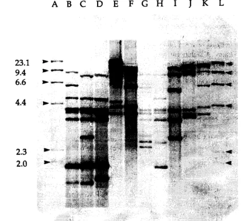
Fig. 1. Ribotype patterns generated from P. intermedia and P. nigrescens strains after treatment of chromosomal DNA with PstI. Lanes B, C, D, H and I, P. nigrescens serotype II strains OMGS 456, OMGS 1449, OMGS 410, OMGS 457, and ATCC 33563. Lane G, P. nigrescens serotype III strain OMGS 447 and lanes E, F, J and K, I? intermedia serotype I strains OMGS 725, ATCC 25611 (obtained from CCUG, GGteborg University), OMGS 754 and ATCC 25611 (obtained from Dental Research Institute, University of Minnesota). Lanes A and L, HindIII fragments of ADNA labeled with digoxigenin.
92 G.G. Dahlia et (11. / FEMS Microbiology Letters 138 f 19961 89-95
3. Results Table 2
3. I. Enzymatic activities
None of the 62 P. intermedia or P. nigrescens reacted positively for aminopeptidase or lipase activ- ities.
3.2. Ribotyping
The ribotypes fell into two main patterns (Fig. I>. Ribotype pattern A which included ATCC 25611 did not show fragments smaller than 3.9 kb, while ribo- type pattern B including both ATCC 25261 and ATCC 33563 consistently produced small, strong bands of 2.8 kb and/or 1.8 kb.
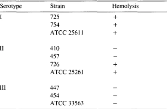
Hemolysis recorded of P. intermedia and P. nigrescens isolates
Serotype Strain Hemolysis
I 125 + 154 + ATCC 25611 + II 410 _ 457 _ 726 + ATCC 2526 I + III 447 _ 454 _ ATCC 33563 _ 3.3. Serotyping
All tested strains did react with the MAbs used and fell into serotype I (32 strains), II (10 strains) and III (20 strains). Of the 7 Kenyan strains 2 were of serotype I while 3 were of serotype II and 2 of serotype III.
964, 868 and 873) did not fit into this pattern. It was not possible to find distinct different ribotype pat- terns between serotype II and III strains. Strains from diseased patients were overrepresented among serotype I isolates, while strains of serotypes II and III belonging to P. nigrescens were about equally recruited from periodontally diseased and healthy individuals.
Table 1 shows the relation between ribotypes and serotypes of the 62 strains. A significant correlation was found between serotype I (P. inter-media) and ribotype A and between serotype II and III (P.
nigrescens) and ribotype B. Only 3 strains (OMGS
3.4. SDS-PAGE
Table I
Ribotyping of P. intermedia and P. nigrescens isolates Serotype Strain category Ribotype Ribotype
pattern A pattern B
I Type strain ATCC 25611 -
Swedish diseased 24 I
Swedish healthy 4 0
Kenyan diseased 2 0
II Type strain - ATCC 2526 I
Swedish diseased 1 2
Swedish healthy 0 4
Kenyan diseased 0 3
III Type strain - ATCC 33563
Swedish diseased I 8
Swedish healthy 0 9
Kenyan diseased 0 2
Total 32 29
The SDS-PAGE patterns of P. intermedia and P. nigrescens type strains ATCC 25611 and ATCC 33563 both showed a strong band of 29 kDa while an additional strong band was displayed by a 25 kDa protein of ATCC 33563.
Comparisons of all isolates by SDS-PAGE showed that the strains grouped as P. intermedia by ribo- and serotyping displayed banding patterns compara- ble to the one seen with P. intermedia ATCC 25611 and that the isolates similarly grouped as P. ni-
grescens corresponded to ATCC 33563. Thus, the latter group consistently showed the band of 25 kDa, which was absent in all P. intermediu isolates. SDS-PAGE pattern confirmed the ribotype pattern for all strains including those 3, which showed sero- logical diversity.
3.5. Hemolysis and hemagglutination
Hemolysis was registered for all P. intermedia strains tested (Tables 2-41, while the pattern was
G.G. Dahl& et al. / FEMS Microbiology Letters 138 (1996) 89-95 93
Table 3 Table 4
Hemagglutination with sheep erythrocytes by P. intermedia and
P. niarescens after culture on various media
Hemagglutination pattern of P. intermedia and P. nigrescens isolates against sheep, human and rabbit erythrocytes
Strain Serotype Cultured on Brucella plate
Sheep Human Rabbit
1269 I - 2-k” 5+ 1286 I 4+ 6+ 1281 I 3-k 6+ 861 II 2+ 2+ 8+ 865 II 2+ 3+ 7+ 868 II 3+ 2+ 6+ 1271 III 2+ 4+ 5+ 1300 III 2+ 3+ 6+
* + denotes the number of 2-fold dilutions of the bacteria1 suspension with a visible agglutination.
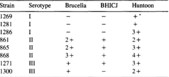
Strain Serotype Brucella BHICJ Huntoon
1269 I 1281 I 1286 I 861 II 865 II 868 II 1271 III 1300 III - - +* - - + - - 3+ 2+ + 2+ 2+ + 3+ 3+ + 4f + + 3+ + - 2-k
* + denotes the number of 2-fold dilutions of the bacterial suspension with a visible agglutination. < /T3 >
inconsistent for the
P. nigrescensstrains. Notably,
all the serotype III strains were negative.
medium showed best over all hemagglutination in-
cluding the strains of
P. intermedia.All
P. inter-mediaand
P. nigrescensstrains ag-
glutinated with rabbit erythrocytes. Agglutination
with human red blood cells was obtained for all
strains, however the reaction was weaker. Five strains
showed weak hemagglutination with sheep erythro-
cytes (Table 3) and all belonged to serotype II and
III
(P. nigrescens),whereas all hemagglutination-
negative strains were serotype I
(P. intermedia)strains. Table 4 shows that the hemagglutination was
influenced by the medium in which the strain was
cultured. Cells cultured in the semifluid Huntoon
3.6.
CoaggregationStrains of both species showed no or very weak
coaggregation with
Streptococcusspp. The two
streptococcal strains S.
oralis 24892and S.
mitis Nshowed a high degree of autoagglutination which
was potentiated particularly with
P. intermediastrains. Presumably,
P. intermediaand
P. ni- grescensisolates did not express receptor specificity
for streptococcal species (data not shown). Both
Table 5
Coaggregation between P. intermedia and I? nigrescens and A. naeslundii strains Ly 7, ATCC 12104 and 2238 Bacterial species Strain Patient category from which Serotype A. naeslundii strain
the strain was isolated
LY 7 ATCC 12104 2238 P. intermedia 864 periodontitis I ++ +++ ++++ 870 periodontitis I + _ + 874 periodontitis I +++ ++++ +++ 1277 gingivitis I ++ ++ - 1281 gingivitis I + - + 1300 gingivitis I + - -
ATCC 25611 type strain I + + ++
P. nigrescens 726 periodontitis (Kenya) II + - _
865 periodontitis II ++ +++ ++++
868 periodontitis II ++ ++ +
904 periodontitis II - - _
ATCC 33563 type strain II + + +
866 periodontitis III _ _ -
1302 gingivitis III - + +
species showed varying coaggregation ability with
A. naeslundii strains (Table 5). Three strains, 2 P.
intermedia and 1 P. nigrescens strains, showed ex- pressed coaggregation with all three A. naeslundii strains. One P. nigrescms (OMGS 866) strain did not coaggregate with any of the A. naeslundii strains. The difference in coaggregation between the A.
naeslundii strains was small.
4. Discussion
Identification of Prelvtella species in the oral cavity has been carried out mainly on the basis of colony morphology and biochemical reaction profile. Thus black-pigmented saccharolytic Prelwtella
species separated into two main groups, one prote- olytic and one non-proteolytic. It has been long been known that the proteolytic group comprises two DNA homology groups, now specified as P. intermedia (homology group I> and P. nigrescens (homology group II).
In spite of a distinct genotypic difference these two species show a remarkable similarity in pheno- typic characteristics, which makes it difficult to rapidly distinguish between isolates of P. intermedia and P. nigrescens. Shah and Gharbia [4] recently proposed to separate these two species after having found appropriate phenotypic variation. However. in our hands, those methods failed to show a distinct difference between P. intermedia and P. nigrescens
isolates.
Most of the isolates studied in the present investi- gation have previously been subjected to biochemi- cal classification and serotyping using rat MAbs, which showed that all strains of the two species expressed one of three serological reactivity patterns [5]. Type I coincided with homology group I (P.
intermedia) and type II and III strains with homol- ogy group II (P. nipscens).
This is confirmed by the present finding and that
P. nigrescens serotypes showed 16s and 25s ribo- typing patterns distinctly different from that of P.
intermedia isolates.
Serological species distinction is based on the binding of MAb 40B13.2.2 to the surface of P.
intermedia but not to P. nigrescens. Two strains belonging to P. nigrescens according to the ribotype
pattern with the use of Pstl and BamHI endonucle- ases, expressed the antigen which binds to MAb 40B13.2.2. P. nigrescens species may therefore con- tain subtypes which share the P. intermedia surface antigen. The antigen detected by this MAb has not been characterized. On the basis of a few preliminary experiments, Gmiir and Wyss [ 1 I] have speculated on the possibility of a cell surface protein of approxi- mately 150 kDa.
In the present study. the outer membrane profile detected by SDS-PAGE showed some distinct differ- ences between the two species. One band of 25 kDa was consistently present in P. nigrescens but lacking in P. intermedia isolates. The distinct P. nigrescens band of 3 1 kDa protein shown by Gharbia et al. [ 121 may correspond to the band of 29 kDa in the present study. However, this band was consistently present in all strains of both species. By SDS-PAGE Frand- sen et al. [ 131 found a band of 20 kDa while the band of 29 kDa was found in strains of both species which was used to separate the strains into P. intermediu and P. nigrescens. This apparent heterogeneity in outer membrane protein profiles may in part be explained by different electrophoretic conditions. More likely it reflects the variations in surface pro- tein expression due to culture conditions which also was found in this study to significantly alter the hemagglutination.
P. nigrescens strains seemed to show slightly more hemagglutination after 3 days of culturing on Brucella agar plates, which may distinguish P. ni- grescens from P. intermedia. This difference was not obtained after culturing in the two different broth media. Variation in adherence to oral epithelial cells and hemagglutination was suggested to be related to types of surface appendages expressed by the two species [ 14- 161. Okuda et al. [ 141 showed a similar hemagglutinating activity for P. intermedia ATCC 2561 I and P. nigrescens ATCC 33563 against intact or neuraminidase treated cells. Nesbitt et al. [ 151 showed coaggregation with various Actinomyces
species by testing 2 strains of P. intermedia belong- ing to the ATCC 25611 DNA hybridization group and differing distinctly in their coaggregation pat- tern. P. nigrescens may also coaggregate with Acti-
nomyes [16], however. the coaggregation was not related to the fibril type of P. intermedia and P.
G.G. DahlGn et al. / FEMS Microbiology Letters 138 (1996) 89-95 95
ing type I or type 2 or both types of fimbriae. Similarly, the differences obtained in outer mem- brane binding proteins in the present study did not give a species related variation in the hemagglutinat- ing activity or coaggregation.
Rapid differentiation between the two species is of interest due to a suggested closer relation of P.
intermediu to pathological conditions in the oral cavity such as periodontal disease and endodontic infections [6,12]. However, even if this association is statistically proven, it may simply be due to environ- mental factors in the periodontal pocket favoring the establishment of P. intermedia. In an experimental study in wound chambers in rabbits a similar abscess formation capability and recovery rate from the ab- scess was obtained for P. intermedia and P. ni-
grescens and the pathological difference between the two species was questioned [17].
It is therefore concluded that despite genetic and serological differences there is still a lack of clear phenotypic differences disclosed for the rapid identi- fication of P. intermedia and P. nigrescens.
Acknowledgements
We thank Mrs. Lisbeth Bengtsson for her techni- cal assistance. Part of this study was performed by Gunnar DahlCn, as Lasby Visiting Professor at the University of Minnesota, USA. This study was sup- ported by a Grant from Swedish Medical Research Council, project B94-24X- 10874-o 1 A.
References 111
121
[31
von Konow, L., Nord, C.E. and Nordenram, A. (1989) Anaerobic bacteria in demoalveolar infections. Int. J. Oral Surg. 10. 313-322.
Finegold, SM., Strong, CA., McTeague, M. and Marina, M. (1993) The importance of black-pigmented Gram-negative anaerobes in human infections. FEMS Immunol. Med. Mi- crobiol. 6. 77.-82.
Johnson. J.L. and Holdeman, L.V. (1983) Bacreroides inter-
medius comb. nav. and description of Bactrroides corporis
sp. nav. and Bacteroides /eL,ii sp. nov. Int. J. Syst. Bacterial. 33, 15-25.
[4] Shah, H.N. and Gharbia, S.E. (1992) Biochemical and chem- ical studies on strains designated Prec,orella infermedia and
proposal of a new pigmented species, Prerorella nigrescens sp. nav. Int. J. Syst. Bacterial. 42, 542-546.
[5] Gmiir. R. and Guggenheim. B. (1983) Antigenic heterogenic- ity of Bacferoides inrermedius as recognized by monoclonal antibodies. Infect. Immun. 42. 459-470.
[6] Dahlen. G., Wikstrom, M., Renvert, S.. Gmiir, R. and Guggenheim, B. (1990) Biochemical and serological charac- terization of Bacferoides infermedius strains isolated from the deep periodontal pocket. J. Clin. Microbial. 28, 2269- 2274.
[7] Dahlin, Cl.. Manji. F., Baelum, V. and Fejerskov, 0. (1992) Putative periodontopathogens in ‘diseased’ and ‘nondiseased’ persons exhibiting poor oral hygiene. J. Clin. Periodontol.
19. 35-42.
[8] Bowden. G., Johnson J. and Schachtele. C. (1993) Character- ization of Actinomyces with genomic DNA fingerprints and rRNA gene probes. J. Dent. Res. 72, 1171-l 179.
[9] Mdller. A.J.R. (1966) Microbiological examination of root canals and periapical tissues of human teeth. Odontol. Tidskr. 74, l-380.
[ 101 Kolenbrander, P.E. and London. J. (1992) Ecological signifi- cance of coaggregation among oral bacteria. Adv. Microbial. Ecol. 12. 183-217.
[I l] Gmilr, R. and Wyss, C. (1985) Monoclonal antibodies to characterize the antigenic heterogeneity of Bacteroides inter- medius. In: Monoclonal antibodies against bacteria (Macario, A.J.L. and Conway de Macario, E., Eds.), pp. 91-119, Academic Press, London.
1121 Gharbia, SE.. Haapasalo, M., Shah, H.N.. Kotiranta, A., Launatmaa. K., Pearce, M.A. and Devine, D.A. (1994) Char- acterization of Prwotella intermedia and Precotella ni- grescens isolates from periodontic and endodontic infections. J. Periodontal. 65, 56-61.
[13] Frandsen, E.V.G., Paulsen, K. and Kilian, M. (1995) Confir- mation of the PreL,otella intermedia and Prerotella ni- grescens. Int. J. Syst. Bacterial. 45, 429-435.
[l4] Okuda. K., Ono, M. and Kato. T. (1989) Neuraminidase enhanced attachment of Bacteroides intermedius to human erythrocytes and buccal epithelial cells. Infect. Immun. 57,
I635- 1637.
1151 Nesbitt. W.E., Fukushima, H., Leung, K.P. and Clark, W.B. (I 993) Coaggregation of Pret,otella infermedia with oral
Acfinomyces species. Infect. Immun. 61, 201 I-2014. [l6] Devine, D.A. and Handley, P.S. (1989) The relationship
between coaggregation properties and surface structures of
Bacreroides inrermedius. Microbial. Ecol. Health Dis. 2, 267-278.
[I 71 Hafstrom, C. and Dahltn, G. (1995) Pathogenicity of Pre-
rotella intermedia and PreL,otella nigrescens isolated from various oral sites. Oral Microbial. Immunol. (submitted).