International Immunology, Vol. 3, No. 2, pp. 127- 134 © 1991 Oxford University Press 0953-8178/91 $3.00
Neonatal tolerance to Mls-1
a
determinants:
deletion or anergy of V/36 + T lymphocytes
depending upon MHC compatibility of
neonatally injected cells
Daniel E. Speiser, Regula Brandle, Rosemary K. Lees1, Reto Schneider,
Rolf M. Zinkernagel, and H. Robson MacDonald1
Laboratory for Experimental Pathology, Institute of Pathology, University Hospital, CH-8091 Zurich, Switzerland
'Ludwig Institute for Cancer Research, Lausanne Branch, CH-1066 Epalinges, Switzerland
Key words tolerance, MHC, Mls-1a, T cells, IL-2 secretion, proliferation, cytotoxic T cells
Abstract
Recent investigations in mice revealed that natural immunologlcal tolerance to endogenous minor lymphocyte-stimulating locus 1a (Mls-1») antigen correlates primarily with deletion of Mls-1
•-speciflc V36+ T lymphocytes In the thymus. Similar mechanisms account for acquired tolerance
in some Instances since the neonatal injection of Mls-1 "-expressing MHC compatible cells in neonatal mice within the first 24 h of life causes clonal deletion of V36+ T cells. Here we
demonstrate that V^6+ T cells are not deleted In mice neonatally treated with Mls-1* spleen cells
expressing allogenelc H-2 molecules. However, when such non-deleted Ve6+ T cells were tested In vitro, no interleukln 2 (IL-2) secretion or proliferation was observed after Mls-1* stimulation.
This non-responsive state could be overcome by addition of exogenous IL-2, consistent with the fact that Vp6+ cells enlarged and expressed IL-2 receptors upon Mls-1 ° stimulation. Furthermore, the same neonatally treated mice showed In vitro functional unresponsiveness of cytotoxic T cells but not of IL-2-secreting cells specific for the tolerated allogeneic MHC antigens. Taken together, our data Indicate that neonatal tolerance to Mls-1* can be accomplished by either clonal deletion or clonal anergy, and that it does not necessarily correlate with tolerance to MHC determinants.
Introduction
Antigen-specific lymphocytes capable of responding to pathogens must simultaneously be unresponsive to self antigens. Tolerization of T cell precursors may take place by their physical elimination during differentiation in the thymus (1). Although clonal deletion has been well described in both normal ( 2 - 4 ) and transgenic (5 - 8) mouse models, it is conceivable that alternative tolerizing mechanisms exist. Possible mechanisms include clonal mactivation or anergy, suppression, and lack of induction of autoantigen specific T cells because self determinants are expressed in anatomically 'privileged sites' or because they are expressed only on non-lymphoid cells that are incapable of effective antigen presentation. Recently, in vitro clonal anergy has been characterized in detail for certain antigen-specific T cell clones (9 -11) as well as for T cells obtained from normal (12-16) or transgenic (17-20) mice. However, it is not clear whether
'anergic' T lymphocytes exist in vivo and how this functional anergy is regulated.
The minor lymphocyte-stimulating 1a (Mls-1a) antigen (21 -24), although molecularly not defined, provides a useful model system for studying self tolerance. Because Mls-1a -specific T cells express particular TCR Vfl domains such as Vp6 (3), V (25), Vp8.1 (4), and V ^ (26), their fate can be followed by serological means using vyspecific monoclonal antibodies. In Mls-1b mice, Mls-1 "-specific Vj6+ cells range between 4 and 15% of the total T cell pool, whereas most Mls-1a mice show < 1 % Vp6+ cells due to clonal deletion. Interestingly, recent studies in immunized (13) and chimeric (12,14-16) mice have shown that Vff6+ cells need not be deleted to achieve
unresponsiveness to Mls-1a in vitro. Instead, VP6+ cells were functionally unresponsive, indicating that clonal inactivation or
Correspondence to: D. E. Speiser, Institut fur Palhologie, AbteSung fur experimentelle Pathologie, Urtiversitatsspital Zurich, Sternwartstrasse 2, CH-8091 Zurich, Switzerland
128 Neonatal tolerance to Mls-1"
anergy was an alternative means for maintaining tolerance to Mls-1a.
Ever since Bilhngham et al. initially showed that the injection of allogeneic cells into a newborn mouse induces specific immunological tolerance for the donor's tissues and organs (27), this approach has been widely used to study antigen-specific immunological unresponsiveness. It was reasoned that neonatal mice may recognize foreign molecules as 'self because of the immaturity of their immune systems. Accordingly, we recently demonstrated that Mls-1b mice clonally deleted their V^6+ T cells after neonatal injection of Mls-1a spleen cells (28), thus exhibiting a phenotype of acquired tolerance similar to natural tolerance to self Mls-1a.
In this study we show that deletion of V ^ * cells occurred only when the Mls-1a spleen cells injected were mouse MHC (H-2) compatible with the newborn Mls-1b mouse. In contrast, when H-2-incompatible Mls-1a spleen cells were inoculated, practically no or only limited deletion of V ^ * T cells was found. Never-theless, functional unresponsiveness to Mls-1a in vitro was detected, indicating that the VP6+ cells present in mice neonatally treated with H-2-incompatible spleen cells were anergic. Possible mechanisms involved in deletion or anergy of Mls-1a-specific cells depending upon MHC compatibility of neonatally injected cells are discussed.
Methods
Animals
Inbred DBA/2 ( H - ^ , BALB/c (H-2"), B10 D2 (H-2d), B10.BR (H-211), and C57BL/6 (H-213) mice were purchased from the Institut fur Zuchthygiene, Tierspital, University of Zurich, Switzerland. B10.G (H-201) and DBA/1 (H-2") mice were obtained from Olac, Bicester, Oxon, UK. BALB.D2-MIS8 (29) breeders were kindly provided by Dr Hilliard Festenstein, London Hospital Medical College, UK. BALB.D2-Mlsa (also referred to as BALB.D2) and hybrid F, mice were bred locally. Characteristics of these strains relevant to the present study are summarized in Table 1.
Neonatal tolerization
Spleen cells (10s) from untreated donor mice were washed and injected i.p. in 100 y.\ Hank's balanced salt solution within 24 h of birth.
Cytofluorographic analysis
Aliquots of thymocytes or lymph node cells were stained at 4°C with rat mAb 44-22-1 (V^6-specific) (30) or KJ16-133 ( V ^ . I /V08.2-specific) (4) followed by fluorescein isothiocyanate-conjugated goat anti-rat IgG (Tago Inc., Burlingame, CA). The PE-conjugated, CD4-specific mAb GK 1.5 (31) (Becton-Dickinson, Mountain View, CA) was used for double staining. To assess the chimerism of neonatally transfused mice, haplotype-specific mouse lgG2a mAbs K7-309 (Kb-specific) (32) or 34-2-12 (Dd-specific) (33) was used followed by a fluorescent goat-anti mouse lgG2a reagent (Southern Biotechnology Associates Inc., Birmingham, AL). Viable cells (10,000 per sample) were analyzed by flow cytometry on a Epics Profile Analyzer (Coulter Electronics Inc., Hialeah, FL) with logarithmic scales. Percentages after subtraction of backgrounds (0.0-1.4%) obtained with the fluorescein conjugate alone are indicated.
Mixed lymphocyte reactions
Responder lymphocytes (3 x 10⁶) were incubated with
irradiated (1000 rad) anti-Thy-1.2 mAb (AT-83) (34) plus rabbit complement-treated splenic stimulator cells (5 x 10⁶) in 2 ml Iscove's modified Dulbecco's medium supplemented with 19 mM L-glutamme, 105 U/l penicillin-streptomycin solution, 5 x 10~5 M 2-mercaptoethand, and 10% heat-inactivated FCS in 24-well plates at 37°C in 5% CO2 plus air. Cultures used for the assessment of [3H]thymidine uptake and interleukin 2 (IL-2) secretion contained 5 x 10⁵ responder cells and 1-10 x 10s stimulator cells in 96-well flat-bottomed plates. As controls, responder cells and stimulator cells respectively were cultured alone, in the presence of concanavalin A (5 fig/ml), or together with the Mls-1 "-specific T cell hybrid RG17.16 (22). In cultures with exogenous IL-2 either human recombinant (r)IL-2 (200 U/ml; corresponding to 60 ng/ml) or 10% supernatant of concanavalin A-stimulated rat spleen cells was added.
IL-2 measurement
II-2 contents of 48 h mixed lymphocyte reaction (MLR) supernatants were assayed on CTLL-2 cells (35) as described (36). IL-2 values were calculated using OD 405 nm measurements after background subtraction, rlL-2 was used to calibrate a standard curve where 50% of the maximal OD 405 nm value was arbitrarily defined as 100 U of IL-2/ml. Consequently, IL-2 values were calculated as follows: (Dilution factor of sample supernatant at 50% max. OD) •+• (Dilution factor at 50% max. OD of rlL-2 standard) x 100 U/ml. In representative control experiments growth of CTLL-2 cells in MLR supernatants was always completely blocked by the IL-2-specific mAb S4B6, proving that the only factor measured was IL-2 (some CTLL-2 cells also show a minor sensitivity to IL-4).
51Cr release assay
Mice were killed and responder spleen cells stimulated in mixed lymphocyte cultures with irradiated (2000 rad) stimulator spleen cells at a ratio of 4 x '\0BA x 106 cells in 24-well plates. After 5 days effector cells were harvested and tested for cytotoxic activity on 51Cr-labeled target fibroblasts as described in detail elsewhere (37,38). MC57G (H-2d), D2 (H-2C)), or DBA/1 (H-2«) target cells (established methylcholanthrene induced or SV40 transformed murine cell lines) were placed (1 Orwell) in round-bottomed microtiter plates (Flacon Labware, Division of Becton-Dickinson, Oxnard, CA) and co-incubated with titrated effector cells for 4.5 h.
Results
Mls-1b mice neonatally treated with H-2-compatible but not
those treated with H-2-incompatible Mls-1" spleen cells deleted
Vfi* T cells
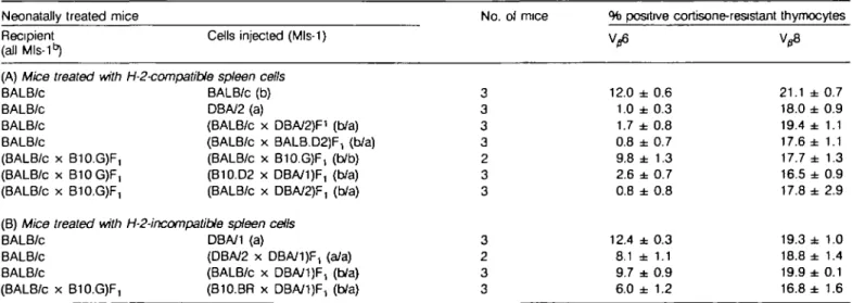
In this study we compared two different regimes for neonatal tolerance induction. The first was the injection of spleen cells which only express H-2 molecules compatible with the recipient's MHC (referred to as 'H-2-compatible cells'); the second was the injection of spleen cells expressing foreign MHC molecules (referred to as 'H-2 incompatible cells'). Newborn BALB/c (H-2d/Mls-1b) mice were treated i.p. with spleen cells from the various donor strains within 24 h of birth. Two to six weeks later,
Neonatal tolerance to Mls-1" 129
cortisone-resistant thymocytes were analyzed by flow cytofluorometry. As expected, injection of syngeneic (Mls-1") spleen cells did not affect V ^ expression (Table 2A). However, as described earlier (28), strongly reduced Vp6 percentages were observed when the spleen cells injected were from DBA/2 (H-2d/Mls-1a) mice. Efficient deletion of Vp6+ cells was also found in BALB/c mice neonatally treated with Mls-1a spleen cells from H-2dxd F, mice [i.e. from (BALB/c x DBA/2)F, or from (BALB/c x BALB.D2-Mlsa)F1 respectively]. Furthermore,
comparable deletion of V36+ cells occurred when
(BALB/c x B10.G)F, (H-2*^ mice were neonatally transfused from Mls-1a spleen cells from H-2-compatible (B10.D2 x DBA/1 )F, mice or from (BALB/c x DBA/2)F1 (H-2dxd) mice. Concerning the latter combination, earlier studies (39) had shown that Mls-1a spleen cells from donors not tolerant to MHC deter-minants of the neonatal host were capable of inducing clonal deletion of V^6+ cells despite overt graft versus host disease. Finally, control treatment with syngeneic Mls-1b spleen cells did
Table 1. Characteristics of mouse strains used in this study
Mouse strain BALB/c B10.D2 BALB.D2-Mlsa(BALB.D2) DBA/2 C57BL/6 B10G DBA/1 B10BR (C57BL/6 x BALB/c)F, (C57BU6 x DBA/2)F, (BALB/c x B10G)F, (BALB/c x DBA/1 )F, Mls-1 b b a a b b a b b/b b/a b/b b/a H-2 d d d d b q q k b/d b/d d/q d/q %V06+/CD4 + 124 9.3 0.4 0.4 7.4 3.8 4.2 8.8 12.0 0.5 11.3 0.7
Lymph node cells were analyzed by two-color immunofluorescence. Data (means of three individual mice; SEM < 0.8) represent <W/J6+ cells of
the total CD4+ population, calculated as follows: (%Vp6+CD4 +
+ %CD4+) x 100.
not alter Vp6 expression significantly when compared to untreated controls (Tables 1 and 2A).
We next injected H-2-incompatible spleen cells and made the following surprising observation: the injection of Mls-1a spleen cells from fully allogeneic DBA/1 (H-201) mice or from F, mice heterozygous at the H-2 locus did not induce elimination of Vp6+ lymphocytes. For example, BALB/c mice neonatally treated with (BALB/c x DBA/1 )F, or (DBA/2 x DBA/1 )F, (both HB-2dX(VMIs-1a) spleen cells exhibited 9.7 or 8 . 1 % Vg6 + lymphocytes respectively. Newborn (BALB/c x B10.G)F, mice which received H-2-semi-allogeneic (B10.BR x DBA/1 )F, (H-2kxq) spleen cells showed some deletion but still had quite high percentages (6.0%) of V^6+ mature thymocytes. Thus, Mls-1b mice neonatally treated with H-2-(semi)allogeneic Mls-1a spleen cells showed much impaired deletion of Vg6+ cells. As positive controls, normal lymphocyte maturation in the mice studied was documented by stainings with monoclonal antibody KJ16-133, specific for a population of lymphocytes which developed largely (but not entirely) independently of Mls-1a.
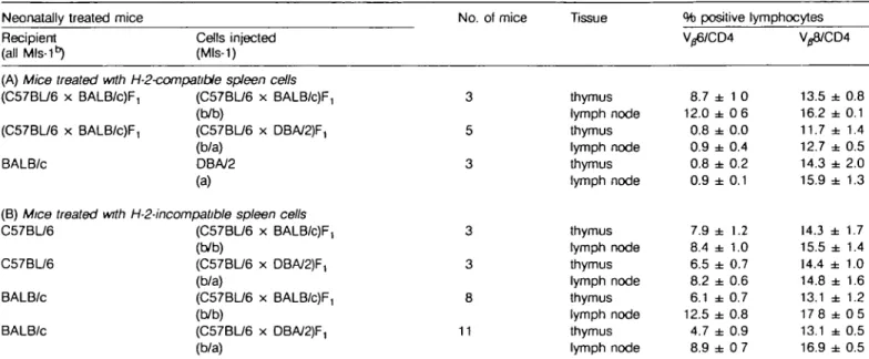
Similar analyses were performed by injection of H-2bxd F, spleen cells in neonatal C57/BL/6 (H-25) or BALB/c (H-201) mice, respectively. Expression of V36 and Vj8 by CD4 + CD8~ thymocytes or lymph node cells are shown in Table 3. In parallel to the findings described above, C57BL/6 or BALB/c mice (both Mls-1 ^ treated with H-2-incompatible Mls-1a spleen cells from (C57BL/6 x DBA/2)F, mice showed practically no or only partially reduced percentages of Vg6+ cells when compared to controls transfused with Mls-1b F, spleen cells (Table 3B). Furthermore, lymph node cells from BALB/c mice treated with Mls-1a F, or Mls-1b F, spleen cells, respectively, did not differ significantly in V^6 fluorescence intensity, suggesting that their TCRs were not specifically down-regulated in response to Mls-1a.
Since tolerogen-specific lymphocytes showed reduced levels of accessory molecules in some systems (40), the expression of CD4 on peripheral T cells was analyzed. No decrease in either the number of CD4+ cells or the surface density of CD4
Table 2. Expression of Vp6 and V^8 in mice neonatally treated with H-2d/q spleen cells
Neonatally treated mice Recipient
(all Mls-1 ^
(A) Mice treated with BALB/c BALB/c BALB/c BALB/c (BALB/c x B10.GJF, (BALB/c x B10G)F, (BALB/c x B10.G)F, (B) Mice treated with BALB/c
BALB/c BALB/c
(BALB/c x B10.G)F,
Cells injected (Mls-1)
H-2-compatible spleen cells BALB/c (b) DBA/2 (a)
(BALB/c x DBA/2)F1 (b/a) (BALB/c x BALB.D2JF, (b/a) (BALB/c x B10.G)F, (b/b) (B10.D2 x DBA/1)F, (b/a) (BALB/c x DBA/2)F, (b/a) H-2-incompatible spleen cells
DBA/1 (a)
(DBA/2 x DBA/1 )F, (a/a) (BALB/c x DBA/1 )F, (b/a) (B10.BR x DBA/1)F, (b/a) No. of mice 3 3 3 3 2 3 3 3 2 3 3 % positive cortisone-resistant Vfl6 12.0 ± 0.6 1.0 ± 0.3 1.7 ± 0.8 0.8 ± 0.7 9.8 ± 1.3 2.6 ± 0.7 0.8 ± 0.8 12.4 ± 0.3 8.1 ± 1.1 9.7 ± 0.9 6.0 ± 1.2 thymocytes 21.1 ± 0.7 18.0 ± 0.9 19.4 ± 1.1 17.6 ± 1.1 17.7 ± 1.3 16.5 ± 0.9 17.8 ± 2.9 19.3 ± 1.0 18.8 ± 1.4 19.9 ± 0.1 16.8 ± 1.6 Mice were treated with 10s spleen cells i.p. within 24 h of birth and assayed after 2 - 6 weeks. Indirect immunofluorescence with mAbs 44-22-1
(Vp6-specific) or KJ 16-133 (Vp8-specific) was performed. The values represent mean ± SEM of cortisone-resistant thymocytes (obtained 48 h after a single i.p. injection of 4 mg hydrocortisone acetate).
130 Neonatal tolerance to Mls-1a
Table 3. Expression of V ^ and V ^ in mice neonatally treated with H-2*b spleen cells
Neonatally treated mice Recipient
(all Mls-1")
Cells injected (Mls-1)
No. of mice Tissue % positive lymphocytes V/S8/CD4
(A) Mice treated with H-2-compatibie spleen cells (C57BL/6 x BALB/c)F, (C57BL/6 x BALB/c)F, BALB/c (C57BL/6 x BALB/c)F, (b/b) (C57BU6 x DBA/2)F, (b/a) DBA/2 (a)
(B) Mice treated with H-2-incompatible spleen cells C57BL/6 (C57BL/6 x BALB/c)F,
(b/b)
C57BU6 (C57BU6 x DBA/2)F, (b/a)
BALB/c (C57BL/6 x BALB/c)F, (b/b)
BALB/c (C57BU6 x DBA/2)F, (b/a) 3 5 3 3 3 8 11 thymus lymph node thymus lymph node thymus lymph node thymus lymph node thymus lymph node thymus lymph node thymus lymph node 8.7 12.0 0.8 0.9 0.8 0.9 7.9 8.4 6.5 8.2 6.1 12.5 4.7 8.9 ± ± ± ± ± ± ± ± ± ± ± ± ± ± 1 0 0 6 0.0 0.4 0.2 0.1 1.2 1.0 0.7 0.6 0.7 0.8 0.9 0 7 13.5 16.2 11.7 12.7 14.3 15.9 14.3 15.5 14.4 14.8 13.1 178 13.1 16.9 ± ± ± ± ± ± ± ± ± ± ± ± ± ± 0.8 0.1 1.4 0.5 2.0 1.3 1.7 1.4 1.0 1.6 1.2 0 5 0.5 0.5 Mice were treated with 10s spleen cells i.p. within 24 h of birth and assayed after 2 - 6 weeks. Thymocytes treated with CD8-specific rat IgM mAb
3.168.1 plus complement or untreated lymph node cells were stained with mAbs 44-22-1 (Vp6-specific) or KJ16-133 (V^8-specific), respectively, plus a CD4-specific mAb. Data are mean ± SEM and represent percentages of the total CD4+ population. Chimensm determined with MHC class l-specific
mAbs in mice treated with H-2-incompalible cells was as follows: immunofluorescence on lymph node cells stained with donor MHC-speafic mAb revealed mean values per group between 7.6 and 10.5% positive cells (SEM < 5.0), those stained with host MHC-specific mAb revealed means between 97 3 and 98.3% positive cells (SEM < 0 9).
Table 4. IL-2 response
Neonatally treated mice Recipient
of lymphocytes
Cells injected (Mls-1)
from neonatally tolerant BALB/c mice
Stimulators Response to: IL-2 (U/ml)
BALB/c DBA/2 BALB/c BALB/c BALB/c DBA/2 (a) (C57BL/6 x BALB/c)F, (b/b) (C57BL/6 x DBA/2)F, (b/a) BALB.D2-MIS8 BALB/c C57BL/6 B10.G BALB.D2-MIS8 BALB/c C57BL/6 B10.G BALB.D2-MIS8 BALB/c C57BL/6 B10.G BALB D2-Mls" BALB/c C57BL/6 B10.G BALB 02-Mls8 BALB/c C57BU6 B10.G Mls-1a H-2b H-2" H-2b H-2" Mls-1a H-2b H ^ Mls-1a H-2b H-2" Mls-1a H-2b H-2" 33.0 2.5 32.5 20.1 1.8 3.2 29.2 32.8 0.0 0.0 20.5 12.7 20.3 1.7 9.5 15.3 1.9 2.2 10.8 13.0 ± ± ± ± ± ± ± ± ± ± ± ± ± ± ± ± ± ± 3.3 0.7 0.5 1.1 0 9 1.4 1 2 1.6 0.5 0.8 1.2 0.1 1.1 3.5 0.7 0.5 1.6 2.3 Spleen cefte (5 x 10s) from 2- to 3-week-old neonataly treated or control mice respectively were stimulated with 5 x 10⁵ irradiated T cell-depleted spleen
cells in 96-well plates. Supernatants were harvested after 48 h and assayed for IL-2 content. Data are mean ± SEM of U IL-2/ml culture supernatant of three individual mice and of triplicate cultures.
Neonatal tolerance to Mls-1a 131
molecules on Vj6+ T cell was observed in lymphocytes from BALB/c mice neonatally treated with Mls-1a F, cells when compared to those transfused with Mls-1b F, (data not shown). Lymphohemopoietic chimerism in mice treated with H-2-incompatible cells was between 6.9 and 17.2% donor H-2 class I-expressing lymph node cells when analyzed in 2- or 3-week-old mice and between 0.6 and 5 . 1 % in 4- to 6-week-old mice (data not shown). Furthermore, the degree of chimerism appeared to be independent of Mls-1a expression by the neonatally injected cells.
Finally, in control experiments Mls-1a heterozygous (C57BL/6 x DBA/2)F, spleen cells were injected neonatally into H-2-syngeneic (C57BL/6 x BALB/c)F, hosts. As expected, this resulted in deletion of V ^ T cells (Table 3A). In conclusion, Mls-1b mice neonatally treated with Mls-1a spleen cells efficiently deleted Vfl6+ T cells if H-2-compatible but not if H-2-incompatible spleen cells were used.
Non-deleted V/>+ Tcells from BALB/c (Mls-1b) mice neonatally
treated with H-2-incompatible Mls-1" spleen cells neither secreted IL-2 nor proliferated after Mis-1" stimulation in vitro
Since BALB/c mice neonatally transfused with (C57BL/6 x DBA/2)F, Mls-1a spleen cells did not delete the majority of their
Vfi+ lymphocytes, the question arose whether these
lympho-cytes would respond to an Mls-1a stimulus in vitro. Analyses of lymphocytes from 2- to 3-week-old neonatally treated BALB/c mice revealed practically no proliferation (data not shown) and very low levels of IL-2 secretion after Mls-1a stimulation in vitro (Table 4). In contrast, control untreated mice or mice neonatally treated with (C57BL/6 x BALB/c)F, Mls-1b spleen cells responded strongly to BALB.D2-Mls-1a stimulator cells. This latter response was Mls-1 "-specific since only background IL-2 levels were found upon stimulation with syngeneic BALB/c spleen cells. All the responder populations tested were responsive to third-party H-2-incompatible B10.G stimulator cells. As expected,
lymphocytes from DBA/2 (Mls-1a) mice or BALB/c mice
neonatally given H-2-compatible Mls-1a spleen cells (both lack-ing Vfi+ cells due to clonal deletion) revealed only very low
levels of IL-2 secretion after Mls-1a stimulation in vitro. In summary, these data show that Mls-1b mice neonatally treated with H-2-semi-allogeneic Mls-1a F, spleen cells contained substantial numbers of Ve6+ T cells, which were unresponsive
to Mls-1a stimulation in vitro. However, it has to be mentioned that this Mls-1 "-specific in vitro unresponsiveness was only observed in mice up to the age of 3 weeks; non-deleted V^6+ lymphocytes from neonatally tolerized mice 4 or more weeks old expressed only slightly reduced or even normal IL-2 and proliferative responses to Mls-1a in vitro (data not shown).
Anergic V^6+ T cells responded to Mls-1" in vitro by
blastogenesis and expression of IL-2 receptors but did not proliferate unless IL-2 was added to the cultures
Lymphocytes from thymus or lymph nodes of BALB/c mice neonatally transfused with (C57BL/6 x DBA/2)F, or (C57BL/6 x BALB/c)F, spleen cells respectively were comparable in size as indicated by their mean values of forward light scatter (thymocytes: 23.0 ± 0.7 or 22.7 ± 1.0 respectively; lymph node cells: 25.9 ± 0.3 or 27.0 ± 0.4 respectively). These values were not significantly different from untreated or syngeneically treated control mice (data not shown), suggesting
that no detectable in vivo blastogenesis of tolerogen-specific T cells occurred. However, after 3 day in vitro stimulation with irradiated T cell-depleted spleen cells, as described above (without addition of growth factors), many cells enlarged. In cultures stimulated with Mls-1" cells this subset of large lympho-cytes contained - 4 0 % V^6+ cells (data not shown). Further-more, up to 66% of lymphoblasts stained positive with the IL-2R-specific mAb PC61.51 (41) after in vitro Mls-1a stimulation. Similar observations were made in lymphocyte populations containing responsive or anergic V ^+ T cells, indicating that blastogenesis and expression of IL-2R (but not IL-2 secretion or proliferation) of anergic VJo+ cells occurred after Mls-1"
stimula-tion in vitro.
Since functional anergy in some systems appears to reflect defective IL-2 production (11,13-16), we further analyzed v^S expression in cultures after addition of exogenous IL-2: responding T cell blasts from 3 day MLRs were re-incubated in IL-2-containing medium for an additional 2 days. As shown in Table 5, responder populations from BALB/c mice, whether untreated or neonatally treated with (C57BL/6 x DBA/2)F, or (C57BL/6 x BALB/c)F, spleen cells, respectively, revealed an increased proportion of V/56+/CD4+ cells (to - 4 0 % ) following Mls-1" stimulation. Since viable cell recoveries were - 10-fold greater in response to Mls-1a than in syngeneic controls (Table 5), these data indicate that actual expansion (rather than preferential survival) occurred. In contrast, no significant expansion of the VS6+ subset was detected in CD4 + lymphocytes from mice rendered tolerant to Mls-1" by clonal deletion (i.e. DBA/2 mice or BALB/c mice neonatally treated with DBA/2 spleen cells). In conclusion, non-deleted V ^ T cells from BALB/c mice neonatally treated with (C57BL/6 x DBA/2)F, Mls-1a spleen cells generated blasts and expressed IL-2R after Mls-1a stimulation in vitro but were defective in IL-2 production and therefore apparently did not proliferate unless exogenous IL-2 was added to the cultures.
In-vitro unresponsiveness of allospecific cytotoxic T cell precursors but not of allospecific IL-2-secreting cells from BALB/c mice neonatally treated with H-2bxd spleen cells
In MLRs such as described above, the response to donor allogeneic (H-2b) stimulators was investigated in parallel. The
data in Table 4 show that lymphocytes from H-2d mice
neonatally treated with H-2tad spleen cells secreted significant amounts of IL-2 after H-2b stimulation in vitro, although this response was lower than the response of controls (untreated or treated with H-2d spleen cells). In contrast, spleen cells from BALB/c mice neonatally treated with H-2bxd F, spleen cells did not give rise to H-2b-specific cytotoxic T lymphocytes (CTLs) in standard mixed lymphocyte cultures, but generated CTLs specific for third-party H-2" determinants (Table 6). Thus, neonatal injection of semi-allogeneic spleen cells expressing foreign H-26
molecules rendered allospecific CTLs unresponsive but only partially reduced allospecific IL-2 secretion. In conclusion, tolerance induction to MHC determinants did not parallel Mls-1a tolerance described above.
Discussion
Actively acquired tolerance to MHC-incompatible grafts can be achieved by neonatal injection of the relevant histoincompatible
132 Neonatal tolerance to Mis-1a
Table 5. Mls-1a-specific in vitro proliferation of Vfl6+ cells from neonatally tolerant mice after addition of exogenous IL-2
Neonatally Recipient BALB/c DBA/2 BALB/c BALB/c BALB/c treated mice Cell injected (Mls-1) — DBA/2 (a) (C57BL/6 x BALB/c)F, (b/b) (C57BL/6 x DBA/2)F, (b/a) Stimulators before culture BALB.D2Mlsa (C57BL/6 x DBA/2)F, BALB/c before culture BALB.D^Mte8 BALB/c before culture BALB.D2-MIS8 (C57BU6 x DBA/2)F, BALB/c before culture BALB.D2-MIS8 (C57BL/6 x DBA/2)F, BALB/c before culture BALB.D2-MIS8 (C57BL/6 x DBA/2)F, BALB/c Response to: (Mls-18) (Mls-1a/H-2b) (Mls-I8) (Mls-1a (MIs-^/H^15) (Mis-1s) (Mls-I'VH^13) (Mls-1a (Mls-1 aIH-2^ Cells recovered Relative no. (x10-5/ml) 15 ND 2.3 4.2 3.0 2 8 ND 2.5 176 ND 1 8 16.1 ND 1.5 %V"6+/CD4 + 12 8 ± 0.7 40.8 ± 0.9 27.6 ± 4.0 7.2 ± 3.9 C O C O C O A A A A C O C O C O C O 12 3 ± 0.9 40 5 ± 5.2 26.7 ± 5 6 9 1 ± 0.2 9.6 ± 0.7 37.2 ± 1 6 31.4 ± 3 1 4.4 ± 2 0 Spleen cells ( 3 x 1 0 * ) from 2- to 3-week-old neonataly treated or control mice respectively were stimulated with 5 x 10⁶ irradiated T cell-depleted spleen
cells in 24-well plates. After 3 days, cells were harvested, washed, and re-cultured for an additional 48 h in IL-2-containing medium. Recovered cells were counted and double-stained with V^6 and CD4-specific mAbs Measurements are given as percentages of the total CD4+ cells. The cultures
stimulated with BALB.D2-MIS8 contained < 3 % H-2b+ cells (mAb K7-309) and between 45 and 55% IL-2R+ cells (mAb PC61.51) with the exception
of the cultures with responder lymphocytes from untreated DBA/2 mice or BALB/c mice treated with DBA/2 cells where < 1 7 % cells were IL-2R +
Data are mean (±SEM) of three mice per group. SEM of numbers of recovered cells ranged between 0.8 and 2 8. ND, not done
Table 6. Allo-H-2-specific cytotoxic T cell response of neonatally tolerant BALB/c mice Neonatally treated mice
Recipient BALB/c DBA/2 BALB/c BALB/c BALB/c BALB/c Cells injected _ -(C57BL/6 x BALB/c)F1 (C57BU6 x BALB/c)F, (C57BL/6 x DBA/2)F, (C57BU6 x DBA/2)F,
% specific lysis of target B10.D2 (H-2d) 0/1/0/0 22/17/5/2 0/0/0/0 0/0/0/9 22/7/2/0 2/5/8/2
cells (E:T ratio 25/8/2.5/0.8) MC57G (H-213) 78/56/29/7 91/91/58/23 0/0/0/0 6/0/0/0 0/0/0/0 0/0/0/0 DBA/1 (H-2") 62/42/23/9 73/57/32/18 81/72/56/19 83/69/62/18 84/62/34/14 83/52/34/17 Representative data for two expenments with effector spleen cells from six individual neonatally treated or control mice are shown. Spleen cells (3 x 10⁶)
were stimulated in 24-well plates with 5 x 10⁶ irradiated (2000 rad) spleen cells of BALB/c (H-2<i), C57BL/6 (H-2b), or DBA/1 (H-2^ mice respectively,
according to the H-2 haplotype of the target fibroblasts. After 5 days, cells were harvested and tested in a 51Cr-release assay Test duration was 4.5 h;
spontaneous 51Cr-release of target cells was <25%. E:T ratio, effectontarget ratio.
lymphoid cells (27). More recently, the same protocol has been used to induce specific functional tolerance to Mls-1a
determinants (42,43). With the realization that T cell reactivity to Mls-1a correlates with usage of particular Vp segments (3,4), it
has now become possible to investigate directly whether neonatal tolerance to Mls-1a is obligatorily associated with clonal deletion
of the relevant Mls-1a-specific T cells. We show here that
neonatal tolerance to Mls-1a may be accomplished by either
clonal deletion or clonal unresponsiveness, depending on the MHC molecules co-expressed on the injected cells. Thus,
neonatal injection of MHC-compatible Mls-1a-bearing cells
results in virtually complete clonal deletion, in agreement with earlier reports (28,44). In contrast, inoculation of MHC-incompatibJe cells expressing Mls-1a does not lead to efficient
donal deletion but, rather, causes a transient non-responsiveness of T cells to Mls-1a determinants in vitro.
In this latter case, where H-2 semi-allogeneic Mls-1a spleen
cells were injected in neonatal mice, it could be argued that only Vfi + T cells with low affinity/avidity for Mls-1a have survived.
Neonatal tolerance to Mls-1e 133
expression were inducible in the otherwise unresponsive V^6 + lymphocytes, it appears that their TCR affinity was sufficient for Mls-1a-specific interactions. Therefore it seems likely that the neonatal injection of H-2 semi-allogeneic Mls-1a spleen cells induced a state of clonal anergy in host Ve6+ cells.
Previous studies indicated that antigen-specific clonal paralysis may be induced in vivo in adult mice by injection of chemically fixed accessory cells (10) or MHC class ll-bearing L cell trans-fectants (45). Anergy of Vp6+ T cells to Mls-1a was subsequently demonstrated directly by Qin et al. (12), who showed specific
in vitro unresponsiveness of V/36+ lymphocytes from adult mice after in vivo treatment with T cell-specific mAbs plus Mls-1a -bearing hemopoietic cells. Similarly, Rammensee ef al. (13) described in vitro anergic Vp6 + cells from adult Mls-1b mice after
in vivo immunization with Mls-1a spleen cells. Furthermore,
in vitro unresponsive Vg6+ T cells were observed in irradiated Mls-1a mice reconstituted with certain (I -E~) bone marrow stem cells (14-16). Mechanisms of T cell unresponsiveness in these models may be similar to the neonatally treated mice described in this study. The generation of antigen-specific blasts expressing IL-2R in the absence of IL-2 secretion and proliferation are common features of these systems (11,13-16) However, the cellular and molecular interactions responsible for this anergic state remain to be elucidated (11).
The limited data on MHC tolerance obtained in this study do not allow detailed conclusions except that there was no direct correlation with Mls-1a tolerance. The differential induction of allospecific functional tolerance in CTLs (presumably MHC dass l-specific) but not in IL-2 producers (presumably MHC class II-spedfic) observed in mice treated neonatally with semi-allogeneic spleen cells is consistent with some examples of split tolerance (43,46), whereas other studies revealed successful neonatal tolerance induction to both allogeneic MHC class I and II determinants (47,48). In any event, interpretation of split tderance is difficult since allogeneic MHC responses do not correlate with usage of particular TCR Vfl segments and hence lack of responsiveness (as observed for MHC class I) may result from clonal deletion, clonal anergy, or other unspecified mechanisms. It has been suggested that neonatal tolerance depends upon persistence of antigen in vivo (49,50). Our evidence that peripheral lymphocytes of neonatally treated mice contained ~ 10% donor MHC class l-expressing cells when analyzed after 2 weeks and still - 3 % of such cells when tested at the age of 5 weeks may indicate long-lasting persistence of neonatally administered MHC class l-expressing cells. Unfortunately, persistence of donor Mls-1a expressing cells is more difficult to monitor in vivo and it is possible that such cells may have been rejected more rapidly, thus causing the observed transient tolerance to Mls-1a.
In conclusion, the present results emphasize the complexity inherent in establishing neonatal T cell tolerance to foreign Mls-1a and MHC determinants. Nevertheless, the availabiltiy of a model system in which several distinct tderogenic mechanisms (i.e. donal deletion and clonal anergy) operate for a single antigen (Mls-1a) should facilitate further experimentation.
Acknowledgements
We thank M. Condrau, C. Knabenhans, P. Zaech, and M. Zimmermann for flow cytometry; A. Aebischef, H. Hengartner, Th. Kundig, T. Pedrazzini, and H . P. Pirctter for conceptual contributions; and A. Porret, A. Althage,
S. Cooper, and R. Lang for excellent technical advice. This work was supported in part by grants from the Swiss National Science Foundation and the Radium Stiftung Zurich (to R. M. Zinkerernagel).
Abbreviations APC CTL H-2 rlL-2 IL-2R MLR Mls-1 References antigen-presenting cell cytotoxic T lymphocyte mouse MHC recombinant mterleukin 2 interleukin 2 receptor mixed lymphocyte reaction
minor lymphocyte-stimulating locus 1
1 Sprent, J., Lo, D., Gao, K. E., and Ron, Y. 1988. T cell selection in the thymus Immunol. Rev. 101:173.
2 Kappler, J,. W., Roehm, N , and Marrack, P. 1987. T cell tolerance by clonal elimination in the thymus. Cell 49:273.
3 MacDonald, H. R , Schneider, R., Lees, R. K., Howe, R C , Acha-Orbea, H., Festenstein.H., Zinkernagel, R. M., and Hengartner, H. 1988. T cell receptor V/3 use predicts reactivity and tolerance to MIs"-encoded antigens. Nature 33240.
4 Kappler, J. W., Staerz, U. D., White, J., and Marrack, P 1988 Self tolerance eliminates T cells specific for Mis-modified products of the major histocompatibdity complex. Nature 33235
5 Kisielow, P., Bluthmann, H., Staerz, U. D., Steinmetz, M., and von Boehmer, H. 1988. Tolerance in T cell receptor transgenic mice involves deletion of nonmalure CD4 + 8+ thymocytes. Nature
333742.
6 Sha, W. C , Nelson, C. A., Newberry, R. D., Kranz, D. M., Russell, J H., and Loh, D. Y. 1988. Positive and negative selection of an antigen receptor on T cells in transgenic mice. Nature 336:73. 7 Berg, L. J., Fazekas de St Groth, B , Pullen, A. M and Davis, M. M.
1989. Phenotypic differences between a0 versus /3 T-ce)l receptor transgenic mice undergoing negative selection. Nature 340:559. 8 Pircher, H. P., Burki, K , Lang, R , Hengartner, H., and Zinkernagel,
R 1989 Tolerance induction in double specific T-cell receptor trans-genic mice varies with antigen. Nature 342.559.
9 Lamb, J, R , Skidmore, B. J., Green,N., Chiller, J M., and Feldmann, M 1983. Induction of tolerance in influenza virus-immune T lymphocyte clones with synthetic peptides of influenza hemagglutinin. J. Exp Med. 157:1434.
10 Jenkins, M. K. and Schwartz, R. H. 1987. Antigen presentation by chemically modified splenocytes induces antigen-specific T cell unresponsiveness in vitro and in vivo. J. Exp. Med. 165.302 11 Schwartz, R. H 1990. A cell culture model for T lymphocyte clonal
anergy. Science 248:1349.
12 Qin, S. X., Cobbold, S, Benjamin, R., and Waldmann, H. 1989. Induction of classical transplantation tolerance in the adult. J. Exp. Med. 169779.
13 Rammensee, H -G., Kroschewski, R., and Frangoulis, B 1989. Clonal anergy induced in mature V ^+T lymphocytes on immunizing Mls-1b
mice with Mls-1" expressing cells. Nature 339:541.
14 Ramsdell, F , Lantz, T., and Fowlkes, B. J. 1989. A nondeletional mechanism of thymic self tolerance. Science 246:1038.
15 Roberts. J. L, Sharrow, S. O., and Singer, A. 1990. Clonal deletion and ctonal anergy in the thymus induced by afferent cellular elements. J. Exp. Med. 171 935.
16 Speiser, D. E., Chvatchko, Y., Zinkernagel, R. M., and MacDonald, H.R 1990. Distinct fates of self specific T cells developing in irradiation bone marrow chimeras, clonal deletion, clonal anergy or in vitro responsiveness to self Mls-1a controlled by hemopoietic cells in the
thymus. J. Exp. Med 172:1305.
17 Lo, D., Burtdy, L. C , Widera, G., Cowing, C. Flavell, R. A., Palmiter, R. D., and Brinster, R. L. 1988. Diabetes and tolerance in transgenic mice expressing class II MHC molecules in pancreatic 0 cells. Cell 53:159.
18 Allison, J., Campbell, I. L., Morahan, G., Mandel, T. E., Harrison, L. C , and Miller, J. F. A. P. 1988. Diabetes in transgenic mice resulting from over-expression of class I histocompatibiDty molecules in
134 Neonatal tolerance to Mls-1a
pancreatic /3 cells. Nature 333 529.
19 Blackman, M. A., Gertiard-Burgert, H., Woodland, D. L , Palmer, E , Kappler, J. W , and Marrack, P. 1990. A role for clonal inactivation in T cell tolerance to Mls-1a. Nature 345:540.
20 Burkly, L. C , Lo, D., and Flavell, R. A. 1990. Tolerance in transgenic mice expressing major histocompatibility molecules extrathymically in pancreatic cells. Science 248:1364.
21 Festenstein, H 1976. The Mis systems. Transplant. Proc. 8.339. 22 MacDonald, H. R., Glasebrook, A. L , Schneider, R., Lees, R. K.,
Pircher, H. P., Pedrazani, T., Kanagawa, O., Nicolas, J. F., Howe, R. C , Zinkernagel, R. M., and Hengartner, H. 1989. T cell reactivity and tolerance to Mis8 encoded antigens, Immunol. Rev. 107:89.
23 Abe, R. and Hodes, R J. 1989. T-cell recognition of minor lymphocyte stimulating (Mis) gene products. Annu. Rev. Immunol 7:683 24 Webb, S. R. and Sprent, J. 1989. T-cell responses and tolerance to
Mis0 determinants. Immunol. Rev. 107:141.
25 Okada, C. Y., Hctanann, B., Guidos, C , Palmer, E. and Weissman, I. L 1990 Characterization of a rat monoclonal antibody specific for a determinant encoded by the V/37 gene segment. Depletion of V07 positive T cells in mice with Mls-1a haplotype. J. Immunol. 1443473
26 Vacchio, M. S. and Hodes, R J 1989. Selective decreases in T cell receptor V/3 expression. J. Exp. Med. 170'1335.
27 Billingham, R. E., Brent, L, and Medawar, P. B. 1953. Actively acquired tolerance of foreign cells. Nature 172.603
28 MacDonald, H. R., Pedrazzini, T , Schneider, R , Louis, J. A., Zinkernagel, R. M., and Hengartner, H. 1988. Intrathymic elimina-tion of Mls"-reactive (V06+) cells during neonatal tolerance induction
to Mlsa-encoded antigens, J Exp. Med. 167:2005.
29 Festenstein, H. and Berumen, L 1974. BALB D2-Mlsa, a new
congenic mouse strain. Transplantation 37 322.
30 Payne, J., Huber, B T., Cannon, N A., Schneider, R., Schilham, M. W., Acha-Orbea, H., MacDonald, H. R., and Hengartner, H 1988. Two monoclonal rat-antibodies with specificity for the V/S6 region of the munne T cell receptor. Proc Natl Acad. Set USA 85:7695. 31 Dialynas, D P., Wilde, D. B., Marrack, P., Pierres, A., Wall, K. A.,
Havran, W., Otten, G., Loken, M. R., Pierres, M , Kappler. J W., and Fitch, F. W. 1983 Characterization of the murine antigenic determinant, designated L3T4a, recognized by monoclonal antibody GK1 5' expression of L3T4a by functional T cell clones appears to correlate primarily with class II MHC antigen reactivity Immunol. Rev. 7429
32 Hammerling, G. J., Rusch, E., Tada, N., Kimura, S., and Hammerling, U. 1982. Localization of allodeterminants on H-2Kb antigens
determined with monoclonal antibodies and H-2 mutant mice. Proc. Natl Acad. Sci. USA 79:4737.
33 Ozato, K., Mayer, N. M , and Sachs, D. H. 1982. Monoclonal antibodies to mouse major histocompatibility complex antigens. IV. A series of hybridoma clones producing anti-H-^ antibodies and an examination of expression of H-2d antigens on the surface of these
cells. Transplantation 34.113.
34 Sarmiento, M., Loken, M. R., and Fitch, F., W. 1981. Structural
differences in cell surface T25 polypeptides from thymocytes and cloned T cells. Hybridoma 1:13.
35 Baker, P. E., Gillis, S., and Smith, K. A. 1979. Monoclonal cytolytic T-cell lines. J Exp. Med. 149 273.
36 Landegren, U. 1984. Measurement of cell number using an endogenous enzyme, hexoseamintdase Applications for the detection of lymphokines and cell surface antigens. J Immunol. Methods 67:379.
37 Cerottini, J. C. and Brunner, K. T. 1974. Cell-mediated cytotoxicity, allograft rejection and tumor immunity. Adv. Immunol. 18.67. 38 Speiser, D. E., Zurcher, Th., Ramseier, H , Hengartner, H., Staeheli,
P., Haller, O., and Zinkernagel, R. M. 1990 Nuclear myxovirus-resistance protein Mx is a minor histocompatibility antigen. Proc. Natl Acad.Sa. USA 87 2021.
39 Speiser, D. E., Schneider, R., Hengartner, H., MacDonald, H. R., and Zinkernagel, R.M. 1989. Clonal deletion of self-reactive T cells in irradiation bone marrow chimeras and neonatally tolerant mice, evidence for intercellular transfer of Mis8. J. Exp. Med 170:595.
40 Teh, H.-S., Kishi, H., Scott, B., and von Boehmer, H. 1989. Deletion of autospectfe T cells in T cell receptor (TCR) transgenic mice spares cells with normal TCR levels and low levels of CDS molecules J Exp. Med. 169.795.
41 Lowenthal, J. W , Corthesy, P . Tougne, C , Lees, R., MacDonald, H. R., and Nabholz, M. 1985. High and low affinity IL 2 receptors, analysis by IL 2 dissociation rate and reactivity with monoclonal anti-receptor antibody PC61. J. Immunol. 135 3988.
42 Macphail, S., Ishizaka, S. T., Bykowsky, M. J , Lattime, E. C, and Stutman, O 1985 Specific neonatally induced tolerance to Mis locus determinants. J. Immunol. 135:2967
43 Hosono, M , Kina, T., Hosokawa, T., and Katsura, Y 1986 Neonatal tolerance induction in the thymus to MHC-class ll-associated antigens I Preferential induction of tolerance to Mis antigens and resistance to allo-MHC-antigens. Cell Immunol. 1031
44 Webb, S. R. and Sprent, J. 1990. Induction of neonatal tolerance to Mis8 antigens by CD8+ T cells Science 248:1643.
45 Madsen, J. C, Superina, R A., Wood, K J., and Morris, P. J. 1988. Immunological unresponsiveness induced by recipient cells transfected with donor MHC genes. Nature 332:161.
46 McCarthy, S A. and Bach, F. H. 1983. A comparison of the neonatal tolerance-inducing capacities of H-2 class I and class II antigens. J. Immunol. 131:1670.
47 Wood, P. J. and Streilein, J. W. 1982. Ontogeny of acquired immune-logical tolerance to H-2 alloantigens. Eur. J. Immunol. 12:188. 48 Carnaud, C , Ishizaka, S. T., and Stutman,O. 1984. Early loss of
pre-cursors of CTL and IL 2-producing cete in the development of neonatal tolerance to alloantigens. J. Immunol., 133:45.
49 Lubaroff, D. M. and Silvers, W K. 1973. The importance of chimerism in maintaining tolerance of skin allografts in mice. J. Immunol. 111:65 50 Morrissey, P. J., Sharrow, S. O., Kohno, Y., Berzofsky, J.A., and
Singer, A. 1985. Correlation of intrathymic tolerance with intrathymic chimerism in neonatally tolerized mice. Transplantation 40.68.