M A J O R A R T I C L E
Epidemiology and Outcome of Fungemia in a
Cancer Cohort of the Infectious Diseases Group
(IDG) of the European Organization for Research
and Treatment of Cancer (EORTC 65031)
Oliver A. Cornely,1,2,3Bertrand Gachot,4,5Hamdi Akan,6Matteo Bassetti,7Omrun Uzun,8Christopher Kibbler,9
Oscar Marchetti,10Peter de Burghgraeve,11Safaa Ramadan,11Liisa Pylkkanen,11Lieveke Ameye,12Marianne Paesmans,12 and Peter J. Donnelly13; on behalf of the EORTC Infectious Diseases Group
1
Cologne Excellence Cluster on Cellular Stress Responses in Aging-Associated Diseases (CECAD), University of Cologne,2Department I of Internal Medicine, University Hospital Cologne, and3Clinical Trials Centre Cologne, ZKS Köln, BMBF 01KN1106, and German Centre for Infection Research (DZIF), partner site Bonn-Cologne, Cologne, Germany;4Institut Gustave Roussy, and5Département de Soins Aigus, Institut Gustave-Roussy, Villejuif, France;6Ankara University School of Medicine, Turkey;7Santa Maria Misericordia Hospital, Udine, Italy;8Department of Medicine, Section of Infectious Diseases, Hacettepe University Medical School, Ankara, Turkey;9University College London, United Kingdom;10Infectious Diseases Service, Department of Medicine, Lausanne University Hospital, Switzerland;11European Organisation for Research and Treatment of Cancer (EORTC), and12Institut Jules Bordet, Université Libre de Bruxelles, Belgium; and13University Medical Centre St Radboud, Nijmegen, The Netherlands
Background. Anti-cancer treatment and the cancer population have evolved since the last European Organisation for Research and Treatment of Cancer (EORTC) fungemia survey, and there are few recent large epidemiological studies. Methods. This was a prospective cohort study including 145 030 admissions of patients with cancer from 13 EORTC centers. Incidence, clinical characteristics, and outcome of fungemia were analyzed.
Results. Fungemia occurred in 333 (0.23%; 95% confidence interval [CI], .21–.26) patients, ranging from 0.15% in patients with solid tumors to 1.55% in hematopoietic stem cell transplantation recipients. In 297 evaluable patients age ranged from 17 to 88 years (median 56 years), 144 (48%) patients were female, 165 (56%) had solid tumors, and 140 (47%) had hematological malignancies. Fungemia including polymicrobial infection was due to: Candida spp. in 267 (90%), C. albicans in 128 (48%), and other Candida spp. in 145 (54%) patients. Favorable overall response was achieved in 113 (46.5%) patients by week 2. After 4 weeks, the survival rate was 64% (95% CI, 59%–70%) and was not significantly dif-ferent between Candida spp. Multivariable logistic regression identified baseline septic shock (odds ratio [OR] 3.04, 95% CI, 1.22–7.58) and tachypnoea as poor prognostic factors (OR 2.95, 95% CI, 1.66–5.24), while antifungal prophylaxis prior to fungemia (OR 0.20, 95% CI, .06–.62) and remission of underlying cancer (OR, 0.18; 95% CI, .06–.50) were protective. Conclusions. Fungemia, mostly due to Candida spp., was rare in cancer patients from EORTC centers but was as-sociated with substantial mortality. Antifungal prophylaxis and remission of cancer predicted better survival.
Keywords. candida; candidemia; cancer; leukemia.
Candidemia occurs frequently, is a severe clinical com-plication, and is associated with high morbidity and mortality, particularly in patients being treated for can-cer [1–3]. Yet the epidemiology of fungemia in these pa-tients has not been fully elucidated [4].
In the 1990s the European Organisation for Research and Treatment of Cancer (EORTC) conducted a study of fungemia in patients undergoing treatment of solid tumors or hematological cancers [5]. Of 270 episodes, 92% were caused by Candida spp., and key results
Received 25 November 2014; accepted 4 April 2015; electronically published 13 April 2015.
Presented in part: ECCMID 2012 in London: Cornely, O. A., Gachot, B., Akan, H., Bassetti, M., Uzun, O., Kibbler, C. C., Marchetti, O., Bille, J., de Burghgraeve, P., Pylkkanen, L., Ameye, L., Paesmans, M. and Donnelly, P. J. Epidemiology and mor-tality of fungaemia in cancer patients - a clinical cohort of the Infectious Diseases Group (IDG) of the European Organization for Research and Treatment of Cancer (EORTC 65031), CMI:18 Suppl s3;9 (O109).
Correspondence: Oliver A. Cornely, MD, University Hospital of Cologne, Kerpener Str. 62, 50937 Cologne, Germany (oliver.cornely@uk-koeln.de).
Clinical Infectious Diseases® 2015;61(3):324–31
© The Author 2015. Published by Oxford University Press on behalf of the Infectious Diseases Society of America. All rights reserved. For Permissions, please e-mail: journals.permissions@oup.com.
showed an association of Candida glabrata infection, advanced age, and disease severity with mortality. Since then epidemiol-ogy may have changed for several reasons: A changing distribu-tion of Candida species has been reported [6], additional antifungal drugs have become available, particularly the echino-candin class [7,8], indications for immunosuppressive therapy, and for hematopoietic stem cell transplantation (HSCT) in par-ticular have increased [9], and early antifungal treatment in-cluding prophylaxis has been adopted [10–12].
We have conducted a second study of the epidemiology of fun-gemia in cancer. Objectives of this study were to determine fungal pathogen distribution, prognostic factors for outcome, and crude and attributable mortality. An additional aim of this study was to describe the incidence of fungemia in relation to the number of hospital admissions of adult cancer patients in Europe.
MATERIAL AND METHODS
This intergroup study was sponsored by the EORTC ( protocol 65031) and conducted in collaboration with the Infectious Dis-ease Working Party of the German Society for Hematology and Oncology and the Infectious Disease Working Party of the Eu-ropean Group for Blood and Marrow Transplantation. Centers affiliated with these Infectious Diseases Groups were invited to participate.
The protocol was approved by the ethics committees and in-stitutional review boards of the participating centers. Two groups of patients were selected. Group A comprised all admis-sions to the participating wards of patients≥18 years of age with a diagnosis of a solid tumor, hematological malignancy, and/or recipients of any type of HSCT. Admission was defined as ≥1 night’s hospital stay. Group B, a subgroup of group A, in-cluded all patients who had a fungus isolated from ≥1 blood culture and for whom there was signed informed consent. Once the eligibility criteria were fulfilled, patients from group B were prospectively registered at the EORTC Data Center by telephone or web access. Patients could only be registered once. Case report forms were completed either in paper form or by electronic remote data capture. For this epidemiologic study approximately 300 fungemia patients were expected over a study period of 2 years. As this was a noninterventional study, no randomization or stratification was done. and no ad-verse events were to be reported.
Fungal isolates were sent to the EORTC-IDG Mycology Ref-erence Laboratory for Yeasts, Institute of Microbiology, Lau-sanne University Hospital, LauLau-sanne, Switzerland for purity check, and confirmation of identification, which overruled identification done by the study sites. Only Candida isolates were tested for their susceptibility tofluconazole, voriconazole, posaconazole, amphotericin B by EUCAST, and caspofungin by the CLSI method.
Criteria evaluated for group A were reporting period (date of first patient in to date of last patient out), number of admissions of patients with a diagnosis of malignant disease or HSCT, type of transplantation, number of admissions of patients by type of solid tumor or hematological malignancy during the reporting period and number of admissions of patients with a document-ed fungemia by underlying disease type. These summary data were collected at regular time intervals during the reporting pe-riod. Additional information collected for group B patients were demographics, details of malignant disease and predisposing factors for the development of fungemia, such as previous sur-gery, radiotherapy, antibiotics, total parenteral nutrition, major organ dysfunction, presence of neutropenia, antifungal prophy-laxis and treatment, date of diagnosis of fungemia, number and source of positive blood cultures, clinical signs and symptoms of fungemia, and organ involvement.
Group B patients were followed up to 12 weeks from diagno-sis of fungemia, and data were collected on antifungal treat-ment, clinical and microbiological response, and survival. Treatment was considered adequate when the isolate was sus-ceptible to the initial treatment. Response to treatment was de-termined at weeks 2, 4, and 12. Clinical evaluation of response was categorized as follows: Complete response (complete reso-lution of clinical signs and symptoms of fungemia), partial re-sponse (significant but incomplete resolution of clinical signs and symptoms), stable disease (no significant improvement in clinical signs and symptoms), and progressive disease (wor-sening of clinical signs and symptoms). Microbiological evalu-ation of response was categorized as complete microbiological response (3 consecutive negative blood cultures), no microbio-logical response ( persistently positive blood cultures), and microbiological relapse (complete microbiological response fol-lowed by a positive blood culture within the 12 week follow-up). If no follow-up blood cultures were obtained, microbiological response was categorized as not assessable. Global response was defined as complete or partial clinical response with com-plete microbiological response.
Survival data were graphically presented by Kaplan–Meier curves and compared by using log rank test. We considered 2 binary outcomes: death within 4 weeks, and favorable overall re-sponse at 2 weeks. Differences in categorical variables were as-sessed withχ2and Fisher exact tests, differences in continuous variables were assessed with t-test and Mann–Whitney test. Multivariable logistic regression models were built with step-wise variable selection. All statistical analyses were done with SAS 9.3 (SAS Institute Inc., Cary, North Carolina).
Explanatory variables considered for the outcomes of “favor-able overall response at 2 weeks” and “death within 4 weeks” were: age, gender, underlying disease and its status, time inter-val between hospital admission to onset of fungemia, HSCT, treatment given within 30 days before diagnosis (chemotherapy,
radiation therapy, immunosuppressive drugs, major surgical procedure, total parenteral nutrition, antibacterials, antifungals), neutropenia, colonization at baseline, signs and symptoms, organ involvement, catheter correlation (whether after removal of the central venous catheter the same pathogen was found as previously isolated in blood culture), and the pathogen. RESULTS
From 1 January 2005 to 2 November 2009, a total of 145 030 cancer patients were admitted to 13 participating centers in 8 countries. Fungemia was diagnosed in 333 of these patients. The overall incidence rate was 0.23%, ranging from 0.15% in solid tumor patients to 1.55% in HSCT recipients. Incidence rates according to underlying malignancy are listed in Table1. We excluded 36 patients for whom no detailed data were ob-tained, mostly because they did not provide informed consent. Of the remaining 297 patients with fungemia (Group B), 165 (56%) had a solid tumor, 140 (47%) patients had a hematolog-ical malignancy, and 50 (17%) underwent HSCT. Baseline char-acteristics including potential risk factors for fungemia are detailed in Table2.
General signs and symptoms at diagnosis of fungemia were fever >37°C (98.6°F) in 93% (275/297),≥38°C (100.4°F) in 76% (225/297), septic shock in 10% (30/297), tachycardia in 70% (204/292), tachypnoea in 33% (95/290), myalgia in 20% (53/271), and chills in 30% (88/293) of the patients.
At baseline, organ involvement was evaluated in 265 (89%) patients, whereas data were missing for 32 (11%) patients. Organs were involved in 83 of 265 (31%) patients. These were skin in 13 (5%), liver/spleen in 12 (5%), kidney/urinary tract in 10 (4%), other intraabdominal involvement in 9 (3%), endocardium in 6 (2%), eye in 6 (2%), vascular in 3 (1%), and central nervous system (CNS) in 2 (<1%) patients. Organ involvement decreased from 31% (83/265) at baseline to 16% (9/55) in week 12.
Of 251 (85%) patients with central vascular device the cath-eter was retained in 84 (33%), whereas it was removed in 167 (67%) patients, a median of 3 days after diagnosis of fungemia (range, 0–112 days). Removed catheters were cultured in 91% (152/167) of patients. Of 152 catheter cultures, 69 (45%) showed fungal growth. In 67 (97%) of them the same fungal species as in the initial blood culture was found.
Fungemia was caused by a single pathogen in 288 (97%) pa-tients, 9 (3%) patients had infections due to more than 1 species, resulting in a total of 306 isolates. Pathogens are listed according to underlying diseases and HSCT in Table3. Candida species accounted for 274 (90%) isolates. Central review of fungal path-ogens was offered to all centers except the reference center itself (35 isolates), for which correct identification was taken for granted. Of the remaining 271 pathogens, central reviews for
172 (63%) were available. Isolate identification by the sites was confirmed by the reference laboratory in 164 (95%) cases. Misidentified isolates were found in 8 cases, and these were evenly distributed across study sites; details are given in Table3. Full susceptibility tests were done for 141 isolates of Candida spp. All 63 C. albicans and 17 C. parapsilosis isolates were sus-ceptible to all antifungals. Of 27 C. tropicalis all but 2 strains were susceptible to all the antifungals, whereas the 2 remaining strains were only susceptible to posaconazole and amphotericin B. EUCAST deems that there is insufficient evidence to consider C. glabrata and C. krusei as good targets for treatment with
Table 1. Fungemia Incidence Rates by Underlying Malignancy (Fungemia Patients per Admissions)
Patient Group Incidence [% (95% CI)] Patients With Fungemia per Observed Group; N/N Overall 0.23% (.21–.26) 333/145 030 Solid tumor without
HSCT 0.15% (.13–.18) 174/114 811 Gastro-intestinal 0.37% (.30–.46) 88/23 718 Lung 0.05% (.01–.11) 5/10 976 Breast 0.05% (.02–.10) 8/16 137 Genito-urinary 0.20% (.14–.27) 42/21 389 Head and Neck 0.13% (.08–.20) 24/18 248 Other 0.03% (.01–.06) 7/24 343 Solid tumor with HSCT 1.55% (.19–5.49) 2/129
Allogeneic HSCT 1/5 Autologous HSCT 0.81% (.02–4.41) 1/124 Hematological malignancies without HSCT 0.42% (.35–.50) 114/27 195 ALL 0.64% (.38–1.01) 18/2801 AML 0.89% (.63–1.21) 39/4403 CLL 0.29% (.09–.67) 5/1738 CML 0.37% (.04–1.32) 2/543 MDS 0.57% (.19–1.33) 5/875 Lymphoma 0.29% (.21–.40) 38/12 933 Multiple myeloma 0.15% (.05–.35) 5/3356 Other 0.37% (.04–1.32) 2/546 Hematological malignancies with HSCT 1.46% (1.06–1.97) 42/2871 Allogeneic HSCT– related donor 2.10% (1.18–3.44) 15/715 Allogeneic HSCT– unrelated donor 1.99% (1.00–3.54) 11/552 Autologous HSCT 1.00% (.57–1.61) 16/1604 HSCT without associated malignancies 1/24
Abbreviations: ALL, acute lymphoblastic leukemia; AML, acute myeloid leukemia; CI, confidence interval; CLL, chronic lymphocytic leukemia; CML, chronic myeloid leukemia; HSCT, hematopoietic stem cell transplantation; MDS, myelodysplastic syndrome.
azole antifungals. The 19 C. glabrata and all 15 C. krusei were susceptible to amphotericin B, but 3 C. glabrata and 5 C. krusei were resistant to caspofungin.
The median time from blood culture sampling to initiation of antifungal treatment was 2 days, with a minimum of 0 and a maximum of 21 days. A total of 242 (81%) patients received treatment for fungemia, and 228 started with antifungal treat-ment within thefirst week. Of these 6 (3%) received antifungal combination therapy, 53 (23%) received >1 antifungal sequen-tially in thefirst week, and 169 (74%) received a single antifungal: 32 (14%) intravenous amphotericin B, 51 (22%) echinocandin, 79 (35%)fluconazole, 6 (3%) voriconazole, and 1 (0.4%) other. Only 14 of 228 (6%) patients receiving initial intravenous treatment were switched to an oral antifungal within thefirst week after di-agnosis. The pathogens cultured fell within the spectrum of the antifungals chosen for treatment in 226 of 297 (76%) cases. Overtly incorrect treatment decisions, such as initiating an anti-fungal despite known resistance, were infrequent. Overall, 55 (19%) patients did not receive antifungal treatment, and 14 pa-tients received treatment more than 7 days post diagnosis of fun-gemia. No antifungal treatment at all was given to 27 of 118 (23%) patients with C. albicans fungemia and to 20 of 140 (14%) patients with candidemia due to other species (P = .08).
Breakthrough fungemia, defined as patients being treated for ≥1 day before the first positive blood culture was drawn, oc-curred in 69 (23%) patients. Most of these infections were caused by a single species; 2 were due to more than 1 species. Breakthrough candidemia was due to: C. albicans (14; 20%) and Candida other than C. albicans (38; 55%), namely C. krusei (15; 22%), C. glabrata (5; 7%), C. tropicalis (6; 9%), C. parapsi-losis (5; 7%), C. norvegensis (2; 3%), C. dubliniensis (1; 1%), C. kefyr (2; 3%), and Candida spp. (2; 3%). Two infections were caused by more than 1 species: C. albicans & C. glabrata and C. albicans and C. parapsilosis. In 10 (14%) patients break-through pathogens were noncandida yeast: Trichosporon spp. (5; 7%), Saprochaete capitata (formerly Geotrichum capitatum) (2; 3%), S. clavata (1; 1%), Cryptococcus laurentii (1; 1%), and Saccharomyces sp. (1; 1%). Other positive blood cultures classi-fied as breakthrough infections grew molds in 1 patient each: Fusarium sp., Syncephalastrum racemosum, Rhizopus oryzae,
Table 2. Baseline Characteristics of European Patients With Cancer and Fungemia (N = 297)
Characteristic Value N/N
Age, median (min– max) 56 (17–88)
Sex, male 153/297 (52%)
Days from hospital admission to diagnosis of fungemia, mean ± std
23 ± 21 Neutropenia <500 cells/µL at time
of diagnosis of fungemia
110/286 (38%) Vascular access device upon
fungemia diagnosis
280/297 (94%) Central venous catheter 238/297 (80%) Peripheral catheter 29/297 (10%) Both 13/297 (4%) Underlying disease Solid Tumora 165/297 (56%) Gastro-intestinal 74/165 (45%) Lung 6/165 (4%) Breast 9/165 (5%) Genito-urinary 41/165 (25%)
Head and neck 23/165 (14%)
Other 11/165 (7%)
Unknown 1/165 (0.6%)
Hematologicala 140/297 (47%)
Acute lymphoblastic leukemia 23/140 (16%) Acute myelogenous leukemia or
myelodysplastic syndrome
60/140 (43%) Lymphoma incl. chronic
lymphocytic leukemia 44/140 (31%) Other 13/140 (9%) HSCT without associated malignancy 1/297 (0.3%) Status of malignancy Solid Tumora At diagnosis 29/165 (18%)
Complete or partial remission 29/165 (18%) No change or progressive disease 107/165 (65%) Hematological malignancya
Onset 19/140 (14%)
Complete remission 20/140 (14%) Partial remission, bone marrow
hypoplasia, refractory, or relapse
101/140 (72%) Treatment at fungemia diagnosis
HSCT 50/297 (17%) Allogeneic HSCT 28/297 (9%) Autologous HSCT 22/297 (7%) Chemotherapyb 142/296 (48%) Radiation therapyb 22/296 (7%) Immunosuppressive drugsb 88/293 (30%) Major surgical procedureb 69/297 (23%) Total parenteral nutritionb 118/295 (40%)
Stopped prior to fungemia diagnosis
36/295 (12%) On-going at fungemia diagnosis 82/295 (28%)
Antibioticsb 255/297 (86%) Table 2 continued. Characteristic Value N/N Antifungalsb 89/297 (30%) Prophylactic 32/297 (11%) Empiric or curative 57/297 (19%)
Abbreviation: HSCT, hematopoietic stem cell transplantation. a
9 patients had both a solid tumor and a hematological malignancy. b
and Paecilomyces species. Breakthrough fungemia was treated in 57 (83%) patients, while 12 (17%) received no antifungal drugs. The median time to new antifungal treatment was 2 days (range 0 to 21 days).
Global response was achieved in 87 (36%) and 26 (11%) evaluable patients by week 2. Seven (6%) patients had a micro-biological relapse, which occurred between day 29 and day 62. At week 2, treatment response was not assessed in 46 (15%) pa-tients, and 8 (3%) patients were lost to follow-up. Of the re-maining 243 patients, 69 (28%) had died and thus were regarded as treatment failures, and 61 (25%) patients had either failed treatment or fungemia had relapsed. In total, treatment failure was observed in 130 of 243 (53.5%) evaluable patients. In
a multivariable logistic regression model baseline characteristics predicting a higher risk of failure were tachycardia (odds ratio [OR] 2.07, 95% confidence interval [CI], 1.10–3.92, P = .03), myalgia (OR 2.37, 95% CI, 1.13–4.98, P = .02), and septic shock (OR 3.57, 95% CI, 1.30–9.80, P = .01).
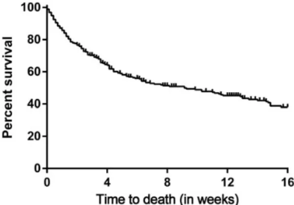
Overall mortality was 35% and 49% at weeks 4 and 12 (Fig-ure1). For patients who had achieved clinical complete re-sponse, partial rere-sponse, or stable disease 2 weeks after diagnosis of fungemia the crude week 4 survival rate was 86%. In contrast, 4 week survival in patients with progressive fungal disease was 58% (P < .001).
Investigators attributed 50 (72%) of the deaths within thefirst 2 weeks to fungemia. Survival rates did not differ between
Table 3. FungiaIsolated From Blood Cultures in European Patients With Cancer, by Underlying Disease and Treatment Groups (N = 297)
Pathogen Isolated by Treating Center Total n = 297 (%) Solid Tumorb n = 165 (%) Hematological Malignancyb n = 140 (%) Allogeneic HSCT n = 28 (%) Autologous HSCT n = 22 (%) No Transplant n = 247 (%) Single pathogen 288 (97.0%) 159 (97%) 137 (98%) 27 (96.4%) 22 (100%) 239 (96.8%) Candida albicans 120 (40.4%) 92 (56%) 31 (22%) 2 (7.1%) 7 (31.8%) 111 (44.9%) Non-albicans candida 138 (46.5%) 59 (36%) 82 (59%) 21 (75.0%) 12 (54.6%) 105 (42.5%) C. glabrata 29 (9.8%) 25 (15.2%) 6 (4.3%) 1 (3.6%) 1 (4.5%) 27 (10.9%) C. tropicalis 39 (13.1%) 11 (6.7%) 30 (21.4%) 2 (7.1%) 4 (18.2%) 33 (13.3%) C. parapsilosis 28 (9.4%) 16 (9.7%) 11 (7.9%) 5 (17.9%) 2 (9.1%) 21 (8.5%) C. krusei 25 (8.4%) 5 (3.0%) 20 (14.3%) 6 (21.4%) 4 (18.2%) 15 (6.1%) C. kefyr 7 (2.4%) 1 (<1%) 6 (4.3%) 2 (7.1%) 1 (4.5%) 4 (1.6%) C. norvegensis 3 (1.0%) . . . 3 (2.1%) 1 (3.6%) . . . 2 (0.8%) C. dubliniensis 2 (<1%) . . . 2 (1.4%) 1 (3.6%) . . . 1 (0.4%) C. guilliermondii 2 (<1%) 1 (<1%) 1 (<1%) 1 (3.6%) . . . 1 (0.4%) C. rugosa 1 (<1%) . . . 1 (<1%) . . . 1 (0.4%) Other Candidac 2 (<1%) . . . 2 (1.4%) 2 (7.1%) . . . . Non-candida yeastd 17 (5.7%) 5 (3%) 13 (9%) 2 (7.1%) 2 (9.1%) 13 (5.3%) Cryptococcus sp. 4 (1.3%) 1 (<1%) 3 (2%) . . . 4 (1.6%) Mold, NOSe 7 (2.4%) 1 (<1%) 7 (5%) 2 (7.1%) 1 (4.6%) 4 (1.6%) Trichoderma longibrachiatum 2 (<1%) 1 (<1%) 1 (<1%) . . . 2 (<1%)
Two pathogens isolated 9 (3%) 6 (3%) 3 (2%) 1 (3.6%) . . . 8 (3.2%)
Candida albicans and non-albicans candidaf
6 (2.0%) 4 (2.4%) 2 (1.4%) . . . 6 (2.4%)
Candida albicans and
non-candida yeastg 2 (<1%) 2 (1.2%) . . . 2 (<1%)
Non-albicans candidah 1 (<1%) . . . 1 (<1%) 1 (3.6%) . . . . . .
Abbreviations: HSCT, hematopoietic stem cell transplantation; NOS, not otherwise specified. a
Identified by local laboratory. b
9 had both, a solid tumor and a hematological malignancy. c
2 NOS. d
8 Trichosporon spp., 4 Saprochaete capitata, 1 S. clavata, 2 Saccharomyces spp., 1 S. cerevisiae, 1 NOS. e
3 Fusarium spp., 1 Rhizopus oryzae, 1 Syncephalastrum racemosum, 1 Paecilomyces sp., 1 NOS. f
4 C. glabrata, 2 C. parapsilosis. g
2 NOS. h
1 C. norvegensis, 1 C. inconspicua. The following isolates were reclassified by the central laboratory: C. albicans→ C. dubliniensis, C. glabrata → C. krusei, C. rugosa→ G. capitatum, C. dubliniensis → C. albicans, C. albicans → C. parapsilosis, C. albicans → C. tropicalis, C. krusei → C. parapsilosis, Trichosporon asahii→ C. tropicalis. For mold infections other than fusariosis this study could not rule out contamination rather than blood stream infection.
patient groups with fungemia due to C. albicans, non-albicans Candida and noncandida yeasts. When comparing survival rates for fungemia caused by the most frequent Candida spp., no difference was found between C. albicans, C. glabrata, C. tropicalis, C. parapsilosis, and C. krusei (Figure2).
For 33 (11%) patients follow-up was less than 1 month, so that 264 (89%) patients underwent analysis of baseline factors potentially prognostic for 28-day survival. Multivariable logistic regression identified presence of septic shock (OR 3.04, 95% CI, 1.22–7.58) and tachypnoea as negative prognostic factors (OR 2.95, 95% CI, 1.66–5.24). Positive prognostic factors were remis-sion of underlying malignancy (OR 0.18, 95% CI, .06–.50) and antifungal prophylaxis at any time within 30 days prior to diag-nosis of fungemia (OR 0.20, 95% CI, .06–.62). Median time to central venous catheter removal was 3 days and removal vs no removal had no impact on 28-day survival. Comparing patients with deep seated organ involvement (CNS, eye, heart/endocar-ditis, kidney/urinary tract infection, liver/spleen, skin) and
patients without documented organ involvement, risk of death at day 28 did not differ.
DISCUSSION
In a large multinational study in cancer patients the overall in-cidence of fungemia was 0.23% and ranged from 0.15% in solid tumor patients to 1.55% in HSCT recipients [13]. In gastrointes-tinal cancer, fungemia rates were comparably higher than with other tumors, which reflects abdominal surgery as a risk factor for invasive candidiasis [14].
An epidemiological shift from C. albicans to other Candida spp. is an ongoing discussion, but published data are inconsis-tent, probably reflecting local epidemiology, rather than global trends [15,16]. The proportion of C. albicans among candide-mias (47%) was similar to the previous EORTC study (49%) [5]. In both studies non-albicans Candida species were more fre-quent in hematological disease than with solid tumors [5]. One potential explanation is higher selection pressure due to more extensive antifungal exposure in hematology [17]. Molds other than Fusarium spp. isolated from blood cultures techni-cally fulfil the definition of fungemia, but contamination cannot be excluded, if isolated only from a single blood culture [18]. A limitation of our noninterventional study is the absence of a standardized evaluation of organ involvement, which may have been underestimated.
Contrary to the overall species distribution, pathogens found in breakthrough fungemia were C. albicans in 20% only. A re-cent overview of >100 clinical trials evaluating antifungal pro-phylaxis found similar results, likely because C. albicans is effectively treated by the systemically active antifungals used in these trials as well as in our study [19,20]. Anotherfinding of our study is the continuing low resistance rate of C. albicans in this population, despite high azole usage. This has been de-scribed previously in a longitudinal evaluation during long-term azole exposure [19].
Central catheters were removed after a median of 3 days, reflecting a median of 2 days from obtaining blood cultures to observing fungal growth. Current guidelines recommend removing any indwelling lines once fungemia is diagnosed be-cause of likely biofilm formation [21–23]. But central venous devices present at onset of fungemia were retained in a third of patients, and 19% of all fungemia patients did not receive any antifungal treatment. These remarkablefindings may be ex-plained by our broad enrolment criteria including patients with minimized treatment interventions in palliative settings. Treat-ment response in our study was lower than in recent large ran-domized clinical trials, again emphasizing healthier patient populations in phase 3 trials [24–26]. Mortality rates were com-parably higher in our study, but mortality did not correlate with fungal species, although in the previous EORTC study
Figure 1. Overall survival in 297 European cancer patients with fungemia.
Figure 2. Species-specific survival in 275 European cancer patients with candidemia (P value .14, overall test).
C. glabrata was associated with worse outcome [5]. Both anti-fungal prophylaxis and remission of malignant disease indepen-dently protected from adverse outcome. Between the 2 study periods, antifungal management underwent major develop-ments, such as the introduction and more widespread use of broad spectrum antifungals, which are now frequently used. It is also encouraging that fungal species were correctly identified on-site in the vast majority of cases.
This epidemiological study covers the years 2005–2009. Whether allfindings are applicable to 2015 is not clear, and ep-idemiological developments need continuous observation.
In summary, we have defined the fungemia rate in patients with cancer for thefirst time and described recent changes in prognostic factors [17,21,23].
Notes
Acknowledgments. The following investigators contributed data to this study: D. Nemet, Univ. Hospital Rebro, Zagreb, Croatia (15), P. Hamal, Med Fac Palacky Univ, Olomouc, Czech Republic (2), B. Lebeau, Centre hospital-ier universitaire de Grenoble (CHUG) Michallon, Grenoble, France (10); B. Gachot, Gustave Roussy, Villejuif, France (74), O. A. Cornely, Universi-tätsklinik Köln, Köln, Germany (9), M. Bassetti, Istituto di ricovero e cura a carattere scientifico Azienda Ospedaliera Universitaria San Martino IST, Genoa, Italy (40), L. Drgona, National Cancer Inst, Bratislava, Slovak Re-public (1), M. Rovira, Hosp. Clinic Univ., Barcelona, Spain (5), A. Droll, Kantonsspital Basel, Basel, Switzerland (2), A. Gratwohl, Kantonsspital Basel, Basel, Switzerland (9), J. Garbino, Hôpitaux Universitaires de Genève Cluse Roseraie, Geneva, Switzerland (17), O. Marchetti, Centre hospitalier universitaire vaudois Lausanne, Lausanne, Switzerland (35), H. Akan, An-kara University School of Medicine, AnAn-kara, Turkey (41), O. Uzun, Hacet-tepe Univ/medic, Ankara, Turkey (40), V. Korten, Marmara Univ Hosp., Istanbul, Turkey (2).
Financial support. This study was an academic study supported with unrestricted educational grants from Fujisawa (now Astellas), Pfizer, and Schering-Plough (now Merck). This publication was supported by Fonds Cancer from Belgium.
Author contributions. Conception and design: O. A. C., M. B., C. K., P. d. B. Collection and assembly of data: O. A. C., B. G., H. A., M. B., C. K., P. d. B., L. P. Data analysis and interpretation: O. A. C., P. d. B., L. P., L. A., M. P., P. J. D. Manuscript writing: All authors. Final approval of manuscript: All authors.
Potential conflicts of interest. O. A. C. reports grants and personal fees from Actelion, Astellas, Cubist, Gilead, Merck/Merck Sharp & Dohme (MSD), Optimer, Pfizer, grants from 3M, Bayer, Celgene, Genzyme, GlaxoSmithKline, Miltenyi, Viropharma, personal fees from Da Volterra, Daiichi Sankyo, F2G, Sanofi Pasteur, Summit, Vifor outside the submitted work. B. G. reports nonfinancial support from MSD, nonfinancial support from Gilead, nonfinancial support from Pfizer, nonfinancial support from Astellas outside the submitted work. M. B. reports grants and personal fees from Angelini, Astellas, Cubist, Pfizer, Novartis, MSD, grants from Gilead during the conduct of the study. O. U. reports other from Pfizer, other from Gilead, other from Merck outside the submitted work. C. K. reports personal fees from Astellas, personal fees from Gilead, grants and personal fees from MSD, personal fees from Pfizer outside the submitted work. O. M. reports grants from Funginos Foundation, Leenaards Founda-tion, Fammid FoundaFounda-tion, Bio-Merieux, Associates of Cape Cod, European Community’s Seventh Framework program (FP7-2007-2013) under grant agreement n_HEALTH-F2-2010-26033-ALLFUN, grants from Essex-Schering-Plough, Gilead, Merck, Novartis, Pfizer, Roche Diagnostics outside the submitted work. P. J. D. reports grants and personal fees from MSD,
grants and personal fees from Pfizer, grants and personal fees from Gilead outside the submitted work. All other authors report no potential conflicts. All authors have submitted the ICMJE Form for Disclosure of Potential Conflicts of Interest. Conflicts that the editors consider relevant to the con-tent of the manuscript have been disclosed.
References
1. Martin GS, Mannino DM, Eaton S, Moss M. The epidemiology of sepsis in the United States from 1979 through 2000. N Engl J Med 2003; 348:1546–54.
2. Wisplinghoff H, Bischoff T, Tallent SM, Seifert H, Wenzel RP, Edmond MB. Nosocomial bloodstream infections in US hospitals: analysis of 24,179 cases from a prospective nationwide surveillance study. Clin Infect Dis 2004; 39:309–17.
3. Bassetti M, Taramasso L, Nicco E, Molinari MP, Mussap M, Viscoli C. Epidemiology, species distribution, antifungal susceptibility and out-come of nosocomial candidemia in a tertiary care hospital in Italy. PLoS One 2011; 6:e24198.
4. Wenzel RP, Edmond MB. The impact of hospital-acquired bloodstream infections. Emerg Infect Dis 2001; 7:174–7.
5. Viscoli C, Girmenia C, Marinus A, et al. Candidemia in cancer patients: a prospective, multicenter surveillance study by the Invasive Fungal In-fection Group (IFIG) of the European Organization for Research and Treatment of Cancer (EORTC). Clin Infect Dis 1999; 28:1071–9. 6. Pfaller MA, Diekema DJ. Twelve years offluconazole in clinical practice:
global trends in species distribution andfluconazole susceptibility of bloodstream isolates of Candida. Clin Microbiol Infect 2004; 10(suppl 1): 11–23.
7. Kullberg BJ, Sobel JD, Ruhnke M, et al. Voriconazole versus a regimen of amphotericin B followed byfluconazole for candidaemia in non-neutropenic patients: a randomised non-inferiority trial. Lancet 2005; 366:1435–42.
8. Mora-Duarte J, Betts R, Rotstein C, et al. Comparison of caspofungin and amphotericin B for invasive candidiasis. N Engl J Med 2002; 347: 2020–9.
9. Gyurkocza B, Rezvani A, Storb RF. Allogeneic hematopoietic cell trans-plantation: the state of the art. Expert Rev Hematol 2010; 3:285–99. 10. Cornely OA, Maertens J, Winston DJ, et al. Posaconazole vsfluconazole
or itraconazole prophylaxis in patients with neutropenia. N Engl J Med 2007; 356:348–59.
11. Ullmann AJ, Lipton JH, Vesole DH, et al. Posaconazole orfluconazole for prophylaxis in severe graft-versus-host disease. N Engl J Med 2007; 356:335–47.
12. Walsh TJ, Teppler H, Donowitz GR, et al. Caspofungin versus liposomal amphotericin B for empirical antifungal therapy in patients with persis-tent fever and neutropenia. N Engl J Med 2004; 351:1391–402. 13. Wey SB, Mori M, Pfaller MA, Woolson RF, Wenzel RP. Risk factors for
hospital-acquired candidemia: a matched case-control study. Arch In-tern Med 1989; 149:2349–53.
14. Eggimann P, Francioli P, Bille J, et al. Fluconazole prophylaxis prevents intra-abdominal candidiasis in high-risk surgical patients. Crit Care Med 1999; 27:1066–72.
15. Tortorano AM, Dho G, Prigitano A, et al. Invasive fungal infections in the intensive care unit: a multicentre, prospective, observational study in Italy (2006–2008). Mycoses 2012; 55:73–9.
16. Montagna MT, Caggiano G, Lovero G, et al. Epidemiology of invasive fungal infections in the intensive care unit: results of a multicenter Ital-ian survey (AURORA Project). Infection 2013; 41:645–53.
17. Ullmann AJ, Akova M, Herbrecht R, et al. ESCMID guideline for the diagnosis and management of Candida diseases 2012: adults with hae-matological malignancies and after haematopoietic stem cell transplan-tation (HCT). Clin Microbiol Infect 2012; 18(suppl 7):53–67. 18. de Pauw B, Walsh TJ, Donnelly JP, et al. Revised definitions of invasive
fungal disease from the European Organization for Research and Treat-ment of Cancer/Invasive Fungal Infections Cooperative Group and the
National Institute of Allergy and Infectious Diseases Mycoses Study Group (EORTC/MSG) Consensus Group. Clin Infect Dis 2008; 46:1813–21.
19. Mann PA, McNicholas PM, Chau AS, et al. Impact of antifungal pro-phylaxis on colonization and azole susceptibility of Candida species. Antimicrob Agents Chemother 2009; 53:5026–34.
20. Tacke D, Buchheidt D, Karthaus M, et al. Primary prophylaxis of inva-sive fungal infections in patients with haematologic malignancies. 2014 update of the recommendations of the Infectious Diseases Working Party of the German Society for Haematology and Oncology. Ann Hematol 2014; 93:1449–56.
21. Cornely OA, Bassetti M, Calandra T, et al. ESCMID guideline for the diagnosis and management of Candida diseases 2012: non-neutropenic adult patients. Clin Microbiol Infect 2012; 18(suppl 7):19–37. 22. Davey ME, O’Toole GA. Microbial biofilms: from ecology to molecular
genetics. Microbial Mol Biol Rev 2000; 64:847–67.
23. Pappas PG, Kauffman CA, Andes D, et al. Clinical practice guidelines for the management of candidiasis: 2009 update by the Infectious Dis-eases Society of America. Clin Infect Dis 2009; 48:503–35.
24. Kuse ER, Chetchotisakd P, da Cunha CA, et al. Micafungin versus lipo-somal amphotericin B for candidaemia and invasive candidosis: a phase III randomised double-blind trial. Lancet 2007; 369:1519–27.
25. Pappas PG, Rotstein CM, Betts RF, et al. Micafungin versus caspofungin for treatment of candidemia and other forms of invasive candidiasis. Clin Infect Dis 2007; 45:883–93.
26. Reboli AC, Rotstein C, Pappas PG, et al. Anidulafungin versus flucon-azole for invasive candidiasis. N Engl J Med 2007; 356:2472–82.
APPENDIX
List of where and when the study has been presented in part elsewhere, if applicable:
ECCMID 2012 in London: Cornely, O.A., Gachot, B., Akan, H. Bassetti, M., Uzun, O., Kibbler, C. C., Marchetti, O., Bille, J., de Burghgraeve, P., Pylkkanen, L., Ameye, L., Paesmans, M., and Donnelly, P. J. Epidemiology and mortality of fungaemia in cancer patients—a clinical cohort of the Infectious Diseases Group (IDG) of the European Organization for Research and Treatment of Cancer (EORTC 65031), CMI:18 Suppl s3;9 (O109).