HAL Id: tel-00565897
https://tel.archives-ouvertes.fr/tel-00565897
Submitted on 15 Feb 2011HAL is a multi-disciplinary open access archive for the deposit and dissemination of sci-entific research documents, whether they are pub-lished or not. The documents may come from teaching and research institutions in France or abroad, or from public or private research centers.
L’archive ouverte pluridisciplinaire HAL, est destinée au dépôt et à la diffusion de documents scientifiques de niveau recherche, publiés ou non, émanant des établissements d’enseignement et de recherche français ou étrangers, des laboratoires publics ou privés.
Contribution of Geometry
Tomoko Muranaka
To cite this version:
Tomoko Muranaka. Fragmentation Dynamics of Triatomic Molecules : Contribution of Geometry. Atomic Physics [physics.atom-ph]. Université de Caen, 2007. English. �tel-00565897�
UNIVERSITE de CAEN/BASSE-NORMANDIE
U.F.R. des SCIENCES ECOLE DOCTORALE SIMEM
THÈSE
présenté par
Mlle. Tomoko MURANAKA
et soutenue le 30 octobre 2007 en vue de l'obtention du
DOCTORAT de l'UNIVERSITE de CAEN
Spécialité: Milieux dilués et optique fondamentale Arrêté du 07 août 2006
Dynamique de la fragmentation de molécules tri-atomiques:
contribution de la géométrie
Membres du JURY
Mme. Karine WOHRER-BEROFF, Directrice de Recherche CNRS, Orsay (Rapporteur) M. Haruo SHIROMARU, Professeur des Universités, Tokyo (Japon) (Rapporteur) M. Jean-Pierre GRANDIN, Directeur de Recherche CNRS, Caen
Acknowledgements
This thesis is the result of three years of work in the laboratory CIRIL where I have been fu-eled and supported by many people. People sometimes compare life to a voyage. For me, especially, these three years in France was literally a long voyage, not always with moderate winds. It is a pleasant task to express my gratitude to all those who made this voyage possible.
The first person I would like to thank is my supervisor Dr. Amine Cassimi. With his enthu-siasm, his great knowledge and skill of experiment, and his practical suggestion not only for re-search but also for life, he helped to navigate this voyage correct. I owe him lots of gratitude for having me show his way of research. It is difficult to overstate how much I have learned from him.
I owe my sincere gratitude to Dr. Serge Bouffard, Director of CIRIL, who accepted me to join this excellent laboratory. I would like to thank also for Dr. Emmanuel Balanzat, Vice Director of CIRIL for their administrative affaires. Also their interests for this research encouraged me a lot. I am indebted to Prof. Dr. Lamri Adoui, who gave me the opportunity to come to France, for his kind assistance for joining AMA group of CIRIL. If he had not participated to the conference where we met!
My warmest thanks are due to Dr. Brigitte Ban-d'Etat, for her gracious suggestion. Her calm and thoughtful consideration has a deep impression on me.
I am grateful for all the member of CIRIL: researchers for their advice, technicians for their help (and well organized coffee time!), secretaries for administrative procedures… I wish to thank my many young colleagues who made "French life" attractive, especially Dr. Jimmy Rangama for his encouragement (with beers).
My sincere thanks are due to Dr. Karine Wohrer-Beroff, Prof. Dr. Haruo Shiromaru and Dr. Jean-Pierre Grandin for being a member of the jury. Their pointed questions and meaning discus-sions are deeply appreciated.
The financial supports of the French Government and of ITSLEIF are gratefully acknowl-edged. I would like to express my gratitude to Prof. Dr. Bernt Huber, Coordinator of ITSLEIF, for his consideration.
I appreciate the interest and contribution of Japanese professors, especially Prof. Dr. Toshi-yuki Azuma, my former supervisor, and Prof. Dr. Yasunori Yamazaki.
Finally, I would like to express my loving thanks to my friends and family. Without their en-couragement and understanding, it would have been impossible to finish this work and continue again the voyage.
Table of Contents
I Introduction 1
I.1 Fragmentation Process⋅⋅⋅⋅⋅⋅⋅⋅⋅⋅⋅⋅⋅⋅⋅⋅⋅⋅⋅⋅⋅⋅⋅⋅⋅⋅⋅⋅⋅⋅⋅⋅⋅⋅⋅⋅⋅⋅⋅⋅⋅⋅⋅⋅⋅⋅⋅⋅⋅⋅⋅⋅⋅⋅⋅⋅⋅⋅⋅⋅⋅⋅⋅⋅⋅⋅⋅⋅⋅⋅⋅⋅⋅⋅⋅⋅⋅⋅⋅⋅⋅⋅⋅⋅⋅⋅⋅⋅⋅⋅⋅⋅⋅⋅⋅⋅⋅⋅⋅⋅⋅⋅1 I.2 Experimental Backgrounds⋅⋅⋅⋅⋅⋅⋅⋅⋅⋅⋅⋅⋅⋅⋅⋅⋅⋅⋅⋅⋅⋅⋅⋅⋅⋅⋅⋅⋅⋅⋅⋅⋅⋅⋅⋅⋅⋅⋅⋅⋅⋅⋅⋅⋅⋅⋅⋅⋅⋅⋅⋅⋅⋅⋅⋅⋅⋅⋅⋅⋅⋅⋅⋅⋅⋅⋅⋅⋅⋅⋅⋅⋅⋅⋅⋅⋅⋅⋅⋅⋅⋅⋅⋅⋅⋅⋅⋅⋅⋅⋅⋅⋅⋅⋅4
I.3 Previous Results and Scopes of Present Works⋅⋅⋅⋅⋅⋅⋅⋅⋅⋅⋅⋅⋅⋅⋅⋅⋅⋅⋅⋅⋅⋅⋅⋅⋅⋅⋅⋅⋅⋅⋅⋅⋅⋅⋅⋅⋅⋅⋅⋅⋅⋅⋅⋅⋅⋅⋅⋅⋅⋅⋅⋅⋅⋅⋅⋅⋅⋅⋅⋅⋅⋅⋅⋅4 I.3.1 Fragmentation Dynamics⋅⋅⋅⋅⋅⋅⋅⋅⋅⋅⋅⋅⋅⋅⋅⋅⋅⋅⋅⋅⋅⋅⋅⋅⋅⋅⋅⋅⋅⋅⋅⋅⋅⋅⋅⋅⋅⋅⋅⋅⋅⋅⋅⋅⋅⋅⋅⋅⋅⋅⋅⋅⋅⋅⋅⋅⋅⋅⋅⋅⋅⋅⋅⋅⋅⋅⋅⋅⋅⋅⋅⋅⋅⋅⋅⋅⋅⋅⋅⋅⋅⋅⋅⋅⋅4 I.3.2 Branching Ratio of HDO2+ Dissociation⋅⋅⋅⋅⋅⋅⋅⋅⋅⋅⋅⋅⋅⋅⋅⋅⋅⋅⋅⋅⋅⋅⋅⋅⋅⋅⋅⋅⋅⋅⋅⋅⋅⋅⋅⋅⋅⋅⋅⋅⋅⋅⋅⋅⋅⋅⋅⋅⋅⋅⋅⋅⋅⋅⋅⋅⋅⋅⋅⋅⋅9
II Experimental Specifics 11
II.1 Projectiles: Highly Charged Ions⋅⋅⋅⋅⋅⋅⋅⋅⋅⋅⋅⋅⋅⋅⋅⋅⋅⋅⋅⋅⋅⋅⋅⋅⋅⋅⋅⋅⋅⋅⋅⋅⋅⋅⋅⋅⋅⋅⋅⋅⋅⋅⋅⋅⋅⋅⋅⋅⋅⋅⋅⋅⋅⋅⋅⋅⋅⋅⋅⋅⋅⋅⋅⋅⋅⋅⋅⋅⋅⋅⋅⋅⋅⋅⋅⋅⋅⋅⋅⋅⋅11 II.1.1 High Energy Projectile Ions from GANIL Facility⋅⋅⋅⋅⋅⋅⋅⋅⋅⋅⋅⋅⋅⋅⋅⋅⋅⋅⋅⋅⋅⋅⋅⋅⋅⋅⋅⋅⋅⋅⋅⋅⋅⋅⋅⋅⋅⋅⋅⋅⋅⋅12 II.1.2 Low Energy Projectile Ions from ARIBE Facility⋅⋅⋅⋅⋅⋅⋅⋅⋅⋅⋅⋅⋅⋅⋅⋅⋅⋅⋅⋅⋅⋅⋅⋅⋅⋅⋅⋅⋅⋅⋅⋅⋅⋅⋅⋅⋅⋅⋅⋅⋅⋅⋅13 II.2 Target Molecules: Supersonic Jet⋅⋅⋅⋅⋅⋅⋅⋅⋅⋅⋅⋅⋅⋅⋅⋅⋅⋅⋅⋅⋅⋅⋅⋅⋅⋅⋅⋅⋅⋅⋅⋅⋅⋅⋅⋅⋅⋅⋅⋅⋅⋅⋅⋅⋅⋅⋅⋅⋅⋅⋅⋅⋅⋅⋅⋅⋅⋅⋅⋅⋅⋅⋅⋅⋅⋅⋅⋅⋅⋅⋅⋅⋅⋅⋅⋅⋅⋅⋅⋅14
II.2.1 Principle and Setup⋅⋅⋅⋅⋅⋅⋅⋅⋅⋅⋅⋅⋅⋅⋅⋅⋅⋅⋅⋅⋅⋅⋅⋅⋅⋅⋅⋅⋅⋅⋅⋅⋅⋅⋅⋅⋅⋅⋅⋅⋅⋅⋅⋅⋅⋅⋅⋅⋅⋅⋅⋅⋅⋅⋅⋅⋅⋅⋅⋅⋅⋅⋅⋅⋅⋅⋅⋅⋅⋅⋅⋅⋅⋅⋅⋅⋅⋅⋅⋅⋅⋅⋅⋅⋅⋅⋅⋅⋅⋅⋅14 II.2.2 NO2 Preparation⋅⋅⋅⋅⋅⋅⋅⋅⋅⋅⋅⋅⋅⋅⋅⋅⋅⋅⋅⋅⋅⋅⋅⋅⋅⋅⋅⋅⋅⋅⋅⋅⋅⋅⋅⋅⋅⋅⋅⋅⋅⋅⋅⋅⋅⋅⋅⋅⋅⋅⋅⋅⋅⋅⋅⋅⋅⋅⋅⋅⋅⋅⋅⋅⋅⋅⋅⋅⋅⋅⋅⋅⋅⋅⋅⋅⋅⋅⋅⋅⋅⋅⋅⋅⋅⋅⋅⋅⋅⋅⋅⋅⋅⋅⋅⋅17
II.2.3 HDO Preparation⋅⋅⋅⋅⋅⋅⋅⋅⋅⋅⋅⋅⋅⋅⋅⋅⋅⋅⋅⋅⋅⋅⋅⋅⋅⋅⋅⋅⋅⋅⋅⋅⋅⋅⋅⋅⋅⋅⋅⋅⋅⋅⋅⋅⋅⋅⋅⋅⋅⋅⋅⋅⋅⋅⋅⋅⋅⋅⋅⋅⋅⋅⋅⋅⋅⋅⋅⋅⋅⋅⋅⋅⋅⋅⋅⋅⋅⋅⋅⋅⋅⋅⋅⋅⋅⋅⋅⋅⋅⋅⋅⋅⋅⋅⋅18 II.2.4 Characteristics⋅⋅⋅⋅⋅⋅⋅⋅⋅⋅⋅⋅⋅⋅⋅⋅⋅⋅⋅⋅⋅⋅⋅⋅⋅⋅⋅⋅⋅⋅⋅⋅⋅⋅⋅⋅⋅⋅⋅⋅⋅⋅⋅⋅⋅⋅⋅⋅⋅⋅⋅⋅⋅⋅⋅⋅⋅⋅⋅⋅⋅⋅⋅⋅⋅⋅⋅⋅⋅⋅⋅⋅⋅⋅⋅⋅⋅⋅⋅⋅⋅⋅⋅⋅⋅⋅⋅⋅⋅⋅⋅⋅⋅⋅⋅⋅⋅⋅⋅18 II.3 Time Of Flight Spectrometer⋅⋅⋅⋅⋅⋅⋅⋅⋅⋅⋅⋅⋅⋅⋅⋅⋅⋅⋅⋅⋅⋅⋅⋅⋅⋅⋅⋅⋅⋅⋅⋅⋅⋅⋅⋅⋅⋅⋅⋅⋅⋅⋅⋅⋅⋅⋅⋅⋅⋅⋅⋅⋅⋅⋅⋅⋅⋅⋅⋅⋅⋅⋅⋅⋅⋅⋅⋅⋅⋅⋅⋅⋅⋅⋅⋅⋅⋅⋅⋅⋅⋅⋅⋅⋅⋅⋅19 II.3.1 Spectrometer Design and Construction⋅⋅⋅⋅⋅⋅⋅⋅⋅⋅⋅⋅⋅⋅⋅⋅⋅⋅⋅⋅⋅⋅⋅⋅⋅⋅⋅⋅⋅⋅⋅⋅⋅⋅⋅⋅⋅⋅⋅⋅⋅⋅⋅⋅⋅⋅⋅⋅⋅⋅⋅⋅⋅⋅⋅⋅⋅⋅⋅⋅20
II.3.2 Time Of Flight Equations⋅⋅⋅⋅⋅⋅⋅⋅⋅⋅⋅⋅⋅⋅⋅⋅⋅⋅⋅⋅⋅⋅⋅⋅⋅⋅⋅⋅⋅⋅⋅⋅⋅⋅⋅⋅⋅⋅⋅⋅⋅⋅⋅⋅⋅⋅⋅⋅⋅⋅⋅⋅⋅⋅⋅⋅⋅⋅⋅⋅⋅⋅⋅⋅⋅⋅⋅⋅⋅⋅⋅⋅⋅⋅⋅⋅⋅⋅⋅⋅⋅20 II.3.3 Time Focusing Condition⋅⋅⋅⋅⋅⋅⋅⋅⋅⋅⋅⋅⋅⋅⋅⋅⋅⋅⋅⋅⋅⋅⋅⋅⋅⋅⋅⋅⋅⋅⋅⋅⋅⋅⋅⋅⋅⋅⋅⋅⋅⋅⋅⋅⋅⋅⋅⋅⋅⋅⋅⋅⋅⋅⋅⋅⋅⋅⋅⋅⋅⋅⋅⋅⋅⋅⋅⋅⋅⋅⋅⋅⋅⋅⋅⋅⋅⋅⋅⋅⋅21 II.3.4 3D Focusing Condition⋅⋅⋅⋅⋅⋅⋅⋅⋅⋅⋅⋅⋅⋅⋅⋅⋅⋅⋅⋅⋅⋅⋅⋅⋅⋅⋅⋅⋅⋅⋅⋅⋅⋅⋅⋅⋅⋅⋅⋅⋅⋅⋅⋅⋅⋅⋅⋅⋅⋅⋅⋅⋅⋅⋅⋅⋅⋅⋅⋅⋅⋅⋅⋅⋅⋅⋅⋅⋅⋅⋅⋅⋅⋅⋅⋅⋅⋅⋅⋅⋅⋅⋅⋅⋅23 II.3.5 Magnetic field⋅⋅⋅⋅⋅⋅⋅⋅⋅⋅⋅⋅⋅⋅⋅⋅⋅⋅⋅⋅⋅⋅⋅⋅⋅⋅⋅⋅⋅⋅⋅⋅⋅⋅⋅⋅⋅⋅⋅⋅⋅⋅⋅⋅⋅⋅⋅⋅⋅⋅⋅⋅⋅⋅⋅⋅⋅⋅⋅⋅⋅⋅⋅⋅⋅⋅⋅⋅⋅⋅⋅⋅⋅⋅⋅⋅⋅⋅⋅⋅⋅⋅⋅⋅⋅⋅⋅⋅⋅⋅⋅⋅⋅⋅⋅⋅⋅⋅⋅23 II.4 Position Sensitive Detectors (PSD) ⋅⋅⋅⋅⋅⋅⋅⋅⋅⋅⋅⋅⋅⋅⋅⋅⋅⋅⋅⋅⋅⋅⋅⋅⋅⋅⋅⋅⋅⋅⋅⋅⋅⋅⋅⋅⋅⋅⋅⋅⋅⋅⋅⋅⋅⋅⋅⋅⋅⋅⋅⋅⋅⋅⋅⋅⋅⋅⋅⋅⋅⋅⋅⋅⋅⋅⋅⋅⋅⋅⋅⋅⋅⋅⋅⋅⋅25 II.4.1 Micro Channel Plate (MCP) ⋅⋅⋅⋅⋅⋅⋅⋅⋅⋅⋅⋅⋅⋅⋅⋅⋅⋅⋅⋅⋅⋅⋅⋅⋅⋅⋅⋅⋅⋅⋅⋅⋅⋅⋅⋅⋅⋅⋅⋅⋅⋅⋅⋅⋅⋅⋅⋅⋅⋅⋅⋅⋅⋅⋅⋅⋅⋅⋅⋅⋅⋅⋅⋅⋅⋅⋅⋅⋅⋅⋅⋅⋅⋅⋅⋅⋅26 II.4.2 Delay Line Detector (DLD) ⋅⋅⋅⋅⋅⋅⋅⋅⋅⋅⋅⋅⋅⋅⋅⋅⋅⋅⋅⋅⋅⋅⋅⋅⋅⋅⋅⋅⋅⋅⋅⋅⋅⋅⋅⋅⋅⋅⋅⋅⋅⋅⋅⋅⋅⋅⋅⋅⋅⋅⋅⋅⋅⋅⋅⋅⋅⋅⋅⋅⋅⋅⋅⋅⋅⋅⋅⋅⋅⋅⋅⋅⋅⋅⋅⋅⋅⋅⋅⋅⋅⋅27 II.5 Acquisition Setup⋅⋅⋅⋅⋅⋅⋅⋅⋅⋅⋅⋅⋅⋅⋅⋅⋅⋅⋅⋅⋅⋅⋅⋅⋅⋅⋅⋅⋅⋅⋅⋅⋅⋅⋅⋅⋅⋅⋅⋅⋅⋅⋅⋅⋅⋅⋅⋅⋅⋅⋅⋅⋅⋅⋅⋅⋅⋅⋅⋅⋅⋅⋅⋅⋅⋅⋅⋅⋅⋅⋅⋅⋅⋅⋅⋅⋅⋅⋅⋅⋅⋅⋅⋅⋅⋅⋅⋅⋅⋅⋅⋅⋅⋅⋅⋅⋅⋅⋅⋅⋅⋅⋅⋅⋅28 II.5.1 Signal Treatment⋅⋅⋅⋅⋅⋅⋅⋅⋅⋅⋅⋅⋅⋅⋅⋅⋅⋅⋅⋅⋅⋅⋅⋅⋅⋅⋅⋅⋅⋅⋅⋅⋅⋅⋅⋅⋅⋅⋅⋅⋅⋅⋅⋅⋅⋅⋅⋅⋅⋅⋅⋅⋅⋅⋅⋅⋅⋅⋅⋅⋅⋅⋅⋅⋅⋅⋅⋅⋅⋅⋅⋅⋅⋅⋅⋅⋅⋅⋅⋅⋅⋅⋅⋅⋅⋅⋅⋅⋅⋅⋅⋅⋅⋅28 II.5.2 Triggers⋅⋅⋅⋅⋅⋅⋅⋅⋅⋅⋅⋅⋅⋅⋅⋅⋅⋅⋅⋅⋅⋅⋅⋅⋅⋅⋅⋅⋅⋅⋅⋅⋅⋅⋅⋅⋅⋅⋅⋅⋅⋅⋅⋅⋅⋅⋅⋅⋅⋅⋅⋅⋅⋅⋅⋅⋅⋅⋅⋅⋅⋅⋅⋅⋅⋅⋅⋅⋅⋅⋅⋅⋅⋅⋅⋅⋅⋅⋅⋅⋅⋅⋅⋅⋅⋅⋅⋅⋅⋅⋅⋅⋅⋅⋅⋅⋅⋅⋅⋅⋅⋅⋅⋅⋅⋅⋅⋅⋅29 II.5.3 Acquisition control programs⋅⋅⋅⋅⋅⋅⋅⋅⋅⋅⋅⋅⋅⋅⋅⋅⋅⋅⋅⋅⋅⋅⋅⋅⋅⋅⋅⋅⋅⋅⋅⋅⋅⋅⋅⋅⋅⋅⋅⋅⋅⋅⋅⋅⋅⋅⋅⋅⋅⋅⋅⋅⋅⋅⋅⋅⋅⋅⋅⋅⋅⋅⋅⋅⋅⋅⋅⋅⋅⋅⋅⋅⋅⋅⋅30
III Data Analysis Details 33
III.1 Preparation of Data⋅⋅⋅⋅⋅⋅⋅⋅⋅⋅⋅⋅⋅⋅⋅⋅⋅⋅⋅⋅⋅⋅⋅⋅⋅⋅⋅⋅⋅⋅⋅⋅⋅⋅⋅⋅⋅⋅⋅⋅⋅⋅⋅⋅⋅⋅⋅⋅⋅⋅⋅⋅⋅⋅⋅⋅⋅⋅⋅⋅⋅⋅⋅⋅⋅⋅⋅⋅⋅⋅⋅⋅⋅⋅⋅⋅⋅⋅⋅⋅⋅⋅⋅⋅⋅⋅⋅⋅⋅⋅⋅⋅⋅⋅⋅⋅⋅⋅⋅⋅⋅⋅⋅33 III.1.1 Algorithm⋅⋅⋅⋅⋅⋅⋅⋅⋅⋅⋅⋅⋅⋅⋅⋅⋅⋅⋅⋅⋅⋅⋅⋅⋅⋅⋅⋅⋅⋅⋅⋅⋅⋅⋅⋅⋅⋅⋅⋅⋅⋅⋅⋅⋅⋅⋅⋅⋅⋅⋅⋅⋅⋅⋅⋅⋅⋅⋅⋅⋅⋅⋅⋅⋅⋅⋅⋅⋅⋅⋅⋅⋅⋅⋅⋅⋅⋅⋅⋅⋅⋅⋅⋅⋅⋅⋅⋅⋅⋅⋅⋅⋅⋅⋅⋅⋅⋅⋅⋅⋅⋅⋅⋅33 III.1.2 Propagation Time of the Delay Line Anode⋅⋅⋅⋅⋅⋅⋅⋅⋅⋅⋅⋅⋅⋅⋅⋅⋅⋅⋅⋅⋅⋅⋅⋅⋅⋅⋅⋅⋅⋅⋅⋅⋅⋅⋅⋅⋅⋅⋅⋅⋅⋅⋅⋅⋅⋅⋅⋅⋅⋅⋅34 III.1.3 Position on the Detector⋅⋅⋅⋅⋅⋅⋅⋅⋅⋅⋅⋅⋅⋅⋅⋅⋅⋅⋅⋅⋅⋅⋅⋅⋅⋅⋅⋅⋅⋅⋅⋅⋅⋅⋅⋅⋅⋅⋅⋅⋅⋅⋅⋅⋅⋅⋅⋅⋅⋅⋅⋅⋅⋅⋅⋅⋅⋅⋅⋅⋅⋅⋅⋅⋅⋅⋅⋅⋅⋅⋅⋅⋅⋅⋅⋅⋅⋅⋅⋅⋅⋅35 III.2 Calibration of the NO2 Spectrometer⋅⋅⋅⋅⋅⋅⋅⋅⋅⋅⋅⋅⋅⋅⋅⋅⋅⋅⋅⋅⋅⋅⋅⋅⋅⋅⋅⋅⋅⋅⋅⋅⋅⋅⋅⋅⋅⋅⋅⋅⋅⋅⋅⋅⋅⋅⋅⋅⋅⋅⋅⋅⋅⋅⋅⋅⋅⋅⋅⋅⋅⋅⋅⋅⋅⋅⋅⋅⋅⋅⋅⋅⋅⋅⋅37
III.2.1 Time Of Flight⋅⋅⋅⋅⋅⋅⋅⋅⋅⋅⋅⋅⋅⋅⋅⋅⋅⋅⋅⋅⋅⋅⋅⋅⋅⋅⋅⋅⋅⋅⋅⋅⋅⋅⋅⋅⋅⋅⋅⋅⋅⋅⋅⋅⋅⋅⋅⋅⋅⋅⋅⋅⋅⋅⋅⋅⋅⋅⋅⋅⋅⋅⋅⋅⋅⋅⋅⋅⋅⋅⋅⋅⋅⋅⋅⋅⋅⋅⋅⋅⋅⋅⋅⋅⋅⋅⋅⋅⋅⋅⋅⋅⋅⋅⋅⋅⋅37 III.2.2 Electric Field⋅⋅⋅⋅⋅⋅⋅⋅⋅⋅⋅⋅⋅⋅⋅⋅⋅⋅⋅⋅⋅⋅⋅⋅⋅⋅⋅⋅⋅⋅⋅⋅⋅⋅⋅⋅⋅⋅⋅⋅⋅⋅⋅⋅⋅⋅⋅⋅⋅⋅⋅⋅⋅⋅⋅⋅⋅⋅⋅⋅⋅⋅⋅⋅⋅⋅⋅⋅⋅⋅⋅⋅⋅⋅⋅⋅⋅⋅⋅⋅⋅⋅⋅⋅⋅⋅⋅⋅⋅⋅⋅⋅⋅⋅⋅⋅⋅⋅⋅40
III.2.2-a Influence of the Post-Acceleration⋅⋅⋅⋅⋅⋅⋅⋅⋅⋅⋅⋅⋅⋅⋅⋅⋅⋅⋅⋅⋅⋅⋅⋅⋅⋅⋅⋅⋅⋅⋅⋅⋅⋅⋅⋅⋅⋅⋅⋅⋅⋅⋅⋅⋅⋅⋅⋅⋅⋅⋅⋅⋅40 III.2.2-b Extraction Strength Determination for NO2 Experiment⋅⋅⋅⋅⋅⋅⋅⋅⋅⋅⋅⋅⋅⋅⋅⋅⋅⋅⋅41
III.2.3 Collision Point⋅⋅⋅⋅⋅⋅⋅⋅⋅⋅⋅⋅⋅⋅⋅⋅⋅⋅⋅⋅⋅⋅⋅⋅⋅⋅⋅⋅⋅⋅⋅⋅⋅⋅⋅⋅⋅⋅⋅⋅⋅⋅⋅⋅⋅⋅⋅⋅⋅⋅⋅⋅⋅⋅⋅⋅⋅⋅⋅⋅⋅⋅⋅⋅⋅⋅⋅⋅⋅⋅⋅⋅⋅⋅⋅⋅⋅⋅⋅⋅⋅⋅⋅⋅⋅⋅⋅⋅⋅⋅⋅⋅⋅⋅⋅⋅⋅41 III.2.4 Velocity Components Determination⋅⋅⋅⋅⋅⋅⋅⋅⋅⋅⋅⋅⋅⋅⋅⋅⋅⋅⋅⋅⋅⋅⋅⋅⋅⋅⋅⋅⋅⋅⋅⋅⋅⋅⋅⋅⋅⋅⋅⋅⋅⋅⋅⋅⋅⋅⋅⋅⋅⋅⋅⋅⋅⋅⋅⋅⋅⋅⋅⋅⋅⋅43 III.3 Calibration of the 3D-focusing Spectrometer⋅⋅⋅⋅⋅⋅⋅⋅⋅⋅⋅⋅⋅⋅⋅⋅⋅⋅⋅⋅⋅⋅⋅⋅⋅⋅⋅⋅⋅⋅⋅⋅⋅⋅⋅⋅⋅⋅⋅⋅⋅⋅⋅⋅⋅⋅⋅⋅⋅⋅⋅⋅⋅⋅⋅⋅⋅⋅⋅⋅⋅⋅45
III.3.1 Time Of Flight and the Extraction Field Strength⋅⋅⋅⋅⋅⋅⋅⋅⋅⋅⋅⋅⋅⋅⋅⋅⋅⋅⋅⋅⋅⋅⋅⋅⋅⋅⋅⋅⋅⋅⋅⋅⋅⋅⋅⋅⋅⋅⋅⋅⋅⋅45 III.3.2 Collision Point⋅⋅⋅⋅⋅⋅⋅⋅⋅⋅⋅⋅⋅⋅⋅⋅⋅⋅⋅⋅⋅⋅⋅⋅⋅⋅⋅⋅⋅⋅⋅⋅⋅⋅⋅⋅⋅⋅⋅⋅⋅⋅⋅⋅⋅⋅⋅⋅⋅⋅⋅⋅⋅⋅⋅⋅⋅⋅⋅⋅⋅⋅⋅⋅⋅⋅⋅⋅⋅⋅⋅⋅⋅⋅⋅⋅⋅⋅⋅⋅⋅⋅⋅⋅⋅⋅⋅⋅⋅⋅⋅⋅⋅⋅⋅⋅⋅46
III.3.3 Low Energy Projectile Experiment HDO⋅⋅⋅⋅⋅⋅⋅⋅⋅⋅⋅⋅⋅⋅⋅⋅⋅⋅⋅⋅⋅⋅⋅⋅⋅⋅⋅⋅⋅⋅⋅⋅⋅⋅⋅⋅⋅⋅⋅⋅⋅⋅⋅⋅⋅⋅⋅⋅⋅⋅⋅⋅⋅⋅50
IV NO2Fragmentation 51
IV.1 Identification of dissociation channels⋅⋅⋅⋅⋅⋅⋅⋅⋅⋅⋅⋅⋅⋅⋅⋅⋅⋅⋅⋅⋅⋅⋅⋅⋅⋅⋅⋅⋅⋅⋅⋅⋅⋅⋅⋅⋅⋅⋅⋅⋅⋅⋅⋅⋅⋅⋅⋅⋅⋅⋅⋅⋅⋅⋅⋅⋅⋅⋅⋅⋅⋅⋅⋅⋅⋅⋅⋅⋅⋅⋅⋅⋅51 IV.2 NO+/O+ dissociation channel⋅⋅⋅⋅⋅⋅⋅⋅⋅⋅⋅⋅⋅⋅⋅⋅⋅⋅⋅⋅⋅⋅⋅⋅⋅⋅⋅⋅⋅⋅⋅⋅⋅⋅⋅⋅⋅⋅⋅⋅⋅⋅⋅⋅⋅⋅⋅⋅⋅⋅⋅⋅⋅⋅⋅⋅⋅⋅⋅⋅⋅⋅⋅⋅⋅⋅⋅⋅⋅⋅⋅⋅⋅⋅⋅⋅⋅⋅⋅⋅⋅⋅⋅⋅⋅⋅⋅⋅58 IV.3 Analysis Method of Three-Body fragmentation of Triatomic Molecules⋅⋅⋅⋅⋅⋅⋅⋅⋅⋅⋅⋅⋅⋅⋅⋅⋅⋅⋅⋅60 IV.3.1 Angular Distributions⋅⋅⋅⋅⋅⋅⋅⋅⋅⋅⋅⋅⋅⋅⋅⋅⋅⋅⋅⋅⋅⋅⋅⋅⋅⋅⋅⋅⋅⋅⋅⋅⋅⋅⋅⋅⋅⋅⋅⋅⋅⋅⋅⋅⋅⋅⋅⋅⋅⋅⋅⋅⋅⋅⋅⋅⋅⋅⋅⋅⋅⋅⋅⋅⋅⋅⋅⋅⋅⋅⋅⋅⋅⋅⋅⋅⋅⋅⋅⋅⋅⋅⋅⋅60 IV.3.2 Newton Diagram⋅⋅⋅⋅⋅⋅⋅⋅⋅⋅⋅⋅⋅⋅⋅⋅⋅⋅⋅⋅⋅⋅⋅⋅⋅⋅⋅⋅⋅⋅⋅⋅⋅⋅⋅⋅⋅⋅⋅⋅⋅⋅⋅⋅⋅⋅⋅⋅⋅⋅⋅⋅⋅⋅⋅⋅⋅⋅⋅⋅⋅⋅⋅⋅⋅⋅⋅⋅⋅⋅⋅⋅⋅⋅⋅⋅⋅⋅⋅⋅⋅⋅⋅⋅⋅⋅⋅⋅⋅⋅⋅60 IV.3.3 Dalitz Plot⋅⋅⋅⋅⋅⋅⋅⋅⋅⋅⋅⋅⋅⋅⋅⋅⋅⋅⋅⋅⋅⋅⋅⋅⋅⋅⋅⋅⋅⋅⋅⋅⋅⋅⋅⋅⋅⋅⋅⋅⋅⋅⋅⋅⋅⋅⋅⋅⋅⋅⋅⋅⋅⋅⋅⋅⋅⋅⋅⋅⋅⋅⋅⋅⋅⋅⋅⋅⋅⋅⋅⋅⋅⋅⋅⋅⋅⋅⋅⋅⋅⋅⋅⋅⋅⋅⋅⋅⋅⋅⋅⋅⋅⋅⋅⋅⋅⋅⋅⋅⋅⋅61 IV.4 N+/ O+/ O+ dissociation channel⋅⋅⋅⋅⋅⋅⋅⋅⋅⋅⋅⋅⋅⋅⋅⋅⋅⋅⋅⋅⋅⋅⋅⋅⋅⋅⋅⋅⋅⋅⋅⋅⋅⋅⋅⋅⋅⋅⋅⋅⋅⋅⋅⋅⋅⋅⋅⋅⋅⋅⋅⋅⋅⋅⋅⋅⋅⋅⋅⋅⋅⋅⋅⋅⋅⋅⋅⋅⋅⋅⋅⋅⋅⋅⋅⋅⋅⋅⋅⋅⋅⋅63 IV.4.1 Identification of the dissociation channel⋅⋅⋅⋅⋅⋅⋅⋅⋅⋅⋅⋅⋅⋅⋅⋅⋅⋅⋅⋅⋅⋅⋅⋅⋅⋅⋅⋅⋅⋅⋅⋅⋅⋅⋅⋅⋅⋅⋅⋅⋅⋅⋅⋅⋅⋅⋅⋅⋅⋅⋅⋅⋅⋅⋅⋅⋅⋅⋅⋅⋅63 IV.4.2 Angular Distribution⋅⋅⋅⋅⋅⋅⋅⋅⋅⋅⋅⋅⋅⋅⋅⋅⋅⋅⋅⋅⋅⋅⋅⋅⋅⋅⋅⋅⋅⋅⋅⋅⋅⋅⋅⋅⋅⋅⋅⋅⋅⋅⋅⋅⋅⋅⋅⋅⋅⋅⋅⋅⋅⋅⋅⋅⋅⋅⋅⋅⋅⋅⋅⋅⋅⋅⋅⋅⋅⋅⋅⋅⋅⋅⋅⋅⋅⋅⋅⋅⋅⋅⋅⋅⋅⋅⋅⋅⋅⋅⋅⋅64 IV.4.3 Newton Diagram⋅⋅⋅⋅⋅⋅⋅⋅⋅⋅⋅⋅⋅⋅⋅⋅⋅⋅⋅⋅⋅⋅⋅⋅⋅⋅⋅⋅⋅⋅⋅⋅⋅⋅⋅⋅⋅⋅⋅⋅⋅⋅⋅⋅⋅⋅⋅⋅⋅⋅⋅⋅⋅⋅⋅⋅⋅⋅⋅⋅⋅⋅⋅⋅⋅⋅⋅⋅⋅⋅⋅⋅⋅⋅⋅⋅⋅⋅⋅⋅⋅⋅⋅⋅⋅⋅⋅⋅⋅⋅⋅⋅⋅⋅⋅⋅⋅⋅65 IV.4.4 Kinetic Energy Release⋅⋅⋅⋅⋅⋅⋅⋅⋅⋅⋅⋅⋅⋅⋅⋅⋅⋅⋅⋅⋅⋅⋅⋅⋅⋅⋅⋅⋅⋅⋅⋅⋅⋅⋅⋅⋅⋅⋅⋅⋅⋅⋅⋅⋅⋅⋅⋅⋅⋅⋅⋅⋅⋅⋅⋅⋅⋅⋅⋅⋅⋅⋅⋅⋅⋅⋅⋅⋅⋅⋅⋅⋅⋅⋅⋅⋅⋅⋅⋅⋅⋅⋅⋅⋅⋅⋅⋅66 IV.4.5 Dalitz Plot⋅⋅⋅⋅⋅⋅⋅⋅⋅⋅⋅⋅⋅⋅⋅⋅⋅⋅⋅⋅⋅⋅⋅⋅⋅⋅⋅⋅⋅⋅⋅⋅⋅⋅⋅⋅⋅⋅⋅⋅⋅⋅⋅⋅⋅⋅⋅⋅⋅⋅⋅⋅⋅⋅⋅⋅⋅⋅⋅⋅⋅⋅⋅⋅⋅⋅⋅⋅⋅⋅⋅⋅⋅⋅⋅⋅⋅⋅⋅⋅⋅⋅⋅⋅⋅⋅⋅⋅⋅⋅⋅⋅⋅⋅⋅⋅⋅⋅⋅⋅⋅⋅⋅⋅⋅⋅⋅⋅⋅67 IV.5 N+/ O+/ Odissociation channel⋅⋅⋅⋅⋅⋅⋅⋅⋅⋅⋅⋅⋅⋅⋅⋅⋅⋅⋅⋅⋅⋅⋅⋅⋅⋅⋅⋅⋅⋅⋅⋅⋅⋅⋅⋅⋅⋅⋅⋅⋅⋅⋅⋅⋅⋅⋅⋅⋅⋅⋅⋅⋅⋅⋅⋅⋅⋅⋅⋅⋅⋅⋅⋅⋅⋅⋅⋅⋅⋅⋅⋅⋅⋅⋅⋅⋅⋅⋅⋅⋅⋅⋅⋅⋅70 IV.5.1 Identification of the dissociation channel⋅⋅⋅⋅⋅⋅⋅⋅⋅⋅⋅⋅⋅⋅⋅⋅⋅⋅⋅⋅⋅⋅⋅⋅⋅⋅⋅⋅⋅⋅⋅⋅⋅⋅⋅⋅⋅⋅⋅⋅⋅⋅⋅⋅⋅⋅⋅⋅⋅⋅⋅⋅⋅⋅⋅⋅⋅⋅⋅⋅⋅70 IV.5.2 Angular Distribution⋅⋅⋅⋅⋅⋅⋅⋅⋅⋅⋅⋅⋅⋅⋅⋅⋅⋅⋅⋅⋅⋅⋅⋅⋅⋅⋅⋅⋅⋅⋅⋅⋅⋅⋅⋅⋅⋅⋅⋅⋅⋅⋅⋅⋅⋅⋅⋅⋅⋅⋅⋅⋅⋅⋅⋅⋅⋅⋅⋅⋅⋅⋅⋅⋅⋅⋅⋅⋅⋅⋅⋅⋅⋅⋅⋅⋅⋅⋅⋅⋅⋅⋅⋅⋅⋅⋅⋅⋅⋅⋅⋅72 IV.5.3 Kinetic Energy Release⋅⋅⋅⋅⋅⋅⋅⋅⋅⋅⋅⋅⋅⋅⋅⋅⋅⋅⋅⋅⋅⋅⋅⋅⋅⋅⋅⋅⋅⋅⋅⋅⋅⋅⋅⋅⋅⋅⋅⋅⋅⋅⋅⋅⋅⋅⋅⋅⋅⋅⋅⋅⋅⋅⋅⋅⋅⋅⋅⋅⋅⋅⋅⋅⋅⋅⋅⋅⋅⋅⋅⋅⋅⋅⋅⋅⋅⋅⋅⋅⋅⋅⋅⋅⋅⋅⋅⋅72 IV.5.4 Newton diagram⋅⋅⋅⋅⋅⋅⋅⋅⋅⋅⋅⋅⋅⋅⋅⋅⋅⋅⋅⋅⋅⋅⋅⋅⋅⋅⋅⋅⋅⋅⋅⋅⋅⋅⋅⋅⋅⋅⋅⋅⋅⋅⋅⋅⋅⋅⋅⋅⋅⋅⋅⋅⋅⋅⋅⋅⋅⋅⋅⋅⋅⋅⋅⋅⋅⋅⋅⋅⋅⋅⋅⋅⋅⋅⋅⋅⋅⋅⋅⋅⋅⋅⋅⋅⋅⋅⋅⋅⋅⋅⋅⋅⋅⋅⋅⋅⋅⋅⋅73 IV.5.5 Dalitz Plot⋅⋅⋅⋅⋅⋅⋅⋅⋅⋅⋅⋅⋅⋅⋅⋅⋅⋅⋅⋅⋅⋅⋅⋅⋅⋅⋅⋅⋅⋅⋅⋅⋅⋅⋅⋅⋅⋅⋅⋅⋅⋅⋅⋅⋅⋅⋅⋅⋅⋅⋅⋅⋅⋅⋅⋅⋅⋅⋅⋅⋅⋅⋅⋅⋅⋅⋅⋅⋅⋅⋅⋅⋅⋅⋅⋅⋅⋅⋅⋅⋅⋅⋅⋅⋅⋅⋅⋅⋅⋅⋅⋅⋅⋅⋅⋅⋅⋅⋅⋅⋅⋅⋅⋅⋅⋅⋅⋅⋅79 IV.6 Conclusion⋅⋅⋅⋅⋅⋅⋅⋅⋅⋅⋅⋅⋅⋅⋅⋅⋅⋅⋅⋅⋅⋅⋅⋅⋅⋅⋅⋅⋅⋅⋅⋅⋅⋅⋅⋅⋅⋅⋅⋅⋅⋅⋅⋅⋅⋅⋅⋅⋅⋅⋅⋅⋅⋅⋅⋅⋅⋅⋅⋅⋅⋅⋅⋅⋅⋅⋅⋅⋅⋅⋅⋅⋅⋅⋅⋅⋅⋅⋅⋅⋅⋅⋅⋅⋅⋅⋅⋅⋅⋅⋅⋅⋅⋅⋅⋅⋅⋅⋅⋅⋅⋅⋅⋅⋅⋅⋅⋅⋅⋅⋅⋅⋅⋅⋅⋅79
V Water Dication Fragmentation 83
V.1 Identification of dissociation channels⋅⋅⋅⋅⋅⋅⋅⋅⋅⋅⋅⋅⋅⋅⋅⋅⋅⋅⋅⋅⋅⋅⋅⋅⋅⋅⋅⋅⋅⋅⋅⋅⋅⋅⋅⋅⋅⋅⋅⋅⋅⋅⋅⋅⋅⋅⋅⋅⋅⋅⋅⋅⋅⋅⋅⋅⋅⋅⋅⋅⋅⋅⋅⋅⋅⋅⋅⋅⋅⋅⋅⋅⋅83 V.2 Fragmentation of dication water molecules⋅⋅⋅⋅⋅⋅⋅⋅⋅⋅⋅⋅⋅⋅⋅⋅⋅⋅⋅⋅⋅⋅⋅⋅⋅⋅⋅⋅⋅⋅⋅⋅⋅⋅⋅⋅⋅⋅⋅⋅⋅⋅⋅⋅⋅⋅⋅⋅⋅⋅⋅⋅⋅⋅⋅⋅⋅⋅⋅⋅⋅⋅⋅⋅⋅86 V.3 Branching ratios and Kinetic Energy Release⋅⋅⋅⋅⋅⋅⋅⋅⋅⋅⋅⋅⋅⋅⋅⋅⋅⋅⋅⋅⋅⋅⋅⋅⋅⋅⋅⋅⋅⋅⋅⋅⋅⋅⋅⋅⋅⋅⋅⋅⋅⋅⋅⋅⋅⋅⋅⋅⋅⋅⋅⋅⋅⋅⋅⋅⋅⋅⋅⋅⋅⋅87
V.4 Comparison with Theoretical Calculations⋅⋅⋅⋅⋅⋅⋅⋅⋅⋅⋅⋅⋅⋅⋅⋅⋅⋅⋅⋅⋅⋅⋅⋅⋅⋅⋅⋅⋅⋅⋅⋅⋅⋅⋅⋅⋅⋅⋅⋅⋅⋅⋅⋅⋅⋅⋅⋅⋅⋅⋅⋅⋅⋅⋅⋅⋅⋅⋅⋅⋅⋅⋅⋅⋅⋅92 V.5 Comparison with Low Energy Projectile Experiment⋅⋅⋅⋅⋅⋅⋅⋅⋅⋅⋅⋅⋅⋅⋅⋅⋅⋅⋅⋅⋅⋅⋅⋅⋅⋅⋅⋅⋅⋅⋅⋅⋅⋅⋅⋅⋅⋅⋅⋅⋅⋅⋅⋅⋅⋅⋅⋅⋅98 V.6 Conclusion⋅⋅⋅⋅⋅⋅⋅⋅⋅⋅⋅⋅⋅⋅⋅⋅⋅⋅⋅⋅⋅⋅⋅⋅⋅⋅⋅⋅⋅⋅⋅⋅⋅⋅⋅⋅⋅⋅⋅⋅⋅⋅⋅⋅⋅⋅⋅⋅⋅⋅⋅⋅⋅⋅⋅⋅⋅⋅⋅⋅⋅⋅⋅⋅⋅⋅⋅⋅⋅⋅⋅⋅⋅⋅⋅⋅⋅⋅⋅⋅⋅⋅⋅⋅⋅⋅⋅⋅⋅⋅⋅⋅⋅⋅⋅⋅⋅⋅⋅⋅⋅⋅⋅⋅⋅⋅⋅⋅⋅⋅⋅⋅⋅⋅103
VI Summary 105
References 109
Appendix A 113
Introduction
Ce travail de thèse a été réalisé à la suite des études des collisions Ion-Atome, Ion-Molécule menées au laboratoire CIRIL. Après de premiers résultats concernant la molécule diatomique de monoxyde de carbone CO, nous avons abordé l’étude de la dynamique de fragmentation de molécules poly-atomiques.
Lors de l'interaction entre un ion multi-chargé rapide et une molécule, la durée de l’interaction est plusieurs ordres de grandeur plus brève que les temps caractéristiques des mouvements nucléaires dans la molécule. Ainsi plusieurs états électroniques de la cible sont peuplés alors que les noyaux conservent leur position initiale, celle de la molécule neutre. Ensuite la molécule ionisée se dissocie en libérant l'énergie potentielle excédentaire sous la forme d'énergie cinétique. Nous mesurons cette énergie cinétique par la technique COLTRIMS (COLd Target Recoil Ion Momentum Spectroscopy), alliant mesure de temps de vol et imagerie en multi-coïncidence.
Une cible poly-atomique simple pour commencer l'étude de la dynamique du processus de fragmentation moléculaire est le dioxyde de carbone CO2, une molécule tri-atomique linéaire. Lors
d’une précédente expérience, nous avons réussi à identifier le scénario de rupture de la liaison selon le degré d’ionisation.
Le CO2 triplement ionisé, se dissociant par la voie C+/O+/O+, présente une dynamique de type
"fragmentation concertée synchrone", c'est-à-dire que les deux liaisons C-O sont rompues simultanément. En raison de la structure symétrique de CO23+, les deux ions O+ sont émis dans des
directions opposées alors que l'ion central C+ est presque au repos (figure f-1a).
Figure f- 1 Diagramme de Newton de la fragmentation de CO2 en a) C+/O+/O+ et b) C+/O+/O. La quantité de mouvement du C+ est choisie comme axe Px, le O+ (le plus énergétique pour le cas de C+/O+/O+) est placé dans la partie supérieure du diagramme (Py>0).
Contrairement au cas de la fragmentation de la molécule triplement ionisée qui suit un mécanisme simple de type "explosion coulombienne", le CO2 doublement ionisé présente un
comportement plus compliqué. Notamment, le canal de dissociation le plus fréquemment observé est la fragmentation à deux corps: CO22+ → CO+ + O+. Le diagramme de Newton de la dissociation
complète CO22+ → C+ + O+ + O montre clairement que l’atome d’oxygène neutre est émis dans la
même direction que l'ion C+ (figure f-1b). Ceci peut être la manifestation de l'existence d'une étape a)
intermédiaire CO+/O+ avant la dissociation complète en trois fragments. Ce type de fragmentation, qui n'est ni une "fragmentation concertée synchrone" ni une "fragmentation séquentielle pure" est appelé "fragmentation concertée asynchrone".
Afin d’étudier l’influence de la géométrie sur la dynamique de la fragmentation, nous avons décidé de conduire le même type d'expériences avec la molécule de dioxyde d’azote NO2 qui,
contrairement au CO2, n’est pas linéaire.
Une autre approche pour révéler le mécanisme de fragmentation d’une molécule tri-atomique est discutée dans cette thèse. Le dication de la molécule d’eau deutérée HDO2+ présente une rupture préférentielle de la liaison OH. Avant ce travail de thèse, deux valeurs différentes du rapport de branchement (H+ + OD+)/(D+ + OH+) ont été obtenues expérimentalement pour une fragmentation induite par impact d’ion rapide (CIRIL et KSU). Deux approches théoriques différentes ont également permis d’évaluer ce rapport. Notre précédent résultat a été comparé au calcul semi-classique s’appuyant sur la surface d'énergie potentielle de l'état fondamental de HDO. Bien que les résultats expérimental et théorique soit en accord pour ce qui concerne la rupture préférentielle de la liaison OH, ainsi que l’énergie cinétique libérée (KER) par chacune de ces deux voies de fragmentation H+ + OD+ et D+ + OH+, des différences importantes subsistent pour ce qui est du rapport de branchement entre ces deux voies.
Les questions encore ouvertes au début de ce travail de thèse sont les suivantes: - Y a-t-il une contribution des états excités du dication sur cet effet isotopique?
- Quelle est la distribution d’énergie cinétique pour chacune des voies de dissociation? - Comment les autres isotopomères (H2O, D2O) se comportent-ils?
Dans cette thèse, pour répondre à ces questions, nous avons développé un spectromètre adapté à ces voies spécifiques de la dissociation à deux corps de la molécule d’eau doublement chargée.
Dispositif expérimental
Dans l’étude de la dynamique de fragmentation d’une molécule, l'information primaire est la distribution d’énergie cinétique emportée par les fragments (KER: Kinetic Energy Release). Ces dernières années, notre groupe a réalisé la mesure de KER en haute résolution à l’aide de la technique la plus récente utilisée dans le domaine de la spectroscopie d’impulsion d'ions reculs "COLTRIMS". Notre dispositif expérimental est constitué de cinq parties principales:
- un faisceau d'ions projectiles fourni par l’accélérateur GANIL
- un jet supersonique de faible diamètre délivrant un faisceau monocinétique de molécules - un spectromètre de vitesse permettant la détection des fragments avec un angle solide de 4π stéradians
- deux détecteurs sensibles en position placés en vis à vis pour la détection des ions reculs et des électrons en coïncidence
- un système d'acquisition permettant une détection multi-hit en mode événement par événement.
Projectiles:
Dans ce travail de thèse, nous avons utilisé un faisceau d'ions S15+ à 13,7 MeV/u pour l'expérience dédiée à la molécule HDO et Ne8+ à 4,7 MeV/u pour l'expérience dédiée à la molécule NO2. L'intensité du faisceau est ajustée afin d’obtenir un nombre moyen d'événements de
coïncidence de l'ordre de 2000 coups par seconde et ainsi assurer la corrélation des électrons avec les ions détectés.
Jet supersonique:
L'objectif de la mesure étant de déterminer les vecteurs impulsion initiaux des fragments en haute résolution, la température des molécules cibles doit être basse et le diamètre de la zone de collision doit être faible. Les jets moléculaires de type "supersonique" satisfont pleinement ce cahier des charges.
NO2: La manipulation du dioxyde d’azote NO2 est plutôt délicate. Tout d'abord c'est un gaz
fortement toxique. De plus, NO2 est liquide à température ambiante et se polymérise à basse
température (présence du dimère N2O4). En pratique, nous avons chauffé le générateur de jet à une
température d’environ 100°C, permettant ainsi de conserver la proportion de NO2 à 89% dans la
cible.
HDO: La molécule de HDO est obtenue à partir de la vapeur du mélange liquide de H2O et
D2O en proportions égales. Le jet se compose alors des trois molécules HDO, H2O et D2O dans un
rapport de proportion de 2:1:1 respectivement. Ce mélange de trois isotopomères permet une comparaison directe des résultats respectifs de fragmentation. La pression de vapeur a été maintenue à environ 550mbars correspondant à une température de 84°C.
Spectromètre:
Le spectromètre se compose de deux parties: l’une dédiée à la détection des ions reculs et l’autre à celle des électrons. Dans ce travail de thèse, nous avons adapté le spectromètre à chacune des expériences : NO2 et HDO.
NO2: Afin d'observer différentes voies de dissociation, comprenant les voies impliquant un
fragment neutre telle que N+/O+/O, nous avons adopté un spectromètre en condition "focalisation en temps" (figure f-2). Cette configuration permet de s’affranchir du volume de la zone de collision et ainsi obtenir une résolution suffisante même si tous les fragments ne sont pas détectés. C’est la configuration standard de toutes les expériences de spectroscopie d’impulsion (COLTRIMS).
Figure f- 2 Schéma du spectromètre de temps de vol: type "focalisation de temps"
HDO: Une alternative, appelée "focalisation en position" et utilisable uniquement pour les événements complets pour lesquels tous les fragments sont détectés, consiste à s’affranchir de la zone de vol libre dans le spectromètre. Dans ce cas, puisque le détecteur sensible en position peut être monté plus prés du point collision, une meilleure résolution est obtenue. Les évènements complets auxquels nous nous sommes intéressés sont les voies H+ + OD+, D+ + OH+, H+ + OH+ et D+ + OD+. En imposant la conservation de la quantité de mouvement, ces voies sont clairement séparées.
Détecteur:
Afin d'obtenir l'information sur le partage de quantité de mouvement entre les fragments, nous devons mesurer leur vecteur vitesse. Cette mesure est réalisée en utilisant les détecteurs sensibles en position (PSD) fournissant deux types d'observables: l'information temporelle correspondant au temps d'arrivée des particules sur le détecteur (galettes de micro-canaux, MCP), et la position de leur point d'impact par des anodes à ligne à retard montées à l’arrière des MCP. L’efficacité de détection nominale de ce PSD est environ 50%.
Système d'acquisition:
Les signaux rapides issus des détecteurs constituent un évènement multi-paramétrique et nécessitent un système de traitement électronique sophistiqué. Nous avons construit un programme d'acquisition et d'analyse en temps réel fondé sur le système d'acquisition du GANIL au standard CAMAC et VME. Les événements sont sélectionnés par le "trigger" lors d’une détection en coïncidence des trois particules : projectile-electron-ion recul.
Analyse des données
L'étape d'analyse des données nous permet d’effectuer la calibration du dispositif puis de déterminer les quantités physiques à mesurer. Un des avantages de la calibration d’un spectromètre COLTRIMS est qu’elle est réalisée à partir des données mesurées elles-mêmes. L'algorithme que nous avons mis en œuvre est le suivant:
-Préparation des données
Il s’agit, dans un premier temps, de trier les évènements pour ne garder que ceux qui pourront être analysés. C’est également au cours de ce tri que les données sont converties du format du logiciel d’acquisition (format IN2P3) au format du logiciel de dépouillement ROOT.
-Détermination des paramètres de calibration
La deuxième partie du traitement des données consiste à déterminer les paramètres de calibration que sont le point d'origine de détecteur ainsi que les constantes de conversion des codeurs afin de remonter finalement aux quantités physiques.
-Sélection des événements
Afin d'éviter les événements correspondant à des fausses coïncidences, les données enregistrées sont filtrées et choisies sur des critères de multiplicité des différents signaux.
-Etalonnage des spectromètres
Cette étape indispensable de l'analyse consiste à déterminer les valeurs du champ électrique régnant dans le spectromètre ainsi que la longueur de celui-ci. Ces valeurs permettent de remonter à la vitesse des différents fragments, à partir du temps de vol et de la position du point d’impact mesurés sur le détecteur. D'autre part, la position et le diamètre du faisceau projectile et du faisceau de molécules cibles sont obtenus.
Spectromètre NO2 "condition de focalisation en temps":
Pour obtenir des temps de vol absolus, il faut soustraire au temps mesuré un décalage systématique que nous appelons Toffset. Or nous avons vu que le temps de vol d'un ion dans ce
type de spectromètre comporte deux termes: l'un dépendant de la vitesse initiale et contribuant à la largeur des pics de masse, l'autre, du rapport masse sur charge de l'ion et déterminant la position de ces pics. C’est de la position de certains de ces pics de masse, associés à des fragments identifiés, que la valeur du champ électrique et de Toffset sont obtenues.
La détermination de la projection du point de collision dans le plan du détecteur est nécessaire pour calculer des vitesses des fragments dans le plan du détecteur. L’extension spatiale de cette projection nous permet de déterminer le volume de collision correspondant à la zone de recouvrement du jet supersonique et du faisceau d'ions projectiles.
Spectromètre HDO "condition focalisation en position 3D":
L'avantage de ce dispositif est que chaque voie de dissociation peut être complètement séparée des autres en tirant avantage de la conservation d'impulsion, avantage rendu possible par la détection complète de tous les fragments. Le point de collision peut être calculé à posteriori pour chaque événement à partir des mesures. Par conséquent, la résolution en position n'est pas limitée par le volume de point collision, comme dans le cas précédent, mais uniquement par la résolution finale du détecteur PSD (300μm). Comme on pouvait s'y attendre, la résolution en énergie cinétique s’en trouve grandement améliorée jusqu’à atteindre une valeur aussi faible que 100meV.
Résultats NO2
L’objectif de ce travail est d’étudier le rôle de la géométrie d’une molécule sur la dynamique de sa fragmentation en comparant le cas d’une molécule tri-atomique coudée, NO2, et à celui d’une
molécule linéaire, CO2, dont les résultats sont présentés dans l'introduction.
La dissociation de l’ion moléculaire NO23+ en trois fragments (N+/O+/O+) montre clairement
que la dynamique de la fragmentation est de type "concertée synchrone", comme nous l’avions montré dans le cas de la molécule linéaire CO23+ → C+ + O+ + O+. Les différences géométriques ont
des effets significatifs et forts sur la dynamique de dissociation: dans le cas de NO2, l'atome central,
l’azote, participe fortement dans le bilan de quantité de mouvement.
Le diagramme de Newton résume graphiquement ce bilan (figure f-3). La quantité de mouvement emportée par chacun des deux fragments O+ est semblable à celle mesurée dans le cas de CO23+.
Cependant, le fragment central N+ a une valeur finie (comprise entre 100-200a.u.) dans la direction opposée aux deux fragments O+, contrairement du dioxyde de carbone pour lequel le fragment C+ est pratiquement au repos (figure f-1a). La distribution d’énergie cinétique emportée par les fragments du NO23+ est centrée autour de 35 eV. La résolution expérimentale est ∼250meV
(figure f-4).
D'autre part, le cas de la dissociation du dication NO22+ montre plusieurs structures
spécifiques additionnelles. Dans le cas de la dissociation à deux corps NO22+ → NO+ + O+, les
événements sont aisément identifiés et séparés. La distribution de KER mesurée pour cette voie est en bon accord avec les travaux précédents. Par contre, lors de la dissociation complète du dication (NO22+ → N+/O+/O), il n'est pas possible de séparer parfaitement, par les méthodes d’analyse de
données conventionnelles, les événements associés à cette voie de ceux associés aux voies à trois corps provenant d’ions moléculaires plus chargés. Cependant, les différences de KER correspondant à ces voies de fragmentation ont permis cette séparation: la contribution de la voie de dissociation (N+/O+/O) est dominante pour des valeurs de KER inférieures à 20eV (figure f-5). Dans cette région, nous avons observé trois dynamiques de dissociation différentes:
Figure f-4 Distribution de KER de la voie N+/O+/O+ sur NO
2 Figure f-3 Diagramme de Newton de la
fragmentation de NO2 en N+/O+/O+. La quantité de mouvement N+ est choisie
comme axe Px, le O+ le plus énergétique est placé dans la partie supérieure du diagramme (Py>0)
1. L'atome d'oxygène neutre est émis vers l'ion azote.
2. L'atome d'oxygène neutre est pratiquement au repos et la quantité de mouvement est partagée entre les ions azote et oxygène.
3. Les fragments chargé O+ et neutre O peuvent avoir les mêmes quantités de mouvement et leur somme est compensée par l'ion azote N+.
La représentation dite de "Dalitz" donne une autre confirmation pour ces scénarios. La figure f-7 montre le diagramme de Dalitz où sont reportés tous les évènements associés à la détection en coïncidence des ions N+ et O+. La distribution la plus importante au centre de la figure correspond au cas de la fragmentation concertée synchrone de l’ion moléculaire NO23+. La région A
n’apparaît que pour les valeurs de KER inférieures à 20eV (voir figure f-6). Elle correspond à l’extension asymétrique des liaisons N+-O et de N+-O+.
La région B, visible à la périphérie du cercle, indique que l'oxygène neutre n'a aucune quantité de mouvement. La région B est associée au pic situé entre 5-15eV sur la distribution d’énergie cinétique libérée (KER).
Figure f-7 Diagramme de Dalitz de la fragmentation de NO2 en coïncidence N+/O+.
Figure f-6 Distribution de KER de la voie de coïncidence N+/O+, NO+/O+,
N+/O+/O+
sur NO2 Figure f-5 Diagramme de Newton de la
fragmentation de NO2 en coïncidence N+/O+. La quantité de mouvement N+ est
choisie comme axe Px, le O+ détecté est placé dans la partie supérieure du diagramme (Py>0)
Enfin, La région C correspond au cas où les deux fragments oxygène, neutre et simplement ionisé, ont des quantité de mouvement pointant dans la direction opposée à celle du fragment N+, lançant supposer que la dissociation implique une étape intermédiaire du type (N+/O2+). Le KER
correspondant à cette observation est dans l’intervalle 7.5-20eV (principalement 10-15eV).
En conclusion, la dissociation à trois corps de la molécule NO2 doublement ionisée conduit
à une dynamique complexe dans laquelle la quantité de mouvement se répartit indifféremment sur les trois fragments, contrairement au cas plus simple d’une molécule linéaire. C'est évidemment la signature d'une fragmentation de type «concerté asynchrone».
Résultats HDO
Un des intérêts principaux de ce travail est de comparer le mécanisme de dissociation du dication HDO2+ induit par impact d'ion lourd rapide avec les prédictions théoriques s’appuyant sur les surfaces d’énergie potentielle (Potential Energy Surface, PES) de cet ion moléculaire. Les deux voies de dissociation de l’ion moléculaire HDO2+ (H+ + OD+ et D+ + OH+), ainsi que celles des deux isotopomères symétriques D2O (D+ + OD+) et H2O (H+ + OH+), sont analysées aux vues
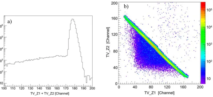
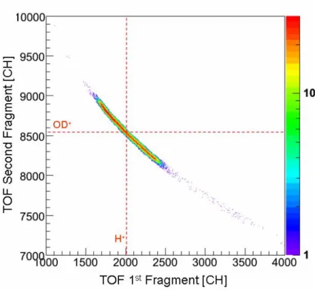
des résultats du calcul. Ces quatre voies de dissociation de ces dications peuvent être clairement séparés par notre technique expérimentale, comme le montre bien la carte de coïncidence présentée ci-dessous (figure f-8).
La fragmentation du HDO2+ fait l’objet d’une attention importante dans de nombreuses expériences récentes du fait de la forte sélectivité isotopique de la rupture des liaisons OH et OD. Deux valeur du rapport de branchement (H+ + OD+)/(D+ + OH+) ont été mesurées par des méthodes expérimentales différentes et sont en désaccord de 25% (6,5 ± 0,5 [réf. V.1] et 5,2 ± 0,3 [réf. V.2). Dans ce travail de thèse, nous avons obtenu une valeur expérimentale intermédiaire. Utilisant une technique d’imagerie ainsi qu’un spectromètre de temps de vol particulièrement adapté à cette expérience, il a été possible d'avoir un accès direct aux distributions d’énergie cinétique emportée par les fragments (KER) et à la proportion de chacune des deux voies avec une haute résolution. Il s’agit de la première expérience permettant l’étude simultanée de tous les isotopomères de la molécule d’eau. En effet, la dissociation de H2O2+ et de D2O2+ ont été analysées de la même
manière que HDO2+ afin d’être directement comparables. Les proportions des différents fragments et les rapports de branchement dans le cas de l’impact d'ions S15+ de 13,7MeV/u sont présentés dans le tableau1. La rupture de la liaison OH s'avère être 5,7 fois plus probable que celle de la liaison OD.
Voie de dissociation Nombre d'événements Proportion de la voie
H+ + OD+ 29984 5,7 ± 0,1
D+ + OH+ 5294 1
H+ + OH+ 16830 3,2 ± 0,1
D+ + OD+ 17222 3,3 ± 0,1
Tableau 1 Les proportions des différents fragments et les rapports de branchement dans le cas de l’impact d'ions S15+ de 13.7MeV/u
Figure f-8 Carte de coïncidence des premier et deuxième ions détectés lors de la fragmentation des dications de la molécule d'eau, après nettoyage par le filtre en position 3D.
Un autre aspect important de la fragmentation à deux-corps de HDO2+ est la différence des valeurs moyennes de l’énergie cinétique libérée (KER) par chacune des deux voies de dissociation. Les valeurs obtenues lors d’un précédent travail sont respectivement 6eV pour la voie D+ + OH+ et 7eV pour la voie H+ + OD+ avec une résolution de 0,5eV. Nous avons amélioré la précision sur la mesure de ces valeurs pour chacune des quatre voies de dissociation des dications de l'eau. Les valeurs moyennes de KER sont les suivantes : 6,5±0,1eV pour les trois voies D+ + OH+, H+ + OH+ et D+ + OD+ , valeur conforme au résultat de Siegmann et al [réf.V.3] pour la voie de H+ + OH+, et 6,9±0,1eV pour la voie H+ + OD+ (figure f-9).
Les caractéristiques principales du processus sont bien reproduites par un calcul semi-classique. En effet, le rapport de branchement théorique de 7,2 est en bon accord avec la valeur expérimentale de 5,7. De plus, il est remarquable que les écarts entre les valeurs moyennes de KER des différentes voies de fragmentation soit bien reproduite. Néanmoins, la simulation ne tient compte que de l’état fondamental de l’ion moléculaire. La différence observée entre l'expérience et le calcul peut éventuellement venir du fait que la collision peut peupler un ou plusieurs états électroniques excités du dication.
Pour tester cette hypothèse, nous avons réalisé une autre expérience avec un ion projectile de basse énergie cinétique: Ne10+ à 10keV/q. Dans ce régime de vitesse de collision, l'ionisation de la molécule se fait par l’intermédiaire de la capture d’électrons par le projectile. L’idée sous jacente est de tenter d’obtenir le dication dans un état électronique différent en le produisant par l’intermédiaire de processus différents : la capture électronique ou l’ionisation directe. Nous avons obtenu des rapports de branchement ainsi que des distributions d’énergie cinétique très similaires à ceux mesurés pour le projectile à haute vitesse et ainsi donc pas de différence notable.
Figure f-9 Distribution de KER des voies de fragmentation H+/OD+, D+/OH+
pour HDO2+, H+/OH+ et D+/OD+ pour
§ I Introduction
Numerous studies have already been devoted to molecular fragmentation. In the case of ion-induced fragmentation, one can address many fundamental aspects of molecule. In CIRIL labora-tory, ion-atom and ion-molecule collision have been investigated by using specially designed super-sonic gas jet apparatus and multi-charged ion produced by GANIL for a decade. Besides, the recent evolution of experimental technique, called COLTRIMS which is now ubiquitous in the field of atomic physics research, opened doors for further studies of spectroscopy. The study of the frag-mentation dynamics of triatomic molecule is, therefore, one of the grand sum of these achieve-ments.
I.1 Fragmentation Process
The analysis of molecular fragmentation is based on the Franck-Condon principle. The quali-tative picture of the principle is illustrated in figure I-1. This illustration shows a ground state mo-lecular energy curve and two dissociative electronic states of diatomic molecule AB. The duration of interaction between a fast multi-charged ion and a target molecule is estimated to be order of the 10-18-10-17 second which is quite shorter than molecular characteristic times: vibration (10-14-10-12) and rotation (10-12-10-9). As a result, the nuclear locations remain unchanged during electronic tran-sition so the ionized molecule keeps the equilibrium internuclear distance of the neutral molecule. In a second step, since most of multiply charged molecular ions are not stable, nuclear motion starts off along the potential energy curves. When resulting ionized molecule (AB)q+ dissociates into Aq1+ + Bq2+ (q=q1+q2), potential energy difference between transient molecular ion state (AB)q+ and final state (Aq1+ + Bq2+) is transferred into kinetic energy shown as Ek1 in the figure, named Kinetic
informa-tion on the fragmentainforma-tion process of the molecule corresponding to the excited states of the molecu-lar ion.
In the case of diatomic molecules, the charge of the transient molecular ion is generally shared equally into two fragments [ref. I.1-3]. This is because the electron rearrangement in the transient molecule occurs in a much shorter time (≈10-16) than the dissociation process.
In the early experiments on diatomic molecular fragmentation, the mean KER value could only be estimated due to the low resolution and results were in fair agreement with simple Coulomb Explosion Model (CEM) predictions [ref.I-4, 5 for example]. In this model, the KER in the dia-tomic molecular fragmentation into (Aq1 + Bq2) is assumed to be due to the Coulomb repulsion en-ergy: r q q E 1 2 ker 4 . 14
= [eV], where r [Å] is the internuclear distance of the neutral molecule at equilib-rium.
More recently, the KER distribution have been measured with better resolution both high en-ergy (1-100MeV) and low enen-ergy (several keV/q) ion impact. The results obtained by high enen-ergy ion impact experiments show that the mean KER values disagree with the predictions of CEM and include high energy components [ref. I.3,6,7]. These high energy components were attributed to highly excited molecular ions. At low energy experiments, contrary, the reaction channels and mean KER values lower than the CEM predictions have been determined [ref.I.8,9]. Furthermore, the measured KER values were well accorded with the theoretical results from potential energy curves obtained a quantum chemical calculation. In CIRIL, both low energy and high energy ion induced fragmentation of CO molecule have successfully conducted. The resolution of KER has been
im-Figure I- 1 Schematic potential energy curves of a diatomic molecule.
proved down to 250 meV leading to clear structures on the spectra. These structures have been in-terpreted by the use of "exact" molecular potential energy curves [ref. I.10]. These results indicate that the fragmentation process is completely driven by dissociative excited states of the transient molecular ion produced during the collision.
The reactions of triatomic molecules are, however, not able to be described by such a simple picture. Following potential energy "surfaces", no more simple curves, many reaction channels are involved in the fast ion-molecule collision. The reaction channels which end into complete three-body dissociation may hold the different scenarios. According to Maul and Gericke [ref.I.11] termi-nology and its expansion proposed by Hsieh and Eland [ref.I.12], the different fragmentation mechanisms are distinguished into three types as follows:
1) Pure sequential fragmentation
2) Synchronous concerted fragmentation 3) Asynchronous concerted fragmentation
The first case corresponds to two independent two-body dissociation reactions. In other word, the first atomic fragment has no interaction with the other fragments when the second dissociation step of the intermediate diatomic fragment takes place:
+ + + + + + + → + → 4 3 2 2 1 C B (BC) BC) ( A (ABC) q q q q q q then
The second fragmentation type is defined as the one for which the two bond breakages occur simul-taneously and with a symmetric behavior.
+ +
+
+ →A 1 +B 2 +C 3
(ABC)q q q q
Then the third one includes reactions from instantaneous dissociation via asymmetric stretch to non pure sequential dissociation, meaning a second step influenced by the primary fragment.
+ +
+ →A 1 +(BC) 2
(ABC)q q q
→Aq1+ +Bq3+ +Cq4+
The following discussion in the present thesis for three-body fragmentation relies on this categori-zation.
I.2 Experimental Backgrounds
The recent growth in research on atomic and molecular physics field has strongly benefited from the recent evolution of the Recoil Ion Momentum Spectroscopy (RIMS) technique1 [ref.I.13]. This method, originally designed for ion-atom collision experiments [ref.I.14], is now widely used either for electron impact [ref. I.15 for review], synchrotron radiation [ref.I.16 for review], or femto-second laser [e.g. ref.I.17] experiments. Apart from the use of supersonic jet target, one of the most important things among numerous technical developments is the multi-hit detection of particles. This "breakthrough" offers unprecedented opportunities for coincidence measurements of fragments from a molecule. The spectrometer can be modified easily to different specifications as well. The time-focusing condition allows to reconstruct momentum components even for undetected particles [ref.I.18]. Position-focusing condition avoids the problem of the resolution limitation due to the volume of the collision region [ref.I.19,20]. Another significant development which helps to obtain the complete kinematics of molecules is the imaging technique. Using a 2D position sensitive detector while the longitudinal velocity is deduced by time of flight measurement, molecular coordinates can be reconstructed in a simple manner.
The localised target, coincidence, multi-hit detection and imaging techniques allow now to investigate the mechanisms of molecular fragmentation with kinematically complete data set.
I.3 Previous Results and Scopes of Present Works
I.3.1 Fragmentation DynamicsFor the last few years, a number of studies have been devoted to polyatomic molecular fragmentation with RIMS techniques. Most of experimental studies have approached molecular characteristics through photoionization. After the successful investigation of diatomic molecules, our group has decided to go further taking advantage of fast ion impact, i.e. the momentum transfer between the projectile ion and target molecules is negligible. Because of this characteristics, ion induced fragmentation can give direct information of momentum sharing among molecular frag-ments in different dissociation channels which cannot be done easily with laser excitation since fragments may start moving during the light pulse if too long.
The linear triatomic molecule CO2 was the first candidate [ref.I.21] in order to reveal the role
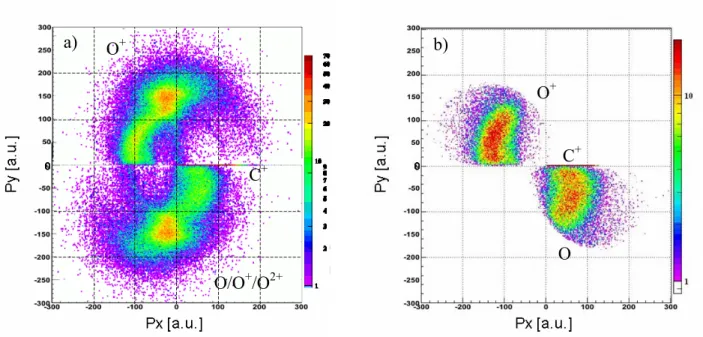
of an internal degree of freedom, such as stretching. A slight bend angle of the triatomic molecule leads the fragments to move in a plane, rather than on an axis as in the case of diatomic molecules. The main results obtained from the previous experiment concerned the dissociation channels of tri-ply and doubly ionized molecules corresponding to the main "island" in the figure I-2:
O O C CO O O C CO 2 2 3 2 + + → + + → + + + + + + +
Figure I- 2 Coincidence Map of 1st and 2nd fragments from CO
2 collision with 8MeV/u Ni24+
In order to show the shared momentum balance among the fragments, Newton diagram, rep-resenting the momentum balance in the molecular frame, is an usual way to present it. Following to the definition (section I.1), these momentum vector coordinates can be used to discriminate the
dis-sociation process. In figure I-3 and I-4, the center atom C+ momentum direction is chosen as the x-axis while the more energetic/detected O+ fragment defines the positive y axis (see section IV.3 for detail).
The Newton diagram corresponding to triple ionized dissociation channel, on figure I-3, shows a symmetric emission of the two oxygen fragments while the carbon ion has smaller momen-tum (0-100 a.u.) in the molecular frame. This is characteristic of a synchronous concerted fragmen-tation. Due to the simultaneous bond breakages of C-O, the center atom ion C+ stays nearly at rest.
The case of CO22+ dissociation is more complex. Since the neutral fragments can not be
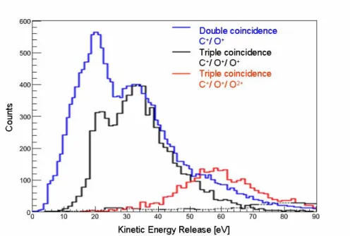
de-tected, one has to separate the relevant events (C+/O+/O) form double coincidence measurements (C+/O+) including "incomplete" events from (C+/O+/O+) and (C+/O+/O2+), etc (figure I-4.a to I-4.b, see figure I-5 for additional information). The figure I-4.b shows no features corresponding to the synchronous concerted fragmentation. What one can observe here is that the neutral O fragment is emitted towards the C+ momentum direction. This fact enables to assume that the dissociation pro-ceeds through the intermediate step CO++ O+. Moreover, comparing the KER distributions of the
dissociation channel which ends into CO++O+ and C+ + O+ +O, there is a certain threshold where the complete dissociation starts to be observed and become dominant (figure I-6). A simple model could be evoked for this dissociation channel relying on an "asymmetric stretch picture". The first bond break leads to (C+-O) and O+, then due to the recoil of C+, the internal vibrational energy of the molecular fragment CO+ increases. This transferred energy from the first step introduces the threshold effect observed in the KER distribution: if the energy is larger than the CO+ binding en-ergy, the dissociation ends into complete three-body fragments (C+/O+/O), otherwise two-body fragmentation (CO+/O+) is detected. Consequently the process of the CO22+ fragmentation is the
non-pure sequential categorized in asymmetric concerted fragmentation.
Figure I- 3 Newton diagram of the fragmentation of CO23+ forming C+/O+/O+.
O+ O+
In order to go further, these interpretations have to extend into a more general triatomic mole-cule case with a bending angle. Thus NO2, a bent molecule having similar mass ratio to CO2 has
been considered to be an ideal candidate for the next step. The molecular characteristics of equilib-rium CO2 and NO2 are shown in table I-1.
Figure I- 4 Newton diagram of a) all measured C+/O+ double coincidence events consisting mainly in (C+/O+/O), (C+/O+/O+) and (C+/O+/O2+) dissociation channels b) separated C+/O+/O dissociation channel from a).
A particular attention is given to the dication, for which the fragmentation scenario may become even more complicated. The rotation of the intermediate molecular fragment should participate more in the dissociation energy sharing among all degrees of freedom than for a linear molecule.
The mean KER value when the dissociation of NO22+ results in the formation of the charged
pair (NO+/O+) has been first measured to be 8.03 eV by R. G. Cooks et al. in 1974 [ref.I.22] with electron impact. Then in PIPICO (PhotonIon-PhotonIon-COincidence) and PEPIPICO (PhotonElectron-PhotonIon-PhotonIon Coincidence) double photo-ionization measurements, total KER of 8.6 and 7.1±1 eV have been measured, respectively by J.H.D. Eland et al. in1987 and 1988 [ref.I.23,24]. Authors also estimated an energy release of 6.1±1 eV and 6.3±3 eV for (N+/O+/O) and (N/O+/O+) dissociation channels, respectively. At the same period, theoretical calculations have also been reported by the same authors [ref.I.25]. More recently, Masuoka and Kobayashi [ref.I.26] have measured same dissociation channels as Eland et al by PIPICO measurements in better resolution. However, these studies focus on the thresholds of double ionization and dissociation, electronic states, or partial cross sections, but not at the dynamics of the fragmentation. The present study will
O/O+/O2+ O+ C+ O O+ C+ a) b)
then give the first direct results of the NO22+ fragmentation dynamics. Moreover, the systematic
understanding of geometrical contribution on molecular fragmentation may hopefully be used as a probe of the electric processes involved, in such fields of cluster fragmentation or radiation damage.
Figure I- 5 Comparison of three KER distributions obtained by double coincidence event C+/O+ (blue) and completely identified triple coincidence events C+/O+/O+ (black) and C+/O+/O2+ (red) dissociation channels. To emphasize the contribution of C+/ O+/ O+ and C+/ O+/ O2+ channels on double coincidence spectrum, peak heights of both spectra are adjusted arbitrarily.
Mass r (A-O) [Å] Bond angle [deg.]
CO2 44 1.163 Two double bonds 172
NO2 46 1.197 One unpaired electron 134
I.3.2 Branching Ratio of HDO2+ Dissociation
Another way to get information on triatomic molecular ion potential energy surfaces is to take advantages of isotopomers. Asymmetric HDO2+ deuterated water dication fragmentation has been given special attention in recent experiments [ref.I.27,28] and particularly to the two-body fragmen-tation channel leading to an atomic and a molecular ion as final products. The exciting question is about the nature of the atomic fragment. Is it equivalent to get H+ or D+ as the atomic fragment? In other words, is the OH bond weaker than the OD bond? The answer is that a strong isotopic prefer-ence has been observed in favor of the cleavage of OH bond over OD bond in HDO dication disso-ciation. Two values have been measured for the branching ratio (H+ + OD+) / (D+ + OH+) which disagree by 25% ([ref. I.27]: 6.5 ± 0.5, [ref. I.28]: 5.2 ± 0.3). In order to find out the underlying process leading to this selective behavior, calculations of this ratio have also been performed, either semi-classical [ref. I.27] or wave-packet propagation calculations [ref. I.28], both restricted to the lowest HDO2+ potential energy surface.
Aiming to test them in details and understand the reason for this strong isotopic selection, we measured branching ratio and Kinetic Energy Release (KER) distributions with a high resolution. Taking advantage of the detection of both fragments, we used a COLTRIM spectrometer without the field-free region of the usual Wiley-MacLaren configuration. This allows to decrease the ex-tracting electric field down to 20V/cm and thus to improve the KER resolution (see section II.3.4).
In order to be as complete as possible, H2O and D2O were analyzed in the same way as
HDO giving a direct calibration of relative cross sections within the same experiment. Figure I- 6 KER distributions of double ionization dissociation channel forming CO+/O+ (black) and C+/O+/O (blue).
§ II Experimental Specifics
To investigate the dynamics of fragmentation process, primary key information is the Kinetic Energy Release (KER) distribution of the system. In recent years our group has achieved high reso-lution measurement of KER using the Recoil Ion Momentum Spectroscopy technique [ref. II.1-3].
Our experimental setup consists in three main parts: 1. projectile ion provided by GANIL beam lines, 2. target supersonic molecular gas jet apparatus attached to the main chamber and 3. recoil ion and electron spectrometer with position sensitive detector on each ends. The improved new acquisition system is also described in this chapter.
II.1 Projectiles: Highly Charged Ions
Figure II- 1 Photograph of Recoil Ion Spectrometer in the main chamber. The projectile ion comes from left-hand side and collides with target molecules from a supersonic jet generator installed on the backside of the reaction chamber.
II.1.1 High Energy Projectile Ions from GANIL Facility
Most of our experiments have been conducted at the SME beam line located at GANIL accel-erator facility. SME means medium energy exit (Sortie Moyenne Energie) designed for interdisci-plinary research (atomic and molecular physics, solid state physics). As illustrated in figure II-2, projectile ions are produced by an ECR ion source and pre-accelerated by a compact cyclotron(C01 or C02), then are injected into the first Cyclotron with Separated Sectors (CSS1) for acceleration up to 4 to 13MeV/amu. Since these cyclotrons have the same specifications, the charge state of ex-tracted ion has to be changed before going into the second accelerator (CSS2) to be accelerated fur-ther. Therefore after the exit of CSS1, extracted ions pass through a thin carbon foil for charge stripping. After this stripper, one charge state is sent into SME beam line while another one is di-rected to CSS2 for high energy (24-96MeV/amu) acceleration. Due to the function principle of the cyclotron accelerators, GANIL provides a pulsed beam which frequency depends on the type and the energy of the circulating ion.
Figure II- 2 GANIL beam facility
In the present work, we used 13.7MeV/amu S15+ for one of HDO experiments and 4.7MeV/amu Ne8+ for NO2 experiment as projectile ions1. These high energy highly charged ions
enable us to observe directly the dynamics of molecular fragmentation. Since the velocities of pro-jectile ions are so high, the momentum exchange between propro-jectiles and target molecules are
ligible. For all experiments, the beam intensity is adjusted to have an average number of coinci-dence events around 2000cps in order to avoid random coincicoinci-dences.
II.1.2 Low Energy Projectile Ions from ARIBE2 Facility
One of the HDO fragmentation experiments has been conducted at ARIBE beam line. This low energy facility located at GANIL has started providing low energy ion beams since 2005. There are 7 experimental areas in total as showed in figureII-3 for conducting heavy ion experiments. Highly charged heavy ions with high intensity are provided from an ECR ion source and extracted up to 25keV/q.
Figure II- 3 ARIBE beam line
In contrast with high energy ion impact, we can no more neglect the momentum transfer be-tween the target and the projectile during collision. Using the position sensitive detector (see II.4), we have observed the charge-exchange on the projectile 1 meter downstream after collision. The projectile delivered to the line L4 passes through two collimators of 10mm in diameter placed at 1.5m and just before the reaction chamber respectively. Then inside the chamber, a third collimator cuts the beam to 0.6mm in diameter to let it cross the collision point. After collision, the multi-charged projectile ions are bent and charge separated by passing through electrostatic deflector plates. Then a drift tube, equipped with a Position Sensitive Detector at the end, is installed after the reaction chamber. Its angle can be modified easily to be adjusted to the chosen projectile charge state [ref. II.2, 4].
We have used Ne10+ with an energy of 17keV/q, i.e. 0.56a.u. in velocity. The deflectors are biased to -310V for the upper one and of +310V for the other in order to be able to separate
ent ion charge states. The detector is set 10cm higher than the beam line in the case of Ne9+ or lower charged projectile.
Figure II- 4 Schematic drawing of the projectile detection principle and a typical detector image for Ne10+ projectile after a charge exchange reaction: Ne9+, Ne8+ and Ne7+ are detected on the detector.
II.2 Target Molecules: Supersonic Jet
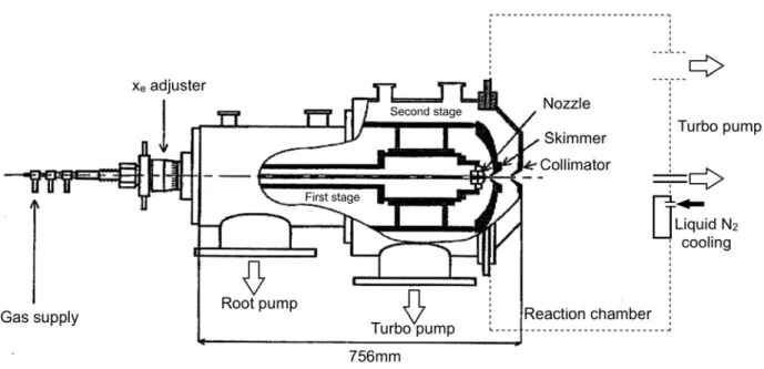
II.2.1 Principle and SetupThe cold and dense gas target is produced by specially designed supersonic gas jet apparatus (Campargue [ref. II.5, 6]). The use of supersonic gas jets allows to achieve high resolution meas-urements that would be impossible with a standard gas cell target at room temperature. Indeed, a small sized target and a reduced thermal motion are necessary for our imaging technique (see fol-lowing sections).
The supersonic jet gas is produced by the expansion from a strong pressure P0 through a small
nozzle to a low pressure stage (Figure II-6). During this adiabatic expansion, the initial disordered momentum is transformed into the directed momentum along the nozzle axis. The supersonic gas jet bordered by a shock-front is formed. The gas jet flowed in the first stage is extracted by a sharp skimmer placed in the “zone-of-silence” without breaking its characteristics. To obtain reasonable gas jet properties, the product of P0 · D should be larger than 1 mbar · cm, where D is the nozzle
di-ameter [ref. II.1, 7]. Practically, the performances of gas jets are mainly fixed by the pumping speed in the first stage and the distance between nozzle and skimmer at the exit to the collision cham-ber.The nozzle position is easily adjustable in order to obtain the best performance for different gas species and driving pressures. Then a high quality jet goes into the collision chamber through a col-limator at the exit of the second stage. The second stage has an important role in maintaining a good vacuum in the reaction chamber.
Figure II- 6 Schematic drawing of the principle of supersonic gas jet generator (Campargue type)
To make this system work properly, specific pumps for each stage are required. For the first stage, we used a combination system of three pumps: ROOTS pump of 2050m3/h, ROOTS pump of
253m3/h and rotary pump of 40m3/h. The vacuum in second stage is obtained with a turbo pump of Figure II- 5 Comparison of Ar+ flight time peaks obtained with a gas cell (red circle) and a supersonic gas jet (green line). The width of these distributions is due to the ini-tial thermal motion of the target atoms.
400l/s associated with a rotary pump. The vacuum in the collision chamber is kept by a turbo pump and liquid nitrogen trap. Finally, to avoid diffusion of the jet, a beam dump is located after the colli-sion region and is pumped with a 70l/s turbo molecular pump. Between the collicolli-sion chamber and the gas generator device is inserted a valve which allows the jet to flow into the chamber or isolates it rapidly and efficiently.
Figure II- 7 Mechanical aspects of the supersonic gas jet generator and pumping systems
Figure II- 8 Photograph and schematic drawing of the source gas injection apparatus. Temperatures at 5 points along the gas flow and of the room are surveyed and limited to certain values. Whole apparatus is winded by heating wires and covered by Al foil.
In order to be able to use molecules from a liquid at room temperature, an oven has been added to the setup. The vapor has to be transported from the oven to the nozzle through heated stainless steel tubes. The oven has to be the coldest point in order to avoid condensation. Heating and temperature monitoring is remotely controlled by a Lavbiew program through a Field Point in-terface.
II.2.2 NO2 Preparation
Handling NO2 is rather complicated. First of all, it's a highly toxic gas. An NO2 detector has
been used for that purpose. The other problems are due to the chemical and physical properties of NO2. Nitrogen dioxide is a liquid in normal atmospheric condition (Figure II-9a). Thus heating is
necessary in order to obtain a high enough pressure for the supersonic gas jet. Furthermore, the main problem is due to the fact that NO2 almost does not exist as a pure molecule. At low
tempera-ture, it polymerizes to the dimer N2O4. To form the monomer NO2, temperature has to be raised
(Figure II-9b) to change the relative proportion of each compound. It is only above the critical point at 158°C that the gas consists only of monomer NO2. In practice however, it is pretty hard to keep
all equipment at this high temperature. The nozzle was then kept at around 100°C and experiments have been performed at different injection pressures (2 and 5 bars) in order to estimate the contribu-tion of the dimers.
Figure II- 9 a) The relationship temperature versus pressure for N2O4. b) Phase diagram of NO2↔ N2O4. The red line indicates the applied nozzle temperature and corresponding ratio of NO2 expected in the present ex-periment.