HAL Id: inserm-02980617
https://www.hal.inserm.fr/inserm-02980617
Submitted on 27 Oct 2020HAL is a multi-disciplinary open access archive for the deposit and dissemination of sci-entific research documents, whether they are pub-lished or not. The documents may come from teaching and research institutions in France or abroad, or from public or private research centers.
L’archive ouverte pluridisciplinaire HAL, est destinée au dépôt et à la diffusion de documents scientifiques de niveau recherche, publiés ou non, émanant des établissements d’enseignement et de recherche français ou étrangers, des laboratoires publics ou privés.
Targeting iron-mediated retinal degeneration by local
delivery of transferrin
Émilie Picard, Quentin Le Rouzic, Antonin Oudar, Marianne Berdugo,
Mohamed El Sanharawi, Charlotte Andrieu-Soler, Marie-Christine Naud,
Laurent Jonet, Chloé Latour, Christophe Klein, et al.
To cite this version:
Émilie Picard, Quentin Le Rouzic, Antonin Oudar, Marianne Berdugo, Mohamed El Sanharawi, et al.. Targeting iron-mediated retinal degeneration by local delivery of transferrin. Free Radical Biology and Medicine, Elsevier, 2015, 89, pp.1105-1121. �10.1016/j.freeradbiomed.2015.08.018�. �inserm-02980617�
Elsevier Editorial System(tm) for Free Radical Biology and Medicine Manuscript Draft
Manuscript Number:
Title: Targeting iron-mediated retinal degeneration by local delivery of transferrin Article Type: Original Research/ Original Contribution
Keywords: Neurodegenerative diseases, Neuroprotection, Iron, Oxidative stress; Inflammation, Transferrin
Corresponding Author: Dr. Emilie Picard, Ph.D Corresponding Author's Institution: INSERM First Author: Emilie Picard, Ph.D
Order of Authors: Emilie Picard, Ph.D; Quentin Le Rouzic, Master; Antonin Oudar, Master; Marianne Berdugo, PhD; Mohamed El Sanharawi, MD; Charlotte Andrieu-Soler, PhD; Marie-Christine Naud; Laurent Jonet; Chloé Latour; Christophe Klein; Stéphane Galiacy, PhD; François Malecaze, PhD; Hélène Coppin, PhD; Marie-Paule Roth, PhD; Jean-Claude Jeanny, PhD; Yves Courtois, PhD; Francine Behar-Cohen, PhD, MD
Abstract: Iron is essential for retinal function but contributes to oxidative stress-mediated
degeneration. Iron retinal homeostasis is highly regulated and transferrin (Tf), a potent iron chelator, is endogenously secreted by retinal cells. In this study, therapeutic potential of a local Tf delivery was evaluated in animal models of retinal degeneration.
After intravitreal injection, Tf spread rapidly within the retina and accumulated in photoreceptors and retinal pigment epithelium, before reaching the blood circulation. Tf injected in the vitreous prior and, to a lesser extent, after light-induced retinal degeneration, efficiently protected the retina histology and function. We found an association between Tf treatment and the modulation of iron homeostasis resulting in a decrease of iron content and oxidative stress marker. The immunomodulation function of Tf could be seen through a reduction in macrophage/microglial activation as well as modulated
inflammation responses. In a mouse model of hemochromatosis, Tf had the capacity to clear abnormal iron accumulation from retinas. And in the slow P23H rat model of retinal degeneration, a sustained release of Tf in the vitreous via non-viral gene therapy efficently slowed-down the photoreceptors death and preserved their function.
These results clearly demonstrate the synergistic neuroprotective roles of Tf against retinal degeneration and allow identify Tf as an innovative and not toxic therapy for retinal diseases associated with oxidative stress.
Suggested Reviewers: Carole Peysonneaux PhD
Chargé de recherche , U1016, Institut Cochin, INSERM carole.peyssonnaux@inserm.fr
In the field of Hypoxia and iron homeostasis Maria Ugarte PhD, MD
Medical Retinal Fellow, Department of Medical Retina Unit, Moorfields Eye Hospital NHS Foundation Trust ·
mugarte@doctors.org.uk In the field of metals in retina Isabelle Ranchon-Cole PhD
researcher, UMR 1107 - Equipe Biophysique Neurosensorielle, INSERM - Facultés de Médecine et de Pharmacie
isabelle.ranchon-cole@udamail.fr
In the field of neuroprotection in models of retinal degeneration Torben Moos PhD
Professor, Department of Health Science and Technology, Aalborg University tmoos@hst.aau.dk
Emilie Picard
INSERM
Centre de recherche des Cordeliers
UMRS1138 team Behar-Cohen
15 rue de l’école de médecine
75006 Paris
France
e-mail :
picardemilie@gmail.com
Editor, Free Radical Biology and Medicine
Paris, July 10th 2015
Dear Editors,
Please find enclosed an original research article entitled “Targeting iron-mediated retinal
degeneration by local delivery of transferrin” by E. Picard et al., which we would like to submit for
publication in Free Radical Biology and Medicine journal. This work has not been previously published
in any form, nor is it under consideration for publication elsewhere.
Iron accumulation and subsequent oxidative stress have been implicated in the pathogenesis of
retinal degeneration of various origins including Age-related Macular Degeneration (AMD). The
efficacy of systemic chemical iron chelators have been evaluated for neurodegenerative retinal diseases
in animal models. However the administration of systemic iron chelation therapy in the absence of body
iron overload raises many safety concerns.
In this study, the therapeutic potential of the endogenous iron chelator, Transferrin, with local
administration via protein and non-viral gene delivery methods has been evaluated in two animal
models of retinal degeneration. Local administration of Transferrin showed promising results in terms of
retina structure and function preservation, furthermore modulation of iron metabolism, inflammatory
responses and apoptosis were observed.
Combined these results indicate that Transferrin has a high therapeutic benefit with low systemic
and visually significant risks in patients with retinal degeneration as AMD. This delivery method and
therapeutic strategy may have an important impact on the therapeutic strategies of other
neurodegenerative diseases, such as Alzheimer’s or Parkinson’s disease, which have also been
associated with iron accumulation-mediated oxidative stress. Considering this possibility we believe that
these results will be of interest to a wide spectrum of your readership.
All authors have reviewed the manuscript, agreed on its content, and approved its
submission to your journal for consideration for publication. We would be pleased if Enrique
Cadenas could be the editor for this article.
Thank you, for your time and consideration.
Yours sincerely,
Picard Emilie, PhD
Cover LetterTargeting iron-mediated retinal degeneration by local delivery of
transferrin
Emilie Picard 1-3, Quentin Le Rouzic 1-3, Antonin Oudar 1-3, Marianne Berdugo 1-3, Mohamed El
Sanharawi 1-3, Charlotte Andrieu-Soler 1-3, Marie-Christine Naud 1-3, Laurent Jonet 1-3, Chloé Latour 4,
Christophe Klein 5, Stéphane Galiacy 6, François Malecaze 6, Hélène Coppin 4, Marie-Paule Roth 4,
Jean-Claude Jeanny 1-3, Yves Courtois 1-3, Francine Behar-Cohen 1-3,7.
1 INSERM, UMRS 1138, team Behar-Cohen, From physiopathology of ocular diseases to clinical
development, Centre de Recherche des Cordeliers, Paris, France.
2 Université Pierre et Marie Curie-Paris 6, Centre de Recherche des Cordeliers UMRS 1138, Paris,
France.
3 Université René Descartes, Centre de Recherche des Cordeliers UMRS 1138, Paris, France.
4 INSERM, U1043, Toulouse, France; CNRS, U5282, Toulouse, France; Université de Toulouse,
UPS, Centre de Physiopathologie de Toulouse Purpan (CPTP), Toulouse, France.
5 INSERM, U1138, CICC, Université René Descartes Sorbonne Paris Cité, Université Pierre et Marie
Curie Paris, Centre de Recherche des Cordeliers, Paris, France.
6 INSERM U563, Centre de Physiopathologie de Toulouse Purpan, Toulouse, France; Department of
Ophthalmology, Purpan Hospital, Toulouse, France.
7 Jules Gonin Ophthalmic Hospital, Lausanne, Switzerland.
Corresponding author:
Tel: +33 1 44 27 81 82; Fax: +33 1 44 27 81 77;
E‐mail: emilie.picard@crc.jussieu.fr
Running title:
Transferrin protects against neurodegeneration Manuscript
Keywords
Neurodegenerative diseases, Neuroprotection, Iron, Oxidative stress; Inflammation, Transferrin
Nonstandard abbreviations used:
AMD: age-related macular degeneration; ET: electrotransfer; IVT: intravitreal; MC: microglial cell;
RPE: retinal pigment epithelium; Tf: transferrin; PR: photoreceptor
Conflict of interest
Highlights:
• Transferrin injection in the eye has no deleterious effect on retinal structure
• Local delivery of transferrin protects photoreceptors and preserves retinal electrophysiological activities in light-induced retinal degeneration model
• Protective mechanisms of tranferrin result from control of iron-induced death decrease and modulation of inflammation
• Intraocular administration of transferrin has therapeutic potential in retinal degenerative diseases
ABSTRACT
Iron is essential for retinal function but contributes to oxidative stress-mediated degeneration. Iron
retinal homeostasis is highly regulated and transferrin (Tf), a potent iron chelator, is endogenously
secreted by retinal cells. In this study, therapeutic potential of a local Tf delivery was evaluated in
animal models of retinal degeneration.
After intravitreal injection, Tf spread rapidly within the retina and accumulated in photoreceptors
and retinal pigment epithelium, before reaching the blood circulation. Tf injected in the vitreous prior
and, to a lesser extent, after light-induced retinal degeneration, efficiently protected the retina
histology and function. We found an association between Tf treatment and the modulation of iron
homeostasis resulting in a decrease of iron content and oxidative stress marker. The
immunomodulation function of Tf could be seen through a reduction in macrophage/microglial
activation as well as modulated inflammation responses. In a mouse model of hemochromatosis, Tf
had the capacity to clear abnormal iron accumulation from retinas. And in the slow P23H rat model of
retinal degeneration, a sustained release of Tf in the vitreous via non-viral gene therapy efficently
slowed-down the photoreceptors death and preserved their function.
These results clearly demonstrate the synergistic neuroprotective roles of Tf against retinal
degeneration and allow identify Tf as an innovative and not toxic therapy for retinal diseases
INTRODUCTION
Oxidative stress and inflammation are recognized as major pathological processes in
neurodegenerative diseases and iron is identified as a catalyzer of both processes (1-3). In the retina,
iron is essential for photoreceptors (PR) activity as it is a cofactor of phototransduction enzymes (4-5).
Iron content within the retina is generally controlled through locally expressed iron-related proteins
(6-7). Nevetheless, iron excess can be found in the retina of elderly people (8) and is known to
accumulate in retinal diseases (2, 9). For instance, in age-related macular degeneration (AMD), the
common leading cause of blindness in industrialized countries, iron deposits can be found in PR
segments, retinal pigment epithelium (RPE) and in drusen (sub-RPE deposits characteristic of AMD)
(10). Moreover, increased iron concentration level are usually detected in the aqueous humor AMD
patients (11). Free iron is shown to be highly toxic for PR (12), which loss lead to irreversible damage
and thus to blindness. Similarly, we showed that iron accumulation and alteration of iron metabolism
are both correlated with the progression of PR degeneration in animal models of retinal degeneration
(rd10 mice and Royal College of Surgeons (RCS) rats) (13-15). Therefore, iron chelation allowing the
removal of excess iron may very likely be a therapeutic target (16-17). Indeed, the use of chemical
iron chelators such as deferoxamine and deferiprone administered by systemic or oral routes, were
reported to be efficient in preventing PR death in animal models of retinal degeneration (14, 18-21).
Nevertheless, the long-term systemic delivery of iron chelator may cause refractory iron depletion and
may not be the best treatment option especially in elderly patients frequently suffering from anemia
(22-23).
Our previous investigations showed that human transferrin (Tf), whether continuously expressed or
administered intraperitoneally, preserve more than 60% of PR from death in rd10 mice (24-25).
Indeed, Tf, the main iron transporter, is a natural iron chelator essential for homeostasis control (24,
26). Our study is the first attempt to directly injected an iron chelator into the eye. We performed
intravitreal injections of human Tf in two different rat models of retinal degeneration and subsequently
analyzed the pathways involved in its protective effect. We first used a light-induced retinal
degeneration model in which oxidative stress is prominent (27-28). This allowed us to screen the
the effects of Tf on iron metabolism, apoptosis and inflammation responses. In a second step, we used
P23H rats suffering from slow retinal degeneration and used a non-viral gene therapy to evaluate
whether sustained intra-ocular Tf production could delay the inherited degeneration.
This study highlights the therapeutic potentiel of local delivery of Tf on neuronal loss, oxidative stress
and inflammation, the pathological features commonly observed in neurodegenerative diseases.
RESULTS
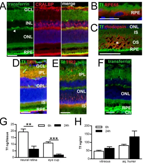
Time-related distribution of transferrin after intravitreal injection
We used Tf labeled with fluorochrom Alexa 488 (Tf-Alexa) to follow the fate of Tf in the rat retina
after intravitreal (IVT) injection. Two hours after injection, Tf-Alexa could be detected throughout the
retina, and was localized in retinal capillaries (arrowhead) and in the retinal pigment epithelium (RPE)
(Figure 1A, left panel). A combination of Müller glial cells marker (Figure 1A, middle panel) with
Tf-Alexa allowed us to show that Tf-Tf-Alexa was present along those cells, with largest concentrations in
their end feet facing the vitreous (Figure 1A, right panel). Confocal microscopy conferring higher
resolution allowed us to localize Tf-Alexa in microvillosities of RPE (Figure 1B) and in the inner and
outer segments of rod PR (Figure 1C). Tf receptor 1 and TfR2 were widely expressed in the retina (26,
29). Tf-Alexa was co-localized with TfR1 in the inner plexiform layer (IPL), outer plexiform layer
(OPL), inner segments (IS) and RPE (Figure 1D), and with TfR2 in IPL, ONL and IS (Figure 1E). Six
hours after injection, Tf-Alexa had the same pattern, and was additionally detected within
choriocapillaries (Figure 1F).
We used a specific transferrin (Tf)-ELISA assay to quantify Tf levels in ocular fluids and tissues after
IVT injection of human Tf in rat eyes. ELISA assays were performed at 6, 24 hours and 7 days after
injection (Figure 1G-H). When comparing Tf content 6 and 24 hours after injection, we observed a
significant decrease of 67.3% in the neural retina and of 82.6% in the eye cup (Figure 1G). In ocular
fluids, Tf concentration was higher in aqueous humor than in the vitreous, and increased with time
(Figure 1H). Tf levels measured in the systemic circulation were of 56.6 ng/ml ±0.32 and 46.5 ng/ml
after injection. These data indicate that IVT injected Tf reach the retina in a short amount of time and
most likely via the Tf receptor 1. It subsequently accumulate in RPE cells and finally disappear
through choroidal blood circulation.
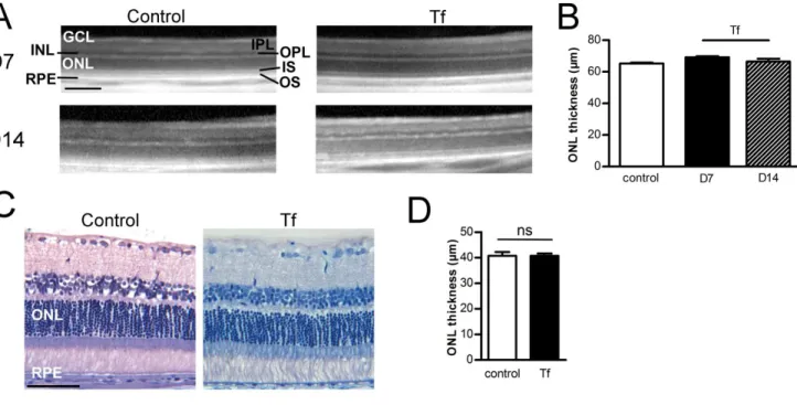
Transferrin intravitreal injection has no deleterious effect on normal retina
We injected Tf in vitreous of rats and analyzed the retina and its histology 7 days later. Some rats
obtained a single injection and others received a second injection 7 days after the first one. In vivo
optical coherence tomography (OCT) imaging revealed no retinal structure modifications after one or
two injections (Figure 2A). We used OCT pictures to measure ONL thickness which is usually
correlated to PR nuclei number, and observed no differences among control not injected rats, and
animals injected once or twice with Tf (Figure 2B). Moreover, when compared to eyes from control
rats, rat eyes injected twice with Tf showed no substantial histological modifications in any layer
(Figure 2C). Finally, an analysis of ONL thickness on sections crossing the optic nerve showed no
significant difference between control and treated rats (Figure 2D) and thus confirm results obtained
from OCT recording.
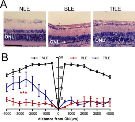
Transferrin intravitreal injection protects photoreceptors nuclear layer
Light-induced damage is a commonly used model to evaluate neuroprotective drugs on PR
degeneration (28, 30-31). We injected Tf in the vitreous directly after a dark adaptation period of 18
hours. We further exposed the rats to intense light during 24 hours. We performed morphological
analysis 8 days after light exposure using OCT recordings as well as histological semi-fine sections.
OCT images realized at superior pole of rats injected with control solution (BLE group) showed a
reduction in ONL thickness. Tf treatment (TfLE group) seemed to preserve the retina against light
effects (Figure 3A). The ONL thickness measured on OCT images (Figure 3B) showed a significant
decrease of 56% in BLE rats as compared to rats neither injected nor exposed to light (NLE group).
There was no significant difference in ONL between TfLE rats and NLE rats. Histology confirmed
OCT observations and measurements (Figure 3C-D). The most severe light-induced damage of BLE
rats treated with Tf had a preservation of ONL, IS, OS and RPE as compared to BLE rats (Figure 3C).
ONL thickness measured throughout the retina showed that 70% of the PR nuclei at the superior pole
and 92% at the inferior pole were protected with Tf injection (Figure 3D).
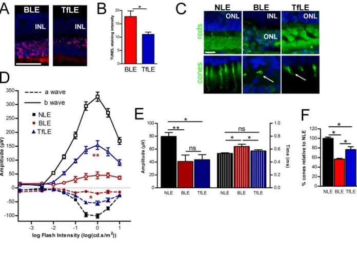
Transferrin preserves PR structures and functions.
We assessed apoptotic cells by TUNEL staining on eye cryosections to better evaluate the preservation
of PR nuclear layer with Tf treatment (Figure 4A). One day after light exposure, many TUNEL
positive PR were present in the ONL of BLE rats. TUNEL intensity staining quantification in ONL
throughout the retina demonstrated a significant reduction of 40% in the number of apoptotic PR in
TfLE retinas as compared to BLE (Figure 4B). We stained rods by rhodopsin immunodetection and
cone by lectin-peanut labeling 8 days after light exposure to confirm Tf-treatment PR morphological
preservation. When compare to unexposed eyes (NLE), BLE retinas had a close to complete loss of
both types of PR (Figure 4C, middle panel), whereas Tf-treatment retinas (TfLE) showed preserved
structures in both rods and cones (arrows) (Figure 4C, right panel).
We performed full-field electroretinograms 16 days after light-exposure to determine whether the
above-described results correlate with the maintenance of retinal function. In scotopic conditions,
light-exposure drastically lowered a- and b-wave amplitudes in BLE rats, meaning a decreased
rod-driven response (Figure 4D). With Tf treatment, these amplitudes were significantly higher compared
to BLE group (Figure 4D). In an extensive analysis in the flash intensity ranging from -0.5 to 0.47
log(cd.s/m2), the mean a-wave amplitude of TfLE reached 55.18% of NLE, and the mean b-wave
amplitudes in TfLE rats were 47% of NLE values. The protection of scotopic a- and b-wave
amplitudes were confirmed by the mixed rod-cone responses recording (Supp. Figure 1). Tf
significantly preserved 85% of PR response (a-wave amplitude; Supp. Figure 1A left panel), and
54.8% of internal retina response (b-wave amplitude; Supp. Figure 1A right panel) relative to
unexposed rats. The time-to-peak for mixed ERG a- and b-waves were not statistically different in the
three rat groups, in spite of a tendency toward a Tf-induced preservation of implicit times (Supp. Fig
1B). Following a rod-suppressing light adaptation, the photopic b-wave responses (Figure 3E, left
BLE and TfLE. These were lower compared to NLE. The implicit time (right panel) was significantly
longer in BLE as compared to NLE, and significantly restored in TfLE rats.To better characterize this
effect on cones, we quantified cones in overall retina (Figure 4F). Eight days after light exposure, there
was a 43% cone loss in BLE retinas and a 23% cone loss in TfLE retinas when expressed as % of
cones within the control NLE group. In conclusion, IVT injection of Tf was effective in protecting
cones and rods from death and allowed to preserve the physiological activities of the retina against
light-induced retinal damages.
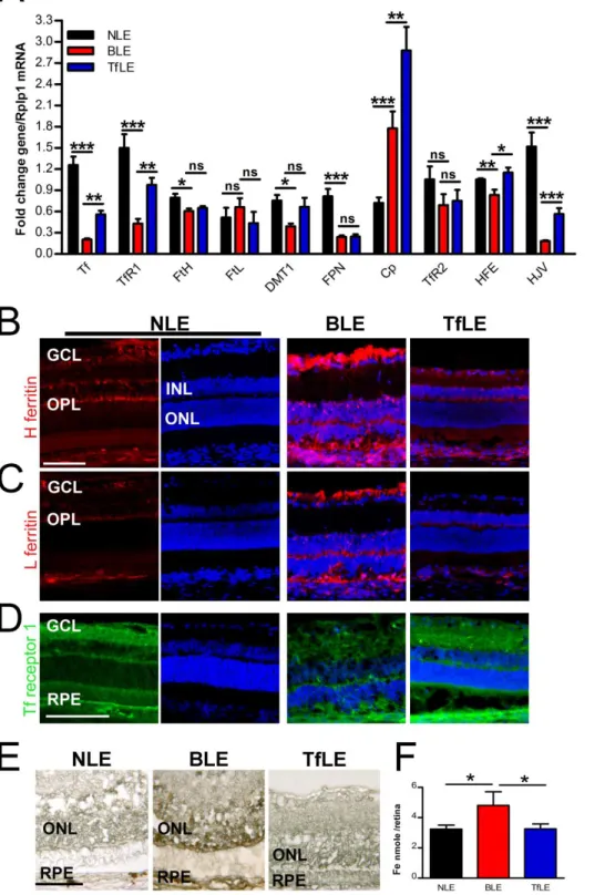
Iron metabolism deregulation in light-damage retina is restored by transferrin IVT injection Previously publications showed that intense light-exposure induces an acute and rapid modification of
iron metabolism that persists for several days after light damage (25, 32). To evaluate the effects of Tf
in iron homeostasis, we investigated the regulation of iron-related genes and proteins as well as the
iron content and distribution after light-exposure.
One day after light exposure, quantitative RT-PCR (Figure 5A) on neural retinas monitored expression
of genes related to iron transport (Tf and TfR1), storage (heavy and light Ferritins (FtH and FtL)),
export (Divalent metal transporter 1 (Dmt1), Ferroportin (Fpn) and Ceruloplasmin (Cp)), and
regulation (TfR2, Human hemochromatosis protein (Hfe) and Hemojuvelin (Hjv)). Except for Cp and
FtL, transcripts of all analyzed iron-related genes significantly decreased in BLE rats as compared to
control NLE rats. In Tf treated retinas, the expression of Tf, TfR1, Dmt1, Hfe and Hjv mRNA was
partially but significantly restored as compared to BLE retinas. The light-induced expression of Cp
was significantly increased in TfLE rat retinas. Tf treatment had no significant effect on FtH, FtL, Fpn
and TfR2 transcription when compared to BSS injected rat retinas.
Using immunohistochemistry, we observed the expression of the two ferritins and TfR1 proteins at
differents time points after the light-exposure (Figure 5B-D). The expression of these proteins are
known to correlated with the retina iron content, regulated by iron regulatory proteins IRP1 and IRP2
(25, 33). One day after light exposure, no modification of the two ferritins staining was observed in
BLE and TfLE treated groups as compared to NLE (data not shown). At day 3 and 8, ferritin staining
modification was observed in the retina of TfLE rats when compared to NLE rats (right compared to
left column). While TfR1 decreased in the retinas of BLE rat (Figure 5D, central panel), it remained
similar to the retinas of NLE rats in the TfLE rat eyes (right panel). Given the previous results, we
explored whether Tf treatment had an impact on iron accumulation in the retina, and revealed
non-heminic iron using Perls Prussian blue reaction intensified with diaminobenzidine (Figure 5E). Iron
accumulated in the outer retina of BLE rat at day 3 and 8 after light exposure (Figure 5E, middle
panel). Such an accumulation could not be detected in the retinas of TfLE rat at any time point (Figure
5E, right panel). This was confirmed with the total non-heme iron quantification determined in neural
retinas at 8 days after light exposure (Figure 5F) and demonstrates that Tf significantly prevent free
iron content increase. To investigate the effect of Tf on oxidative stress, we assessed heme oxygenase
(Hmox1) mRNA expression using qPCR on neural retina one day after light-exposure. Light-induced
iron overload significantly increases Hmox1 in the BLE group. This upregulation was less important in
the retinas of rats treated with Tf (Figure 6). These results demonstrated that IVT of Tf allows a rapid
and efficient control of iron homeostasis thereby avoiding iron accumulation and oxidative stress.
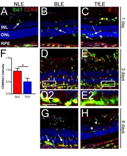
Light-induced retinal inflammation is controlled by transferrin.
Microglial cells (MC), specific macrophage/monocyte of the retina, are activated by light exposure
and migrate towards PR debris (34-35). We used ionized calcium-binding adapter molecule (Iba1) and
CD68 as markers to reveal macrophage/monocyte. Both are constitutively expressed but CD68 is
restricted to lysosomal membranes of non-activated cells, and is expressed in the cytoplasm of
activated cells, allowing the discrimination of the activation state at 1, 3 or 8 days after light-induced
damage (Figure 7A-H). We localized non-activated MC in the inner retina of NLE rats (arrow, Figure
7A). Twenty-four hours after light-damage, Iba1 positive cells had migrated towards the outer retina in
both rats and most of them were CD68- (arrows, Figure 7B-C). At 3 days, numerous round Iba1+ cells
increased in outer retina in BLE and TfLE rats (Figure 7D-E). Higher magnification of subretinal
space revealed that the Iba1+ cells were CD68+ in BLE rats, while Iba1+/CD68- cells reprensented the
majority in TfLE rats (Figure 7D2-E2). CD68+/Iba1+ intensity ratio quantifed in cells staining in the
true only for half the cells within the TfLE group. At 8 days (Figure 7G-H), numerous Iba1+/CD68+
amoeboid cells remained in the outer part of retina of BLE rats (arrows), while Iba1+ non-activated
cells were localized in the inner retina in the TfLE group (arrowheads).
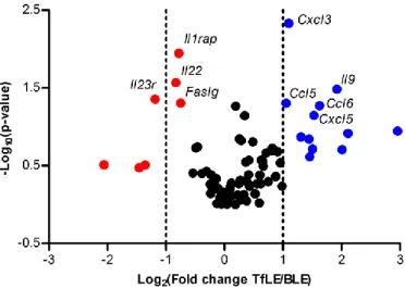
To get a better picture of the effect of Tf on light-induced retinal inflammation, we performed a PCR
array on 84 genes of inflammatory mediators on the neural retina, 1 day after light exposure. In the
BLE group, 34 genes were up-regulated and 13 down-regulated when compared to the NLE control
group. When comparing the TfLE group to the NLE group, 42 genes were up-regulated and 9
down-regulated (Table 1). Figure 8 shows gene distribution differentially down-regulated in TfLE when compared
to BLE according to theirs p-value versus fold change. Ccl5, Ccl6, Cxcl3, Cxcl5, Il9 genes were
significantly upregulated, whereas Il23r, Il22, Il1rap and Faslg were down-regulated. List of genes
modified by one-third by Tf injection are indicated in Supplemental Table 2. All these data indicate
that IVT of Tf control inflammatory cell activities through the modulation of inflammatory mediators
induced during light-exposure.
Transferrin has a long-term effect and efficacy in advanced disease models.
To evaluate the effectiveness of Tf in animals models presenting pathogenic characteristics such as
iron accumulation, photoreceptors degeneration and long-term disease evolution: 1- we administered
Tf treatment when part of the retina had already been damaged through light exposure; 2- we induced
sustained expression of Tf, via ciliary muscle electroporation of a plasmid encoding human Tf, in the
P23H rat model of slow retinal degeneration; 3- we tested the efficacy of Tf in removing iron deposits
from the retina of the Bmp6-/- mouse genetic model presenting iron overload.
1- Effect of IVT Tf treatment on partially degenerated retina. In retinal disease, PR death begins
before diagnosis, and therefore prospective treatments would have to be initiated in deleterious
conditions. The onset of light-induced retinal damage is rapid, occurring in about 2 hours after light
exposure. Once started PR degradation continues for a 2 days period (36).To demonstrate that Tf can
protect PR even after the degenerative process has begun, we performed IVT injection subsequent to
light-exposure. Eight days after the injection, ONL was mainly destroyed in BLE rats (Figure 9A,
39.43µm ±1.10; BLE: 5.94µm ±0.40) (Figure 9B). In the TfLE group, Tf was not able to protect the
PR layer at the superior pole, which was largely destroyed. However, relative to NLE rats, 50% of PR
nuclei were preserved in the inferior pole (mean ONL thickness of NLE: 40.43µm ±1.51; TfLE:
20.3µm ±2.72). This suggests that Tf injection may have a protective effect on partially damaged
retinas.
2- Sustained production of Tf by gene therapy in slow retinal degeneration model. IVT injections are
routinely used for eye treatment. In the context of chronic diseases such as dry AMD, frequent
intraocular injections may contribute to reduce treatment compliance. Our laboratory has developed a
non-viral gene therapy strategy based on ciliary muscle cells transduction via electrotransfer (ET)
thereby serving as a biofactory for sustained production of therapeutic proteins in the vitreous
(37). Using a disposable device and a minimally invasive procedure, this gene therapy technique can
be applied to secrete proteins of any molecular weight for several months (38-39). We produced
pVAX1-Tf plasmid through subcloning the cDNA of human Tf in a pVAX1 backbone under a
cytomegalovirus-β promoter which is known to be efficient for ET protocol (38). We transfected
human immortalized RPE cell line (ARPE-19) that does not synthesize Tf with pVAX1-Tf and
immunoblotted the culture media. The size of the produced Tf was similar to Tf isolated from human
plasma (Supp. Figure 2A). We used Tf-ELISA assay to determine the amount of Tf produced in the rat
eye 3 days after after ET. We detected human Tf in all compartments of the eye, including the
vitreous, aqueous humor and neural retina as well as the eye cup (Supp. Figure 2B). When performing
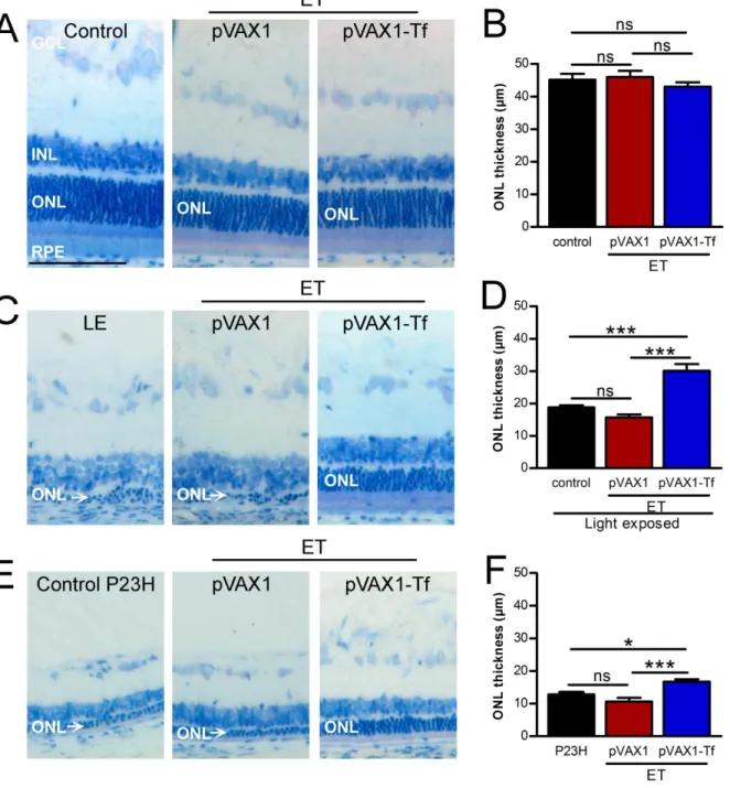
retinal histology 12 days after ET, we did not observe any difference in eyes ET with empty pVAX1
as compared to control eyes and did not see any variation of ONL thickness for eyes in which we
induced Tf production (control: 45.11µm ±1.82; ET pVAX1: 45.97µm ±1.92; ET pVAX1-Tf:
43.03µm ±1.31) (Figure 10A-B). We performed ET 3 days before exposing rats to light to evaluate in
oculo the efficacy of produced Tf on PR degeneration (Figure 10C-D). ET of empty pVAX1 plasmid did not have a significant impact on light-induced retinal damage (mean of ONL thickness of rat
exposed to light (LE): 18.78µm ±0.69 compared to rat injected with pVAX1 and exposed to light:
to rats not exposed (rat injected with pVAX1-Tf and exposed to light: 30.10µm ±2.08 versus control
not light-exposed figure 10B).
We further tested the efficiency of Tf production in the P23H rhodopsin transgenic rat model of
gradual PR loss, a model of autosomal dominant retinitis pigmentosa (40). We induced ET with
pVAX1-Tf or empty-plasmid when PR loss had already begun (4 weeks-old rats) and collected the
eyes 4 weeks later. Eight week-old P23H control rats presented nearly one fourth of ONL thickness as
compared to control wild-type rats (Figure 10E-F compare to 10A-B) (P23H: 12.83µm ±0.72).
Synthesis of Tf in P23H eye significantly preserved PR nuclei as compared to P23H eyes
electrotransfered with empty pVAX1 (P23H rats ET with pVAX1: 10.61µm ±1.17; P23H rats ET with
pVAX1-Tf: 16.15µm ±0.71) (Figure 10 E-F). These results demonstrate that Tf expressed in the eye
via gene therapy can protect the retina from retinal degeneration whether acute or progressive.
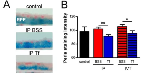
3- Iron removal in a mouse model of iron retinal overload. In AMD, iron accumulates in RPE, drusen
and outer segments of PR (8). We used mice knock-out for the Bone Morphogenetic Protein 6 (Bmp6)
gene, a model of hemochromatosis to validate the effect of Tf in reducing iron deposits in the retina.
By binding with its specific receptor, BMP6 protein activates transcription of the hepcidin gene. This
peptidic hormone blocks iron cellular exportation by activating ferroportin proteolysis. In Bmp6 KO
mice eyes, we detected iron deposits in the RPE at 22 weeks and also in inner segments of PR in older
mice (data not shown). Bmp6 KO mice develop severe retinal degeneration by 41 weeks (41). We used
32 week-old mice already presenting iron overload in the RPE to test the efficacy of iron-removal by
Tf (Figure 11A-B). We used two different protocols of Tf adminstration: 3 intraperitoneal injections
per weeks over a one-month period, or a single IVT injection 7 days before eyes collection. As
observed in Figure 11A, iron deposits on the basolateral side of RPE were partially removed with
systemic infusion. Both treatment protocols significantly reduced iron deposits in the RPE by nearly
10% (IP BSS: 102.1 ±2.6; IP Tf: 91.47±2.35; IVT BSS: 105.5 ±3; IVT Tf: 97.71 ±3.74) (Figure11B).
DISCUSSION
In the present study, we investigated whether intravitreal injection of the endogenous iron chelator,
transferrin, is efficient to prevent and protect PR against degeneration in several animal models.
The pathogenic role of iron is recognized in neurogenerative retinal diseases and the use of iron
chelators (deferoxamine, deferiprone or deferasirox) has been already evaluated (42-45). Oral
deferiprone has been the most studied drug in different murine models of retinal degeneration
(hereditary, light- or iron-induced) and had a protective effect on PR (19-20, 46-47). However, in the
absence of iron overload, systemic iron chelation therapy raises safety issues including chelator
overdose toxicity, iron and other essential metal deficiencies as well as diseases associated with such
metabolic imbalance. Ocular toxicity including cataract, maculopathy, and optic neuritis has been
reported in patients treated with deferoxamine or deferasirox during an extended period (48-50).
Aside from the synthetic (as deferiprone or deferasirox) and phytochemical chelators (curcumin,
R-lipoic acid), Tf is an endogenous and potent iron chelator locally produced by ocular cells. Its main
property is to control iron content in order to avoid any free labile iron, known to be a source of
oxidative stress and cell death. Systemic administration of Tf exerted strong neuroprotective effects on
rodent models of retinal dystrophies (14), positioning Tf supplementation as a therapeutic option for
patients with retinal degeneration. For clinical use, parenteral administration is not adapted,
particularly if long-term treatment is envisaged. We therefore conducted this extensive study to
explore and better understand the effects of Tf administered locally through an intravitreous route.
When injected into the vitreous, Tf rapidly penetrated into the retina through Müller glial cell end feet
at the vitreous border. The presence of Tf in choroidal capillaries within a few hours after its
intravitreal injection suggests that Tf follows a transretinal elimination route through Tf-receptors
localized on vascular endothelial cell. Indeed, we detected Tf in plasma already 6 hours and up to 7
days after IVT injections, demonstrating a quick turn-over of Tf in the retina. This led us to inject Tf
just before or just after a 24 hours continuous light exposure.
In acute models of light-induced retinal degeneration, PR death occurred within one day and was
associated to retinal inflammation and iron homeostasis remodeling (14, 32, 35). The local
(Figure 3C, 4C). Electroretinograms recording allowed us to confirmed the functionality of PR cells
with 50% of rod-driven electrical responses and an improved cone response time, indicating
preservation of cone and rod responses and synaptic connections with retinal neurons (Figure 4D-F).
These protective effects of IVT of Tf were associated with a decrease in apoptotic cells (Figure 4A)
and a reduction of oxidative stress-induced heme oxygenase expression (Figure 6).
Pre-treatment with Tf also protected against the early iron imbalance induced by light exposure, as
detected on iron-related genes expression 1 day, and on iron retinal tissue staining 3 days after light
exposure (Figure 5). Indeed, the retinal expression of iron-related proteins regulated through the
IRP/IRE system, dependent on the intracellular iron status, was modified rapidly during light exposure
(25). Here, we propose that pre-treatment with Tf avoids iron retinal overload through the
down-regulation of TfR1, Fpn, ferritins, and Dmt1 mRNA. Similarly, oral deferiprone and deferoxamine
regulated Hmox1, TfR1 and ferritin expression in models of retinal degeneration (19-21, 46-47). Tf
exerted a much stronger regulation of other iron-related gene expressions, Tf, Cp, HFE, and HJV,
which do not depend on IRP/IRE systems. Hepcidin is a peptidic hormone synthesized in the retina
that enhances ferroportin proteolysis under iron excess and inflammation, decreasing iron released
(51-52). Its transcription is controlled by several signaling pathways, including Hemojuvelin
(HJV)/BMP-6, IL6 and TfR2/HFE (53-54). Previously, Hadziahmetovic M. et al. demonstrated that
IVT injection of Tf rapidly activated ERK1/2 signaling pathways through Tf/TfR2/HFE binding,
which reduced iron export (55). One day after light exposure, Hfe, TfR2 and Hjv expression were
down-regulated in the retina. This may decrease Hepcidin transcription and enhance iron release and
PR death. The IVT injection of Tf prevented the light-induced Hfe down-regulation, which potentially
enhanced the TfR2-HFE-Hepcidin pathway, and thus decreased iron exportation from cells. The role
of TfR2 in iron metabolism is not fully understood, and its intracellular regulation, known to be
independent of IRP/IRE system is not well characterized (56). In the retina, TfR2 is expressed in all
nuclear layers, Tf injected co-localized with TfR2 in IPL, ONL and inner segments (Figure 1D-E).
Recently, Wysokinski et al. showed an association between a polymorphisms of the TfR2 gene and the
occurrence of AMD in obese patients (57). And Tf neuroprotective effect has also been demonstrated
exerts a complex and interesting regulation of several proteins involved in retinal iron trafficking,
resulting in a further reduction of iron-mediated oxidative toxicity.
Another mechanism of Tf neuroprotective effects on light-induced retinal degeneration is
related to the regulation of retinal inflammation. Studies already demonstrated that Tf modulated the
cytokines profile in animal models of inflammation (61-62) and reduced the ageing-associated
immunologic decline in mice (63). Tf was also shown to be efficient in an ocular model of intraocular
inflammation (64). In our model, the intense cold white LED exposure induced an early retinal
expression of genes involved in MC/macrophages recruitment (day 1) (Ccl2, Ccl17, Ccl12, Ccr2,
Ccr4, and Il1a). This was followed by the massive infiltration of activated Iba1+ cells in the outer
retina at 3 days (65-66) and the possible recruitment of circulating monocytes attracted by molecules
up-regulated in the illuminated retina (Csf1, CD14, Interleukins, Toll-like receptors group, Cxcl
chemokines and Cxcr receptors family genes) (67). Tf treatment induced a reduction of CD68
intensity staining in Iba1 positive cells (Figure 7F) suggesting deactivation of MC/macrophages in the
retina, known to be neurotoxic for PR and RPE cells (68-70). Tf also reduced retinal inflammation by
the up-regulation of the anti-inflammatory cytokines Il1rn and Il9 (71) and the down-regulation of
pro-inflammatory cytokines Ccl1, Il1rap, Il22, Il23r (72-73). This supports an additional neuroprotective
mechanism of Tf in relation with inflammatory responses. In this experiment, the modulation of
cytokines profile expression paralleled the iron-related genes modification and the exact relation
between these two mechanisms remains to be studied.
There are a number of considerations to use local delivery of iron chelator in a context of
retinal disorders such as dry AMD. Beyond retinal protection, PR preservation in deleterious condition
such as iron overload and chronic PR loss needed to be investigated. In this study, we showed that Tf
not only protected the retina prior to light-exposure but was still partially effective after light exposure.
Mechanisms could include reduction of reactive oxygen species production, iron accumulation, and
lipids peroxydation. The therapeutic potential of Tf is also suggested by its ability to “clear” the outer
retina and RPE from accumulated iron (Figure 11). Thus Tf could be adminstered not only for
retinal degeneration diseases are chronic diseases needing treatment over long period, a non-viral gene
transfer technique was used to produce Tf locally in a sustained manner. The ciliary muscle
electrotranfer of a plasmid encoding Tf not only showed a significant effect preventing PR
degeneration in the acute light-induced retinal degeneration model (Figure 10 C-D) but also
demonstrated a protective effect in a genetic model of slow retinal degeneration, the P23H rat (Figure
10 E-F). This indicates that the slow and continuous intraocular release of Tf may very likely be
beneficial to treat progressive retinal degeneration diseases.
CONCLUSION
Many strategies are being developed to delay retinal degeneration and proving their efficacy in
clinical trials is a real challenge. Whatever the cause of retinal degeneration, oxidative stress is
involved in PR death and, in this context, iron neutralization is one of the strategies present in all
neurodegenerative diseases. However, due to the redox activity of iron, it is important to consider
whether iron chelators can provide the cellular specificity required to remove excess of iron from the
appropriate tissue, without affecting systemic iron homeostasis. Here, we demonstrated that the local
administration of Tf, the endogenous iron chelator naturally present in the eye, responded to this
criteria. We showed that Tf protects PR against intense cell-death and preserves retinal functions
through several mechanisms: oxidative stress, iron homeostasis balance, inflammation control and
reduction of apoptosis. Therefore, we propose local ocular Tf administration as a high benefit/risk
ratio therapeutic candidate to be evaluated to limit PR cell death in patients and with further potential
applications including degenerative retinal diseases such as dry AMD.
METHODS Animals
Animals were fed with a standard laboratory diet and ad libitum tap water in a temperature-controlled
room at 21-23°C. The cyclic light environment consisted of 12 hours light per day (6am-6pm).
Animals were sacrificed by carbon dioxide inhalation. All experimental procedures in rats were
Paris Descartes. Experiments were performed in accordance with the Association for Research in
Vision and Ophthalmology (ARVO) statement for the use of animals in Ophthalmic and Vision
Research.
Photic injury
During the 3 weeks prior to light exposure, to reduce PR sensibility variability between rats, Wistar
rats (7 to 10-months old) (Janvier laboratory, Le Genest St Isle, France) were adapted in ventilated
cages at the bottom rows of the rack, where the level of light was controlled below 250 lux (74).
The day before light exposure, rats were dark-adapted 18 hours from 6 pm. The next day, pupils were
dilated with 1% atropine (Alcon, Norvartis, Rueil Malmaison, Fr) under dim light, and rats were
isolated in separate cages containing enough food for one day. Then light-exposure was performed
with commercial cold white LED panel generating 2,300 lumens during 24 hours. The LED panel was
placed above 8 transparent cages, placed on white surfaces, leaving enough space for air circulation
and constant temperature maintenance at 23°C. The luminance measured at the rats’ eyes position was
6,500 lux (Photometre DT-8809A, CEM, China). After light exposure, rats were replaced under cyclic
light (12h/day, 250 lux) for 1, 3, 8 or 16 days. Control animals were also dark adapted, and then
returned to rearing cyclic light conditions.
Rats were randomly separated into 3 groups (n= 4-5 rats per group): a control group not exposed to
light and untreated (NLE), a group treated with intravitreous injection of sterile saline buffer (BSS,
Bausch and Lomb, Montpellier, Fr) and exposed to light (BLE), and a group treated with an
intravitreous injection of human Tf solution and exposed to light (TfLE).
Intravitreal injections were performed after dark adaptation period under dim red light, or after light
exposure under normal light condition. Lyophilized apotransferrin (iron free transferrin (Tf)) isolated
from human plasma was dissolved in sterile BSS solution at a concentration of 48mg/ml (T5391,
Sigma Aldrich Chemical Co, Saint-Quentin en Yvelines, Fr). Rats were anesthetized using
intramuscular injection of 35:25 mix of ketamine (Virbac, Carros, Fr) and xylazine (Bayer, Lyon, Fr)
(1ml/100mg) and intravitreal injections (5µl,) were performed in inferior quadrant of both rat eyes
efficient to protect photoreceptors (data not shown). Rats were sacrificed at different time points after
light exposure. For in vivo analysis and histology, rats were examined or sacrificed 8 or 16 days after
light exposure. Additional time points were used for immunohistochemistry and RT-qPCR at 1, 3 or 8
days after light exposure.
Kinetic of transferrin eye distribution after intravitreal injection
At 6, 24 hours and 7 days after intravitreous injection of apotransferrin (5µl at 48mg/ml in BSS;
240µg), rats were sacrificed and eyes were enucleated, rinsed in 0.9% NaCl and dried. Ocular media
and tissues were separated: aqueous humor and vitreous, cornea, iris/ ciliary body, neural retina, and
RPE/choroid/sclera (eyecup). Retina and eyecups were analyzed individually whilst vitreous and
aqueous humor from same eyes were pooled. Tissues were incubated in lysis buffer [15 mM Tris, pH
7.9, 60 mM KCl, 15 mM NaCl, 2 mM EDTA, 0.4 mM phenylmethylsulphonyl fluoride (PMSF)
(Perbio Science, Brebiers, Fr)]. After four freeze/thaw cycles, lysates were centrifuged at 5,000g for
10 min and supernatants were stored at –20°C. Ocular fluids were diluted to a sufficient volume for
assay. Human Tf was quantified by an antibody-sandwich ELISA as described (14).
Immunohistochemistry
Freshly enucleated eyes (n=3-4 per time point) (superior pole tagged with suture) were fixed for 2
hours with 4% paraformaldehyde (PAF, Inland Europe, Conflans sur Lanterne, Fr) in 1X
phosphate-buffered saline (PBS, Gibco distributed by Life Technologies), washed with PBS, infiltrated in
gradients sucrose series and then, mounted in Tissue Tek O.C.T. (Siemens Medical, Puteaux, Fr).
Immunohistochemistry was performed on 10µm-thick sections as previously described (14, 25).
Cryosections were incubated with different primary antibodies: rabbit polyclonal anti-Tf receptor 1
(Serotec, Oxford, UK), rabbit polyclonal specific for the High and Light subunits of Ferritin (P.
Santambrogio); rabbit cellular retinaldehyde binding protein (CRALBP; J. Saari); rabbit
anti-RPE65 (AbCys, Courtaboeuf, Fr); rabbit anti-ionized calcium-binding adapter molecule 1 (Iba1, Wako
Pure Chemical Industries, Neuss, Germany); mouse anti-CD68 (Bio-Rad AbD Serotec GmbH,
were respectively labeled with anti-rhodopsin (Rho4D2, R.S. Molday) and peanut agglutinin
conjugated with fluorescein isothiocyanate (Sigma). Control sections were incubated with rabbit
non-immune serum (Invitrogen, Cergy Pontoise, Fr) or without primary antibodies. The corresponding
Alexa–conjugated secondary antibodies (Invitrogen) were used to reveal the primary antibodies, and
sections were counterstained with 4.6-diamidino-2-phenylindole (DAPI; Sigma). The sections were
viewed with a fluorescence microscope (BX51, Olympus, Rungis, Fr) or confocal microscope (LSM
510 laser scanning microscope Zeiss, Carl Zeiss, Le Pecq, Fr) and photographed using identical
exposure parameters for all samples to be compared.
Quantification of cones number 7 days after the end of light-exposure was realized throughout the
superior pole of retinal sections and reported to control rats as 100%. Microglia cells activation 3 days
after end of light-exposed was determined in outer plexiform layer, outer nuclear layer and segments
layers, identified after DAPI nuclei stain. Measurement of microglia/macrophages staining was
performed on pictures (60X) taken in the overall retina with same exposure time. The rapport of CD68
intensity staining to total microglia cells revealed by Iba1 intensity staining was measured with ImageJ
software.
Histology and outer nuclear layer thickness measurement
Oriented ocular globes werefixed with 4% PAF, 0.5% glutaraldehyde (LADD, Inland Europe, Fr) in
PBS for 2 hours. After fixation, samples were washed, dehydratedand transferred into the infiltration
solution of the Leica Historesinembedding kit (Leica, Nanterre, France) over night at 4°C. Samples
were embedded in resin (Leica) and 5 µm thick sections passing through the optic nerve head were
prepared along the superior and inferior pole of the eye using a microtome (Leica), stained with 1%
Toluidin Blue solution. Sections were observed on a Leitz microscope and photographed with a Leica
camera. Thicknesses of outer nuclear layer (ONL) were measured every 500 µm using Visilog 5.3
software (Noesis, Courtabouef, Fr). Histological sections measurements were performed across the
whole retina, considering the inferior pole to 0 from -4,000µm of optic nerve and superior pole to 0
from 4,000µm of optic nerve. Thickness profiles along the retina were generated by averaging, for
Optical Coherence Tomography
For in vivo analysis, rats were anesthetized, pupils were dilated and pictures of retinas of both rat eyes
were performed at superior pole using spectral domain OCT (SD-OCT; Spectralis device; Heidelberg)
adapted for small animal eyes.Each volume scan consisted of at least 100 B-Scans recorded at 30°
field of view centered on the superior pole, which were used to measure retinal thickness across the
scanned retinal area.
Electroretinography
Full-field ERG responses were recorded 16 days after the end of light exposure. Rats were
dark-adapted over a period of 18 hours – for scotopic recordings – and anesthetized by an intramuscular
injection of a mixture of ketamine and xylazine. The cornea was desensitized with a drop of
oxybuprocaine (Novesine© Novartis Ophthalmics, Basel, Switzerland) and the pupils were dilated
with a drop of tropicamide (Tropicamide©, Novartis Ophthalmics). Gold wire ring electrodes were
placed on the corneas of both eyes and stainless steel needle electrodes inserted into the forehead
served as working electrodes and references electrodes, respectively. A needle was subcutaneously
inserted at the base of the animal tail for grounding. All these manipulations were performed under
dim red light, without bringing the animal into ambient light after dark adaptation. Measurements were
performed simultaneously in both rat eyes using the commercial Ganzfeld VisioSystem device (Siem
Biomedicale, Nîmes, Fr). For scotopic electroretinograms in the dark-adapted state, flash intensities
ranged from 0.0003 to 10 cd.s/m2. Five flashes per stimulation intensity were applied at a 0.5
Hz-frequency, and corresponding responses were averaged. Flash duration was 10ms (–30 to 0 dB) except
for 10 cd.s/m2 (0 dB) it was 30ms. Following the scotopic recordings, a rod-suppressing background
light of 25 cd.s/m2 was turned on for 10 minutes. A cone-stimulating light flash was then applied, the
light intensity being 10 cd.s/m2 (flash duration 79 ms), for photopic ERGs recording. Mixed (rods +
cones) ERG measurements were performed simultaneously on both eyes, with flash intensity of 3
cd.s/m2, flash duration of 40 ms, and the amplifier set to 0.5 Hz. Five responses were averaged.
trough, and b-wave amplitudes (positive waves) were measured from the bottom of the a-wave trough
to the peak of the b-wave. Implicit times of the a- and b- waves were measured from time of stimulus
to peaks. Results were expressed in microvolts (µV) for amplitudes and milliseconds (ms) for implicit
times. The data obtained from each eye belonging to the same experimental group were averaged.
RT-PCR Inflammation-focused genes array and data analysis
Neural retinas were isolated on ice and directly frozen until RNA isolation. Total RNA was isolated
with RNeasy mini kit (Qiagen, Courtaboeuf, Fr) according to the manufacturer’s protocols. RNA
concentration, purity, and integrity were determined with an Agilent Bio-analyzer. All RNA used had
RNA integrity number superior to 8. First-strand cDNA was generated by reverse transcription using
0.7µg total RNA and the RT2 First Strand Kit (Qiagen). Genomic DNA was removed on RNeasy
columns and before reverse transcription according to the manufacturer’s protocols. Expression of 84
inflammatory-related genes and 5 housekeeping genes were evaluated in a 96-well plate (including
five housekeeping and seven control genes) by the Rat Inflammatory-Autoimmunity RT2 Profiler
PCR Array (PARN-077Z, SABiosciences, Qiagen). Gene arrays were processed according to the
manufacturer’s instructions. Each PCR reaction was performed following manufacturer’s instructions,
and dissociation curves were performed to control product of amplification. The threshold for
calculating cycle threshold (Ct) values was calculated automatically using RQ software
(SABiosciences). A fixed threshold was assigned manually, as suggested by the manufacturer. The
relative expression of each gene was calculated using the ∆∆Ct method. Ct values were analyzed with
Web-Based PCR Array Data Analysis tools: (http://www.sabiosciences.com/pcr/arrayanalysis.php,
SABiosciences) to determine the best housekeeping gene (Rplp1), the fold change and p value of each
gene (t-test Student). Fold changes ≥ ±2-fold were defined as biologically relevant changes.
In vivo ciliary muscle electroporation of a plasmid encoding transferrin in rats
Plasmids construction
Human Tf cDNA was extracted by NotI digestion from pCMV6-hTf plasmid (Origene, Rockville,
Technologies), downstream of a cytomegalovirus-β promoter, to give pVAX1-Tf. pVAX1 empty was
used as a negative control in all experiments.All plasmids were amplified in Escherichia coli bacteria
and endotoxin-free prepared (EndoFree Plamid Kit; Qiagen, Courtaboeuf, France). Plasmids were
diluted in endotoxin-free water containing 77 mM of NaCl (half saline) (saline, NaCl 0.9%, Versol, Laboratoire Aguettant, Lyon, Fr) as previously described (37). The concentration of DNA was
determined by spectroscopy measurements (optical density at 260 nm).
In vivo electroporation of the rat ciliary muscle
Wistar adult males rats came from Janvier Laboratory. P23H-line 1 rats (40) were kindly provided by
Mathiew Lavail (UCSF, Scool of Medecine, Beckman Vision Center), breeding at homozygous.
Wistar rats (Janvier laboratory) were used as control. Rats were anesthetized as described, before
ciliary muscle electrotransfer (ET). Thirty micrograms of plasmid (pVAX1, pVAX1-Tf), in a total
volume of 10 µl, were injected in the ciliary muscle using a 30-gauge disposable needle (BD
Micro-Fine syringe, NM Médical, Asnière, Fr) transsclerally posterior to the limbus then electric pulses were
delivered as described (37). Adult Wistar rats were sacrificed for ELISA Tf quantification in ocular
media and tissues 3 days after ET and for retinal histology analysis 12 days after ET. For light
experiment, ET were realized 3 days before light protocol realized as described above, and rats were
sacrificed 8 days later. P23H rats were submitted to ET at 4 weeks of age (an age preceding major
onset of PR loss in dystrophic rats), and killed at 8 weeks of age. All eyes were managed as described
previously for histological analysis. ONL thickness were measured every 500µm of each pole the
retina and values were averaged. Mean of ONL thickness at superior pole was representative of both
poles and represented on figures.
Statistical analysis
Results are presented as mean ± Standard error on the mean (SEM). Analysis were performed using
GraphPad Prism 5 software. Normal distribution of data was checked by the Shapiro–Wilk test.
Comparisons between 2 groups were analyzed by unpaired two-tailed Student's t-test, and multiples
comparisons by one-way ANOVA followed by Bonferroni post-test as appropriate. Non‐normally
distributed were analysed using nonparametric Kruskal–Wallis test. p<0.05 was considered
Acknowledgements
We thank Dr. Torriglia and Dr. de Kozack (UMRS1138, Centre de Recherche des Cordeliers,
INSERM) for their guidance and stimulating discussion. We thank Dr. Reed and Mélanie Glaettli for
the english correction and the staff at the animal facility at Centre de Recherche des Cordeliers. The
authors wish to thank Paolo Santambrogio (Department Biological and Technological Research,
Instituto de Ricovero e Cure a Carattere Scientifico, San Rafaelle, Milan, Italy) for rabbit polyclonal
specific for the High and Light subunits of Ferritin; John Saari (University of Washington, Seattle,
USA) for rabbit anti-cellular retinaldehyde binding protein and Robert S. Molday (University of
British Columbia, Vancouver, Canada) for anti-rhodopsin antibody. This study was supported by
INSERM, ANR Emergence 2012 (R11086DD), Retina France and Fondation de France-Fondation de
References
1. Andersen HH, Johnsen KB, and Moos T. Iron deposits in the chronically inflamed central nervous system and contributes to neurodegeneration. Cell Mol Life Sci. 2014;71(9):1607-22. 2. Wong RW, Richa DC, Hahn P, Green WR, and Dunaief JL. Iron toxicity as a potential factor in
AMD. Retina. 2007;27(8):997-1003.
3. Kell DB. Iron behaving badly: inappropriate iron chelation as a major contributor to the aetiology of vascular and other progressive inflammatory and degenerative diseases. BMC Med Genomics. 2009;2(2.
4. Moiseyev G, Chen Y, Takahashi Y, Wu BX, and Ma JX. RPE65 is the isomerohydrolase in the retinoid visual cycle. Proc Natl Acad Sci U S A. 2005;102(35):12413-8.
5. Shichi H. Microsomal electron transfer system of bovine retinal pigment epithelium. Exp Eye Res. 1969;8(1):60-8.
6. Gnana-Prakasam JP, Martin PM, Smith SB, and Ganapathy V. Expression and function of iron-regulatory proteins in retina. IUBMB Life. 2010;62(5):363-70.
7. Song D, and Dunaief JL. Retinal iron homeostasis in health and disease. Front Aging Neurosci. 2013;5(24.
8. Hahn P, Ying GS, Beard J, and Dunaief JL. Iron levels in human retina: sex difference and increase with age. Neuroreport. 2006;17(17):1803-6.
9. Ciudin A, Hernandez C, and Simo R. Iron overload in diabetic retinopathy: a cause or a consequence of impaired mechanisms? Exp Diabetes Res. 2010;2010(
10. Hahn P, Milam AH, and Dunaief JL. Maculas affected by age-related macular degeneration contain increased chelatable iron in the retinal pigment epithelium and Bruch's membrane. Arch Ophthalmol. 2003;121(8):1099-105.
11. Junemann AG, Stopa P, Michalke B, Chaudhri A, Reulbach U, Huchzermeyer C, Schlotzer-Schrehardt U, Kruse FE, Zrenner E, and Rejdak R. Levels of aqueous humor trace elements in patients with non-exsudative age-related macular degeneration: a case-control study. PLoS One. 2013;8(2):e56734.
12. Rogers BS, Symons RC, Komeima K, Shen J, Xiao W, Swaim ME, Gong YY, Kachi S, and Campochiaro PA. Differential sensitivity of cones to iron-mediated oxidative damage. Invest Ophthalmol Vis Sci. 2007;48(1):438-45.
13. Yefimova MG, Jeanny JC, Keller N, Sergeant C, Guillonneau X, Beaumont C, and Courtois Y. Impaired retinal iron homeostasis associated with defective phagocytosis in Royal College of Surgeons rats. Invest Ophthalmol Vis Sci. 2002;43(2):537-45.
14. Picard E, Jonet L, Sergeant C, Vesvres MH, Behar-Cohen F, Courtois Y, and Jeanny JC. Overexpressed or intraperitoneally injected human transferrin prevents photoreceptor degeneration in rd10 mice. Mol Vis. 2010;16(2612-25.
15. Deleon E, Lederman M, Berenstein E, Meir T, Chevion M, and Chowers I. Alteration in iron metabolism during retinal degeneration in rd10 mouse. Invest Ophthalmol Vis Sci. 2009;50(3):1360-5.
16. Boddaert N, Le Quan Sang KH, Rotig A, Leroy-Willig A, Gallet S, Brunelle F, Sidi D, Thalabard JC, Munnich A, and Cabantchik ZI. Selective iron chelation in Friedreich ataxia: biologic and clinical implications. Blood. 2007;110(1):401-8.
17. Devos D, Moreau C, Devedjian JC, Kluza J, Petrault M, Laloux C, Jonneaux A, Ryckewaert G, Garcon G, Rouaix N, et al. Targeting chelatable iron as a therapeutic modality in Parkinson's disease. Antioxid Redox Signal. 2014;21(2):195-210.
18. Hadziahmetovic M, Pajic M, Grieco S, Song Y, Song D, Li Y, Cwanger A, Iacovelli J, Chu S, Ying GS, et al. The Oral Iron Chelator Deferiprone Protects Against Retinal Degeneration Induced through Diverse Mechanisms. Transl Vis Sci Technol. 2012;1(3):2.
19. Hadziahmetovic M, Pajic M, Grieco S, Song Y, Song D, Li Y, Cwanger A, Iacovelli J, Chu S, Ying GS, et al. The Oral Iron Chelator Deferiprone Protects Against Retinal Degeneration Induced through Diverse Mechanisms. Transl Vis Sci Technol. 2012;1(2):7.
20. Hadziahmetovic M, Song Y, Wolkow N, Iacovelli J, Grieco S, Lee J, Lyubarsky A, Pratico D, Connelly J, Spino M, et al. The oral iron chelator deferiprone protects against iron overload-induced retinal degeneration. Invest Ophthalmol Vis Sci. 2011;52(2):959-68.
21. Obolensky A, Berenshtein E, Lederman M, Bulvik B, Alper-Pinus R, Yaul R, Deleon E, Chowers I, Chevion M, and Banin E. Zinc-desferrioxamine attenuates retinal degeneration in the rd10 mouse model of retinitis pigmentosa. Free Radic Biol Med. 2011;51(8):1482-91.
22. Baath JS, Lam WC, Kirby M, and Chun A. Deferoxamine-related ocular toxicity: incidence and outcome in a pediatric population. Retina. 2008;28(6):894-9.
23. Wong A, Alder V, Robertson D, Papadimitriou J, Maserei J, Berdoukas V, Kontoghiorghes G, Taylor E, and Baker E. Liver iron depletion and toxicity of the iron chelator deferiprone (L1, CP20) in the guinea pig. Biometals. 1997;10(4):247-56.
24. Picard E, Fontaine I, Jonet L, Guillou F, Behar-Cohen F, Courtois Y, and Jeanny JC. The protective role of transferrin in Muller glial cells after iron-induced toxicity. Mol Vis. 2008;14(928-41.
25. Picard E, Ranchon-Cole I, Jonet L, Beaumont C, Behar-Cohen F, Courtois Y, and Jeanny JC. Light-induced retinal degeneration correlates with changes in iron metabolism gene expression, ferritin level, and aging. Invest Ophthalmol Vis Sci. 2011;52(3):1261-74.
26. Yefimova MG, Jeanny JC, Guillonneau X, Keller N, Nguyen-Legros J, Sergeant C, Guillou F, and Courtois Y. Iron, ferritin, transferrin, and transferrin receptor in the adult rat retina. Invest Ophthalmol Vis Sci. 2000;41(8):2343-51.
27. Noell WK, Walker VS, Kang BS, and Berman S. Retinal damage by light in rats. Invest Ophthalmol. 1966;5(5):450-73.
28. Marc RE, Jones BW, Watt CB, Vazquez-Chona F, Vaughan DK, and Organisciak DT. Extreme retinal remodeling triggered by light damage: implications for age related macular degeneration. Mol Vis. 2008;14(782-806.
29. Martin PM, Gnana-Prakasam JP, Roon P, Smith RG, Smith SB, and Ganapathy V. Expression and polarized localization of the hemochromatosis gene product HFE in retinal pigment epithelium. Invest Ophthalmol Vis Sci. 2006;47(10):4238-44.
30. Margrain TH, Boulton M, Marshall J, and Sliney DH. Do blue light filters confer protection against age-related macular degeneration? Prog Retin Eye Res. 2004;23(5):523-31.
31. Wielgus AR, Collier RJ, Martin E, Lih FB, Tomer KB, Chignell CF, and Roberts JE. Blue light induced A2E oxidation in rat eyes--experimental animal model of dry AMD. Photochem Photobiol Sci. 2010;9(11):1505-12.
32. Hadziahmetovic M, Kumar U, Song Y, Grieco S, Song D, Li Y, Tobias JW, and Dunaief JL. Microarray analysis of murine retinal light damage reveals changes in iron regulatory, complement, and antioxidant genes in the neurosensory retina and isolated RPE. Invest Ophthalmol Vis Sci. 2012;53(9):5231-41.
33. Hentze MW, Muckenthaler MU, Galy B, and Camaschella C. Two to tango: regulation of Mammalian iron metabolism. Cell. 2010;142(1):24-38.
34. Rutar M, Provis JM, and Valter K. Brief exposure to damaging light causes focal recruitment of macrophages, and long-term destabilization of photoreceptors in the albino rat retina. Curr Eye Res. 2010;35(7):631-43.
35. Santos AM, Martin-Oliva D, Ferrer-Martin RM, Tassi M, Calvente R, Sierra A, Carrasco MC, Marin-Teva JL, Navascues J, and Cuadros MA. Microglial response to light-induced photoreceptor degeneration in the mouse retina. J Comp Neurol. 2010;518(4):477-92. 36. Organisciak DT, and Vaughan DK. Retinal light damage: mechanisms and protection. Prog
Retin Eye Res. 2010;29(2):113-34.
37. Touchard E, Kowalczuk L, Bloquel C, Naud MC, Bigey P, and Behar-Cohen F. The ciliary smooth muscle electrotransfer: basic principles and potential for sustained intraocular production of therapeutic proteins. J Gene Med. 2010;12(11):904-19.