HAL Id: hal-03092511
https://hal.archives-ouvertes.fr/hal-03092511
Submitted on 5 Jan 2021HAL is a multi-disciplinary open access archive for the deposit and dissemination of sci-entific research documents, whether they are pub-lished or not. The documents may come from teaching and research institutions in France or abroad, or from public or private research centers.
L’archive ouverte pluridisciplinaire HAL, est destinée au dépôt et à la diffusion de documents scientifiques de niveau recherche, publiés ou non, émanant des établissements d’enseignement et de recherche français ou étrangers, des laboratoires publics ou privés.
Study of fibronectin adsorption on plasma-enhanced
deposition of silver nanoparticles embedded in a silica
matrix
Laurine Martocq, P Chevallier, M Laurent, G Laroche, K Makasheva
To cite this version:
Laurine Martocq, P Chevallier, M Laurent, G Laroche, K Makasheva. Study of fibronectin adsorption on plasma-enhanced deposition of silver nanoparticles embedded in a silica matrix. 35th Annual Meeting of the Canadian Biomaterials Society, May 2019, Quebec city, Canada. �hal-03092511�
35th Annual Meeting of the Canadian Biomaterials Society 21-24 May 2019
Study of fibronectin adsorption on plasma-enhanced deposition of silver
nanoparticles embedded in a silica matrix
L Martocq
1-2, P Chevallier
1, M Laurent
1, G Laroche
1, K Makasheva
21
Laboratoire d’Ingénierie de Surface, Département de génie des mines, de la métallurgie et des
matériaux, Université Laval, Québec, QC, Canada
2
LAPLACE, Université de Toulouse, CNRS, UPS, INPT, Toulouse, France
INTRODUCTION: Every year, 1 out of 9 patients in Canada hospitals gets a potentially fatal healthcare-associated infection (HCAI), which makes it the 4th leading cause of death in the
country. The development of HCAIs are mainly due to the material’s surface colonization by microorganisms. Adherent bacteria organize themselves to form a biofilm, once the extracellular polymeric matrix made of proteins, polysaccharides and lipids is formed [1]. The biofilm structure renders it highly resistant to existing antibacterial agents and accordingly, there is a growing need to develop new strategies to inhibit this bacterial structuration. This project takes advantage of the known potential of silver nanoparticles (AgNPs) to deliver Ag+ ions to kill bacteria [2]. Protection of the
AgNPs from oxidation and homogeneous NPs distribution were ensured through a coating with a plasma-deposited silica matrix [3,4]. This study will focus on the adsorption of fibronectin (FN) on plasma-deposited silica (SiO2) surfaces containing
AgNPs, as protein adsorption is the first step toward bacterial film formation.
METHODS: A customized axially-asymmetric radiofrequency plasma reactor was used to deposit a SiO2 matrix and AgNPs on silicon wafers. The
matrix was obtained using a gas mixture of hexamethyldisiloxane (HMDSO), oxygen and argon, while the AgNPs were synthesized by sputtering of the upper silver electrode in an argon plasma [3,4]. A droplet of 10 µL of human FN solution in HEPES buffer at two different concentrations, 6 and 200 µg/mL, was deposited on the plasma-deposited SiO2 surfaces. The adsorption
lasted for 1h and was followed by 3 successive washings in nanopure water with vortex and drying. AFM, XPS, FTIR analyses and ELISA tests were used to characterize the FN surface density and conformation.
RESULTS: XPS spectra clearly put in evidence the FN adsorption, thanks to the presence of the N1s feature, characteristic of proteins, while no N1s peak was detected on the plasma-deposited SiO2
matrix (data not shown). The FN adsorption is further confirmed through the decrease of Si surface
concentration with the amount of adsorbed FN (Table 1).
Table 1. Elemental composition obtained by XPS
Elemental composition (%)
Sample O Si C N
FN [6 µg/mL]/SiO2/Si 50.6 25.0 22.0 2.4
FN [200 µg/mL]/SiO2/Si 33.1 14.0 44.0 9.0
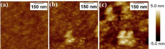
AFM images confirmed the presence of FN as round objects on the investigated surfaces. Besides, a higher FN density is observed when using the 200 g/mL FN solution compared to the one at 6 g/mL (Fig. 1).
Fig. 1: AFM images (700 nm x 700 nm) of (a) SiO2/Si, (b) FN [6 µg/mL]/SiO2/Si and (c) FN
[200 µg/mL]/SiO2/Si.
DISCUSSION & CONCLUSIONS: The FN presence after adsorption was confirmed by XPS and AFM analyses. In addition, the rather circular shape of the protein indicates that FN had a low interaction with the surface and accordingly, adopted a globular conformation. The FN conformation will be further analyzed by ImageJ analyses. In addition, FTIR analyses and ELISA tests will be performed. Finally, FN density, conformation, and bioactivity will be assessed and compared with AgNPs based surfaces in order to evaluate the FN adsorption behaviors on these antimicrobial surfaces.
REFERENCES: 1M. Habash, and G. Reid (1999), J. Clin. Pharmacol., 39:887-98. 2 M. Rai, et al.
(2008) Biotechnol. Adv., 27:76-83. 3 A. Pugliara, et al. (2016) Sci. Total Environ., 565:863-871. 4 A.
Scarangella, et al. (2019) Nanotechnology, 30:165101.