HAL Id: hal-01137420
https://hal.archives-ouvertes.fr/hal-01137420
Submitted on 30 Mar 2015HAL is a multi-disciplinary open access
archive for the deposit and dissemination of sci-entific research documents, whether they are pub-lished or not. The documents may come from teaching and research institutions in France or abroad, or from public or private research centers.
L’archive ouverte pluridisciplinaire HAL, est destinée au dépôt et à la diffusion de documents scientifiques de niveau recherche, publiés ou non, émanant des établissements d’enseignement et de recherche français ou étrangers, des laboratoires publics ou privés.
Binding modes of thrombin binding aptamers
investigated by simulations and experiments
Ana Trapaidze, Aurélien Bancaud, Marie Brut
To cite this version:
Ana Trapaidze, Aurélien Bancaud, Marie Brut. Binding modes of thrombin binding aptamers investi-gated by simulations and experiments. Applied Physics Letters, American Institute of Physics, 2015, 106 (4), pp.043702. �10.1063/1.4906594�. �hal-01137420�
Binding modes of thrombin binding aptamers investigated by simulations
and experiments
A. Trapaidze1,2, A. Bancaud1,2, M. Brut1,3,a)
1CNRS, LAAS, 7 avenue du colonel Roche, F-31400 Toulouse, France 2Université de Toulouse, LAAS, F-31400 Toulouse, France
3Université de Toulouse, UPS, LAAS, F-31400 Toulouse, France
a) Author to whom correspondence should be addressed. Electronic mail: [email protected].
ABSTRACT. Thrombin binding aptamers HD1 and HD22 are the most studied aptamers, both
for therapeutic and sensing purposes. Yet there is still no commercialized aptamer-based sensor device for thrombin detection, suggesting that the binding modes of these aptamers remain to be precisely described. Here we investigate thrombin-aptamer interactions with molecular dy-namics simulations, and show that the different solved structures of HD1-thrombin complex are energetically similar and consequently possibly co-existing. Conversely HD22 folding is much more stable and its binding energy with thrombin is significantly largerthan that of HD1 com-plexes. These results are confronted to experiments, which consist in monitoring aggregation of aptamer-functionalized gold nanoparticles triggered by thrombin. HD1 alone, but not HD22, can trigger aggregation, meaning that this aptamer has multiple sites of interactions with thrombin. Furthermore, pre-incubation of HD22 with thrombin impedes HD1 aggregation, sug-gesting that HD1 and HD22 have competing affinities for the same binding site. Altogether this study shows that thecharacterization of aptamer-thrombin interactionsby structural and kinetic experiments joined to simulations is necessary for the development of biosensors.
Aptamers are single-stranded nucleic acids able to bind to a variety of targets with high speci-ficity and affinity1-3. Their selection is achieved through the SELEX protocol, which consists in consecutivecycles of selection and amplification, starting from a large library of oligonuc-leotides (1013–1018 sequences) 4. Due to their attractive properties in term of ease of synthesis, low-cost, chemical stability, and high affinity and specificity for a broad range of targets, ap-tamers have attracted much attention, particularly as potential therapeutic inhibitors and sens-ing elements for a future generation of biosensors5. Thrombin binding aptamers (TBA) HD1 and HD22 have been the most studied aptamers for biosensing, representing ~20% of the 5000 papers published about the use of aptamers for analytical technologies 6.Surprisingly there is little consensus on their binding properties in the literature. A rapid survey of Surface Plasmon Resonance (SPR) data shows that the reaction constant of HD1 and HD22 spans ~2
decades from 6 nM7 to 170 nM 8 and from 2 nM 9 to 110 nM8 for HD1 and HD22, respective-ly. Efforts to characterize the interaction of aptamers with thrombin are thus needed to im-prove their performances as sensing layers in a device, and definitively establish aptamer-based technologies.
HD1, the 15-mer DNA (5’-GGTTGGTGTGGTTGG-3’) TBA that binds the fibrinogen site, also known as exosite I, was the first to be described in 1992 10 and structurally characterized in 1993 11. HD1 was shown to adopt a G-quadruplex structure with one TGT and two TT loops (see Fig. 1). The loops have been shown to bind thrombin in different ways, according to X-ray and NMR models respectively 12, andthe determination of multiple crystal structures of the thrombin – HD1 complex has left a doubt on the existence of a unique complex vs.several co-existing states 13. On the other hand, only one structure of thrombin-HD22 com-plex has been reported in the literature. This 29-mer (5’-AGTCCGTGGTAGGGCAGGTTGGGGTGACT-3’), which presents a duplex-quadruplex folding, binds to the heparin site, known as exosite II, with a higher affinity 14 than HD1.
In this study, we considered all TBA structures available in the Protein Data Bank (PDB), namely four HD1- and one HD22- thrombin structures (Fig. 1). Our goal was to compare their energy in their equilibrium configuration, as well as their binding energy to thrombin. We first focused on the two HD1 low-resolution structures (2.80Å) solved in 1996 12 with NMR-based and X-ray-NMR-based models (PDB code: 1HAO and 1HAP respectively). In 1HAO, G8 and T9 nucleic bases from the TGT loop are stacked on the G-quadruplex while T7 is rejected outside the loop. Interaction with thrombin is established through both TT loops. In 1HAP, the same configuration of the TGT loop can be observed but it is directly interacting with thrombin. Two higher resolution X-ray structures of HD1 (1.80Å) solved more recently 15 in the presence of Na+ and K+ (PDB code: 4DIH and 4DII respectively) were also examined. 4DIH and 4DII chains follow the same orientation as 1HAO, and interaction with thrombin occurs through the TT loops. However the TGT loop undergoes a different configuration be-cause T7 and G8 bases are now stacked on the G-quadruplex whereas T9 is rejected outside. The orientation of HD1 on thrombin is rotated by 180° around the helix axis (Fig.1).The other meaningful difference comes from the cation position, which is inserted between the G-quartets anddrastically increases the inter-strand distance from ~12 Å in 1HAO and 1HAP to ~21 Å in 4DIH and 4DII. More details about interaction modes have already been described
15,16
. Concerning HD22, the X-ray structure recently solved by Krauss et al. with a 2.80 Å resolution was used in the calculations (PDB code: 4I7Y) 14.
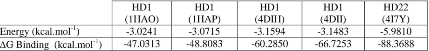
All calculations were performed with Amber 10 suite of programs 17 and the parm99/bsc0 force field 18. As a first step, TBAs were submitted to molecular mechanics energy minimiza-tion. Simulations were performed in implicit solvent (Generalized Born approach 19), and us-ing the sander module of Amber, with 200000 steps of conjugate-gradient minimization with-out restraints and a cutoff of 12 Å. Resulting energies are presented in TableI. Despite changes in folding associated to the lower compaction of4DII and 4DIH vs. 1HAO and 1HAP, the four HD1 energies are similar, spanning -3.15 to -3.02 kcal.mol-1. It is therefore impossible to ensure that one HD1 conformation prevails over the other ones. Moreover, the energy of HD22 is -5.98 kcal.mol-1, i.e. twice as large as that of HD1. This result is expected due tothe existence of a longer G-quadruplex motif in comparison to HD1, plus four base-pair duplex. For this reason, HD22 is less likely to undergo conformational changes; however, there is no other PDB data to support this hypothesis.
As a second step, the binding energy of TBA with thrombin was estimated with molecular mechanics Poisson-Boltzmann surface area (MM-PBSA) method. This method combines mo-lecular mechanics with continuum solvation models to exploit multiple snapshots extracted from molecular dynamics trajectories. It presents the advantage to be faster than other usual techniques 20,21. Each aptamer-thrombin complex was solvated in a cubic box of water mole-cules extending at least 10Å outside the structures (TIP3P model 22). We first equilibrated the solvated complexes with a short 2000 steps of conjugate-gradient minimization run, followed by 50 ps of heating up to 300K with MD, 50 ps of density equilibration with weak restraints (2 kcal.mol-1.Å-2) and 500 ps of constant pressure equilibration with no restraint. All simula-tions were run using a cutoff of 8Å. The equilibration and subsequent production were run with Langevin dynamics for temperature control, and the shake algorithm to impose con-straints on bond lengths involving hydrogen atoms, allowing for a 2 fs time step. The produc-tion runs were performed during 10 ns of fully unrestrained MD and the collected results for each run, recorded every 5 ps, were used to estimate the aptamer binding energy. The result-ing estimated bindresult-ing energies are reported in Table I. The bindresult-ing energies of 1HAO and 1HAP complexes is very similar (-47.03 and -48.81 kcal.mol-1, respectively), but approx-imately twice lower than that of HD22-thrombin complex(-88.37kcal.mol-1). However, in presence of cations and more specificially of K+, HD1-thrombin complexes gains in stability
with binding energies of -60.29 and -66.73 kcal.mol-1.Because monovalent cations are ubi-quitously used in molecular biology assays, HD1 is unlikely to be found unbound to cations. Therefore4DIH and 4DII complexes are expected to prevail over 1HAO and 1HAP, and at least two states likely co-exist in experiments. At this step we wished to test this idea further with simple experiments.
We thus carried out experiments involvingTBA-capped gold nanoparticlesof 80 nm(AuNPs). We monitored the dynamics of aggregation, which occurs whenever thrombinmediates the formation of bonds between two functionalized AuNPs23,24. Aggregation kinetics was moni-tored by dynamic light scattering (DLS), which provides measurements of average hydrody-namic radius over time.In a typical experiment, TBA-AuNPs diluted in 100µL PBS buffer at the final concentration 0.1 OD at 540 nmwere covered with 0.85nmol ofthiol modified TBA(Eurogentec). The initial size of the NP was measured as the control assuring that there was no aggregation without thrombin (Sigma-Aldrich), which was then introduced ata con-centration of 50 nM. Thrombin injection into HD1-AuNP and HD22-AuNP mixture led to rapid aggregation,as expected and shown in many other experiments(Fig. 2). Nextthe same experiment carried out with only one TBA on AuNPs showed rapid aggregation for HD1 and a slight onset in average diameter of 3±0.5 nm for HD22, which is consistent with the binding of thrombin to NPs.This result therefore shows the existence of multiple binding sites for HD1, in agreement with earlier reports on the weak association of HD1 to thrombin exosite II12,25.Further complementing experiments were conducted to clarify whether the second bind-ing site for HD1 was indeed exosite II. We reasoned that the specificity of HD22 bindbind-ing to thrombin exosite II could be used to block the access to this site by pre-incubation of throm-bin withan excess of HD22for 20 min.This HD22-thromthrom-bin complex was theninjected into the HD1-AuNP solution, and aggregation did not take place. Rather the average diameterin-creased by 15±2 nm, in agreement with the formation of ternary complex between HD1-AuNP, thrombin and HD22. Altogether our results indicate that HD1 has binding sites with thrombin towards exosite I and also at the vicinity of exosite II. We made a final round of experiments to estimate the binding energy of HD1 to these two sites. HD1-AuNP was incu-bated with excess of single-stranded DNA complementary to HD1 (ssHD1’), and this mixture was then injectedto a solution of HD22-AuNP and thrombin or only thrombin. Aggregation occurred normally in the latter case, but not in the former one (not shown).The successful aggregation suggests that HD1 binding to exosite 1 is favored energetically over HD1-HD1’ pairing, whereas thrombin could not displace HD1’ for binding HD1 to exosite II leading to a
constant hydrodynamic radius. Given the energy of HD1-HD1’ pairing of -29.35 kcal.mol-1 (OligoAnalyzer IDT), our simulations are consistent with these estimates of HD1 binding to exosite 1,since all calculated binding energies range from -66.73 to -47.03kcal.mol-1. Moreo-ver the low energy of the secondary binding site of HD1 to exosite IIof less than 30 kcal.mol
-1
likely accounts the absence of crystals of this complex.
In this paper, a theoretical study of all different thrombin-aptamer complexes available in the PDB is conducted to investigate the possible binding modes of HD1 and HD22 to thrombin. Simulations show that HD1 has multiple modes of interaction tothrombin exosite I, which possibly co-exist, and HD22 has higher affinity for exosite II. Experiments confirm that the binding landscape of HD1 to thrombin is not limited to exosite I, and suggest a secondary low-affinity interaction to the vicinity of exosite II. These observations call into question the relevance of HD1 for sensing thrombin in real conditions involving abundant proteins with residual non specific interactions. Consequentlythe selection of aptamers through the SELEX protocol is not sufficient to guarantee the successful design of biosensing systems. This result is consonant with our previous observation that the attachment of an aptamer to a surface was interfering with its folding and potentially with its interaction properties 26. Altogether, we argue that structural data, molecular modeling, and kinetic experiments are required to collect the full benefits of aptamers in technological developments.
REFERENCES 1
K-M. Song, S. Lee and C. Ban. Sensors 12, 612 (2012).
2
A. D. Ellington and J. W. Szostack, Nature 346, 818 (1990).
3
C. Tuerk and L. Gold, Science 249, 505 (1990).
4
J. L. Boots, K. Matylla-Kulinska, M. Zywicki, B. Zimmermann, and R. Schroeder. Genomic SELEX. Handbook of RNA Biochemistry: Second, Completely Revised and Enlarged Edition, 1185-1206 (2014).
5
M. Mascini, I. Palchetti and S. Tombelli. Angew. Chem. Int. Ed. 51, 1316 (2012).
6
B. Deng, Y. Lin, C. Wang, F. Li, Z. Wang, H. Zhang, X.-F. Li and X. C. Le. Anal. Chim. Acta 837, 1 (2014).
7
S. Davis. Biacore J.1, 29 (2014).
8
P.-H. Lin, R.-H. Chen, C.-H. Lee, Y. Chang, C.-H. Chen and W.-Y. Chen.Colloids Surf. B Biointer-faces 88, 552 (2011).
9
J. Müller, D. Freitag, G. Mayer and B. Pötzsch, B.J. Thromb. Haemost. JTH 6, 2105 (2008).
10
L. C. Bock, L. C. Griffin, J. A. Latham, E. H. Vermaas, and J. J. Toole. Nature 356, 564 (1992).
11
R. F. Macaya, P. Schultze, F. W. Smith, J. A. Roe, and J. Feignon, Proc. Natl. Acad. Sci. USA 90, 3745 (1993).
12
K. Padmanabhan and A. Tulinsky, Acta Cryst. D 52, 272 (1996).
13
I. Russo Krauss, A. Merlino, C. Giancola, A. Randazzo, L. Mazzarella and F. Sica. Nucleic Acids Res. 39, 7858 (2011).
14
I. Russo Krauss, A. Pica, Merlino, A. Randazzo, E. Novellino, L. Mazzarella and F. Sica. Acta Cryst. D69, 2403 (2013).
15
I. Russo Krauss, A. Merlino, A. Randazzo, E. Novellino, L. Mazzarella and F. Sica. Nucleic Acids Res. 40, 8119 (2012).
16
A. Pica, I. Russo Krauss, A. Merlino, S. Nagatoishi, N. Sugimoto and F. Sica. FEBS Journal 280, 6581 (2013).
17
D.A. Case, T.A. Darden, T.E. Cheatham, C.L. Simmerling, J. Wang, R.E. Duke, R. Luo, R.C. Walker, W. Zhang, K.M. Merz, B. Wang, S. Hayik, A. Roitberg, G. Seabra, I. Kolossvary, K.F. Wong, F. Paesani, J. Vanicek and L. Jian. Eur. Phys. J. E 35, 75 (2012).
18
A. Perez, I. Marchan, D. Svozil, J. Sponer, T.E. Cheatham, C.A. Laughton and M. Orozco. Biophys. J. 92, 3817 (2007).
19
C.S. Rapp, and R.A. Friesner. Proteins 35, 173 (1999).
20
P.A. Kollman, I. Massova, C. Reyes, B. Kuhn, S. Huo, L. Chong, M. Lee, T. Lee, Y. Duan, W. Wang, O. Donini, P. Cieplak, J. Srinivasan, D.A. Case and T.E. Cheatham. Acc. Chem. Res. 33, 889 (2000).
21
22
W.L. Jorgensen, J. Chandrasekhar, J.D. Madura, R.W. Impey and M.L. Klein. J. Chem. Phys. 79, 926 (1983).
23
V. Pavlov, Y. Xiao, B. Shlyahovsky and I. Willner. J.A.C.S. 126, 11768 (2004).
24
C.-C. Huang, Y.-F. Huang, Z. Cao, W. Tan and H.-T. Chang. Anal. Chem. 77, 5735 (2005).
25
B. Pagano, L. Martino, A. Randazzo and C. Giancola. Biophys. J. 94, 562 (2008).
26
M. Brut, A. Trapaidze, A. Estève, A. Bancaud, D. Estève, G. Landa, M. Djafari Rouhani. Appl. Phys. Lett. 100, 163702 (2012).
ACKNOWLEDGMENTS
We thank the CALMIP Supercomputer Center for CPU resources.
SUPPLEMENTARY MATERIAL
See supplemental material at [URL will be inserted by AIP] for more information about the aggrega-tion assays described in this letter.
Figure 1
FIG. 1. Molecular structures of four HD1-thrombin complexes (1HAO, 1HAP, 4DIH, 4DII) and one HD22-thrombin complex (4I7Y) used in the calculations. Thrombin is colored in gray and aptamers in spectrum colors from blue to red to indicate the sequence orientation (5’ to 3’).
Figure 2
FIG. 2. Aggregation assay of 80 nm AuNPs coated with HD1 or HD22 aptamers. Initial time corres-ponds to the addition of 50 nM thrombin in each vial. Aggregation is monitored by dynamic light scat-tering, which yields the average hydrodynamic radius over time.
500 400 300 200 100 H y d ro d y n a m ic r a d iu s ( n m ) 50 0 Time (min) AuNP-HD1 / AuNP-HD22 + Thr 50 nM AuNP-HD1 / AuNP-HD1 + Thr 50 nM AuNP-HD1-HD1' / AuNP-HD22 + Thr 50 nM AuNP-HD1 / AuNP-HD1 + Thr 50 nM-HD22 AuNP-HD1-HD1' / AuNP-HD1 HD1' + Thr 50 nM AuNP-HD22 / AuNP-HD22 + Thr 50 nM
Table 1 HD1 (1HAO) HD1 (1HAP) HD1 (4DIH) HD1 (4DII) HD22 (4I7Y) Energy (kcal.mol-1) -3.0241 -3.0715 -3.1594 -3.1483 -5.9810 G Binding (kcal.mol-1) -47.0313 -48.8083 -60.2850 -66.7253 -88.3688 Table I. Minimized energy of five aptamer structures extracted from the PDB, calculated with Amber 10 package, with the parm99/bsc0 force field and in implicit solvent. Binding energies of TBAs to thrombin are estimated with the MM-PBSA method.