HAL Id: hal-01789352
https://hal.archives-ouvertes.fr/hal-01789352
Submitted on 10 May 2018
HAL is a multi-disciplinary open access
archive for the deposit and dissemination of
sci-entific research documents, whether they are
pub-lished or not. The documents may come from
teaching and research institutions in France or
abroad, or from public or private research centers.
L’archive ouverte pluridisciplinaire HAL, est
destinée au dépôt et à la diffusion de documents
scientifiques de niveau recherche, publiés ou non,
émanant des établissements d’enseignement et de
recherche français ou étrangers, des laboratoires
publics ou privés.
Motuporamine Derivatives as Antimicrobial Agents and
Antibiotic Enhancers against Resistant Gram-Negative
Bacteria
Diane Borselli, Marine Blanchet, Jean-Michel Bolla, Aaron Muth, Kristen
Skruber, Otto Phanstiel, Jean Brunel
To cite this version:
Diane Borselli, Marine Blanchet, Jean-Michel Bolla, Aaron Muth, Kristen Skruber, et al..
Motuporamine Derivatives as Antimicrobial Agents and Antibiotic Enhancers against Resistant
Gram-Negative Bacteria.
ChemBioChem, Wiley-VCH Verlag, 2017, 18 (3), pp.276 - 283.
Motuporamine Derivatives as Antimicrobial Agents and
Antibiotic Enhancers against Resistant Gram-Negative
Bacteria
Diane Borselli,
[a]Marine Blanchet,
[b]Jean-Michel Bolla,
[a]Aaron Muth,
[c]Kristen Skruber,
[c]Otto Phanstiel IV,*
[c]and Jean Michel Brunel*
[b]Introduction
Antimicrobial resistance threatens the prevention and treat-ment of an ever-increasing range of infections caused by bac-teria, parasites, viruses, and fungi. An increasing number of governments around the world are devoting efforts to this problem, which is so serious that it threatens the achievements of modern medicine. Far from being an apocalyptic fantasy, a post-antibiotic era in which common infections and minor inju-ries can kill is a real possibility for the 21st century. A recent WHO report makes a clear case that resistance to common bacteria has reached alarming levels in many parts of the world, and that in some settings few, if any, of the available treatment options remain effective for common infections. Another important finding of the report is that surveillance of antibacterial resistance is neither coordinated nor harmonized and there are many gaps in information regarding bacteria of major public health importance.[1]
The intensive use of antibiotics for the treatment of numer-ous bacterial infections is one of the biggest healthcare
advan-ces in modern times. Nevertheless, their widespread use has led to an increasing number of antibiotic-resistant bacteria.[2]
In particular, the emergence of Gram-negative multidrug-resist-ant (MDR) bacteria, such as Pseudomonas aeruginosa and Kleb-siella pneumoniae, has prompted efforts to develop new classes of antibiotics and chemosensitizers (molecules to pro-mote an increase in the internal antibiotic concentration in resistant strains). Thus, diseases caused by MDR Gram-negative bacteria are increasing worldwide,[3,4] and the emergence of
pan drug-resistant (PDR) bacteria (resistant to all classes of an-tibiotics and to quaternary ammonium disinfectants)[5]appears
to have reached a point of no return.[6,7] We have noticed
great concern in the medical community, as numerous recent clinical reports have confirmed that Gram-negative bacteria have developed resistance to polymyxins, the last efficient therapy against PDR Gram-negative bacteria.[8–10]
An appealing target is the unique structure of the bacterial membrane, which is highly conserved among most species of Gram-negative bacteria, and forms an effective barrier to many types of antibiotics.[11]Indeed, the acquisition of resistance to
membrane-active antibiotics has likely required major changes in membrane structure. Ironically, modifications to the bacterial membrane to escape membrane-targeting antibiotics might in-crease the permeability of the barrier and actually inin-crease the susceptibility of the bacteria to hydrophobic antibiotics.
It is well established that most immune responses to Gram-negative bacteria involve recognition of lipopolysaccharides (LPS) and their lipid A anchors, which constitute the major components of the outer membrane.[12–17] The permeability
barrier of the outer membrane is due to the cross-bridging electrostatic interactions between lipid A molecules and diva-lent cations such as calcium or magnesium.[12] We speculated
that cationic peptides[18]and polyamines[19]could out-compete
these divalent cations for their membrane binding sites and disrupt the outer membrane organization, thereby increasing Dihydromotuporamine C and its derivatives were evaluated for
their in vitro antimicrobial activities and antibiotic enhance-ment properties against Gram-negative bacteria and clinical isolates. The mechanism of action of one of these derivatives, MOTU-N44, was investigated against Enterobacter aerogenes by using fluorescent dyes to evaluate outer-membrane
depolari-zation and permeabilidepolari-zation. Its efficiency correlated with in-hibition of dye transport, thus suggesting that these molecules inhibit drug transporters by de-energization of the efflux pump rather than by direct interaction of the molecule with the pump. This suggests that depowering the efflux pump provides another strategy to address antibiotic resistance.
[a] D. Borselli, J.-M. Bolla Aix–Marseille Universit8, IRBA TMCD2 UMR-MD1, Facult8 de M8decine
27 Boulevard Jean Moulin, 13385 Marseille Cedex 05 (France) [b] M. Blanchet, Dr. J. M. Brunel
Centre de Recherche en Canc8rologie de Marseille (CRCM) CNRS, UMR7258, Institut Paoli Calmettes
Aix–Marseille Universit8, UM 105, Inserm, U1068
27 Boulevard Jean Moulin, 13385 Marseille Cedex 05 (France) E-mail: bruneljm@yahoo.fr
[c] A. Muth, K. Skruber, Dr. O. Phanstiel IV
Department of Medical Education, University of Central Florida 12722 Research Parkway, Orlando, FL 32826-3227 (USA) E-mail: otto.phanstiel@ucf.edu
T 2017 The Authors. Published by Wiley-VCH Verlag GmbH & Co. KGaA. This is an open access article under the terms of the Creative Commons At-tribution-NonCommercial-NoDerivs License, which permits use and distribu-tion in any medium, provided the original work is properly cited, the use is non-commercial and no modifications or adaptations are made.
permeability. Because of the promising applications of poly-amine derivatives in medicine,[20–22]we evaluated a series of
hy-drophobic polyamine derivatives for their ability to target the membrane stability of Gram-negative bacteria and increase the sensitivity of these bacteria to known antibiotics.
The motuporamines (originally isolated from the marine sponge Xestospongia exigua)[23] were selected because their
amphiphilic architectures comprise a large hydrophobic macro-cycle with an appended polyamine motif (1–3, Scheme 1). A series of motuporamine derivatives (4–6) was prepared[24, 25]
along with a series of related anthracenyl-polyamine deriva-tives (7a–d). These amphiphilic polyamines have large hydro-phobic substituents to facilitate interaction with the bacterial membrane.
Here, 4–6 and 7a–d were screened for their in vitro antimi-crobial activities and antibiotic-enhancement properties against resistant Gram-negative bacteria. We also explored the mechanism of action of this class of derivatives against Entero-bacter aerogenes (EA289) by using fluorescent dyes, in order to evaluate changes in outer-membrane depolarization and per-meabilization.
Results and Discussion
Our investigations began with the determination of the mini-mum inhibitory concentrations (MICs) of 4–7 in Gram-positive and -negative species, in order to identify the concentrations that produce a direct antibacterial effect and allowed us to rank their relative potencies. We included two Gram-negative bacteria encountered in hospitals, P. aeruginosa and Klebsiella pneumonia, and multidrug-resistant E. aerogenes EA289
(Table 1). Several compounds showed MICs of 100–200 mm for these bacterial strains. The anthracenyl compounds 7a–d had relatively weak antimicrobial activities, whereas their related motuporamine derivatives 4a–b, 5a–b, and 6a–b showed MICs of 1.56–50 mm. Specifically, 6a (MOTU-CH2-33) and 6b
(MOTU-CH2-44) exhibited excellent antimicrobial activities
against many species, including the multidrug-resistant E. aero-genes EA289.
As stated previously, the development of chemo-sensitizing agents, which enhance the intracellular antibiotic concentra-tion in resistant strains (or by other mechanisms) is an attrac-tive approach to overcome bacterial resistance. Thus, we inves-tigated the use of these polyamine derivatives as adjuvants in combination with antibiotics. Success here would provide an exciting approach to increase the potency of current antibacte-rial drugs, even for strains that have developed resistance.
We investigated whether these polyamine agents could re-store the potency of the antibiotic doxycycline at significantly below its MIC. For example, in our hands the MIC of doxycy-cline against P. aeruginosa PAO1 was 16 mgmL@1, so we
investi-gated the use of doxycycline at a significantly lower concentra-tion (2 mgmL@1, corresponding to its pharmacokinetic
proper-ties in humans)[6]in the presence of the polyamine derivatives.
We speculated that the polyamine agents would disrupt bacte-rial membrane integrity and increase antibiotic delivery to the bacteria and thus increase doxocycline potency. Rewardingly, even at this low doxycycline concentration, eight of the poly-amine derivatives restored doxycycline activity against E. aero-genes EA289, P. aeruginosa PAO1, and K. pneumoniae KPC2-ST258; no improvement was observed for 7b (ANT4) or 7a (ANT-N-butyl) even at 40 mm (Table 2). The fact that this effect
Scheme 1. Motuporamine compounds 1–6, anthracenyl compounds 1–7 squalamine 8, and spermine 9.
ChemBioChem 2017, 18, 276 – 283 www.chembiochem.org 277 T 2017The Authors. Published by Wiley-VCH Verlag GmbH & Co. KGaA, Weinheim
was compound-specific was intriguing and ruled out a non-specific detergent effect, especially because no cell lysis was observed.
Several of the effective compounds also acted synergistically with chloramphenicol and erythromycin, particularly against PAO1, but weakly against EA289 and KPC2-ST258 (Table 3). Thus, we identified two groups of compounds: one (7a and 7b) displayed weak or no activity, and the second (e.g., 5a and 5b) increased the antibiotic susceptibility effectively against PAO1. Overall, 5a and 5b appeared the most promis-ing adjuvants for use with doxycycline; 5b (MOTU-N44) was chosen to investigate the mechanism of action of this molecu-lar class.
Within the motuporamine series (4–6) several compounds exhibited moderate to good antibacterial activity as well as potent synergy with different antibiotics against Gram-nega-tive bacteria. We explored the mechanism of action of these compounds and focused on two possible pathways: permeabi-lization and/or disruption of the outer membrane, and inhibi-tion of an efflux pump.
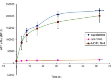
First, we determined the effect of 5b on Staphylococcus aureus ATCC25923 by measuring ATP release for 1 min: there was dramatic disruption of the bacterial membrane, similar to that by squalamine (positive control; Figure 1).[26] Conversely,
no significant effect was found for the polyamine spermine (negative control).
As we observed different compound performance in the assays with S. aureus in Table 1, we speculated that some of these molecules might achieve lethality by increasing the rate
of transport of molecules across the cytoplasmic membrane, whereas others might not. We surmised that compounds like 5b might induce a smaller membrane breach, modestly affect the permeability barrier of the cytoplasmic membrane and cause membrane depolarization. Indeed, a small breach would allow the passage of electric current (thereby causing mem-brane depolarization) without allowing the passage of larger molecules. This alternative mechanism seemed plausible be-cause depolarization would de-energize the efflux pump and also lead to increased potency of the antibiotic agent. There-fore, we investigated whether these molecules generated
Table 1. MIC of motuporamine derivatives against various bacterial strains.
Compound MIC [mm]
S. aureus S. intermedius E. faecalis E. coli P. aeruginosa E. aerogenes K. pneumoniae
ATCC25923 1051997 ATCC29212 ATCC28922 PAO1 EA289 KPC2-ST258
7b, ANT4 >200 200 >200 200 200 100 100 7c, ANT44 50 200 >200 50 100 200 >200 7d, ANT444 12.5 25 200 25 100 100 100 7a, ANT-N-butyl >200 200 >200 >200 >200 >200 >200 6a, MOTU-CH2-33 1.56 3.125 3.125 1.56 6.25 50 100 5a, MOTU-N33 3.125 1.56 12.5 3.125 12.5 100 100 6b, MOTU-CH2-44 1.56 1.56 3.125 1.56 12.5 50 100 4b, MOTU44 100 50 >200 100 100 50 100 4a, MOTU33 50 50 100 50 50 100 100 5b, MOTU-N44 1.56 1.56 6.25 6.25 25 50 50
Table 2. Concentration of motuporamine derivatives necessary to restore doxycycline activity (2 mgmL@1) against EA289, PAO1 and KPC2 ST258 Gram-negative bacterial strains.
Compound Concentration of motuporamine derivative [mm]
EA289 PAO1 KPC2 ST258 7c, ANT44 10 5 5 7d, ANT444 1.25 2.5 1.25 4a, MOTU33 2.5 1.25 1.25 4b, MOTU44 1.25 2.5 1.25 5a, MOTU-N33 2.5 2.5 2.5 5b, MOTU-N44 5 1.25 2.5 6a, MOTU-CH2-33 5 5 2.5 6b, MOTU-CH2-44 2.5 2.5 1.25 7b, ANT4 40 >40 40 7a, ANT-N-butyl >40 >40 >40
MICs of doxycycline against PAO1, EA289, KPC2ST258: 40 mgmL@1
(90 mm), 20 mgmL@1(45 mm), and 10 mgmL@1(22.5 mm), respectively.
Table 3. Concentration of the motuporamine derivative [mm] required to restore chloramphenicol, erythromycin, and cefepime activity (2 mgmL@1) against EA289, PAO1, and KPC2 ST258.
Compound PAO1 EA289 KPC2 ST258
CHL ERY FEP CHL ERY FEP CHL ERY FEP
4a, MOTU33 5 20 n.t. 40 40 40 40 >40 >40
4b, MOTU44 5 40 n.t. 40 >40 > 40 40 >40 >40
5a, MOTU-N33 2.5 10 n.t. 20 20 20 20 40 >40
5b, MOTU-N44 5 >40 n.t. >40 40 > 40 40 >40 40
6b, MOTU-CH2-44 2.5 10 n.t. 20 20 > 40 20 >40 >40
CHL: chloramphenicol, ERY: erythromycin, FEP: cefepime, n.t.: not tested. MIC of FEP against PAO1: 10 mgmL@1. All other antibiotic/strain combinations: >100 mgmL@1.
a smaller breach of the permeability barrier of the cytoplasmic membrane.
Fluorescent cyanine dyes are excellent probes to monitor membrane depolarization. These dyes lose fluorescence inten-sity when in polarized membranes and become highly fluores-cent once polarization is lost.[27]Thus, one can use changes in
dye fluorescence to monitor change in membrane polarization. Interestingly, strong depolarization of S. aureus membranes was observed after 21 minutes as a strong increase in relative fluorescent units (RFU) of the cyanine dye (Figure 2) in the presence of 5b. This suggests that 5b facilitated membrane depolarization.
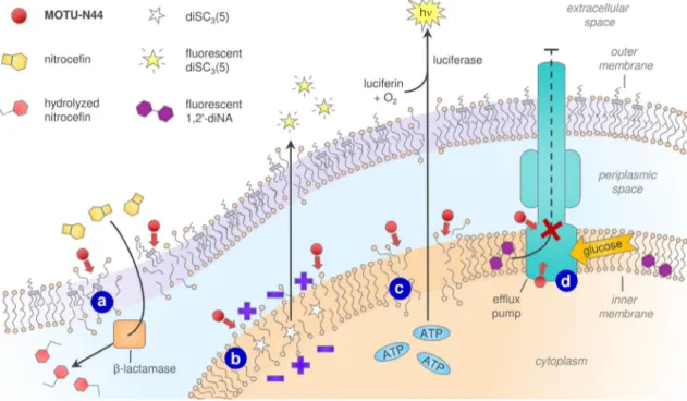
Next, 5b was investigated for its ability to alter the cell outer membrane integrity of E. aerogenes EA289, by using ni-trocefin, a chromogenic b-lactam that is efficiently hydrolyzed by periplasmic b-lactamases, thereby resulting in a significant color change from yellow to red.[28, 29] Thus, colorimetric
changes were used to monitor outer membrane integrity. Even
at a low concentration (3.9 mm), 5b increased the rate of nitro-cefin hydrolysis compared to the spermine-treated or untreat-ed control (Figure 3a). The behavior was similar to that of the positive control polymyxB (PMB) which also produced an in-crease in nitrocefin hydrolysis. All these data suggest that 5b is able to permeabilize or disrupt the outer membrane of Gram-negative bacteria as no cell lysis was observed.
The drug-resistant bacterium EA289 overexpresses the AcrAB-TolC pump,[30] which belongs to the RND efflux pumps
and uses the proton gradient across the inner membrane as an energy source. In order to determine if 5b could act as a disruptor of the transmembrane potential, we used the mem-brane-potential-sensitive probe DiSC3(5) which concentrates at
the inner membrane and self-quenches its fluorescence.[31]
When a compound impairs the membrane potential, the dye is released into the growth medium thus leading to a fluores-cence increase. Treatment with 5b resulted in dose-dependent depolarization after 10 min of incubation (Figure 3b), thus sug-gesting disruption of the proton gradient and an ability to affect efflux pumps from the RND family such as AcrAB-TolC.
A similar outcome was observed when using a biolumines-cence method to determine the release of intracellular ATP. Addition of 5b caused dose-dependent permeabilization (Fig-ure 3c). Interestingly, 10 mgmL@1 5b caused 11% ATP release
into the medium after a few seconds, thus suggesting rapid disruption.
In general, efflux systems employ an energy-dependent mechanism (active transport) to pump out unwanted substan-ces such as toxins, antibiotics, or dyes, through specific efflux pumps.[32] Some efflux systems are drug-specific, whereas
others eject multiple drugs, and thus contribute to MDR. Efflux pumps are proteinaceous transporters in the cytoplasmic membrane of bacteria and are active transporters; thus they require a source of chemical energy. Some are primary active transporters that use ATP hydrolysis as a source of energy, whereas in others (secondary active transporters) transport is coupled to an electrochemical potential difference created by pumping protons or sodium ions from or to the outside of the cell. The transport of a known transport substrate can be used to directly monitor the function of efflux pumps, and 5b was thus tested for its ability to inhibit efflux.
After loading EA289 bacteria with the dye 1,2’-dinaphthyla-mine (1,2’-diNA), which is a substrate of the AcrAB-TolC efflux pump,[33]the bacteria fluoresced. Bacteria were then incubated
with and without 5b at different concentrations before addi-tion of glucose as an energy source. In the absence of 5b, rapid active transport of more than 80 % of the dye was ob-served (Figure 3d, black line). When 5b was present, signifi-cant dose-dependent inhibition was observed (>80 % reten-tion at up to 25 mm 5b; Figure 3c, orange line). These results suggest that 5b inhibits the AcrAB-TolC efflux pump.
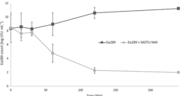
A time-kill assay (Figure 4) and a cell viability assay (Figure 5) were performed in order to evaluate the bactericidal or bacter-iostatic behavior of this compound. As shown in Figure 4, a time kill analysis was performed against the EA289 bacterial strain by using 5b at a four times the MIC: 99.9 % death (de-tection limit) occurred by 2 h.
Figure 1. The effect of squalamine (100 mg mL@1), spermine (100 mgmL@1), and 5b (MOTU-N44, 100 mgmL@1) on ATP release kinetics for Gram-positive bacteria S. aureus.
Figure 2. Depolarization of the bacterial membrane of S. aureus in the pres-ence of 2.6 and 5.2 mm squalamine, spermine, or 5b (MOTU-N44).
ChemBioChem 2017, 18, 276 – 283 www.chembiochem.org 279 T 2017The Authors. Published by Wiley-VCH Verlag GmbH & Co. KGaA, Weinheim
A cell viability assay (Figure 5) was performed by monitoring the irreversible reduction of blue resazurin to red resorufin by viable cells. This conversion is an oxidation–reduction
indica-tion in cell viability assays and can serve as an aerobic respira-tion measurement for bacteria.[34]When using 5b at four times
the MIC, there was clearly no cell viability.
Figure 3. MOTU-N44 (5b) has multiple effects on the cell membrane of the Gram-negative bacterium E. aerogenes EA289. a) Outer-membrane permeabiliza-tion detected by nitroceflin hydrolysis, in a dose-and time-dependent manner. b) Dose-dependent inner-membrane depolarizapermeabiliza-tion quantified by the release of DiSC3(5). c) Membrane disruption revelaed by APT efflux. d) Inhibition of glucose-triggered 1,2’-diNA release via effux pumps.
Thus, the time-kill experiment shows that 5b at four times MIC (200 mm) led to a decrease in live bacteria after 30 min. When the cells were incubated for 60 min at this concentra-tion, the cell viability assay demonstrated total inhibition of respiratory metabolism allowing us to conclude that this de-crease in bacterial count correlates highly with cell death.
The real-time assay demonstrated the ability of 5b to inhibit efflux transport to around 60% by using a sub-inhibitory con-centration (10 mm, MIC/4). The results from the time-kill assay allow us to state that the cells remain viable in the efflux assay conditions (, 30 min) and that the inhibition of the dye trans-port is a consequence of a specific action of the compound.
On the other hand, the nitrocefin hydrolysis and membrane depolarization assays suggest that efflux inhibition is probably due to disruption of membrane integrity thereby leading to proton-motive force dissipation. Indeed, the hydrolysis kinetics observed at a low concentration of 5b demonstrated a slight effect on the membrane, thus correlating with the results ob-tained for the depolarization assay. We noted that outer-mem-brane permeation increased with increasing 5b concentration, and this is likely responsible for cell death at high levels. We also note that the real-time assays required higher concentra-tions than those for fixed incubation times to generate a quan-tifiable signal.
Wang et al. recently described a similar action of the substi-tuted diamine, 1,13-bis (((2,2-diphenyl)-1-ethyl)thioureido)-4,10-diazatridecane.[35] This diamine compound was also shown to
depolarize the cytoplasmic membrane and provide enhanced permeabilization of the outer bacterial membrane. Further structure–activity relationship studies revealed that the central diamine nitrogens were key to bioactivity. In contrast to the N-substituted systems, the unN-substituted diamines (putrescine and cadaverine) had no antibacterial activity, did not affect membrane permeability, and did not cause membrane rupture. Both of the higher polyamines (spermidine and spermine) were found to be inactive against S. aureus RN4220, P. aerugi-nosa PAO1and E. coli ANSI. This, when coupled to our findings, suggests that either mono- or di-substituted polyamine sys-tems can serve as antibacterial agents, whereas the unsubsti-tuted native polyamine systems do not. Taken together, our studies also suggest that the presence of hydrophobic N-sub-stituents is key to the ability of these compounds to target bacterial membranes and elicit a bacteriocidal response.
Conclusion
Several polyamine derivatives were investigated for their intrin-sic antimicrobial activities against positive and Gram-negative bacteria. Derivatives 5a and 5b showed excellent ac-tivities (MIC 1.56–100 mm). In addition, 5b dramatically affected the antibiotic susceptibility of E. aerogenes, P. aeruginosa, and K. pneumoniae MDR strains. We conclude that changes in the transmembrane electrical potential in E. aerogenes EA289 corre-late with permeabilization of the cell membrane by motupora-mine derivatives, thereby leading to (or concomitantly facilitat-ing) an altered proton homeostasis. Finally, motuporamine derivatives such as 5b, that are able to disrupt the proton gra-dient, effectively de-energize the efflux pump and can be con-sidered as efflux-pump inhibitors.
Experimental Section
Bacterial strains: Eight bacterial strains (Institut Pasteur and per-sonal collection) were used in this study. Gram-positive bacteria (S. aureus ATCC25923, S. intermedius 1051997, Enterococcus faecalis ATCC29212) and Gram-negative bacteria (E. coli ATCC28922, P. aeru-ginosa PAO1, E. aerogenes EA289, a Kan derivative of the MDR clini-cal isolate Ea27,[30] and K. pneumoniae KPC2 ST258) were stored at
@808C in glycerol (15%, v/v). Bacteria were grown in Mueller– Hinton (MH) broth at 378C.
Antibiotics: All the antibiotics were purchased from Sigma–Aldrich except fordoxycycline, which was purchased from TCI Europe (Zwijndrecht, Belgium). All antibiotics were dissolved in water. The susceptibility of bacterial strains to antibiotics and compounds was determined in microplates by the standard broth dilution method, according to the recommendations of the Comit8 de l’Antibio-Gramme de la Soci8t8 FranÅaise de Microbiologie (CA-SFM).[36]
Briefly, MICs were determined with an inoculum of 105CFU in of
MH broth (200 mL) containing twofold serial dilutions of each drug. MIC was defined as the lowest concentration to completely inhibit growth after incubation for 18 h at 378C. Measurements were re-peated in triplicate.
Determination of antibiotic MIC in the presence of compounds: Briefly, restoring enhancer concentrations were determined with an inoculum of 5V105CFU in MH broth (200 mL) containing twofold Figure 4. Time-kill curves of 5b (MOTU-N44, 4VMIC) over 4 h against EA289
bacteria.
Figure 5. Cell viability of EA289 in the presence of 5b (MOTU-N44, 4VMIC).
ChemBioChem 2017, 18, 276 – 283 www.chembiochem.org 281 T 2017The Authors. Published by Wiley-VCH Verlag GmbH & Co. KGaA, Weinheim
serial dilutions of each derivative and antibiotic (chloramphenicol, doxycycline, cefepime, or erythromycin; 2 mgmL@1). The lowest
concentration of the polyamine adjuvant that completely inhibited growth after incubation for 18 h at 378C was determined. Measure-ments were repeated in triplicate.
Membrane depolarization assays: Bacteria were grown in MH broth for 24 h at 378C and centrifuged (3600g, 208C). The pellet was washed twice with buffered HEPES (pH 7.2) sucrose (250 mm) and magnesium sulfate (5 mm). The fluorescent dye 3,3’-diethyl-thiacarbocyanine iodide was added (3 mm) and allowed to pene-trate into bacterial membranes by incubation for 1 h of at 378C. Cells were then washed to remove the unbound dye before adding 5b at different concentrations. Fluorescence measurements were performed on a FluoroMax 3 spectrofluorometer (Horiba; slit widths 5/5 nm). The relative corrected fluorescence (RFU) was re-corded at 0, 3, 5, 7, 9, 11, 13, 15, 17, 19, and 21 min. Maximum RFU was that recorded with a pure solution of the fluorescent dye in buffer (3 mm).
Nitrocefin hydrolysis assay: Outer membrane permeabilization was measured by using nitrocefin, a chromogenic substrate of per-iplasmic b-lactamase. MH broth (10 mL) was inoculated with of an overnight culture (0.1 mL) of EA289 and grown at 378C to OD600=
0.5. The remaining steps were performed at room temperature. Cells were recovered by centrifugation (3600 g, 20 min) and washed once with potassium phosphate buffer (PPB; 20 mm, pH 7.2) containing MgCl2(1 mm). After another centrifugation, the
pellet was resuspended in PPB (100 mL) and adjusted to OD600=
0.5. Then, either Polymyxin B (positive control; Sigma–Aldrich) or 5b (50 mL) was added to the cell suspension (100 mL) to final con-centrations of 0.98–500 mm. Nitrocefin (50 mL, 50 mgmL@1; Oxoid)
was added, and its hydrolysis was monitored spectrophotometri-cally by measuring the increase in absorbance at 490 nm. Assays were performed in 96-well plates with an M200 Pro spectropho-tometer (Tecan).
Glucose-triggered 1,2’-diNA efflux assays: Bacteria were grown to stationary phase, collected by centrifugation, and resuspended to OD600=0.25 in PPB (20 mm, pH 7.2) supplemented with carbonyl
cyanide m-chlorophenyl hydrazone (CCCP, 5 mm; Sigma–Aldrich), and incubated overnight with 1,2’-dinaphthylamine (1,2’-diNA, 32 mm; Sigma–Aldrich) at 37 8C. Before addition of compound 5b (100 mm), the cells were washed with phosphate buffer. Glucose (50 mm) was added after 300 s to initiate bacterial energization. Re-lease of membrane-incorporated 1,2’-diNA was followed by moni-toring the fluorescence (lex=370 nm; lem=420 nm) every 30 s at
378C in an Infinite M200 Pro plate reader (Tecan). Assays were per-formed in 96-well plates (half area, black with solid bottom, 100 mL per well; Greiner Bio-One).
Measurement of ATP efflux: Squalamine were prepared in doubly distilled water at different concentrations. A suspension of growing S. aureus or E. aerogenes (EA289) in MH broth was incubated at 378C. The suspension (90 mL) was added to squalamine solution synthesized in our laboratory according reported procedures (10 mL), and the mixture was vortexed for 1 s. Luciferin–luciferase reagent (Yelen, France; 50 mL) was immediately added, and lumi-nescence was quantified with an Infinite M200 microplate reader (Tecan) for 5 s. ATP concentration was quantified by addition of a known amount of ATP (1 mm). A similar procedure was performed for spermine (100 mgmL@1) and for 5b (200 mm, i.e., 4VMIC).
Time-killing assay: Mid-log phase cultures of EA289 with an inocu-lum of 107CFUmL were incubated with 5 b (4VMIC, 200 mm) at
378C with 160 rpm shaking. Bacterial counts were performed after
0, 15, 30, 90, 120 and 240 min by spreading appropriate dilutions on MH agar plates (detection limit 102CFUmL@1). The plates were
incubated overnight at 378C before colonies were counted. The curves from two independent experiments were averaged and ex-pressed as logarithms (mean:SE).
Cell viability assay: An overnight culture of EA289 was diluted 100-fold into MHII broth. An inoculum of 107CFUmL@1was
incu-bated in the presence or absence of 5b (4VMIC, 200 mm) for 1 h at 378C with shaking at 160 rpm. The fluorescence of the cell suspen-sion was monitored after addition of CellTiter-Blue reagent (10%, v/v; Promega). Measurements were performed by using a 96-well Greiner film-bottom black microplate (Greiner Bio-One) and an Infinite M200 microplate reader (Tecan; lex=568 nm and lem=
660 nm). The curves from two independent experiments were combined (mean:SE).
Synthesis of compounds 4–7: The synthesis of 4–7 was previously reported.[15,37–41]
Keywords: antibiotics · antimicrobial agents · bacterial resistance · membranes · motuporamine · polyamine derivatives
[1] Antimicrobial Resistance: Global Report on Surveillance WHO, 2014, http://www.who.int/drugresistance/documents/surveillancereport/en/. [2] P. Fernandes, Nat. Biotechnol. 2006, 24, 1497 –1503.
[3] A. E. Pop-Vicas, E. M. C. D’Agata, Clin. Infect. Dis. 2005, 40, 1792 –1798. [4] J. M. Coelho, J. F. Turton, M. Kaufmann, J. Glover, N. Woodford, M. Warner, M.-F. Palepou, T. L. Pitt, B. C. Patel, D. M. Livermore, J. Clin. Mi-crobiol. 2006, 44, 3623 – 3627.
[5] M. C. Jennings, K. P. C. Minbiole, W. M. Wuest, ACS Infect. Dis. 2015, 1, 288– 303.
[6] C. Y. Wang, J. S. Jerng, K. Y. Chen, L. N. Lee, C. J. Yu, P. R. Hsueh, P. C. Yang, Clin. Microbiol. Infect. 2006, 12, 63– 68.
[7] S. D. Mentzelopoulos, M. Pratikaki, E. Platsouka, H. Kraniotaki, D. Zerva-kis, A. Koutsoukou, S. Nanas, O. Paniara, C. Roussos, E. Giamarellos-Bour-boulis, C. Routsi, S. G. Zakynthinos, Intensive Care Med. 2007, 33, 1524 – 1532.
[8] A. Antoniadou, F. Kontopidou, G. Poulakou, E. Koratzanis, I. Galani, E. Pa-padomichelakis, P. Kopterides, M. Souli, A. Armaganidis, H. Giamarellou, J. Antimicrob. Chemother. 2007, 59, 786– 790.
[9] M. E. Falagas, S. K. Kasiakou, Clin. Infect. Dis. 2005, 40, 1333 –1341. [10] S. Biswas, J.-M. Brunel, J.-C. Dubus, M. Reynaud-Gaubert, J.-M. Rolain,
Expert Rev. Anti-Infect. Ther. 2012, 10, 917 –934.
[11] H. Labischinski, G. Barnickel, H. Bradaczek, D. Naumann, E. T. Rietschel, P. Giesbrecht, J. Bacteriol. 1985, 162, 9– 20.
[12] M. Vaara, Microbiol. Rev. 1992, 56, 395– 411.
[13] H. Nikaido in Escherichia coli and Salmonella: Cellular and Molecular Biology (Eds.: F. C. Neidhardt, R. Curtis III, J. L. Ingraham), 1st ed., ASM Press, Washington, 1996, pp. 29–47.
[14] R. E. W. Hancock, Trends Microbiol. 1997, 5, 37–42.
[15] T. Murata, W. Tseng, T. Guina, S. I. Miller, H. Nikaido, J. Bacteriol. 2007, 189, 7213– 7222.
[16] M. Vaara, Antimicrob. Agents Chemother. 1993, 37, 354– 356. [17] H. Nikaido, Microbiol. Mol. Biol. Rev. 2003, 67, 593– 656. [18] M. Zasloff, Nature 2002, 415, 389– 395.
[19] L. Djouhri-Bouktab, J. M. Rolain, J. M. Brunel, Anti-Infect. Agents 2014, 12, 95– 103.
[20] M. Blanchet, D. Borselli, J. M. Brunel, Future Med. Chem. 2016, 8, 963 – 973.
[21] C. Pieri, D. Borselli, C. Di Giorgio, M. De M8o, J.-M. Bolla, N. Vidal, S. Combes, J. M. Brunel, J. Med. Chem. 2014, 57, 4263 –4272.
[22] J. M. Brunel, A. Lieutaud, V. Lome, J.-M. PagHs, J.-M. Bolla, Bioorg. Med. Chem. 2013, 21, 1174– 1179.
[23] D. E. Williams, P. Lassota, R. J. Andersen, J. Org. Chem. 1998, 63, 4838 – 4841.
[24] A. Muth, V. Pandey, N. Kaur, M. Wason, C. Baker, X. Han, T. R. Johnson, D. A. Altomare, O. Phanstiel IV, J. Med. Chem. 2014, 57, 4023 –4034. [25] A. Muth, M. Madan, J. J. Archer, N. Ocampo, L. Rodriguez, O. Phanstiel
IV, J. Med. Chem. 2014, 57, 348– 363.
[26] K. Alhanout, S. Malesinki, N. Vidal, V. Peyrot, J. M. Rolain, J. M. Brunel, J. Antimicrob. Chemother. 2010, 65, 1688– 1693.
[27] T. Wieder, P. J. Sims, J. Membr. Biol. 1985, 84, 249 –258.
[28] O. Lomovskaya, M. S. Warren, A. Lee, J. Galazzo, R. Fronko, M. Lee, J. Blais, D. Cho, S. Chamberland, T. Renau, R. Leger, S. Hecker, W. Watkins, K. Hoshino, H. Ishida, V. J. Lee, Antimicrob. Agents Chemother. 2001, 45, 105– 116.
[29] Y. Matsumoto, K. Hayama, S. Sakakihara, K. Nishino, H. Noji, R. Iino, A. Yamaguchi, PLoS One 2011, 6, e18547.
[30] M. Mallea, J. Chevalier, C. Bornet, A. Eyraud, A. Davin-Regli, C. Bollet, J.-M. PagHs, Microbiology 1998, 144, 3003 – 3009.
[31] M. Wu, R. E. W. Hancock, J. Biol. Chem. 1999, 274, 29–35.
[32] L. Amaral, A. Martins, G. Spengler, J. Molnar, Front. Pharmacol. 2013, 4, 168.
[33] J. A. Bohnert, S. Schuster, M. Szymaniak-Vits, W. V. Kern, PLoS One 2011, 6, e21196.
[34] A. Mariscal, R. M. Lopez-Gigosos, M. Carnero-Varo, J. Fernandez-Crehuet, Appl. Microbiol. Biotechnol. 2009, 82, 773 –783.
[35] B. Wang, B. Pachaiyappan, J. D. Gruber, M. G. Schmidt, Y.-M. Zhang, P. M. Woster, J. Med. Chem. 2016, 59, 3140– 3151.
[36] Members of the SFM Antibiogram Committee, Int. J. Antimicrob. Agents 2003, 21, 364– 391.
[37] N. Kaur, J.-G. Delcros, B. Martin, O. Phanstiel IV, J. Med. Chem. 2005, 48, 3832 –3839.
[38] C. Wang, J.-G. Delcros, J. Biggerstaff, O. Phanstiel IV, J. Med. Chem. 2003, 46, 2663 –2671.
[39] C. Wang, J.-G. Delcros, J. Biggerstaff, O. Phanstiel IV, J. Med. Chem. 2003, 46, 2672 –2682.
[40] C. Wang, J.-G. Delcros, L. Cannon, F. Konate, H. Carias, J. Biggerstaff, R. A. Gardner, O. Phanstiel IV, J. Med. Chem. 2003, 46, 5129– 5138. [41] O. Phanstiel IV, N. Kaur, J.-G. Delcros, Amino Acids 2007, 33, 305 –313.
Manuscript received: September 30, 2016 Final Article published: January 18, 2017
ChemBioChem 2017, 18, 276 – 283 www.chembiochem.org 283 T 2017The Authors. Published by Wiley-VCH Verlag GmbH & Co. KGaA, Weinheim


