HAL Id: hal-03160488
https://hal.archives-ouvertes.fr/hal-03160488
Submitted on 5 Mar 2021
HAL is a multi-disciplinary open access
archive for the deposit and dissemination of
sci-entific research documents, whether they are
pub-lished or not. The documents may come from
teaching and research institutions in France or
abroad, or from public or private research centers.
L’archive ouverte pluridisciplinaire HAL, est
destinée au dépôt et à la diffusion de documents
scientifiques de niveau recherche, publiés ou non,
émanant des établissements d’enseignement et de
recherche français ou étrangers, des laboratoires
publics ou privés.
Crystal Structure of the GalNAc/Gal-Specific
Agglutinin from the Phytopathogenic Ascomycete
Sclerotinia sclerotiorum Reveals Novel Adaptation of a
β-Trefoil Domain
Gerlind Sulzenbacher, Véronique Roig-Zamboni, Willy Peumans, Pierre
Rougé, Els van Damme, Yves Bourne
To cite this version:
Gerlind Sulzenbacher, Véronique Roig-Zamboni, Willy Peumans, Pierre Rougé, Els van Damme, et al..
Crystal Structure of the GalNAc/Gal-Specific Agglutinin from the Phytopathogenic Ascomycete
Scle-rotinia sclerotiorum Reveals Novel Adaptation of a β-Trefoil Domain. Journal of Molecular Biology,
Elsevier, 2010, 400 (4), pp.715-723. �10.1016/j.jmb.2010.05.038�. �hal-03160488�
HAL Id: hal-03160488
https://hal.archives-ouvertes.fr/hal-03160488
Submitted on 5 Mar 2021
HAL is a multi-disciplinary open access
archive for the deposit and dissemination of
sci-entific research documents, whether they are
pub-lished or not. The documents may come from
teaching and research institutions in France or
abroad, or from public or private research centers.
L’archive ouverte pluridisciplinaire HAL, est
destinée au dépôt et à la diffusion de documents
scientifiques de niveau recherche, publiés ou non,
émanant des établissements d’enseignement et de
recherche français ou étrangers, des laboratoires
publics ou privés.
Crystal Structure of the GalNAc/Gal-Specific
Agglutinin from the Phytopathogenic Ascomycete
Sclerotinia sclerotiorum Reveals Novel Adaptation of a
β-Trefoil Domain
Gerlind Sulzenbacher, Véronique Roig-Zamboni, Willy Peumans, Pierre
Rougé, Els van Damme, Yves Bourne, I Wilson
To cite this version:
Gerlind Sulzenbacher, Véronique Roig-Zamboni, Willy Peumans, Pierre Rougé, Els van Damme, et al..
Crystal Structure of the GalNAc/Gal-Specific Agglutinin from the Phytopathogenic Ascomycete
Scle-rotinia sclerotiorum Reveals Novel Adaptation of a β-Trefoil Domain. Journal of Molecular Biology,
Elsevier, 2010, �10.1016/j.jmb.2010.05.038�. �hal-03160488�
Crystal Structure of the GalNAc/Gal-Specific
Agglutinin from the Phytopathogenic Ascomycete
Sclerotinia sclerotiorum Reveals Novel Adaptation of
a
β-Trefoil Domain
Gerlind Sulzenbacher
1, Véronique Roig-Zamboni
1, Willy J. Peumans
2,
Pierre Rougé
3, Els J. M. Van Damme
2and Yves Bourne
1⁎
1Architecture et Fonction des
Macromolécules Biologiques (AFMB, UMR-6098) CNRS, Université Aix-Marseille, Campus Luminy, Case 932, F-13288 Marseille cedex 09, France
2Laboratory of Biochemistry and Glycobiology, Department of Molecular Biotechnology, Ghent University, Coupure Links 653, Ghent, Belgium
3Surfaces Cellulaires et
Signalisation chez les Végétaux, UMR-CNRS 5546, Pôle de Biotechnologie Végétale, Toulouse, France Received 11 May 2010; accepted 12 May 2010
A lectin from the phytopathogenic ascomycete Sclerotinia sclerotiorum that shares only weak sequence similarity with characterized fungal lectins has recently been identified. S. sclerotiorum agglutinin (SSA) is a homodimeric protein consisting of two identical subunits of ∼17 kDa and displays specificity primarily towards Gal/GalNAc. Glycan array screening indi-cates that SSA readily interacts with Gal/GalNAc-bearing glycan chains. The crystal structures of SSA in the ligand-free form and in complex with the Gal-β1,3-GalNAc (T-antigen) disaccharide have been determined at 1.6 and 1.97 Å resolution, respectively. SSA adopts a β-trefoil domain as previously identified for other carbohydrate-binding proteins of the ricin B-like lectin superfamily and accommodates terminal non-reducing galactosyl and N-acetylgalactosaminyl glycans. Unlike other structurally related lectins, SSA contains a single carbohydrate-binding site at site α. SSA reveals a novel dimeric assembly markedly dissimilar to those described earlier for ricin-type lectins. The present structure exemplifies the adaptability of theβ-trefoil domain in the evolution of fungal lectins.
Edited by I. Wilson
Keywords: fungal lectin; crystallography; galactose; β-trefoil domain; glycan array
Introduction
Numerous mushrooms and other fungi contain carbohydrate-binding proteins commonly known as lectins.1–3 Though not all previously identified lectins have been purified and characterized, the available biochemical, sequence and structural data provide ample evidence that fungi, and more specifically the largest and diverse phylum Asco-mycota, express a heterogeneous mixture of carbo-hydrate-binding proteins.
Fungal lectins can be assigned as storage proteins4,5and are involved in morphogenesis and development of the fungi.6,7Contrary to established roles of bacterial lectins in host–parasite interactions, the functional roles of lectins expressed by phyto-pathogenic fungi are poorly understood. Although many believe that fungal lectins mediate host– parasite interactions8 similar to bacterial adhesins, they have been implicated in the process of specific recognition in mycoparasitism and mediate interac-tions between parasitic and pathogenic fungi and their hosts.5,9However, none of these assigned roles are clearly established.
To further corroborate the diversity of fungal lectins, a cytoplasmic lectin has recently been characterized from the phytopathogenic ascomy-cete Sclerotinia sclerotiorum, a fungal pathogen that has the broadest host range of all known fungi.10
*Corresponding author. E-mail address: yves.bourne@afmb.univ-mrs.fr.
Abbreviations used: SSA, Sclerotinia sclerotiorum agglutinin; EW29, earthworm 29-kDa lectin; MOA, Marasmius oreades agglutinin.
The so-called S. sclerotiorum agglutinin (SSA) can be considered the prototype of a small lectin family, which hitherto has been documented exclusively in the Sclerotiniaceae family. Although orthologs of SSA have been isolated from sclerotes of several Sclerotinia species,11 complete sequences are avail-able only for SSA and the orthologs from Botryotinia fuckeliana, Pyrenophora tritici-repentis and Cochliobo-lus heterostrophus. Sequence comparison reveals that the SSA orthologs represent a novel family of fungal lectins that shares limited sequence similarity with other fungal lectins. Preliminary biochemical and structural characterization has revealed that SSA readily interacts with glycolipid glycans harboring terminal non-reducing Gal or GalNAc residues and galactosylated N-glycans with highest affinity for α1–3 branched mono- and multi-antennary chains.12
Since SSA appears to exhibit sugar-binding proper-ties distinct from other previously characterized fungal lectins, it might identify a new lectin subfam-ily, more widespread within the phylum Ascomycota. A molecular modeling study using threading algorithms has shown that SSA is remotely related to the β-trefoil domain of the non-toxin hemagglu-tinin HA33/A from Clostridium botulinum13 that is classified in the ricin B-like lectin superfamily. The ricin B chain, which belongs to the ricin-like or R-type lectin family, is characterized by the presence of a triple (QXW)3 motif14 and is widespread in bacteria, animals and plants, but has not been studied extensively in fungi. Only a few ricin B-type lectins have been isolated from fungi, the best known examples being the multi-modular lectins from the mushrooms Marasmius oreades15and Poly-porus squamosus,16and the pore-forming lectin from the mushroom Laetiporus sulphureus.17However, we may anticipate that the ricin B domain is also fairly widespread in the newly available fungal genomes. In a further step towards understanding the carbohydrate specificity and the oligomeric assem-bly of this novel fungal lectin, we report the 1.60 and 1.97 Å resolution crystal structure of SSA in the absence or presence of bound Gal-β1,3-GalNAc (T-antigen). The monomer structure of SSA closely resembles that of the ricin B lectin domain, a trefoil-based fold observed in many lectins and carbohydrate-binding domains, but the shape and hydrophobic character of the unique carbohydrate-binding site are markedly modified compared to that of other members of the β-trefoil fold family, consistent with the carbohydrate-binding specific-ity of the lectin. SSA shares highest structural similarity with the β-trefoil domain of the multi-modular Galα1,3Gal-specific lectin from the mush-room M. oreades,18 the hemagglutinin HA1 and HA-17 subcomponents from the C. botulinum type C progenitor toxin,19and a serotype D toxin complex from C. botulinum.20 SSA exhibits a novel dimeric assembly that is rarely observed within members of the ricin B-type lectin family and may participate to multivalent lectin–carbohydrate cross-linking interactions. Altogether, these results suggest that SSA identifies a new lectin subfamily with specific
sequence and carbohydrate-binding properties and might give indications towards the possible involve-ment of this subfamily of fungal lectins in the regulation of morphogenesis or pathogenesis.
Results and Discussion
Quality and overall view of the structure
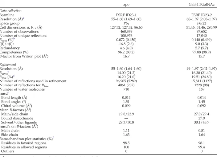
The structures of SSA in the apo form and in complex with the T-antigen disaccharide were solved from crystals grown in two distinct space groups. They show well-defined electron densities for most of the protein regions and bound sugar and exhibit excellent stereochemistry (Materials and Methods;Table 1).
The SSA monomer with overall dimensions of 30 Å × 30 Å × 25 Å belongs to theβ-trefoil fold family and adopts a typical three-lobed organization that consists of 3 four-strandedβ-sheets (β1–β4, β5–β8, and β9–β12), referred to as subdomains α, β, and γ and displaying characteristic pseudo-3-fold symmetry22 (Fig. 1). Structure superposition of the three subdomains reveals that the overall structure of these domains is quite similar to each other with rmsd between subdomains in the 1.55 to 1.66 Å range for ∼40 Cα atoms, with subdomain β being the smallest. In contrast to most of the extracellular R-type lectins that have been extensively studied, SSA lacks the two disulfide bridges that stabilize the β-trefoil fold, a feature that is also observed in most cytoplasmic ricin B lectin domains. Moreover, the key/characteristic signature QXW motif reminiscent of the R-type lectins is present only in the third subdomain of SSA.14
A DALI search for close structural homologs of SSA revealed top-ranked hits (Z-score valuesN20) for the N-terminalβ-trefoil domain of the M. oreades agglutinin (MOA),18 the C-terminal β-trefoil do-main of the HA1 subcomponent of botulinum type C toxin,19 and the 17-kDa HA-17 subcomponent of a serotype D toxin complex from C. botulinum.20 Surprisingly, SSA appears to possess higher struc-tural homology with multi-modular proteins har-boring a β-trefoil domain rather than with individual/isolated R-type lectins. The rmsd's be-tween the SSA structure and those of the three top-ranked homologs above are 1.24, 0.94 and 1.21 Å for 125, 124 and 118 Cα atoms, respectively. The major structural differences between SSA and the three lectin homologs are confined to the shape and dimensions of loop regions connecting β-strands and forging the architecture of the carbohydrate-binding sites.
Glycan array screening
Analysis of the carbohydrate-binding specificity of SSA by glycan array screening revealed strong interaction of the lectin with GalNAc-containing oligosaccharides (Table 2), consistent with previously
Fig. 1. Overall view of the struc-ture of SSA. (a) Overall strucstruc-ture of the SSA monomer showing the three subdomains, denoted α, β, and γ, viewed in two orientations rotated by 90° and colored green, cyan and orange, respectively.β-Strands of a subunit are labeled β1 to β12. Broken lines indicate the disordered β8–β9 loop region in the apo SSA structure. (b) Structure-based se-quence alignment of the three indi-vidual subdomains of SSA; residues involved in carbohydrate binding at siteα are shown by ★.
Table 1. Data collection and refinement
apo Galβ1,3GalNAc
Data collection
Beamline ESRF ID23-1 ESRF ID23-2
Resolution (Å)a 55–1.60 (1.69–1.60) 60–1.97 (2.08–1.97)
Space group P61 P6122
Cell dimensions: a, b, c (Å) 127.32, 127.32, 86.65 51.46, 51.46, 295.99
Number of observations 460,339 97,652
Number of unique reflections 100,976 17,040
Rsym(%)b 0.072 (0.450) 0.140 (0.499)
〈(I)/σ(I)〉 16.8 (2.6) 9.0 (3.3)
Redundancy 4.6 (4.0) 5.7 (5.7)
Completeness (%) 96.2 (80.2) 97.88 (98.9)
B-factor from Wilson plot (Å2) 16.7 15.7
Refinement
Resolution (Å) 55–1.60 (1.64–1.60) 49–1.97 (2.02–1.97)
Rcrystc 14.00 (21.2) 16.30 (21.40)
Rfree(%)d 16.20 (21.0) 19.51 (24.80)
Number of reflections used in refinement 96,905 (5289) 15,811 (1127)
Number of reflections for Rfree 4061 (237) 1228 (98)
Number of water molecules 710 169
rmsde Bond length (Å) 0.014 0.014 Bond angles (°) 1.51 1.45 Chiral volume (Å3) 0.099 0.092 Mean B-factors (Å2) Main/side chain 19.8/22.9 27.0/29.4 Bound disaccharide — 27.9 Solvent/other ligands 29.3/30.8 30.1/43.7 rmsd's on B-factors (Å2) Main chain 1.11 0.81 Side chain 1.63 1.64
Ramachandran plot statistics (%)f
Residues in favored regions 98.5 98.1
Residues in allowed regions 100 99.4
Outliers 0 0
a
Values in parentheses are for the highest-resolution shell. b R
sym=∑hkl(∑i∣Ihkl−〈Ihkl〉∣)/∑hkl∣〈Ihkl〉∣. c
Rcryst=∑hkl∣∣Fo∣−∣Fc∣∣/∑hkl∣Fo∣. d R
freeis calculated for randomly selected reflections excluded from refinement. e
rmsd from ideal geometry. f
Ramachandran plot statistics have been calculated with the MolProbity server.21
3
reported binding data using various pNP-oligosac-charides as potential ligands.12 Although there is a clear preference for terminal non-reducing Gal/ GalNAc residues, the specificity of SSA is rather broad and glycans with internal Gal/GalNAc resi-dues are also recognized. SSA also reacts with branched oligosaccharides, but addition of extra sugar moieties can lead to low-affinity glycans. For instance, glycans withα1,2-fucosyl residues linked to the Gal moiety of the GalNAcα1,4Gal core show a significant response, while those with fucose linked to the terminal Gal, as found in the core H (type 3) blood group antigen, show only a moderate re-sponse. Such an interaction with fucosylated glycans was revealed in a previous binding analysis using version 2 of the printed glycan array.23 While the profile of binding specificity is similar for the three (0.25, 0.5 and 1.0μg/ml) lectin concentrations tested, complex N-glycans were recognized only at the two higher concentrations (Supplementary Fig. S1). The carbohydrate-binding site
The structure of SSA in complex with the Galβ1,3GalNAc disaccharide reveals a well-ordered carbohydrate at only one (site α) out of the three possible sites (Fig. 2) and provides detailed infor-mation about lectin–carbohydrate interactions (Table 3). The non-reducing Gal moiety is tightly bound at the primary binding site within theα site, consistent with the requirement of unsubstituted non-reducing terminal Gal/GalNAc residues for binding activity.12 The aromatic ring of Tyr37 establishes a stacking interaction against the B face of Gal, while the perpendicularly oriented Trp24 indole ring makes additional Van der Waals contacts with the C6–O6 atom pair. The axial O4 hydroxyl group is tightly anchored to the nitrogen backbone atom of Glu25 and the oxygen atom of the Asn22 side chain. The neighboring O3 hydroxyl is hydrogen
bound to the side chains of Asn22 and Asn46 and to Ser44 via a water molecule. The O2 hydroxyl is bound to Asn46 via a water-mediated interaction. The GalNAc moiety is exposed to the solvent and interacts only weakly with residues in the carbohy-drate-binding site (Fig. 2), consistent with the diversity of sugar linkages and moieties allowed beyond the requirement of terminal Gal or GalNAc residues (Table 2). Indeed, only the O4 hydroxyl of GalNAc acts as hydrogen-bond donor to the carbonyl oxygen of Glu25, while the O6 hydroxyl weakly interacts with the Glu25 carboxyl group. The overall conformation of the bound Galβ1,3GalNAc (Φ/Ψ=44°/−10°) falls within the minimum energy area calculated for the disaccharide.26 Structural comparison of the apo and sugar-bound form reveals no major conformational changes upon binding of the disaccharide. In the apo form, a glycerol molecule is observed in siteα and partly mimics a bound Gal moiety (Fig. 2).
Examination of the amino-acid sequence of SSA reveals that the key aromatic side chains (Tyr37 and Trp24) for sugar binding in siteα are absent in sites β and γ and may explain the lack of sugar-binding activity in these two sites (Fig. 1). The carbohydrate-binding site of SSA is shallow and solvent-exposed and may permit accommodation of both theα and β conformation of the 1,3- or 1,4-glycosidic linkage. The carbohydrate-binding site of SSA is filled with 14 well-ordered water molecules that are mostly clustered in the vicinity of the O2 and O3 positions of Gal, suggesting that SSA could accommodate the larger acetamido group of GalNAc, other sugar substituents, or bisecting/multi-antennary glycan chains. Indeed, models of the α-GalNAc complex generated using Autodock Vina27 showed that the docked orientation of α-GalNac is nearly identical with that of Gal in the T-antigen complex observed in the X-ray crystal structure. The N-acetyl group lies in the vicinity of the Glu25-Gly26 backbone
Table 2. Glycan array binding of 0.25μg/ml SSA: the 15 glycan structures are ranked from highest RFU to lowest
Glycan number Glycan Mean RFUa STDEVb SEMc %CVd
9 GalNAcα-Sp8 23,843 2458 1229 10 84 GalNAcα1–3Galβ-Sp8 23,589 221 111 1 90 GalNAcβ1–4GlcNAcβ-Sp0 21,092 348 174 2 85 GalNAcα1–4(Fucα1–2)Galβ1–4GlcNAcβ-Sp8 21,025 1327 664 6 329 GalNAcβ1–3Galα1–4Galβ1–4GlcNAcβ1–3Galβ1–4Glcβ-Sp0 19,851 810 405 4 19 GalNAcβ-Sp8 19,428 1347 673 7 88 GalNAcβ1–3Galα1–4Galβ1–4GlcNAcβ-Sp0 16,809 1706 853 10 298 GalNAcα-Sp15 16,684 634 317 4 91 GalNAcβ1–4GlcNAcβ-Sp8 14,934 582 291 4 300 GalNAcβ1–3Galβ-Sp8 14,658 2082 1041 14 346 Galβ1–4GlcNAcβ1–2Manα1–3(Manα1–6)Manβ1–4GlcNAcβ1–4GlcNAcβ-Sp12 11,530 555 278 5 4 Galβ1–3GlcNAcβ1–2Manα1–3(Galβ1–3GlcNAcβ1–2Manα1–6)Manβ1– 4GlcNAcβ1–4GlcNAcβ-Sp19 7656 571 286 7 83 GalNAcα1–3GalNAcβ-Sp8 7575 54 27 1 55 Fucα1–2Galβ1–3GalNAcβ1–3Galα1–4Galβ1–4Glcβ-Sp9 7122 1485 742 21 59 Fucα1–2Galβ1–3GalNAcβ1–4(Neu5Acα2–3)Galβ1–4Glcβ-Sp9 7074 913 456 13
a Relative fluorescence unit. b
Standard deviation. cStandard error measurement. d
Coefficient of variation.
atoms and the side chains of Ser44 and Asn46 (Fig. 2), indicating that terminal N-acetylgalactosa-minyl moieties could also be easily accommodated into the carbohydrate-binding site of SSA, consistent with the glycan array data (Table 2).
Despite low sequence similarity, the carbohydrate-binding site of SSA reveals structural similarities to other previously reported ricin-like or R-type lectin–
sugar complexes. Although SSA displays only a single carbohydrate-binding site at site α, the mode of binding to Gal resembles that observed in other galactose-binding lectins. Structural compari-son of SSA and the galactose-binding lectin EW29 (earthworm 29-kDa lectin)25 from the earthworm Lumbricus terrestris, which contains two carbohy-drate-binding sites at sitesα and γ (Fig. 2), reveals a
Fig. 2. Carbohydrate-binding site at siteα. (a) Overall view of the dimer with bound Galβ1,3GalNAc in each subunit. (b) Close-up views of the carbohydrate-binding site in (left) the apo form with bound glycerol (orange) and (middle) the complex with bound Galβ1,3GalNAc (orange), viewed in a similar orientation. The 1.95 Å resolution 2mFo−DFcelectron
density map (cyan) is contoured at 1σ. Molecular surface (right) showing the shallow binding site. The position of the dockedα-GalNAc (green) is displayed and overlaid with bound Galβ1,3GalNAc. Side chains that directly interact with the disaccharide are labeled. (c) Overall view (left) of the MOA dimer with bound blood group B Galα1,3(Fucα1,2)Gal trisaccharide (green)24(accession code: 3EF2). The N- and C-terminal domains from each subunit are colored yellow and green, respectively. Close-up stereo view (right) of the overlay of the carbohydrate-binding sites in SSA and MOA. (d) Overall view (left) of the EW29 lectin with bound GalNAc (blue)25(accession code: 2ZQO). Close-up stereo view (right) of the overlay of the carbohydrate-binding sites, oriented as in (c), in SSA and EW29. Side chains that directly interact with the bound carbohydrate in EW29 and MOA are labeled.
5
similar orientation of the terminal Gal moiety in EW29, with an outward 1.6 Å displacement of the sugar ring compared to its position in SSA along with a shift of the β1–β2 loop. Despite the close structural similarities between SSA and theβ-trefoil domain of MOA (rmsd of 1.24 Å for 125 Cα atoms), the Gal moiety adopts a distinct position and orientation in the two structures. In MOA, the non-reducing terminal Gal moiety of the blood group B trisaccharide is shifted by ∼1.5 Å and rotated by ∼30° compared to its position in SSA to favor interactions between the O4 and O6 hydroxyl groups of Gal and MOA (Fig. 2).
A novel dimeric interface
The SSA crystal structure identifies a novel dimer interface for an R-type fungal lectin, consistent with data from gel-filtration experiments (Supplementary Fig. S2). The overall architecture of the SSA dimer consists of a compact assembly with dimensions of 60 Å × 30 Å × 25 Å that buries a flat surface on each β-trefoil monomer. The dimer interface is formed by the tight association of theβ4 and β5 strands from
each monomer with the participation of two peripheral loop regions (β5–β6 and β7–β8) (Fig. 2). In turn, the dimeric assembly of SSA differs from the heterodimeric assembly of the lectin domain of ricins.28 In fact, a structural overlay between SSA and the ricin B chain from the castor bean plant29 reveals that a nine-residue insertion in the β4–β5 loop of SSA prevents formation of a dimeric assembly as seen in the ricin R-type lectin domain. In SSA, the calculated buried surface area to a 1.4 Å probe radius encompasses∼950 A2on each subunit and involves 16 residues that are dominantly apolar. The topology of theβ-strands recruited in this novel dimer interface is conserved in sequence family members of SSA (see below) and residues involved in this assembly are well conserved, suggesting that this type of dimeric association should be preserved in SSA homologs.
Diversity in the oligomeric assembly of various lectins that share a similar fold has been already described and may account for their multivalent carbohydrate-binding specificities.30 In the SSA dimer, the two carbohydrate-binding sites, which are located on the same face of the dimer, are separated by 38 Å, a distance preventing the formation of cross-linking interactions between different antennae of a single N-glycan. Although the nature and biological signification of cross-linking interactions remain to be investigated, the remote location of the two carbohydrate-binding sites in the SSA dimer may favor interaction with distinct glycan chains harboring terminal galactosyl residues.
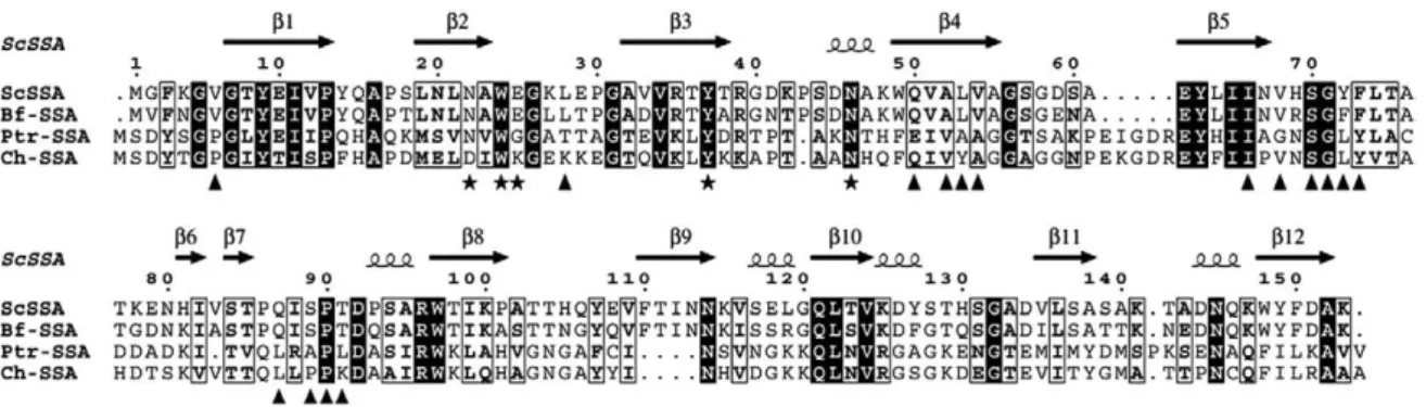
BLAST searches using the SSA sequence as a template identified close homologs in the genome of the three plant pathogenic fungi, B. fuckeliana (Botrytis cinerea), P. tritici-repentis and C. heterostrophus, the former two belonging to the same Pleosporaceae family. These three putative SSA homologs share approximately 77%, 27% and 28% sequence identity, respectively, and may belong to the same family of fungal lectins, as key residues at the carbohydrate-binding site are conserved in siteα along with most of side chains involved in the dimeric assembly (Fig. 3).
Table 3. SSA–Galβ1,3GalNAc interactions
Ligand atoma Direct contact (Å) Interaction partner
Gal O4 2.8 Asn22 OD1
2.8 Glu25 N
Gal O3 2.9 Asn22 ND2
3.0 Asn46 ND2
2.7 Wat25
Gal 02 2.8 Wat45
Gal C6–O6 3.3–3.5 Trp24 indole ring
Gal C3–C4 3.6–3.8 Tyr37 phenol ring
GalNAC O4 2.7 Glu25 O
2.8 Wat98
GalNAc O7 3.0 Wat46
GalNAc O1 2.7 Wat130
2.7 Wat118
Contacts observed in a single subunit within a 3.2 and 4.0 Å distance for hydrogen bonds and hydrophobic carbon–carbon interactions, respectively.
a Hydrophobic interactions are indicated in boldface.
Fig. 3. Sequence alignment of SSA with homologs from B. fuckeliana (Bf-SSA, accession EDN28997.1), P. tritici-repentis (Ptr-SSA, accession EDU42915.1) and C. heterostrophus (Ch-SSA, accession FK679805.1), as putative representative members of this new class of fungal lectins. Identical residues are indicated with a black background while similar residues are boxed. Amino acids involved in carbohydrate binding and dimer assembly of SSA are indicated by★ and ▴, respectively. Secondary-structure elements are indicated above the sequence.
In summary, the crystal structure of SSA reveals that the specific binding to terminal Gal/GalNAc sugars with 1,3 linkages is achieved by both direct and water-mediated hydrogen bonds to the O4 and O3 hydroxyls of Gal at the non-reducing end. The novel dimeric assembly demonstrates that different quaternary structures exist within the R-type lectin family, resulting from a different association of structurally similar domains. Whereas other types of quaternary arrangements may occur within the extended group of fungal lectins, the restricted number of functional binding sites per β-trefoil domain in SSA could dictate the type of quaternary structures within this new family of fungal lectins and affect the ability to establish multiple interac-tions and cross-link glycan receptors. By analogy with the dual storage–defense role attributed to a large group of abundant plant lectins, the Scleroti-niaceae lectins might also combine a likely function as storage protein in the development and morpho-genesis of the fungus, with a possible defense role against predating organisms. Further studies to elucidate/investigate the biological role of this emerging class of fungal lectins are required.
Materials and Methods
Materials
SSA was isolated from mature S. sclerotiorum sclerotes and purified as described previously.11
Crystallization and data collection
For crystallization, ammonium sulfate-precipitated SSA was extensively dialyzed against 10 mM Hepes, pH 7.5, and 150 mM NaCl and concentrated to 7.3 mg/ml. Crystallization experiments were performed by vapor diffusion with Greiner plates (Greiner-BioOne) using a Freedom (Tecan) and Honeybee (Cartesian) robot. Initial crystals of apo SSA were obtained at 20 °C from a condition containing 2.0 M sodium chloride and 0.1 M sodium acetate, pH 4.6, of the Structure Screen 2 (Molecular Dimension Limited). After manual optimiza-tion in Limbro plates, larger crystals appeared within 10 days from typically 1μl of protein solution and 0.5 μl of reservoir containing 2.1 M sodium chloride and 0.1 M sodium acetate, pH 4.6. The SSA–Galβ1,3GalNAc com-plex was made by mixing the protein solution with 2.5 mM Galβ1,3GalNAc followed by preincubation overnight at 4 °C prior to crystallization experiments. Crystals of the complex appeared at 20 °C from a condition containing 1.26 M ammonium sulfate, 0.2 M lithium sulfate and 0.1 M Tris, pH 8.5. Crystals selected for data collection were rapidly transferred in the reservoir solution supplemented with 25% glycerol (apo) or 30% polyethylene glycol 400 (complex), flash cooled at 100 K in a nitrogen gas stream, and stored in liquid nitrogen. Data were collected on the European Synchrotron Radiation Facility beamlines ID23-1 and ID23-2 (Grenoble, France). Oscillation images were integ-rated with MOSFLM31 and scaled and merged with SCALA.32
Structure determination and refinement
Attempts to solve the structure using a Se-labeled
D-Gal-β-SePh derivative were unsuccessful. Initial phases
were obtained by molecular replacement with the Phaser program33 using as search model a structural template generated with the protein homology recognition engine Phyre,34which uses profile–profile matching algorithms
for the detection of homologs of known three-dimensional structure, the so-called template-based homology model-ing or fold recognition. The model generated by the Phyre server was based on the crystal structure of C. botulinum non-toxin hemagglutinin HA33/A13(Protein Data Bank accession number 1YBI) and led to an unambiguous solution in Phaser with a Z-score value of 51 in the 15- to 2.8 Å resolution range. The resulting model, comprising four molecules of SSA in the asymmetric unit, was rebuilt in an automatic fashion with the program ARP/wARP.35 Final refinement was then performed with REFMAC36 using data up to 1.6 Å resolution, and the resulting σA-weighted 2mFo−DFc and mFo−DFc electron density
maps were used to correct the model with the graphics program COOT.37 The structure of the SSA disaccharide complex was solved by molecular replacement with Phaser,33
using apo SSA as a search model. The structure of the complex was subsequently refined and manually adjusted with the programs REFMAC36and Coot,37respectively.
The final apo model comprises residues Gly2 to Lys153 for the four SSA molecules and two glycerol molecules arising form the cryo buffer; that of the disaccharide complex comprises residues Gly2 to Lys153 for a single SSA molecule, a Galβ1,3GalNAc disaccharide, four sulfate ions arising from the crystallization buffer, and two small polyethylene glycol molecules arising form the cryo buffer. In the two structures, the backbone region of residues Gly58 to Ser60 adopts two alternate conforma-tions while, in the apo structure, the surface loop region Thr103-Glu108 is disordered. The rmsd between the apo and sugar-bound form is 0.64 Å for 144 Cα atoms. In the apo form, theβ2–β3 surface loop region containing the residue pair Trp24-Glu25 at site α undergoes large conformational changes in one out of four molecules, but these movements are mediated by the crystal packing environment. The stereochemistry of the models was analyzed with MolProbity;21 no residues were found in
the disallowed regions of the Ramachandran plot. Data collection and refinement statistics are reported inTable 1. Structural sequence alignment was performed with the RAPIDO server.38 Figures 1 and 2were generated with PyMOL.39Figure 3was prepared with ESPript.40 Glycan array screening
The glycan binding specificity of SSA was analyzed by the Consortium for Functional Glycomics using micro-arrays v3.1.23Full details of protocols used and the∼380 glycans included in the array are presented on the Consortium's web site† and described by Paulson et al.23
Lyophilized SSA was dissolved in phosphate-buffered saline at 1 mg/ml and labeled with tetrafluorophenyl-Alexa Fluor 488 using the Invitrogen protein labeling kit following the manufacturer's instructions. Labeled SSA was diluted to 0.25μg/ml in Tris-buffered saline (20 mM Tris, 150 mM NaCl, 2 mM CaCl2, and 2 mM MgCl2,
†http://www.functionalglycomics.org/static/consortium/ resources/resourcecoreh8.shtml
7
pH 7.4) containing 1% bovine serum albumin and 0.05% Tween 20. An aliquot (70μl at 0.25 μg/ml) of the labeled lectin solution was applied to separate microarray slides and incubated under a coverslip for 60 min in a dark, humidified chamber at room temperature. After incuba-tion, the coverslips were gently transferred to a solution of Tris-buffered saline containing 0.05% Tween 20 and successively washed four times by gently dipping the slides in Tris-buffered saline containing 0.05% Tween 20 and deionized water. After the last wash, the slides were spun in a slide centrifuge for approximately 15 s to dry and immediately scanned in a ProScanArray MicroArray Scanner (PerkinElmer) using an excitation wavelength of 488 nm and ImaGene software (BioDiscovery, Inc., El Segundo, CA) to quantify fluorescence. Data are expressed as average relative fluorescence units for binding each glycan calculated by averaging four values after removing the highest and lowest values; STDEV is the standard deviation, SEM is the standard error measurement, and CV is the coefficient of variation (SD/mean) calculated as percentage. Analysis of the carbohydrate specificity of SSA on the glycan array was repeated at lectin concentrations of 0.50 and 1.0μg/ml.
Molecular docking
α-GalNAc was selected as a candidate ligand for automated docking to SSA using Autodock Vina.27 SSA and the carbohydrate ligands were treated as a rigid protein and flexible molecules, respectively. The grid of the docking simulation was defined by a 20 Å × 20 Å × 20 Å cube centered on the SSAα site. The docking simulation was performed using the default parameters. For each ligand, the nine top-ranked generations based on the predicted binding affinity (in kilocalories per mole) were analyzed. Accession numbers
Coordinates and structure factors have been deposited in the Protein Data Bank with accession numbers 2X2S and 2X2T.
Acknowledgements
The authors thank the European Synchrotron Radiation Facility staff for assistance with data collection, Jean-Marie Beau and Dominique Urban for providing us with a selenium-labeled D-
Gal-β-SePh derivative and Miguel Ortiz-Lombardia for modelling studies. This work was supported in part by the Centre National de la Recherche Scientifique (to Y.B.), the Ghent University, and the Fund for Scientific Research-Flanders (FWO grant G.0022.08) (to E.J.M.V.D.). The authors want to thank the Consortium for Functional Glycomics funded by the NIGMS GM62116 for the glycan array analysis.
Supplementary Data
Supplementary data associated with this article can be found, in the online version, atdoi:10.1016/ j.jmb.2010.05.038
References
1. Wang, H., Ng, T. B. & Ooi, V. E. C. (1998). Lectins from mushrooms. Mycol. Res.102, 897–906.
2. Imberty, A., Mitchell, E. P. & Wimmerová, M. (2005). Structural basis of high-affinity glycan recognition by bacterial and fungal lectins. Curr. Opin. Struct. Biol.15, 525–534.
3. Goldstein, I. J. & Winter, H. C. (2007). Mushroom lectins. In Comprehensive Glycoscience: From Chemistry to Systems Biology (Kamerling, J. P., ed.), Elsevier Ltd., Amsterdam, The Netherlands.
4. Kellens, J. T. C. & Peumans, W. J. (1990). Develop-mental accumulation of lectins in Rhizoctonia solanii: potential role as a storage protein. J. Gen. Microbiol. 136, 2489–2495.
5. Rosen, S., Sjollema, K., Veenhuis, M. & Tunlid, A. (1997). A cytoplasmic lectin produced by the fungus Arthrobotrys oligospora functions as a storage protein during saprophytic and parasitic growth. Microbiology, 143, 2593.
6. Cooper, D. N. W., Boulianne, R. P., Charlton, S., Farrell, E. M., Sucher, A. & Lu, B. C. (1997). Fungal galectins, sequence and specificity of two isolectins from Coprinus cinereus. J. Biol. Chem.272, 1514. 7. Yatohgo, T., Nakata, M., Tsumuraya, Y., Hashimoto,
Y. & Yamamoto, S. (1988). Purification and properties of a lectin from the fruiting bodies of Flammulina velutipes. Agric. Biol. Chem.52, 1485–1494.
8. Singh, R. S., Bhari, R. & Kaur, H. P. (2010). Mushroom lectins: current status and future perspectives. Crit. Rev. Biotechnol.30, 99–126.
9. Fukazawa, Y. & Kagaya, K. (1997). Molecular bases of adhesion of Candida albicans. J. Med. Vet. Mycol.35, 87–99.
10. Candy, L., Van Damme, E. J., Peumans, W. J., Menu-Bouaouiche, L., Erard, M. & Rougé, P. (2003). Structural and functional characterization of the GalNAc/Gal-specific lectin from the phytopathogenic ascomycete Sclerotinia sclerotiorum (Lib.) de Bary. Biochem. Biophys. Res. Commun.308, 396–402. 11. Kellens, J. T. C. & Peumans, W. T. (1992). Lectins in
different members of the Sclerotiniaceae. Mycol. Res. 96, 495–502.
12. Van Damme, E. J., Nakamura-Tsuruta, S., Hirabayashi, J., Rougé, P. & Peumans, W. J. (2007). The Sclerotinia sclerotiorum agglutinin represents a novel family of fungal lectins remotely related to the Clostrid-ium botulinum non-toxin haemagglutinin HA33/A. Glycoconjugate J.24, 143–156.
13. Arndt, J. W., Gu, J., Jaroszewski, L., Schwarzenbacher, R., Hanson, M. A., Lebeda, F. J. & Stevens, R. C. (2005). The structure of the neurotoxin-associated protein HA33/A from Clostridium botulinum suggests a reoc-curringβ-trefoil fold in the progenitor toxin complex. J. Mol. Biol.346, 1083–1093.
14. Hazes, B. (1996). The (QxW)3 domain: a flexible lectin scaffold. Protein Sci.5, 1490–1501.
15. Winter, H. C., Mostafapour, K. & Goldstein, I. J. (2002). The mushroom Marasmius oreades lectin is a blood group type B agglutinin that recognizes the Galα 1,3Gal and Galα 1,3Galβ 1,4GlcNAc porcine xeno-transplantation epitopes with high affinity. J. Biol. Chem.277, 14996–15001.
16. Mo, H., Winter, H. C. & Goldstein, I. J. (2000). Purification and characterization of a Neu5Acα2-6Galβ1-4Glc/GlcNAc-specific lectin from the fruiting body of the polypore mushroom Polyporus squamosus. J. Biol. Chem.275, 10623–10629.
17. Tateno, H. & Goldstein, I. J. (2003). Molecular cloning, expression, and characterization of novel hemolytic lectins from the mushroom Laetiporus sulphureus, which show homology to bacterial toxins. J. Biol. Chem.278, 40455–40463.
18. Grahn, E., Askarieh, G., Holmner, A., Tateno, H., Winter, H. C., Goldstein, I. J. & Krengel, U. (2007). Crystal structure of the Marasmius oreades mushroom lectin in complex with a xenotransplantation epitope. J. Mol. Biol.369, 710–721.
19. Nakamura, T., Tonozuka, T., Ide, A., Yuzawa, T., Oguma, K. & Nishikawa, A. (2008). Sugar-binding sites of the HA1 subcomponent of Clostridium botulinum type C progenitor toxin. J. Mol. Biol. 376, 854–867.
20. Hasegawa, K., Watanabe, T., Suzuki, T., Yamano, A., Oikawa, T., Sato, Y. et al. (2007). A novel subunit structure of Clostridium botulinum serotype D toxin complex with three extended arms. J. Biol. Chem.282, 24777–24783.
21. Davis, I. W., Leaver-Fay, A., Chen, V. B., Block, J. N., Kapral, G. J., Wang, X. et al. (2007). MolProbity: all-atom contacts and structure validation for proteins and nucleic acids. Nucleic Acids Res.35, W375–W383. 22. Murzin, A. G., Lesk, A. M. & Chothia, C. (1992). β-Trefoil fold. Patterns of structure and sequence in the Kunitz inhibitors interleukins-1 β and 1 α and fibroblast growth factors. J. Mol. Biol. 223, 531–543. 23. Blixt, O., Head, S., Mondala, T., Scanlan, C., Huflejt,
M. E., Alvarez, R. et al. (2004). Printed covalent glycan array for ligand profiling of diverse glycan binding proteins. Proc. Natl Acad. Sci. USA,101, 17033–17038. 24. Grahn, E. M., Winter, H. C., Tateno, H., Goldstein, I. J. & Krengel, U. (2009). Structural characterization of a lectin from the mushroom Marasmius oreades in complex with the blood group B trisaccharide and calcium. J. Mol. Biol.390, 457–466.
25. Suzuki, R., Kuno, A., Hasegawa, T., Hirabayashi, J., Kasai, K. I., Momma, M. & Fujimoto, Z. (2009). Sugar-complex structures of the C-half domain of the galactose-binding lectin EW29 from the earthworm Lumbricus terrestris. Acta Crystallogr., Sect. D: Biol. Crystallogr.65, 49–57.
26. Weimar, T., Bukowski, R. & Young, N. M. (2000). The conformation of the T-antigen disaccharide bound to Maclura pomifera agglutinin in aqueous solution. J. Biol. Chem.275, 37006–37010.
27. Trott, O. & Olson, A. J. (2010). AutoDock Vina:
improving the speed and accuracy of docking with a new scoring function, efficient optimization, and multithreading. J. Comput. Chem.31, 455–461. 28. Montfort, W., Villafranca, J. E., Monzingo, A. F., Ernst,
S. R., Katzin, B., Rutenber, E. et al. (1987). The three-dimensional structure of ricin at 2.8 Å. J. Biol. Chem. 262, 5398–5403.
29. Rutenber, E., Katzin, B. J., Ernst, S., Collins, E. J., Mlsna, D., Ready, M. P. & Robertus, J. D. (1991). Crystallographic refinement of ricin to 2.5 Å. Proteins, 10, 240–250.
30. Sinha, S., Gupta, G., Vijayan, M. & Surolia, A. (2007). Subunit assembly of plant lectins. Curr. Opin. Struct. Biol.17, 498–505.
31. Leslie, A. G. W. (1992). Recent changes to the MOSFLM package for processing film and image plate data. Joint CCP4 + ESF-EAMCB Newsl. Protein Crystallogr.26.
32. Collaborative Computational Project, Number 4 (1994). The CCP4 suite: programs for protein crystal-lography. Acta Crystallogr., Sect. D: Biol. Crystallogr.50, 760–763.
33. McCoy, A. J. (2007). Solving structures of protein complexes by molecular replacement with Phaser. Acta Crystallogr., Sect. D: Biol. Crystallogr.63, 32–41. 34. Kelley, L. A. & Sternberg, M. J. (2009). Protein
structure prediction on the Web: a case study using the Phyre server. Nat. Protoc.4, 363–371.
35. Perrakis, A., Morris, R. & Lamzin, V. S. (1999). Automated protein model building combined with iterative structure refinement. Nat. Struct. Biol. 6, 458–463.
36. Murshudov, G. N., Vagin, A. A. & Dodson, E. J. (1997). Refinement of macromolecular structures by the maximum-likelihood method. Acta Crystallogr., Sect. D: Biol. Crystallogr.53, 240–255.
37. Emsley, P. & Cowtan, K. (2004). Coot: model-building tools for molecular graphics. Acta Crystallogr., Sect. D: Biol. Crystallogr.60, 2126–2132.
38. Mosca, R. & Schneider, T. R. (2008). RAPIDO: a web server for the alignment of protein structures in the presence of conformational changes. Nucleic Acids Res. 36, W42.
39. Delano, W. L. (2002). The PyMOL molecular graphics system. DeLano Scientific LLC, Palo Alto, CA. 40. Gouet, P., Courcelle, E., Stuart, D. I. & Métoz, F.
(1999). ESPript: analysis of multiple sequence align-ments in PostScript. Bioinformatics,15, 305–308.
9