The
ne w engl and
journal
of
medicine
e s ta b l i s h e d i n 1 8 1 2 j a n ua ry8, 2 0 0 4 v o l . 3 5 0 n o . 2
A Comparison of Vasopressin and Epinephrine for Out-of-Hospital
Cardiopulmonary Resuscitation
Volker Wenzel, M.D., Anette C. Krismer, M.D., H. Richard Arntz, M.D., Helmut Sitter, Ph.D., Karl H. Stadlbauer, M.D., and Karl H. Lindner, M.D.,
for the European Resuscitation Council Vasopressor during Cardiopulmonary Resuscitation Study Group*
a b s t r a c t
From the Department of Anesthesiology and Critical Care Medicine, Leopold-Fran-zens University, Innsbruck, Austria (V.W., A.C.K., K.H.S., K.H.L.); the Department of Medicine, Division of Cardiology–Pulmonol-ogy, Benjamin Franklin Medical Center, Free University, Berlin, Germany (H.R.A.); and the Institute for Theoretical Surgery, Phil-ipps University, Marburg, Germany (H.S.). Address reprint requests to Dr. Lindner at the Department of Anesthesiology and Crit-ical Care Medicine, Leopold-Franzens Uni-versity, Anichstr. 35, 6020 Innsbruck, Aus-tria, or at volker.wenzel@uibk.ac.at. *The investigators who participated in the
study group are listed in the Appendix. N Engl J Med 2004;350:105-13. Copyright © 2004 Massachusetts Medical Society. b a c k g r o u n d
Vasopressin is an alternative to epinephrine for vasopressor therapy during cardiopul-monary resuscitation, but clinical experience with this treatment has been limited. m e t h o d s
We randomly assigned adults who had had an out-of-hospital cardiac arrest to receive two injections of either 40 IU of vasopressin or 1 mg of epinephrine, followed by addi-tional treatment with epinephrine if needed. The primary end point was survival to hospital admission, and the secondary end point was survival to hospital discharge. r e s u l t s
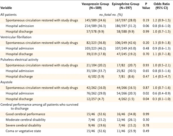
A total of 1219 patients underwent randomization; 33 were excluded because of miss-ing study-drug codes. Among the remainmiss-ing 1186 patients, 589 were assigned to re-ceive vasopressin and 597 to rere-ceive epinephrine. The two treatment groups had simi-lar clinical profiles. There were no significant differences in the rates of hospital admission between the vasopressin group and the epinephrine group either among pa-tients with ventricular fibrillation (46.2 percent vs. 43.0 percent, P=0.48) or among those with pulseless electrical activity (33.7 percent vs. 30.5 percent, P=0.65). Among patients with asystole, however, vasopressin use was associated with significantly higher rates of hospital admission (29.0 percent, vs. 20.3 percent in the epinephrine group; P=0.02) and hospital discharge (4.7 percent vs. 1.5 percent, P=0.04). Among 732 patients in whom spontaneous circulation was not restored with the two injections of the study drug, additional treatment with epinephrine resulted in significant im-provement in the rates of survival to hospital admission and hospital discharge in the vasopressin group, but not in the epinephrine group (hospital admission rate, 25.7 percent vs. 16.4 percent; P=0.002; hospital discharge rate, 6.2 percent vs. 1.7 percent; P=0.002). Cerebral performance was similar in the two groups.
c o n c l u s i o n s
The effects of vasopressin were similar to those of epinephrine in the management of ventricular fibrillation and pulseless electrical activity, but vasopressin was superior to epinephrine in patients with asystole. Vasopressin followed by epinephrine may be more effective than epinephrine alone in the treatment of refractory cardiac arrest.
here are more than 600,000 sud-den deaths in North America and Europe each year. More than half of these deaths occur before 65 years of age, which underscores the need for optimal cardiopulmonary resuscitation (CPR) strategies in order to improve patients’ chanc-es of survival.
Epinephrine has been used during CPR for more than 100 years1 but has become controversial be-cause it is associated with increased myocardial oxygen consumption, ventricular arrhythmias, and myocardial dysfunction during the period after re-suscitation.2 Since it was found that endogenous vasopressin levels in successfully resuscitated tients were significantly higher than levels in pa-tients who died, it was postulated that it might be beneficial to administer vasopressin during CPR.3 Laboratory studies of CPR revealed that vasopres-sin was associated with better blood flow to vital organs,4 delivery of cerebral oxygen,5 chances of re-suscitation,6,7 and neurologic outcome8 than epi-nephrine. In a small clinical study, the use of vaso-pressin resulted in a significantly higher rate of short-term survival than epinephrine,9 indicating that vasopressin may be a reasonable alternative to epinephrine for vasopressor therapy during CPR.
The current international guidelines for CPR rec-ommend the use of epinephrine during cardiac re-suscitation, with vasopressin considered only as a secondary alternative, because clinical data on vaso-pressin therapy have been limited.10,11 We there-fore conducted a clinical trial to assess the effects of vasopressin and epinephrine on survival among adults who have an out-of-hospital cardiac arrest and present with ventricular fibrillation, pulseless electrical activity, or asystole. The null hypothesis was that there would be no differences between the treatment groups in the rates of survival to hospi-tal admission and survival to hospihospi-tal discharge.
s t u d y p a t i e n t s
This study was conducted in 33 communities and involved 44 physician-staffed emergency medical service units in Austria, Germany, and Switzerland. Adult patients who had an out-of-hospital cardiac arrest and presented with ventricular fibrillation, pulseless electrical activity, or asystole requiring CPR with vasopressor therapy were included; the criteria for exclusion were successful defibrillation without the administration of a vasopressor, documented
terminal illness, a lack of intravenous access, hem-orrhagic shock, pregnancy, cardiac arrest after trau-ma, an age of less than 18 years, and the presence of a do-not-resuscitate order.
s t u d y d e s i g n
The study was designed as a double-blind, prospec-tive, multicenter, randomized, controlled clinical tri-al; the primary end point was survival to hospital admission, and the secondary end point was surviv-al to hospitsurviv-al discharge. The protocol was approved by the institutional review board of each participat-ing center. For all patients, the requirement of in-formed consent was waived in accordance with the ethical standards of the local institutional review board and the guidelines for good clinical practice of the European Agency for the Evaluation of Me-dicinal Products.12 The patients’ families and sur-viving patients were informed about the trial, and the protocol specified that if there were any objec-tions, the patient would be withdrawn from the study; there were no objections. Treatment assign-ments to the study drugs were randomly generated in blocks of 10, with stratification according to cen-ter. If all criteria for inclusion were met and none of the criteria for exclusion were met, patients who pre-sented with pulseless electrical activity or asystole underwent randomization immediately; patients with ventricular fibrillation underwent randomiza-tion after the first three attempts at defibrillarandomiza-tion had failed.
When a given patient underwent randomiza-tion, a box containing the study drugs — either two ampules of 1 mg of epinephrine (Suprarenin) or two ampules of 40 IU of vasopressin (Pitressin) — was opened, and either 1 mg of epinephrine or 40 IU of vasopressin was injected. The authenticity of both drugs was confirmed with the use of high-pressure liquid chromatography. If spontaneous circulation was not restored within three minutes after the first injection of the study drug, the same drug at the same dose was injected again. If spon-taneous circulation was still not restored, the pa-tient was given an additional injection of epineph-rine at the discretion of the emergency physician who was managing the CPR attempt. All drugs were injected exclusively intravenously, followed by 20 ml of normal saline.
Investigators and physicians were unaware of the study-drug assignment unless decoding became clinically necessary for management in the period after resuscitation; if this occurred, the data and
t
v a s o p r e s s i n a n d e p i n e p h r i n e f o r c a r d i o p u l m o n a r y r e s u s c i t a t i o n
safety monitoring committee was to be informed. Additional interventions such as the administration of sodium bicarbonate, atropine, lidocaine, or amio-darone and fibrinolysis were used at the discretion of the physician managing the CPR attempt. d o c u m e n t a t i o n
The CPR attempt was documented according to the Utstein style13; data were entered into a data base by one investigator and were subsequently indepen-dently cross-checked twice by two other investiga-tors who were unaware of the treatment-group as-signment. Original data were made available to the data and safety monitoring committee for indepen-dent scrutiny. Neurologic function in the surviving patients was categorized according to a cerebral per-formance score.14
s t a t i s t i c a l a n a l y s i s
An estimation of the number of patients needed was derived during the analysis of another study of out-of-hospital cardiac arrest.15 The calculation was based on a possible drug-related improvement in the outcome of 25 percent, a significance level of 0.05, two-tailed analysis, and a power of 80 percent. According to this calculation, 571 patients per group might be necessary in order to show a clinically sig-nificant difference in the rates of hospital admis-sion between the two treatment groups; the addi-tion of a safety margin of 30 percent resulted in an estimate of 1500 patients for the entire trial. Analysis was performed according to the intention-to-treat principle; the chi-square test was used to determine differences between groups with respect to the pri-mary and secondary end points. Odds ratios and their 95 percent confidence intervals were calculat-ed. Comparisons of patient characteristics and sur-vival outcomes were tested with the chi-square test, the chi-square test for trend, Fisher’s exact test, or Student’s t-test, as appropriate. Logistic-regression analysis was used to control for possible confound-ing effects of variables related to the different end points. All P values are two-sided; no corrections were made for multiple comparisons.
The study was conducted from June 1999 to March 2002; only one internal, blinded administrative in-terim analysis was performed in June 2000 after the randomization of 200 patients, and the results were revealed only to the data and safety monitoring
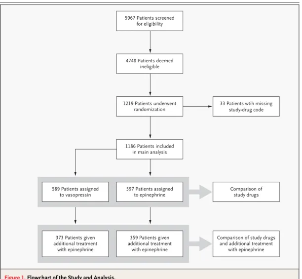
com-mittee. This analysis established that the study was safe, that randomization was working properly, and that no adverse events had been reported. Since funding had ended by December 2001, enrollment was stopped in March 2002. The treatment groups had similar clinical profiles (Tables 1 and 2); 88 of the patients who underwent randomization were later shown to meet criteria for exclusion, but they were included in the final analysis on an intention-to-treat basis. Thirty-three patients had to be exclud-ed from the analysis because of a missing study-drug code (the characteristics of the patients who were included were similar to those of the patients who were excluded), and no significant differences were observed among different centers (Fig. 1).
The rate of survival to hospital admission was higher among patients with a witnessed cardiac ar-rest than among those with an unwitnessed cardiac arrest (352 of 920 patients [38.3 percent] vs. 41 of 255 patients [16.1 percent], P<0.001), and the rate was higher among patients who received basic life support within 10 minutes than among those who received such support more than 10 minutes after the cardiac arrest (291 of 665 patients [43.8 percent] vs. 107 of 517 patients [20.7 percent], P<0.001). The rates of hospital admission were similar between the two treatment groups both for patients with ven-tricular fibrillation and for those with pulseless electrical activity. Patients with asystole, however, were more likely to survive to hospital admission and to hospital discharge if they were treated with vasopressin than if they received epinephrine as initial therapy (Table 3). In an analysis including 732 patients in whom spontaneous circulation was not restored with the administration of the study
r e s u l t s
Table 1. Cardiovascular History of the Patients.
Variable Vasopressin Group (N=589) Epinephrine Group (N=597) P Value no./total no. (%)
Coronary heart disease 176/467 (37.7) 189/463 (40.8) 0.33
Hypertension 84/475 (17.7) 82/474 (17.3) 0.88
Diabetes 78/476 (16.4) 78/477 (16.4) 0.99
Left ventricular failure 59/467 (12.6) 59/468 (12.6) 0.99
Peripheral vascular disease 47/474 (9.9) 53/475 (11.2) 0.53
Cardiac arrhythmias 35/467 (7.5) 29/468 (6.2) 0.43
Pacemaker 20/474 (4.2) 18/474 (3.8) 0.74
Valvular heart disease 13/468 (2.8) 14/468 (3.0) 0.85
drug, additional treatment with epinephrine (me-dian dose, 5 mg; interquartile range, 2 to 10) result-ed in a significant improvement in the survival rate in the vasopressin group (P=0.007 by the chi-square test for trend) but not in the epinephrine group (Ta-ble 4). There was no significant difference between the two groups in cerebral performance (Tables 3 and 4).
With both study drugs, the rate of survival to
hospital admission was significantly improved by both amiodarone treatment (79 of 163 patients [48.5 percent] vs. 321 of 1023 patients [31.4 percent]; P<0.001; odds ratio, 2.1; 95 percent confidence interval, 1.5 to 2.9) and fibrinolysis (45 of 99 pa-tients [45.5 percent] vs. 355 of 1087 papa-tients [32.7 percent]; P=0.01; odds ratio, 1.7; 95 percent con-fidence interval, 1.1 to 2.6). After hospital admis-sion, the code for the study drug was broken (the * Plus–minus values are means ±SD. CPR denotes cardiopulmonary resuscitation.
† Intervals are given separately for the duration of untreated cardiac arrest and the periods from the provision of basic life support to each treatment procedure because bystanders may not have been able to judge the intervals accurately, owing to emotional stress.
Table 2. Base-Line Characteristics of the Patients.*
Characteristic Vasopressin Group (N=589) Epinephrine Group (N=597) P Value Age — yr 66.5±14.4 65.9±14.2 0.45
Male sex — no./total no. (%) 402/580 (69.3) 421/591 (71.2) 0.47
Arrest witnessed — no./total no. (%) 448/583 (76.8) 472/592 (79.7) 0.53
CPR by bystander or family member — no./total. no. (%) 111/589 (18.8) 107/597 (17.9) 0.68
Suspected cause of cardiac arrest — no./total no. (%)
Myocardial infarction 262/454 (57.7) 249/449 (55.5) 0.49
Primary arrhythmia 99/455 (21.8) 109/452 (24.1) 0.40
Pulmonary embolism 64/456 (14.0) 53/455 (11.6) 0.28
Additional treatments given during CPR — no./total no. (%)
Sodium bicarbonate 198/587 (33.7) 205/596 (34.4) 0.81
Atropine 139/587 (23.7) 151/597 (25.3) 0.51
Lidocaine 114/589 (19.4) 114/597 (19.1) 0.90
Amiodarone 75/589 (12.7) 88/597 (14.7) 0.32
Fibrinolysis 54/589 (9.2) 45/597 (7.5) 0.31
Initial cardiac rhythm — no./total no. (%)
Ventricular fibrillation 223/589 (37.9) 249/597 (41.7) 0.18
Pulseless electrical activity 104/589 (17.7) 82/597 (13.7) 0.06
Asystole 262/589 (44.5) 266/597 (44.6) 0.98
Intervals — min†
Duration of untreated cardiac arrest (before basic life support provided)
7.9±6.4 7.9±6.4 0.94
Time from basic life support
To first defibrillation attempt 7.0±6.8 7.7±7.6 0.18
To endotracheal intubation 7.6±6.2 7.9±6.8 0.39
To intravenous cannulation 8.2±6.7 8.5±7.0 0.37
To first injection of study drug 9.6±6.6 10.2±7.4 0.15
To second defibrillation attempt 12.9±7.6 13.9±8.1 0.14
To second injection of study drug 13.3±6.8 13.9±7.9 0.16
To third defibrillation attempt 17.7±8.4 18.4±9.5 0.37
To standard protocol with epinephrine 17.5±7.9 17.6±8.3 0.91
v a s o p r e s s i n a n d e p i n e p h r i n e f o r c a r d i o p u l m o n a r y r e s u s c i t a t i o n
treatment assignment was disclosed) for five pa-tients in order to optimize post-resuscitation care.
Our results did not confirm previous data that showed vasopressin to be more effective than epi-nephrine as adjunctive therapy in the treatment of patients with ventricular fibrillation and pulseless electrical activity.4-9 This discrepancy raises the ques-tion of whether vasopressin improves perfusion pressures during CPR in patients with these condi-tions but does not improve the outcome.16 Similar-ly, although some studies in animals have suggested that high-dose epinephrine during CPR has bene-ficial effects, this strategy caused a hyperadrenergic state and was associated with higher early mortality in other studies that used a preparation for pigs.17
Subsequent clinical studies with high-dose epineph-rine did not show any benefit.2 We were unable to determine whether problems in extrapolating from CPR performed in the laboratory to clinical experi-ence were attributable to differexperi-ences among spe-cies, the fact that our patients had underlying dis-ease whereas the laboratory animals were otherwise healthy, or differences between out-of-hospital CPR and CPR performed under laboratory conditions.
In contrast to the findings regarding patients with ventricular fibrillation or pulseless electrical activity, we found that among patients with asysto-le, those who received vasopressin were about 40 percent more likely than those given epinephrine to reach the hospital alive. The extreme ischemia in pa-tients with asystole may suggest a possible under-lying mechanism. As has been shown in an in vitro study, vasopressin has vasoconstricting efficacy d i s c u s s i o n
Figure 1. Flowchart of the Study and Analysis.
5967 Patients screened for eligibility 4748 Patients deemed ineligible 1219 Patients underwent randomization 1186 Patients included in main analysis 597 Patients assigned to epinephrine Comparison of study drugs 589 Patients assigned to vasopressin 359 Patients given additional treatment with epinephrine
Comparison of study drugs and additional treatment
with epinephrine 373 Patients given
additional treatment with epinephrine
33 Patients wtih missing study-drug code
even in severe acidosis, when catecholamines are less potent.18 Thus, vasopressin may be a more ef-fective vasopressor than epinephrine in patients with asystole, resulting in better coronary perfusion pressure during cardiac resuscitation. Since proved coronary perfusion pressure during CPR im-proves survival,19 vasopressin may be a better option than epinephrine for patients with asystole, who normally have the worst chance of survival of all pa-tients with cardiac arrest. This post hoc observation could be tested in a trial restricted to such patients, for whom few treatment options are available.
In addition, improvement in the rate of surviv-al to hospitsurviv-al discharge among patients who were treated with epinephrine after vasopressin may in-dicate that the interactions among vasopressin, epi-nephrine, and the underlying degree of ischemia during CPR may be more complex than was
previ-ously thought. When prolonged asphyxia has de-pleted endogenous epinephrine levels and caused fundamental ischemia in pigs, the administration of vasopressin combined with epinephrine results in coronary perfusion pressures triple those achieved with either epinephrine or vasopressin alone.20 This finding suggests that the presence of one of these drugs may enhance the effects of the other, especial-ly during prolonged ischemia. These data from ex-perimental CPR are in agreement with the results of our current clinical trial, in which the combina-tion of vasopressin and epinephrine was effective in patients about 25 minutes after cardiac arrest, at a time when a severe degree of ischemia must be as-sumed, but increasing doses of epinephrine alone were not effective.
In a recent study of in-hospital CPR in which vasopressin and epinephrine were reported to have * Eleven patients in the vasopressin group (1.9 percent) and nine in the epinephrine group (1.5 percent) were lost to
fol-low-up before hospital discharge. Eleven of the patients in the vasopressin group and 12 of the patients in the epineph-rine group who survived to hospital discharge (19.3 percent and 20.7 percent, respectively) were lost to follow-up for cerebral performance. P values were not adjusted for multiple comparisons. An odds ratio of less than 1.0 represents an advantage for vasopressin. CI denotes confidence interval.
Table 3. Data on Outcomes in All 1186 Patients and on Cerebral Performance in 115 Patients at Hospital Discharge.*
Variable Vasopressin Group (N=589) Epinephrine Group (N=597) P Value Odds Ratio (95% CI)
All patients no./total no. (%)
Spontaneous circulation restored with study drugs 145/589 (24.6) 167/597 (28.0) 0.19 1.2 (0.9–1.5)
Hospital admission 214/589 (36.3) 186/597 (31.2) 0.06 0.8 (0.6–1.0)
Hospital discharge 57/578 (9.9) 58/588 (9.9) 0.99 1.0 (0.7–1.5)
Ventricular fibrillation
Spontaneous circulation restored with study drugs 82/223 (36.8) 106/249 (42.6) 0.20 1.3 (0.9–1.8)
Hospital admission 103/223 (46.2) 107/249 (43.0) 0.48 0.9 (0.6–1.3)
Hospital discharge 39/219 (17.8) 47/245 (19.2) 0.70 1.1 (0.7–1.8)
Pulseless electrical activity
Spontaneous circulation restored with study drugs 21/104 (20.2) 17/82 (20.7) 0.93 1.0 (0.5–2.1)
Hospital admission 35/104 (33.7) 25/82 (30.5) 0.65 0.8 (0.5–1.6)
Hospital discharge 6/102 (5.9) 7/81 (8.6) 0.47 1.4 (0.5–4.7)
Asystole
Spontaneous circulation restored with study drugs 42/262 (16.0) 44/266 (16.5) 0.87 1.0 (0.7–1.6)
Hospital admission 76/262 (29.0) 54/266 (20.3) 0.02 0.6 (0.4–0.9)
Hospital discharge 12/257 (4.7) 4/262 (1.5) 0.04 0.3 (0.1–1.0)
Cerebral performance among all patients who survived to discharge
Good cerebral performance 15/46 (32.6) 16/46 (34.8) 0.99
Moderate cerebral disability 7/46 (15.2) 12/46 (26.1) 0.30
Severe cerebral disability 9/46 (19.6) 7/46 (15.2) 0.78
v a s o p r e s s i n a n d e p i n e p h r i n e f o r c a r d i o p u l m o n a r y r e s u s c i t a t i o n
similar effects, 87 percent of the patients in the vaso-pressin group also received epinephrine.21 The use-fulness of the deliberate administration of the com-bination of vasopressin and epinephrine during CPR is supported by clinical observations that the ad-ministration of epinephrine followed by vasosin significantly improved coronary perfusion pres-sure,22 the likelihood of restoration of spontaneous circulation,23 and 24-hour survival rates.24 The po-tential of this approach was demonstrated in our study by the improvement in the rates of survival to hospital discharge.
Among patients who needed additional treat-ment with epinephrine, many patients with a good neurologic outcome received the combination of vasopressin and epinephrine, but this strategy also resulted in an increase in the number of comatose
patients as compared with the use of epinephrine alone, although the difference was not statistically significant. This finding indicates that the combi-nation of vasopressin and epinephrine effectively restored heart function but took effect too late to re-store brain function in some patients. When one is starting a CPR attempt, it is difficult to predict what the level of brain function will be after resuscita-tion.25 For example, of five patients with asystole in whom no bystander performed CPR (indicating that they had severe prolonged ischemia) who were resuscitated with the combination of vasopressin and epinephrine, four remained comatose, and only one had good cerebral performance at hospital dis-charge.
A multivariate analysis confirmed the results of previous investigations showing that patients whose * Four patients in the vasopressin group (1.1 percent) and four in the epinephrine group (1.1 percent) were lost to
follow-up before hospital discharge. Three of the patients in the vasopressin grofollow-up and one patient in the epinephrine grofollow-up who survived to hospital discharge (17.4 percent and 16.7 percent, respectively) were lost to follow-up for cerebral per-formance. P values are not adjusted for multiple comparisons. An odds ratio of less than 1.0 represents an advantage for vasopressin. CI denotes confidence interval.
Table 4. Data on Outcomes in 732 Patients Who Initially Received Vasopressin or Epinephrine and Subsequently Received Additional Treatment with Epinephrine and on Cerebral Performance in 29 Patients at Hospital Discharge.*
Variable Vasopressin Group (N=373) Epinephrine Group (N=359) P Value Odds Ratio (95% CI)
All patients no./total no. (%)
Spontaneous circulation restored 137/373 (36.7) 93/359 (25.9) 0.002 0.6 (0.4–0.8)
Hospital admission 96/373 (25.7) 59/359 (16.4) 0.002 0.6 (0.4–0.8)
Hospital discharge 23/369 (6.2) 6/355 (1.7) 0.002 0.3 (0.1–0.6)
Ventricular fibrillation
Spontaneous circulation restored 58/122 (47.5) 40/122 (32.8) 0.02 0.5 (0.3–0.9)
Hospital admission 37/122 (30.3) 25/122 (20.5) 0.08 0.6 (0.3–1.1)
Hospital discharge 13/121 (10.7) 6/121 (5.0) 0.09 0.4 (0.2–1.2)
Pulseless electrical activity
Spontaneous circulation restored 18/64 (28.1) 14/56 (25.0) 0.70 0.8 (0.4–1.8)
Hospital admission 17/64 (26.6) 10/56 (17.9) 0.25 0.6 (0.2–1.4)
Hospital discharge 3/64 (4.7) 0/55 0.10
Asystole
Spontaneous circulation restored 61/187 (32.6) 39/181 (21.5) 0.02 0.6 (0.4–0.9)
Hospital admission 42/187 (22.5) 24/181 (13.3) 0.02 0.5 (0.3–0.9)
Hospital discharge 7/184 (3.8) 0/179 0.008
Cerebral performance among all patients who survived to discharge
Good cerebral performance 8/20 (40.0) 2/5 (40.0) 1.00
Moderate cerebral disability 2/20 (10.0) 2/5 (40.0) 0.17
Severe cerebral disability 2/20 (10.0) 1/5 (20.0) 0.50
cardiac arrest was witnessed had a chance of surviv-al more than twice that of patients who had an un-witnessed cardiac arrest, because CPR could be ini-tiated earlier.26 Correspondingly, the provision of basic life support within 10 minutes after the cardi-ac arrest resulted in a doubling of the rate of survival to hospital admission, validating the fundamental value of the early provision of basic life support.27 In our trial, amiodarone and fibrinolysis were ad-ministered at the discretion of the physician who was managing the CPR attempt. Both of these in-terventions resulted in improved rates of survival to hospital admission, as has also been shown in oth-er studies.28,29
Our study had some important limitations. Few-er patients undFew-erwent randomization than we in-tended, and the primary end point of survival to hos-pital admission is not optimal but is realistic for a trial of this type. The clinical care of successfully resuscitated patients in the emergency room, inten-sive care unit, ward, and rehabilitation facilities may vary among hospitals and could not be standardized by our study protocol, but it may have profoundly influenced outcomes. We did not collect dose– response data, and the cause of cardiac arrest could not be verified; both factors may have affect-ed the success of CPR. Although the rate of survival to hospital discharge (9.7 percent) compares fa-vorably with those cited in other reports, 2.2
per-cent of our patients were comatose at hospital dis-charge before being transferred to a rehabilitation facility. Our data do not show whether hypother-mia during the period after resuscitation could also have improved neurologic recovery, as has recent-ly been described.25
In conclusion, the effects of vasopressin were similar to those of epinephrine in the management of ventricular fibrillation and pulseless electrical ac-tivity, but vasopressin was superior to epinephrine in patients with asystole. The use of vasopressin fol-lowed by epinephrine may be more effective than the use of epinephrine alone in patients with refrac-tory cardiac arrest.
Supported in part by a Founders Grant for Training in Clinical Critical Care Research, Society of Critical Care Medicine, Des Plaines, Ill.; by Science Funds No. 7280 of the Austrian National Bank, Vien-na, Austria; by the Dean’s Office of the Leopold-Franzens University College of Medicine, Innsbruck, Austria; by the Laerdal Foundation for Acute Medicine, Stavanger, Norway; by an Austrian Science Foun-dation grant (P-14169-MED), Vienna, Austria; by Pfizer, Karls-ruhe, Germany; by the Science Foundation of the Tyrolean State Hospitals, Innsbruck, Austria; and by the Department of Anesthesi-ology and Critical Care Medicine, Leopold-Franzens University, Inns-bruck, Austria.
Presented in part at the European Resuscitation Council tific Congress, Florence, Italy, October 3–5, 2002; and at the Scien-tific Sessions of the American Heart Association, Orlando, Fla., November 7–11, 2003.
We are indebted to the paramedics, firefighters, emergency med-ical technicians, nurses, secretaries, and students who participated in the study; to our families for their dedicated help, advice, encour-agement and support; to Drs. Pamela Talalay and Henry R. Halperin for editorial assistance; and to the patients for their trust.
a p p e n d i x
The following investigators participated in the European Resuscitation Council Vasopressor during Cardiopulmonary Resuscitation Study Group (the number of patients enrolled at each center is given in parentheses): Data monitoring committee — D.A. Chamberlain (chair), Uni-versity of Wales College of Medicine, Cardiff; W.F. Dick, Johannes-Gutenberg UniUni-versity, Mainz, Germany; L.L. Bossaert, P. Bruyneel, Ant-werp University, Edegem, Belgium; data analysis — H. Sitter, H. Prünte, Institute for Theoretical Surgery, Philipps-University, Marburg, Ger-many; central coordinating office — V. Wenzel (chair); A.C. Krismer, K.H. Stadlbauer, V.D. Mayr, H.G. Lienhart, Leopold-Franzens University, Innsbruck, Austria, and Dispatchers of the Austrian Red Cross Emergency Medical Service, Innsbruck, Austria; emergency medical service investi-gators — H.R. Arntz, J. Breckwoldt, Benjamin Franklin Medical Center, Free University, Berlin, Germany (126); M.A. Baubin, W.G. Voelckel, Leopold-Franzens University, Innsbruck, Austria (115); M. Toursarkissian, German Red Cross Hospital Westend, Berlin, Germany (92); M.M. Menges, A. Jenner, Humboldt Hospital, Berlin, Germany (73); G. Prause, J. Kainz, Karl-Franzens University, Graz, Austria (65); M. Messelken, Hospital at Eichert, Göppingen, Germany (59); H.P. Milz, A. Röper, City Hospital, Center Campus, Bielefeld, Germany (54); F.L. Bertschat, Humboldt University, Virchow Campus, Berlin, Germany (49); G. Bürkle, F. Koberne, St. Josef’s Hospital, Freiburg, Germany (48); G. Bandemer, A. Callies, Central Hospital Left of the Weser River, Bremen, Germany (47); B. Schmitz, J. Schüttler, Friedrich-Alexander University, Erlangen, Germany (45); T. Wilde, General Hospital Wandsbek, Hamburg, Germany (38); K. Ellinger, S. Burfeind, H.V. Genz-würker, Ruprecht-Karls University, Mannheim, Germany (34); J. Koppenberg, University Hospital, Regensburg, Germany (32); U. Eb-meyer, Otto-von-Guericke University, Magdeburg, Germany (31); B. Dirks, B. Lehle, University Hospital, Ulm, Germany (28); W. Ummen-hofer, R. Albrecht, University Hospital, Basel, Switzerland (27); H. Trimmel, N. Gaberszig, County Hospital, Wiener Neustadt, Austria (27); J. Beneker, Trauma Hospital, Berlin, Germany (26); T. Schlechtriemen, K.-H. Altemeyer, City Hospital Winterberg, Saarbrücken, Germany (26); H. Wauer, T. Geyer, Humboldt University, Campus Charité, Berlin, Germany (25); S. Kleinschmidt, W. Wilhelm, Saarland University, Hom-burg, Germany (22); P. Lauber, R. Cartarius, St. Theresia Caritas Hospital, Saarbrücken, Germany (20); B.W. Böttiger, M. Bujard, Ruprecht-Karls University, Heidelberg, Germany (17); J. Switalski, G. Hemicker, City Hospital, Leverkusen, Germany (17); R. Lenz, County Hospital, St. Gallen, Switzerland (17); J. Koster, Cardiac Center, Bad Krozingen, Germany (14); F.U. Hahne, G. Edelhoff, County Hospital, Emmend-ingen, Germany (11); I. Besmer, County Hospital, Lucerne, Switzerland (11); P. Tietze-Schnur, Emergency Medical Service, Zeven, Germany (11); L. Fischer, Ernst-Moritz-Arndt University, Greifswald, Germany (8); D. Poppelbaum, Oskar-Ziethen Hospital, Berlin, Germany (4).
v a s o p r e s s i n a n d e p i n e p h r i n e f o r c a r d i o p u l m o n a r y r e s u s c i t a t i o n
r e f e r e n c e s
1. Gottlieb R. Ueber die Wirkung der Neb-ennieren Extracte auf Herz und Blutdruck. Arch Exp Path Pharmakol 1896-97;38:99-112.
2. Paradis NA, Wenzel V, Southall J. Pres-sor drugs in the treatment of cardiac arrest. Cardiol Clin 2002;20:61-78.
3. Lindner KH, Strohmenger HU, Ensing-er H, Hetzel WD, Ahnefeld FW, Georgieff M. Stress hormone response during and after cardiopulmonary resuscitation. Anesthesi-ology 1992;77:662-8.
4. Lindner KH, Prengel AW, Pfenninger EG, et al. Vasopressin improves vital organ blood flow during closed-chest cardiopulmonary resuscitation in pigs. Circulation 1995;91: 215-21.
5. Prengel AW, Lindner KH, Keller A. Cebral oxygenation during cardiopulmonary re-suscitation with epinephrine and vasopres-sin in pigs. Stroke 1996;27:1241-8. 6. Wenzel V, Lindner KH, Prengel AW, et al. Vasopressin improves vital organ blood flow after prolonged cardiac arrest with post-countershock pulseless activity in pigs. Crit Care Med 1999;27:486-92.
7. Wenzel V, Lindner KH, Krismer AC, Mil-ler EA, Voelckel WG, Lingnau W. Repeated administration of vasopressin but not epi-nephrine maintains coronary perfusion pres-sure after early and late administration during prolonged cardiopulmonary resuscitation in pigs. Circulation 1999;99:1379-84. 8. Wenzel V, Lindner KH, Krismer AC, et al. Survival with full neurologic recovery and no cerebral pathology after prolonged cardio-pulmonary resuscitation with vasopressin in pigs. J Am Coll Cardiol 2000;35:527-33. 9. Lindner KH, Dirks B, Strohmenger HU, Prengel AW, Lindner IM, Lurie KG. Random-ised comparison of epinephrine and vaso-pressin in patients with out-of-hospital ven-tricular fibrillation. Lancet 1997;349:535-7. 10.Guidelines 2000 for cardiopulmonary resuscitation and emergency cardiovascular care: international consensus on science. Cir-culation 2000;102:Suppl:I-1–I-384. 11.Guidelines 2000 for cardiopulmonary
resuscitation and emergency cardiovascular care: an international consensus on science. Resuscitation 2000;46:1-447.
12.Human Medicines Evaluation Unit. Note for guidance on good clinical practice (Clin-ical Practice Medicinal Products [CPMP]/ International Conference on Harmonization [ICH]/135/95). In: Guidelines for good clin-ical practice. ICH topic E6. London: Europe-an Agency for the Evaluation of Medicinal Products, 1995:1-58.
13.Cummins RO, Chamberlain DA, Abram-son NS, et al. Recommended guidelines for uniform reporting of data from out-of-hos-pital cardiac arrest: the Utstein Style: a state-ment for health professionals from a task force of the American Heart Association, the European Resuscitation Council, the Heart and Stroke Foundation of Canada, and the Australian Resuscitation Council. Circula-tion 1991;84:960-75.
14.The Brain Resuscitation Clinical Trial II Study Group. A randomized clinical trial of calcium entry blocker administration to co-matose survivors of cardiac arrest: design, methods, and patient characteristics. Con-trol Clin Trials 1991;12:525-45.
15.Schneider T, Martens PR, Paschen H, et al. Multicenter, randomized, controlled trial of 150-J biphasic shocks compared with 200-to 360-J monophasic shocks in the resuscita-tion of out-of-hospital cardiac arrest victims. Circulation 2000;102:1780-7.
16.Babar SI, Berg RA, Hilwig RW, Kern KB, Ewy GA. Vasopressin versus epineph-rine during cardiopulmonary resuscitation: a randomized swine outcome study. Resus-citation 1999;41:185-92.
17.Berg RA, Otto CW, Kern KB, et al. High-dose epinephrine results in greater early mortality after resuscitation from prolonged cardiac arrest in pigs: a prospective, random-ized study. Crit Care Med 1994;22:282-90. 18.Fox AW, May RE, Mitch WE. Compari-son of peptide and nonpeptide receptor-mediated responses in rat tail artery. J Car-diovasc Pharmacol 1992;20:282-9. 19.Paradis NA, Martin GB, Rivers EP, et al.
Coronary perfusion pressure and the return of spontaneous circulation in human car-diopulmonary resuscitation. JAMA 1990; 263:1106-13.
20.Mayr VD, Wenzel V, Voelckel WG, et al. Developing a vasopressor combination in a pig model of adult asphyxial cardiac arrest. Circulation 2001;104:1651-6.
21.Stiell IG, Hébert PC, Wells GA, et al. Vasopressin versus epinephrine for inhos-pital cardiac arrest: a randomised controlled trial. Lancet 2001;358:105-9.
22.Morris DC, Dereczyk BE, Grzybowski M, et al. Vasopressin can increase coronary per-fusion pressure during human cardiopulmo-nary resuscitation. Acad Emerg Med 1997;4: 878-83.
23.Lindner KH, Prengel AW, Brinkmann A, Strohmenger HU, Lindner IM, Lurie KG. Vasopressin administration in refractory car-diac arrest. Ann Intern Med 1996;124:1061-4.
24.Grmec √, Kamenik B, Mally √, et al. Vaso-pressin in refractory out-of-hospital ventric-ular fibrillation: preliminary results. Crit Care 2002;6:Suppl 1:P162. abstract.
25.The Hypothermia after Cardiac Arrest Study Group. Mild therapeutic hypothermia to improve the neurologic outcome after car-diac arrest. N Engl J Med 2002;346:549-56. [Erratum, N Engl J Med 2002;346:1756.] 26.Eisenberg MS, Mengert TJ. Cardiac re-suscitation. N Engl J Med 2001;344:1304-13.
27.Hallstrom A, Cobb L, Johnson E, Copass M. Cardiopulmonary resuscitation by chest compression alone or with mouth-to-mouth ventilation. N Engl J Med 2000;342:1546-53. 28.Kudenchuk PJ, Cobb LA, Copass MK, et al. Amiodarone for resuscitation after out-of-hospital cardiac arrest due to ventricular fibrillation. N Engl J Med 1999;341:871-8. 29.Böttiger BW, Bode C, Kern S, et al. Effi-cacy and safety of thrombolytic therapy after initially unsuccessful cardiopulmonary re-suscitation: a prospective clinical trial. Lan-cet 2001;357:1583-5.
Copyright © 2004 Massachusetts Medical Society.
electronic access to the journal’ s cumulative index
At the Journal’s site on the World Wide Web (www.nejm.org), you can search an index of all articles published since January 1975 (abstracts 1975–1992, full text 1993–present). You can search by author, key word, title, type of article, and date. The results will include the citations for the articles plus links to the abstracts of articles published since 1993. For nonsubscribers, time-limited access to single articles and 24-hour site access can also be ordered for a fee through the Internet