RESEARCH OUTPUTS / RÉSULTATS DE RECHERCHE
Author(s) - Auteur(s) :
Publication date - Date de publication :
Permanent link - Permalien :
Rights / License - Licence de droit d’auteur :
Bibliothèque Universitaire Moretus Plantin
Dépôt Institutionnel - Portail de la Recherche
researchportal.unamur.be
University of Namur
Selective Recognition of Neutral Guests in an Aqueous Medium by a Biomimetic Calix[6]cryptamide Receptor
Lascaux, Angelique; De Leener, Gael; Fusaro, Luca; Topic, Filip; Rissanen, Kari; Luhmer, Michel; Jabin, Ivan
Published in:
Org. Biomol. Chem.
DOI:
10.1039/C5OB02067K
Publication date:
2015
Document Version
Peer reviewed version Link to publication
Citation for pulished version (HARVARD):
Lascaux, A, De Leener, G, Fusaro, L, Topic, F, Rissanen, K, Luhmer, M & Jabin, I 2015, 'Selective Recognition of Neutral Guests in an Aqueous Medium by a Biomimetic Calix[6]cryptamide Receptor', Org. Biomol. Chem., pp. -. https://doi.org/10.1039/C5OB02067K
General rights
Copyright and moral rights for the publications made accessible in the public portal are retained by the authors and/or other copyright owners and it is a condition of accessing publications that users recognise and abide by the legal requirements associated with these rights. • Users may download and print one copy of any publication from the public portal for the purpose of private study or research. • You may not further distribute the material or use it for any profit-making activity or commercial gain
• You may freely distribute the URL identifying the publication in the public portal ?
Take down policy
If you believe that this document breaches copyright please contact us providing details, and we will remove access to the work immediately and investigate your claim.
This is an Accepted Manuscript, which has been through the Royal Society of Chemistry peer review process and has been accepted for publication.
Accepted Manuscripts are published online shortly after
acceptance, before technical editing, formatting and proof reading. Using this free service, authors can make their results available to the community, in citable form, before we publish the edited article. We will replace this Accepted Manuscript with the edited and formatted Advance Article as soon as it is available.
You can find more information about Accepted Manuscripts in the
Information for Authors.
Please note that technical editing may introduce minor changes to the text and/or graphics, which may alter content. The journal’s standard Terms & Conditions and the Ethical guidelines still apply. In no event shall the Royal Society of Chemistry be held responsible for any errors or omissions in this Accepted Manuscript or any consequences arising from the use of any information it contains.
Accepted Manuscript
Organic &
Biomolecular
Chemistry
www.rsc.org/obc
This article can be cited before page numbers have been issued, to do this please use: A. Lascaux, G. DeLeener, L. Fusaro, F. Topi, K. Rissanen, M. Luhmer and I. Jabin, Org. Biomol. Chem., 2015, DOI: 10.1039/C5OB02067K.
1
Selective Recognition of Neutral Guests in an Aqueous Medium by a
Biomimetic Calix[6]cryptamide Receptor
Angélique Lascaux,[a] Gaël De Leener,[a,b] Luca Fusaro,[c,d] Filip Topic,[e] Kari Rissanen,[e] Michel Luhmer,[c] and Ivan Jabin [a]*
[a]
Laboratoire de Chimie Organique, Université libre de Bruxelles (ULB), Av. F. D. Roosevelt 50, CP160/06, B-1050 Brussels, Belgium.
[b]
Laboratoire de Chimie et Biochimie Pharmacologiques et Toxicologiques, Université Paris Descartes, Sorbonne Paris Cité, CNRS UMR 8601, 45 rue des Saints Pères, 75006 Paris, France.
[c]
Laboratoire de Résonance Magnétique Nucléaire Haute Résolution, Université libre de Bruxelles (ULB), Av. F. D. Roosevelt 50, CP160/08, B-1050 Brussels, Belgium.
[d]
Unité de Chimie des Nanomatériaux (CNANO), Université de Namur, 61 rue de Bruxelles B-5000 Namur, Belgium.
[e]
University of Jyvaskyla, Department of Chemistry, Nanoscience Center, P.O. Box 35, 40014, Finland.
[email protected] Graphical Abstract
A hydrophilic calix[6]cryptamide decorated with oligo(ethylene glycol) units was synthesized. This compound behaves as a biomimetic receptor for neutral guests in an aqueous medium.
Abstract
The design of artificial receptors that can efficiently work in water is a challenging research area. A possible biomimetic approach for the elaboration of such receptors consists of associating a hydrophobic cavity with a polar polyfunctional binding site. On this basis, a hydrophilic calix[6]cryptamide decorated with oligo(ethylene glycol) units (i.e. 8) was synthesized through an efficient [1+1] macrocyclization reaction as the key-step. The complexation of neutral molecules was evaluated by NMR spectroscopy through competition experiments either in apolar or aqueous media. In both media, host 8 can bind neutral species that display H-bonding acceptor and donor groups such as amides or ureas. Interestingly, the
Organic
&
Biomolecular
Chemistry
Accepted
Manuscript
Published on 10 November 2015. Downloaded by National University of Singapore on 10/11/2015 15:28:32.
View Article Online
2
most polar and acidic molecule is the best guest in chloroform and the worst one in an aqueous medium, highlighting the importance of the environment. As shown by NMR and X-ray diffraction data, the mode of recognition involves a complementary DAAAD‐ADDDA quintuple H‐bonding array between the binding partners as well as multiple CH–
interactions. A comparison of this calix[6]arene-based host-guest system with the binding site of biotin-binding proteins shows strong similarities. Besides, the acid-base control of the binding properties of receptor 8 in aqueous media is highly reminiscent of allosteric processes encountered in natural systems.
Keywords: Calixarenes – Supramolecular chemistry – Neutral guests – NMR – Biomimicry Introduction
Synthetic molecular receptors that can selectively bind a given substrate with high affinity offer many applications in various areas such as biological and environmental analysis, drug delivery, separation science, catalysis and materials chemistry.1 However, the development of artificial receptors that can efficiently work in water remains a challenging task.2 Indeed, on the one hand, fastidious syntheses may be required in order to obtain a water-soluble receptor and, on the other hand, water is a very competitive medium that strongly solvates polar molecules and decreases host-guest electrostatic attractive interactions. A possible strategy consists of exploiting the hydrophobic effect in order to drive apolar guests into the hydrophobic inner space of water-soluble receptors such as cyclodextrins3 or cucurbiturils.4 With natural receptors, however, formation of host-guest complexes also involves multiple weak, non-covalent interactions between complementary functional groups on the binding partners. In particular, combining a buried polar site to a hydrophobic pocket is a recurrent strategy encountered in Nature to efficiently and selectively bind ligands.5 A well-known example is the remarkable affinity of avidin for biotin (vitamin H), which is the strongest known non-covalent interaction between a protein and a ligand (Ka ≈ 1015 M-1).6 Thus, a possible biomimetic approach for the elaboration of artificial receptors consists of associating in close proximity a cavitand, that selects the guest, with a polar polyfunctional binding site that can recognize the guest through specific interactions (H-bonding, charge-charge, coordination to a metal center, etc.).7 Examples of such heteroditopic receptors based on cyclophanes,8 resorcinarenes,9 hemicryptophanes,10 pillararenes11 or calixarenes12 have been described. In this context, calix[6]arenes appear as very promising molecular platforms: they possess a cavity size that is well-adapted to the inclusion of organic guests13 and several methods allowing their selective functionalization have been developed.14 We have previously reported the synthesis of three families of calix[6]arene-based receptors capped by a tris(2-aminoethyl)amine (tren)-based subunit: calix[6]trens,15 calix[6]cryptureas16 and the calix[6]cryptamides 1-317 (Figure 1). These receptors display remarkable host-guest properties for neutral guests with a high selectivity for polar molecules displaying both H-bond donor and acceptor groups such as ureas and in particular imidazolidinones or imidazolones.18 Such ureido heterocycles constitute attractive targets19 since they are pharmacophores of many pharmaceutical drugs such as azlocillin, imidapril, sertindole and
niridazole. However, all the calix[6]tren, calix[6]crypturea and calix[6]cryptamide derivatives
Organic
&
Biomolecular
Chemistry
Accepted
Manuscript
Published on 10 November 2015. Downloaded by National University of Singapore on 10/11/2015 15:28:32.
View Article Online
3
we have developed so far are only efficient in organic solvents due to their lack of solubility in aqueous media.
Herein, we report (i) the synthesis of a hydrophilic biomimetic calix[6]cryptamide comprising oligo(ethylene glycol) (OEG) subunits on the narrow rim and (ii) a comparative NMR study of its host-guest properties toward neutral guests in an apolar solvent and in an aqueous medium.
Figure 1. The calix[6]cryptamides 1-3.
Results and Discussion
Synthesis of hydrophilic calix[6]cryptamide 8. The synthesis of the targeted calix[6]cryptamide 8 exploits the known functionalized C3v molecular platform 5 (Scheme 1).
This sophisticated building-block was obtained in two steps (84 % overall yield) from calix[6]hexa-amine 4 according to a previously reported procedure that allows the selective protection of three amino arms in alternating positions by tert-butyloxycarbonyl (Boc) groups.20 The key-step [1+1] macrocyclization reaction was then performed under conditions that were reported as optimal for the synthesis of various closely related calix[6]cryptamides.21 Thus, 5 was reacted with nitrilotriacetic acid in the presence of an excess of coupling agent 2-(1H-benzotriazol-1-yl)-1,1,3,3-tetramethyluronium tetrafluoroborate (TBTU) and triethylamine (TEA) at 50°C. The reaction was monitored by
1
H NMR spectroscopy and, to our delight, the calix[6]cryptamide 6 could be isolated in 76 % yield. Deprotection of the amino groups yielded compound 7 in quantitative yield and subsequent reaction with 2-(2-(2-methoxyethoxy)ethoxy)acetyl chloride 922 afforded the desired hydrophilic calix[6]cryptamide decorated with OEG units 8 in 76 % yield.
Organic
&
Biomolecular
Chemistry
Accepted
Manuscript
Published on 10 November 2015. Downloaded by National University of Singapore on 10/11/2015 15:28:32.
View Article Online
4
Scheme 1. Synthesis of calix[6]cryptamide 8. i) Zn(OTf)2, TEA, DCM/MeOH, rt, 84 % ii) AcNH2, TEA,
PhCOOH, CHCl3, rt then Boc2O, rt, quant.; iii) N(CH2COOH)3, TBTU, TEA, CHCl3/DMF, 50°C, 76%; iv)
TFA/DCM (2:5), rt then DCM/NaOH (1M), quant.; v) MeO(CH2CH2O)2CH2COCl 9, TEA, DMF, 70°C, 76%.
Characterization of the calix[6]cryptamides 6, 7 and 8. If the three new calix[6]cryptamides 6, 7 and 8 were clearly characterized by ESI-MS analysis, broad and/or complicated 1H NMR spectra were however observed in CDCl3 for these compounds (see
Figure 2a for 8).23 These complex NMR signatures are likely due to the presence of multiple conformations that are in slow exchange on the NMR timescale.24 This was confirmed by the fact that, upon the addition of a few equiv. of imidazolidin-2-one (Imi), well-resolved NMR spectra characteristic of C3v-symmetrical calixarene derivatives were obtained in all cases.
Similarly to calix[6]cryptamides 1-3 and as shown below, this drastic change of the NMR pattern is due to the intra-cavity binding of the neutral urea guest and the formation of the corresponding complex, i.e. 6Imi, 7Imi or 8Imi, as the unique species (see Figure 2b for 8Imi). All the signals of these host-guest complexes were assigned through 2D NMR spectroscopy (COSY, HSQC, HMBC).23
Organic
&
Biomolecular
Chemistry
Accepted
Manuscript
Published on 10 November 2015. Downloaded by National University of Singapore on 10/11/2015 15:28:32.
View Article Online
5
Figure 2. 1H NMR spectra (600 MHz, 298K) of a) 8 in CDCl3; b) 8Imi in CDCl3 obtained after addition of 3
equiv. of Imi to host 8; c) 8Imi in D2O/CD3OD (1:2) obtained after addition of 40 equiv. of Imi to host 8. :
free Imi; ▼: Imi included; w: water; s: residual solvent.
Neutral molecules recognition. The host-guest properties of calix[6]cryptamide 8 toward neutral guests (G) were investigated by 1H NMR spectroscopy. With the compound 8 being soluble in both apolar and aqueous media,25 comparative studies were conducted in CDCl3
and in D2O/CD3OD (1:2). First, in both media, the NMR spectrum of 8 remained unchanged
upon the addition of apolar molecules (CH2Cl2, toluene, THF) as well as butanol or
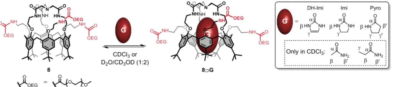
1-heptylamine. In strong contrast, the inclusion of cyclic ureas, i.e. Imi and 1,3-dihydro-2H-imidazol-2-one (DH-Imi), and of the -lactam pyrrolidin-2-one (Pyro) was clearly observed both in CDCl3 and in the D2O/CD3OD mixture (Scheme 2). The inclusion of small primary
amides such as AcNH2 and EtCONH2 was also detected but only in chloroform. In all cases,
the calixarene core of the complexes 8G displayed a flattened cone conformation, as revealed by the difference in chemical shift observed for the aromatic 1H (ΔδArH > 0.73 ppm;
see Figure 2b-c for 8Imi). It is noteworthy that quasi-identical chemical shifts were observed for the NCH2CO protons either in CDCl3 or in D2O/CD3OD (1:2) (NCH2CO = 3.21 or
Organic
&
Biomolecular
Chemistry
Accepted
Manuscript
Published on 10 November 2015. Downloaded by National University of Singapore on 10/11/2015 15:28:32.
View Article Online
6
3.24 ppm respectively for 8Imi), indicating that the basic capping tertiary amino group of the complexes 8G was not protonated in the aqueous medium.23,26 In both media, the high-field region of the 1H NMR spectra showed one or several signal(s) pertaining to the complexed guest, indicating slow in-out exchange on the chemical shift timescale, and integration revealed a 1:1 host-guest stoichiometry. The complexation induced shifts (CISs) for the complexes 8G are displayed in Table 1. For comparison purpose, the CISs were also determined for the parent system 1G in chloroform. First, all the CISs clearly show that the neutral guests are deeply included in the calixarene cavity. In the case of the complex 8Imi in D2O/CD3OD (1:2), the intra-cavity binding was confirmed by a 1D NOESY experiment
conducted with selective excitation of the signal at 0.26 ppm corresponding to the methylenic protons of the included Imi.23 Indeed, significant negative NOE’s were observed between these methylenic protons and the calixarene cavity (i.e. ArHin and tBuin protons).
Interestingly, very similar CISs were observed in the cases of complexes 1G and 8G in CDCl3 or in the D2O/CD3OD mixture (Table 1). This denotes a similar positioning of the
guest in the calixarene cavity whatever the solvent and the nature of the small rim substituents that are projected toward the outside of the cavity. Moreover, in all cases, significant downfield shifts of the NH protons of the tris-amido cap of 8 were observed upon complexation (e.g. δNHcap < 8.90 ppm and δNHcap = 9.67 ppm respectively for 8 and 8Imi in
CDCl3), suggesting a stabilization of the guest by strong H-bonding interactions.27 All in all,
these results show that calix[6]cryptamide 8 can selectively include polar neutral molecules that display H-bonding acceptor and donor groups such as amides or ureas either in apolar or aqueous environments. Similarly to natural systems, this binding of neutral guests through H-bonding interactions in a competitive aqueous medium may be due to the intra-cavity complexation of the guest which, consequently, is protected from the solvent.
Scheme 2. Host-guest properties of calix[6]cryptamide 8 towards polar neutral guests.
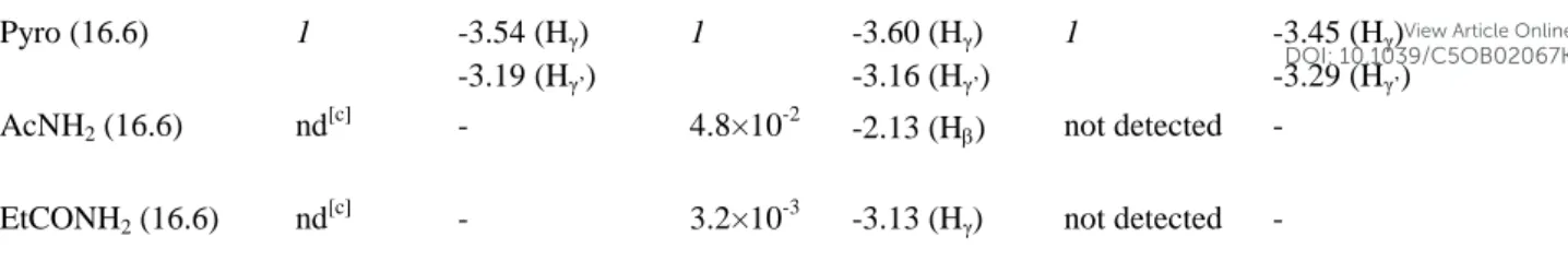
Table 1. Relative affinities (KG/Pyro) and
1
H NMR complexation induced shifts (CISs) in the case of hosts 1 and 8 with neutral guests. The acidity constant (pKa1) of the guests is also indicated.
1G in CDCl3 8G in CDCl3 8G in D2O/CD3OD (1:2) Guest G (pKa1)28 KG/Pyro [a] CIS (ppm)[b] KG/Pyro [a] CIS (ppm)[b] KG/Pyro [a] CIS (ppm)[b] DH-Imi (12.2) 1.3×10² -3.43 (Hγ) 1.1×10² -3.37 (Hγ) 0.46 -3.51(Hγ) Imi (14.6) 17 -3.35 (Hγ) 19 -3.31(Hγ) 3.0 -3.27(Hγ)
Organic
&
Biomolecular
Chemistry
Accepted
Manuscript
Published on 10 November 2015. Downloaded by National University of Singapore on 10/11/2015 15:28:32.
View Article Online
7 Pyro (16.6) 1 -3.54 (Hγ) -3.19 (Hγ’) 1 -3.60 (Hγ) -3.16 (Hγ’) 1 -3.45 (Hγ) -3.29 (Hγ’)
AcNH2 (16.6) nd[c] - 4.8×10⁻² -2.13 (H) not detected -
EtCONH2 (16.6) nd[c] - 3.2×10⁻³ -3.13 (Hγ) not detected -
[a] Relative affinities determined by NMR at 298 K and defined as ([Gin]×[Pyrofree])/([Gfree]×[Pyroin]) where the
subscript “in” stands for “included”. Errors estimated ±15%.
[b]CIS measured at 298 K and defined as Δδ = δ (Gin) – δ (Gfree). The β and γ positions are defined in Scheme 2.
[c] Not determined.
A deeper insight into the recognition process involved in these calix[6]arene-based complexes was gained by the X-ray structure analysis of the complex 1Imi (Figure 3b,c).29 First, the position of the guest into the poly-aromatic cavity and the flattened cone conformation of the host 1 are in good agreement with what was observed in solution by NMR spectroscopy. The guest is stabilized by multiple CH– interactions with the aromatic walls of the calixarene cavity. Besides, the complex exhibits a complementary DAAAD‐ADDDA quintuple H‐ bonding array between the cyclic ureido guest and its host. Note that the amido cap wraps around the guest and adopts a helical shape in order to maximize the number of H-bonding interactions. In other words, the XRD structure reveals that the key factor governing the recognition process is the high degree of complementarity between both the polar and apolar parts of the calixarene-based receptor and its guest. From a biomimetic point of view, the host–guest complex 1Imi shows remarkable similarities with complexes formed between biotin and biotin-binding proteins such as avidin. As a representative example, the XRD structure of one of the two subunits of a homodimeric avidin-biotin complex is given in Figure 3a.6 It shows that, similarly to the complex 1Imi, the avidin and biotin interact through a complementary DAAAD‐ADDDA H‐bonding array between the ureido imidazolidin-2-one ring and the polar side chains of a threonine, a tyrosine and two asparagine residues. Moreover, two tryptophan and a phenylalanine residue in the binding site form a polyaromatic pocket in which the hydrophobic part of the biotin resides and establishes CH- interactions.
Figure 3. a) X-ray diffraction structure of avidin-biotin complex (PDB accession 2AVI); b) and c) side and top
views of the X-ray diffraction structure of the complex 1Imi (obtained from X-ray crystals that were grown by
slow diffusion of pentane vapor into a solution of 1Imi in chloroform at ca. 4 °C). Receptors are represented in
capped sticks model while the guests are represented in balls and sticks or spacefill models. Hydrogen bonds are indicated by dashed lines. All the hydrogen atoms of the complexes, minor disorder components and the solvents
Organic
&
Biomolecular
Chemistry
Accepted
Manuscript
Published on 10 November 2015. Downloaded by National University of Singapore on 10/11/2015 15:28:32.
View Article Online
8
of crystallization are omitted for clarity. Selected distances in the case of the complex 1Imi (CHCl3:pentane):
[d(N…O)]: 2.874, 2.875, 2.934, 2.968 and 2.986 Å.
1
H NMR competitive binding experiment showed that receptors 1 and 8 possess very similar relative affinities toward neutral guests in chloroform (Table 1). Indeed, in both cases, the affinity decreases significantly according to the sequence DH-Imi > Imi > Pyro. Moreover, in the case of 8, the affinity for DH-Imi is at least four orders of magnitude higher than for the primary amido guests AcNH2 and EtCONH2. On the one hand, the affinity depends on the
number of H-bonding interactions that the guest can form; ureas are thus better recognized than amides. On the other hand, the affinity increases with the acidity of the guest (Table 1). Hence, with such a synergistic effect, DH-Imi behaves as the best “key” for receptors 1 and 8. Interestingly, the affinities observed in D2O/CD3OD (1:2) in the case of the hydrophilic
receptor 8 are very different from those observed in chloroform. First, as mentioned above, the complexation of primary amides could not be detected despite the addition of a large excess of these guests. Moreover, a weak discrimination was observed in the case of the cyclic guests (Table 1), DH-Imi being now the worst guest. Such a discrepancy between the results obtained in chloroform and in an aqueous medium is ascribable to the greater solvation in water of the more polar and acidic molecules. Besides, in absence of any guest, receptor 8 adopts a flattened cone conformation very similar to that observed in the case of complexes 8G.23 This behavior can be rationalized by the filling of the receptor by one or more water molecules. In other words, the neutral guests have to compete with water in an aqueous medium. It is noteworthy that an apparent association constant (Kapp) of 300 M-1 could be
estimated for Imi in the 1:2 mixture of D2O and CD3OD (see the Experimental Section). For
comparison purpose, an association constant of 5.2×10³ M-1 for the complex 8Imi was determined in CDCl3,30 showing that the recognition of neutral guests is stronger in a less
competitive medium.
Finally, the host-guest properties of host 8 in presence of acids were evaluated at 298 K by 1H NMR spectroscopy in D2O/CD3OD (1:2). First, a reference spectrum of the protonated ligand 8.H+ was obtained by addition of D2SO4 (0.1 M, 1 equiv.) to host 8.23 The protonation of the
basic cap at the level of the tertiary amino group was clearly visible by the significant downfield shift of the HN+-CH2 protons ( = 0.15 ppm).31 After that, an experiment consisting in the progressive addition of D2SO4 (> 96%) to a solution of the complex 8Imi
was conducted. It led to the release of the urea guest and the formation of the protonated derivative 8.H+, the exchange between the two species being slow on the NMR timescale (Scheme 3). Such a reluctance of the protonated derivative toward polar neutral guests was already observed in the case of the parent receptor 1 in chloroform and was attributed to the competing formation of a stable five membered intramolecular hydrogen-bonded ring between the HN+ and an introverted amido C=O group.17a The HN+ proton can thus be considered as an allosteric inhibitor that induces a conformational reorganization of the trisamido recognition site into an insensitive form of the receptor. Very interestingly, it was also shown that ca. 30% of the complex 8Imi survived the addition of a large amount of D2SO4 (> 96%, 450 equiv.) while only 1 equiv. of this acid was necessary to fully protonate
host 8 in absence of Imi. This result highlights the remarkably high stability of the complex
Organic
&
Biomolecular
Chemistry
Accepted
Manuscript
Published on 10 November 2015. Downloaded by National University of Singapore on 10/11/2015 15:28:32.
View Article Online
9
8Imi in acidic media and suggests a significant pKa shift of the basic cap upon complexation
of the urea guest.
Scheme 3. Acid-triggered release of Imi in an aqueous medium. Inset: aromatic region of the 1H NMR spectra (D2O/CD3OD (1:2), 600 MHz, 298 K) of 8 a) after addition of 18 equiv. of Imi; b) after the subsequent addition
of 450 equiv. of D2SO4 (> 96%); ▼: 8Imi; ♦: 8.H+.
Conclusion
The synthesis of a hydrophilic calix[6]cryptamide decorated with oligo(ethylene glycol) units (i.e. 8) was readily achieved from the known C3v molecular platform 5 through an efficient
[1+1] macrocyclization reaction as the key-step. Host 8 is soluble either in apolar solvents or in aqueous media, allowing us to compare its binding properties toward neutral molecules in two different environments. In both media, it appears that 8 behaves as a remarkable receptor for neutral molecules that display H-bonding acceptor and donor groups such as amides or ureas. As shown by NMR and X-ray diffraction data obtained for the parent receptor 1, the mode of recognition involves a complementary DAAAD‐ADDDA quintuple H‐bonding array between the guest and the calixarene-based host. The fact that such a mode of recognition can operate in an aqueous medium highlights the high preorganization of the tris-amido binding site of 8 and its protection from the external medium by the calixarene-based hydrophobic corridor. Very interestingly, these host-guest systems provide a simple structural model for the binding site of biotin-binding proteins. 1H NMR comparative studies show that the affinity of 8 for neutral molecules greatly depends on the nature of the environment, the most polar and acidic molecule being the best guest in chloroform and the worst one in an aqueous medium. Besides, protonation of the receptor 8 leads to the release of the guest through a conformational reorganization of the tris-amido recognition site. This control of the binding properties in aqueous media is highly reminiscent of allosteric processes encountered in natural systems. Current efforts are now directed toward the use of 8 and related receptors for the sensing of charged and neutral species in an aqueous environment.
Experimental Section
General experimental methods. All reactions were performed under an inert atmosphere. Commercial anhydrous solvents were used. Other solvents and chemicals were of reagent grade and were used without purification. Silica gel (230-400 mesh) was used for flash chromatography separations. 1H NMR spectra were recorded at either 600, 400 or 300 MHz and 13C NMR spectra were recorded at 75 MHz using Varian VNMRS-600, VNMRS-400 or Bruker Avance-300 spectrometers equipped with a 5 mm probe. The solvent was used as internal standard for both 1H and 13C chemical shift referencing (δ 1H = 7.26 ppm for residual
Organic
&
Biomolecular
Chemistry
Accepted
Manuscript
Published on 10 November 2015. Downloaded by National University of Singapore on 10/11/2015 15:28:32.
View Article Online
10
CHCl3 and 3.31 ppm for residual CHD2OD; δ 13C = 77.16 ppm for CDCl3 and 49.00 ppm for
CD3OD). CDCl3 was filtered over a short column of basic alumina in order to remove traces
of DCl. Most of the 1H NMR spectra signals were assigned through 2D NMR analyses (COSY, HSQC, HMBC). Low-resolution mass spectra were recorded on an ESI-MS apparatus (Finnigan ThermoQuest LCQ-Deca) equipped with an ion-trap using the following settings: flow rate: 10 μL.min-1, spray voltage: 5 kV, capillary temperature: 160°C, capillary voltage: 10V, tube lens offset voltage: -5V. High-resolution mass spectra were recorded on an ESI-MS apparatus (Q-TOF 6520 Agilent Technology) equipped with a TOF detector. Otherwise notified, IR analyses were performed with a Bruker IFS-25 on pellets of potassium bromide. Compounds 4,20 5,20 922 were prepared according to procedures already described in the literature.
Calix[6]cryptamide 6. Anhydrous CHCl3 (45 mL) and anhydrous DMF (20 mL) were added
to calix[6]arene 5 (502 mg, 0.328 mmol, 1 equiv.) and nitrilotriacetic acid (156 mg, 0.816 mmol, 2.5 equiv.). A solution of TBTU (524 mg, 1.64 mmol, 5 equiv.) and TEA (220 µL, 1.64 mmol, 5 equiv.) in anhydrous DMF (25 mL) was then added. The reaction mixture was stirred for 15 h at 50 °C and then the solvents were removed under reduced pressure. The residue was dissolved in CH2Cl2 (100 mL) and washed with an aqueous NH4OH solution
(5%, 50 mL). The aqueous layer was extracted with CH2Cl2 (2×25 mL) and the combined
organic layers were washed with H2O (2×50 mL) and concentrated under reduced pressure.
The crude residue was triturated with H2O/EtOH 1:1 and the resulting precipitate was isolated
by filtration and dried under vacuum to give the calix[6]cryptamide 6 (418 mg, 76%) as a beige solid. Mp 190 °C; IR (KBr): υ 3408, 2963, 1685, 1540, 1482, 1458, 1364, 1175, 1049 cm-1; 1H NMR of 6Imi (400 MHz, CDCl3, 298 K): δH (ppm) 0.23 (s, 4H, CH2 Imiin), 0.75
(s, 27H, tBu), 1.39 (s, 54H, tBu + Boc), 3.24 (s, 6H, NCH2CONH), 3.44 (d, J = 15.1 Hz, 6H, ArCH2eq), 3.50 (m, 6H, OCH2CH2NHBoc), 3.91 (sb, 6H, OCH2CH2NHBoc), 4.02 (sb, 6H,
OCH2CH2NHCO), 4.05 (sb, 6H, OCH2CH2NHCO), 4.40 (d, J = 15.1 Hz, 6H, ArCH2ax), 4.64
(s, 2H, NH Imiin), 5.07 (sb, 3H, NHBoc), 6.56 (s, 6H, ArH), 7.30 (m, 6H, ArH), 9.60 (sb, 3H,
NCH2CONH); 13C NMR of 6Imi (75 MHz, CDCl3, 298 K): δC (ppm) 28.5, 29.3, 31.3, 31.8,
34.1, 34.5, 38.5 (Imiin), 40.8, 41.4, 60.8, 72.1, 77.5, 79.9, 123.6, 128.8, 132.1, 132.5, 145.2,
146.4, 151.9, 153.6, 156.1, 165.3 (Imiin), 170.6. HRMS (ESI-TOF) calcd for C99H141N7O15Na
[M+Na]+ 1691.0383, found 1691.0387.
Calix[6]cryptamide 7. Calix[6]cryptamide 6 (100.2 mg, 0.06 mmol) was dissolved in CH2Cl2 (5 mL) and TFA (2 mL) was slowly added. The reaction mixture was stirred
overnight at room temperature and then the solvent was removed under reduced pressure. The crude residue was triturated with diethyl ether (3×2 mL) and the solvent was removed under reduced pressure. The resulting solid was dissolved in CH2Cl2 (10 mL) and washed with
NaOH (1 M, 5 mL) for 1 h. The aqueous layer was extracted with CH2Cl2 (2×10 mL) and the
combined organic layers were washed with H2O (2×10 mL) and concentrated under reduced
pressure to give calix[6]cryptamide 7 (82.2 mg, quant. yield) as a pale yellow solid. Mp 200 °C (dec); IR (KBr): υ 3360, 2959, 1672, 1477, 1459, 1199, 1121, 1051 cm-1
; 1H NMR of 7Imi (300 MHz, CDCl3, 298K): δH (ppm) 0.23 (s, 4H, CH2 Imiin), 0.77 (s, 27H, tBu), 1.40
(s, 27H, tBu), 3.13 (t, J = 5.6 Hz, 6H, CH2NH2), 3.21 (s, 6H, NCH2CONH), 3.46 (d, J = 15.1 Hz, 6H, ArCH2eq), 3.88 (t, J = 5.6 Hz, 6H, OCH2CH2NH2), 4.00 (sb, 6H, OCH2CH2NHCO),
4.09 (sb, 6H, OCH2CH2NHCO), 4.45 (d, J = 15.1 Hz, 6H, ArCH2ax), 4.62 (s, 2H, NH Imiin),
Organic
&
Biomolecular
Chemistry
Accepted
Manuscript
Published on 10 November 2015. Downloaded by National University of Singapore on 10/11/2015 15:28:32.
View Article Online
11
6.61 (s, 6H, ArH), 7.31 (m, 6H, ArH), 9.68 (sb, 3H, NCH2CONH). 13C NMR of 7Imi (75
MHz, CDCl3, 298 K): δC (ppm) 29.3, 31.1, 31.8, 34.1, 34.5, 38.6 (Imiin), 41.0, 42.5, 60.1,
75.3, 77.9, 123.6, 128.7, 132.2, 132.6, 145.1, 146.2, 151.9, 153.8, 165.3 (Imiin), 170.7; HRMS
(ESI-TOF) calcd for C84H118N7O9 [M+H]+ 1368.8991, found 1368.8885.
Calix[6]cryptamide 8. 2-[2-(2-methoxyethoxy)ethoxy]acetyl chloride 9 (36.7 mg, 0.1867 mmol, 3.6 equiv.) was added at 0 °C to a solution of calix[6]cryptamide 7 (71 mg, 0.0519 mmol, 1 equiv.) and TEA (43.4 µL, 0.3112 mmol, 6 equiv.) in anhydrous DMF (4.3 mL). The mixture was stirred at 70 °C for 22 h and then the solvent was removed under reduced pressure. The crude residue was triturated with H2O (3×1 mL) and the resulting precipitate
was isolated by centrifugation and dried under vacuum to give the calix[6]cryptamide 8 (73.2 mg, 76%) as a beige solid. Mp 145 °C; IR (KBr): υ 3325, 2955, 1664, 1541, 1481, 1460, 1362, 1196, 1050 cm-1;1H NMR of 8Imi (600 MHz, CDCl3, 298K): δH (ppm) 0.23 (s, 4H,
CH2 Imiin), 0.75 (s, 27H, tBu), 1.40 (s, 27H, tBu), 3.21 (s, 6H, NCH2CONH), 3.34 (s, 9H, OCH3), 3.47 (d, J = 15.0 Hz, 6H, ArCH2eq), 3.50-3.72 (m, 30H, OCH2OEG + CH2NHCOOEG),
3.93-3.98 (m, 12H, ArOCH2OEG+cap), 4.00 (sb, 6H, OCH2CONHOEG), 4.08 (sb, 6H,
CH2NHCOcap), 4.38 (d, J = 15.1 Hz, 6H, ArCH2ax), 4.56 (s, 4H, NH Imiin) 6.56 (s, 6H, ArH),
7.30 (m, 9H, ArH + CONHOEG), 9.67 (sb, 3H, NCH2CONH). 13C NMR of 8Imi (75 MHz,
CDCl3, 298 K): δC (ppm) 29.3, 31.3, 31.8, 34.1, 34.5, 38.6 (Imiin), 39.3, 40.5,* 59.0, 60.5,*
70.4, 70.5, 70.6, 71.2, 71.6, 72.0, 77.9, 123.7, 128.9, 132.0, 132.5, 145.1, 146.2, 152.2, 153.7, 165.4 (Imiin), 170.6, 170.7; HRMS (ESI-TOF) calcd for C105H154N7O21 [M+H]+ 1849.1198,
found 1849.1166. *: determined by 2D NMR spectroscopy analysis (HSQC, HMBC).
Determination of the relative affinity data KG/Pyro by 1H NMR competitive binding
studies in CDCl3 and in D2O/CD3OD (1:2). To a solution containing 8 (3.0×10⁻³ M) were successively added Pyro (> 1 equiv.) and a second guest G (> 1 equiv.) in such a ratio that a
1
H NMR spectrum recorded at 298 K showed the resonances of both the complexes 8Pyro and 8G besides the signals corresponding to the free guests (Pyro and G). Integration of the signals of the included guests, i.e. Pyroin and Gin, and of the free guests, i.e. Pyrofree and Gfree,
allowed us to calculate the relative affinity KG/Pyro, defined as ([Gin]×[Pyrofree])/([Gfree]×[Pyroin]).
Determination of the apparent association constant Kapp of 8 toward Imi in D2O/CD3OD (1:2). To a D2O/CD3OD (1:2) solution containing 8 (1.6×10⁻³ M) was added Imi in such a
ratio that a 1H NMR spectrum recorded at 298K showed the resonances of both the calixarene 8 and the complex 8⊃Imi besides the signals corresponding to the free guest (Imi). Integration of the signals of these species allowed us to calculate the association constant Kapp
according to the following equation: Kapp = [8⊃Imi]/([8]×[Imi]).
X-ray crystallography, 1Imi (CHCl3:pentane): X-ray crystals were grown by slow
diffusion of pentane vapor into a solution of 1Imi in chloroform at ca. 4 °C. C89.62H124.18Cl7.05N6O10, M = 1695.54 gmol−1, monoclinic, space group P21/n, a = 15.2163(4)
Å, b = 29.5270(15) Å, c = 20.3699(10) Å, = 92.916(3)°, V = 9140.2(7) Ǻ3, Z = 4, 49692 reflections (θmax = 26.993°) measured (19310 unique, Rint = 0.1106, completeness = 96.8%),
Final R indices (I > 2σ(I)): R1= 0.0878, wR2 = 0.2346, R indices (all data): R1= 0.1596, wR2 =
0.2164. GOF = 1.011 for 1115 parameters and 1625 restraints, largest diff. peak and hole 0.772/–0.474 eǺ−3. 1Imi (CHCl3:diisopropyl ether): X-ray crystals were grown by slow
Organic
&
Biomolecular
Chemistry
Accepted
Manuscript
Published on 10 November 2015. Downloaded by National University of Singapore on 10/11/2015 15:28:32.
View Article Online
12
diffusion of diisopropyl ether vapor into a solution of 1Imi in chloroform at ca. 4 °C. C88.35H120.90Cl7.34N6O10.32, M = 1692.16 gmol−1, monoclinic, space group P21/n, a =
15.2333(7) Å, b = 29.5603(13) Å, c = 20.3969(5) Å, = 92.992(2)°, V = 9172.2(6) Ǻ3, Z = 4, 48842 reflections (θmax = 27.476°) measured (20321 unique, Rint = 0.1454, completeness =
93.9%), Final R indices (I > 2σ(I)): R1= 0.1139, wR2 = 0.2857, R indices (all data): R1=
0.2254, wR2 = 0.3056. GOF = 1.021 for 1207 parameters and 1871 restraints, largest diff.
peak and hole 0.554/–0.452 eǺ−3. CCDC 1428618 and 1428619 contain the supplementary crystallographic data for this paper. These data can be obtained free of charge from The Cambridge Crystallographic Data Centre via www.ccdc.cam.ac.uk/data_request/cif.
Supplementary Information: 1D and 2D NMR spectra of all new compounds, competitive NMR binding studies of 8 with Imi and Pyro and their behavior in acidic environment, refinement details of the structure solution and refinement of the crystal structures.
Acknowledgments. This work was supported by the Université libre de Bruxelles - ULB (A.L. Ph.D. grant), the Fonds de la Recherche Scientifique-FNRS (FRFC 2.4.617.10.F Project and G.D.L. Ph.D. grant), the Agence Nationale de la Recherche (ANR10-BLAN-714 Cavity-zyme(Cu) Project), Academy of Finland (K.R.: project no.’s 263256 and 265328) and the University of Jyväskylä, and was undertaken within the framework of the COST Action CM-1005 “Supramolecular Chemistry in Water”.
Corresponding Author
* E-mail: [email protected]; Fax: (+32) 2 650 2799; Tel: (+32) 2 650 3537.
Notes and References
1
(a) J.-M. Lehn, in Supramolecular Chemistry, Wiley-VCH, Weinheim, 1995; (b) J. W. Steed, D. R. Turner and K. J. Wallace, in Core Concepts in Supramolecular Chemistry and
Nanochemistry, Wiley-VCH, Chippenham, 2007; (c) T. D. James and C. J. Ward, J. Chem. Soc. Perkin Trans. 1, 2000, 3155; (d) H.-J. Schneider, in Applications of Supramolecular Chemistry, ed. H.-J. Schneider, CRC Press, Taylor & Francis group, Boca Raton, 2012, 3, 49;
(e) J.-M. Lehn, Science, 2002, 295, 2400.
2
G. V. Oshovsky, D. N. Reinhoudt and W. Verboom, Angew. Chem., Int. Ed., 2007, 46, 2366.
3
J. Szejtli, Chem. Rev., 1998, 98, 1743.
4
J. Lagona, P. Mukhopadhyay, S. Chakrabarti and L. Isaacs, Angew. Chem., Int. Ed., 2005, 44, 4844.
5
H. Dugas, in Bioorganic Chemistry: A Chemical Approach to Enzyme Action, 3rd ed.; Springer-Verlag: New York, 1996; pp 252-387.
6
O. Livnah, E. A. Bayer, M. Wilchek and J. L. Sussman, Proc. Natl. Acad. Sci. USA, 1993, 90, 5076.
7
(a) N. Le Poul, Y. Le Mest, I. Jabin, and O. Reinaud, Acc. Chem. Res., 2015, 48, 2097; (b) R. Gramage-Doria, D. Armspach and D. Matt, Coord. Chem. Rev., 2013, 257, 776; (c) J.-N. Rebilly, B. Colasson, O. Bistri, D. Over and O. Reinaud, Chem. Soc. Rev., 2015, 44, 467; (d)
Organic
&
Biomolecular
Chemistry
Accepted
Manuscript
Published on 10 November 2015. Downloaded by National University of Singapore on 10/11/2015 15:28:32.
View Article Online
13
D. Coquière, S. Le Gac, U. Darbost, O. Sénèque, I. Jabin and O. Reinaud, Org. Biomol.
Chem., 2009, 7, 2485; (e) O. Bistri and O. Reinaud, Org. Biomol. Chem., 2015, 13, 2849.
8
(a) S. B. Ferguson, E. M. Seward, E. M. Sanford, M. Hester, M. Uyeki and F. Diederich
Pure & App. Chem., 1989, 61, 1523; (b) M. Miyake, M. Kirisawa and K. Koga, Chem. Pharm. Bull. 1992, 40, 3124; (c) F. Diederich, Angew. Chem., Int. Ed., 1988, 27, 362; (d) M.
Inouye, K. Fujimoto, M. Furusyo and H. Nakazumi, J. Am. Chem. Soc., 1999, 121, 1452.
9
F. H. Zelder, R. Salvio and J. Rebek Jr., Chem. Commun., 2006, 1280.
10
O. Perraud, V. Robert, H. Gornitzka, A. Martinez and J.-P. Dutasta, Angew. Chem., Int.
Ed., 2012, 51, 504.
11
Y. Ma, M. Xue, Z. Zhang, X. Chi and F. Huang, Tetrahedron, 2013, 69, 4532.
12
(a) K. D. Daze, M. C. F. Ma, F. Pineux and F. Hof, Org. Lett., 2012, 14, 1512; (b) F. Cuevas, S. Di Stefano, J. O. Magrans, P. Prados, L. Mandolini and J. de Mendoza, Chem.
Eur. J., 2000, 6, 3228; (c) E. Brunetti, A. Inthasot, F. Keymeulen, O. Reinaud, I. Jabin and K.
Bartik, Org. Biomol. Chem., 2015, 13, 2931; (d) S. Kunsági-Máté, K. Szabó, I. Bitter, G. Nagy and L. Kollár, J. Phys. Chem. A, 2005, 109, 5237; (e) F. Corbellini, R. M. A. Knegtel, P. D. J. Grootenhuis, M. Crego-Calama and D. N. Reinhoudt, Chem. Eur. J., 2005, 11, 298.
13
A. Ikeda and S. Shinkai, Chem. Rev., 1997, 97, 1713.
14
R. Lavendomme, S. Zahim, G. De Leener, A. Inthasot, A. Mattiuzzi, M. Luhmer, O. Reinaud and I. Jabin, Asian J. Org. Chem., 2015, 4, 710.
15
(a) E. Garrier, S. Le Gac and I. Jabin, Tetrahedron: Asymm., 2005, 16, 3767; (b) I. Jabin and O. Reinaud, J. Org. Chem., 2003, 68, 3416.
16
M. Ménand and I. Jabin, Org. Lett., 2009, 11, 673.
17
(a) A. Lascaux, S. Le Gac, J. Wouters, M. Luhmer and I. Jabin, Org. Biomol. Chem., 2010, 8, 4607; (b) A. Lascaux, G. Delahousse, J. Ghostin, J.-P. Bouillon and I. Jabin, Eur. J. Org.
Chem., 2011, 5272.
18
(a) M. Ménand and I. Jabin, Chem. Eur. J., 2010, 16, 2159; (b) U. Darbost, M-.N. Rager, S. Petit, I. Jabin and O. Reinaud, J. Am. Chem. Soc., 2005, 127, 8517.
19
For artificial receptors devoted to the complexation of urea or biotin derivatives, see: (a) M. D. Santa María, M. Á. Farráan, M. Á. García, E. Pinilla, M. R. Torres, J. Elguero and R. M. Claramunt, J. Org. Chem., 2011, 76, 6780; (b) F. Herranz, M. D. Santa María and R. M. Claramunt, J. Org. Chem., 2006, 71, 2944; (c) V. Hedge, C.-Y. Hung, P. Madhukar, R. Cunningham, T. Höpfner and R. P. Thummel, J. Am. Chem. Soc., 1993, 115, 872.
20
S. Le Gac, J. Marrot and I. Jabin, Chem. Eur. J., 2008, 14, 3316.
21
S. Le Gac and I. Jabin, Chem. Eur. J., 2008, 14, 548.
22
Y. Pan and W. T. Ford, Macromolecules, 2000, 33, 3731.
23
See the Supporting Information.
24
Note that the 1H NMR spectrum of 8 remained unchanged upon dilution in CDCl3. This
result indicates that the multiple conformations are likely due to the presence of an intramolecular H-bonding network between the multiple H-bonding donor and acceptor groups of the receptor.
25
Host 8 is not soluble in pure D2O. 26
In D2O/CD3OD (1:2), the signal of the NCH2CO protons was identified through 2D NMR
spectroscopy analysis (HSQC).
Organic
&
Biomolecular
Chemistry
Accepted
Manuscript
Published on 10 November 2015. Downloaded by National University of Singapore on 10/11/2015 15:28:32.
View Article Online
14
27
Note that a slow deuteriation process was observed in D2O/CD3OD (1:2) for the
exchangeable NH protons of 8, allowing the detection of their 1H signal.
28
The pKa1 values were calculated using the Advanced Chemistry Development (ACD/Labs)
software V11.02 (© 1994-2015 ACD/Labs).
29
X-ray crystals were grown by slow diffusion either of pentane or diisopropyl ether vapors
into a solution of 1Imi in chloroform at ca. 4 °C (see the Experimental Section). Very similar X-ray structures were obtained from both crystals (see the Supporting Information).
30
This value is based on the hypothesis that the influence of residual water in CDCl3 is
negligible.
31
Note that a minor dissymmetrical conformation with a self-included tBu group was also observed.
Organic
&
Biomolecular
Chemistry
Accepted
Manuscript
Published on 10 November 2015. Downloaded by National University of Singapore on 10/11/2015 15:28:32.
View Article Online
![Figure 1. The calix[6]cryptamides 1-3.](https://thumb-eu.123doks.com/thumbv2/123doknet/14512834.721305/5.892.302.592.274.471/figure-the-calix-cryptamides.webp)