HAL Id: tel-00597065
https://tel.archives-ouvertes.fr/tel-00597065
Submitted on 31 May 2011
HAL is a multi-disciplinary open access archive for the deposit and dissemination of sci-entific research documents, whether they are pub-lished or not. The documents may come from teaching and research institutions in France or abroad, or from public or private research centers.
L’archive ouverte pluridisciplinaire HAL, est destinée au dépôt et à la diffusion de documents scientifiques de niveau recherche, publiés ou non, émanant des établissements d’enseignement et de recherche français ou étrangers, des laboratoires publics ou privés.
microtubules XMAP215/ch-TOG
Claudia Paez
To cite this version:
Claudia Paez. Etude fonctionnelle de la protéine associée aux microtubules XMAP215/ch-TOG. Biologie cellulaire. Université de Grenoble, 2011. Français. �NNT : 2011GRENV014�. �tel-00597065�
THÈSE
Pour obtenir le grade de
DOCTEUR DE L’UNIVERSITÉ DE GRENOBLE
Spécialité : Biologie cellulaire
Arrêté ministériel : 7 août 2006
Présentée par
Claudia PAEZ
Thèse dirigée par Monsieur le Professeur François BERGER préparée au sein du Laboratoire Nanomédicine et cerveau.
Grenoble Institut des Neurosciences INSERM U836
dans l'École Doctorale Chimie et Sciences du Vivant
Etude fonctionnel de la Protéine
associée aux Microtubules
XMAP215/ch-TOG
Thèse soutenue publiquement le 29 Avril 2011 devant le jury composé de :
Monsieur le Dr. Marc SAVASTA
Directeur, Ecole doctorale Chimie et Sciences du Vivant, Grenoble. Président du Jury
Monsieur le Dr. Matthieu PIEL
Chef d’équipe à l’Institut Curie, Paris. Rapporteur Monsieur le Dr. Phong TRAN
Chef d’équipe à l’Institut Curie, Paris. Rapporteur Monsieur le Pr. François BERGER
Directeur d´’équipe au Grenoble Institut des Neurosciences (GIN). Examinateur
They (the cosmos explorations) remind us that humans are evolved to wonder, that understanding is a joy, that knowledge is a prerequisite to survival…
Those explorations required skepticism and imagination both. Imagination will often carry us to worlds that never were. But without it we go nowhere. Skepticism enables us to distinguish fancy from fact, to test our speculations. The cosmos is rich beyond measure – in elegant facts, in exquisite interrelationships, in the subtle machinery of awe.
Carl Sagan, Cosmos. Astrophysicist
I
I would like to thanks Professors Matthieu PIEL and Phong TRAN for refereeing this thesis manuscript. It’s a privilege for me that professionals of such international reputation have accepted to judge my work.
I do not have enough words to express my gratitude to the Joseph Fourier University, in particulary to its president, Mr Farid Ouabdesselam. His helping and fair hand made possible that an objective might comme true.
I am really grateful with the members of the Joseph’s Fourier Doctoral School CSV (Chimie and Sciences du Vivant). Especially I would like to extend my warmest gratitude to its director, Dr Marc Savasta. Thanks a lot for his professional and human support that finally allow me to reach the end.
To Dr François Berger at the Grenoble Institut of Neurosciences (GIN), I do express my gratitude. Thanks for accepting me in to your group, an acceptation made with open arms. Thanks for your support and kindness through all this time.
I would like to thank, Dr Andrei Popov at the GIN. His accurate guidance through the course of my PhD studies was a key element in my development as a scientist.
Thanks to each one of the members of team 7 at the GIN. These professionals assumed day after day the heroic and courageous effort to deal with my franglish, decorated with an ethnic touch of colombian spanish.
Thanks to the great professionals and scientist that made all this work possible:
Thanks to Emannuelle Neumann at the IBS institute. A great scientist that made possible the electron microscopy experiments. Thanks for your kindness, professionalism and open minded criteria.
Thanks to Alexei Grichine, at the IAB institute. A well recognize international authority in the imaging domain, but above all a great human being with a huge sense of humor. The laughs allow me to survive many afternoons of bored and almost never ending imaging analysis. Thanks for your inconditional support. Thanks to Dr Didier Grundwald at the CEA (Grenoble). His help, kindness, entousiasm and professionalism during the acquisition of the confocal images were determinant for this research.
Thanks to those brave scientists that using the sense of sacrifice and courage read this up-side-down manuscript. Because of their efforts, time, good will and patience, they succeeded to transform all those chaotic pages in to a scientific dissertation. Thanks to Michelle, Jean Paul and Jean-Marc. Please guys, the next time do not forget to check the bibliography...
II
also a home, a family and great friends. I must say thanks to each one of the members of this wonderful and giving family that expressed an unlimited generosity, they tolerate an alien among them and this grateful alien was treated as one of them. God bless you all.
Thanks to Constanza, the brain and heart behind this project. Thanks for being so stubborn, not accepting a no for an answer. Thanks for listening, for your prayers, for being there… for so many years already. Thanks for the advices, the trust, the free sharing since the early years of this adventure.
Thanks to Svetlana, pure energy coming from the distant Siberia. Thanks for the inner peace, wisdom and compassion message transmited systematically through years... мышь
спасибо
много
велемудрая
.
Thanks to Slaveia… for the adulthood minded crieria, the faith, freedom and for keeping this friendship safe through the distance and time.
Thanks to Mailyn. I just can say: God bless you. You where a light in the dark… only a true friend dares to do so. Gracias
Thanks to the selected members of the Pick-Assietes Grenoble Club. To Vincent, Carmen, Claude, Stephanie, Carole, Fabienne, Nicole and Jean-Jacques. Thanks for accepting my modest candidature to enjoy you taxes….
Thanks to Nicole. Thanks for the free thinking and the open minded sharing, the unlimited generosity, the freedom, the solidarity, the complexity and simplicity of the friendship… thanks for everything.
Thanks to Soline, that funny little and generous fairy that made this manuscript and my thesis defence possible... 100% pure handcraft.Thanks to Aurelien, Jao and Eliseo… for their laughs.
I must thank my beloved third world. Thanks a lot for the ever lasting hope, for the wisdom of the chaos, for the never-ending positivism, for the short term memory law, for the warm and friendly no reason every day smile...
I must thanks to my beloved ones. Thanks to Mom and Dad, my angels. You encouraged me to follow my dreams at my way. Thanks for the freedom to growing up as a wild-type human being, thanks for the respect for each one of my projects and for the non question inner law. Thanks to every single member of this loving and mad family that excerced individually or collectivelly as a hybrid among Sigmund Freud and a Tibetan monk… thanks a lot for the brigtest words in the darkest times.
At the end, it is time to say thanks to my beginning. Thanks to the light, thanks to the love, thanks to Abba...
III
ACKNOWLEDGEMENTS
I
LIST OF ABBREVIATIONS
III
CONTENTS
IV
FIGURES LIST
XI
TABLES LIST
XII
SUMMARY
1
RESUME
3
GENERAL INTRODUCTION
5
1. The cell and its cell cycle
7
1.1 The cell
7
1.2 The cell cycle
7
1.3 Mitosis phases
9
1.3.1 Interphase
9
1.3.2 Prophase
9
1.3.3 Prometaphase
9
1.3.4 Metaphase
10
1.3.5 Anapahase
10
1.3.6 Telophase
10
1.3.7 Cytokinesis
10
2. Cytoskeleton
13
2.1 Microtubules
15
2.1.1 The dynamic instability model
17
IV
3.1 Differential mechanism
23
3.2 EB1 (Ending Binding Protein 1)
25
3.2.1 EB1 Structure
26
3.2.2 EB1 Interactions
27
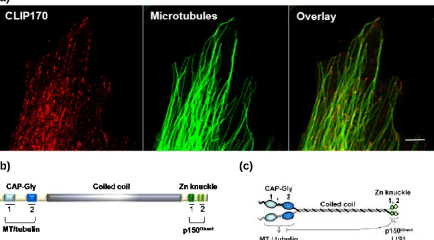
3.3 CLIP170 (Cytoplasmic Linker Protein 170)
28
3.3.1 CLIP170 structure
28
3.3.2 CLIP170 interactions
29
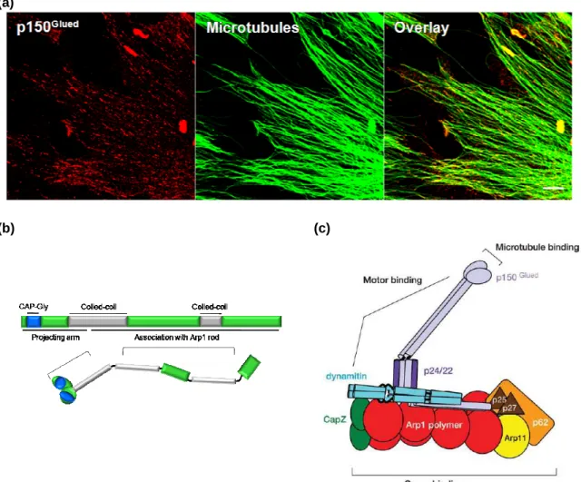
3.4 p150
Glued30
3.4.1 p150
Gluedstructure
32
3.4.2 p150
Gluedinteractions
32
3.5 Regulation of + TIPs interactions
33
3.5.1 Specific sequences
33
3.5.2 Phosphorylation
34
3.5.3 Mechanical implications
36
3.5.4 α-tubulin tyrosination-detyrosination
36
3.5.5 The GTP cap
37
3.5.6 Autoinhibition complexes or co-regulation?
38
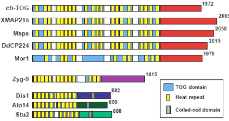
4. The XMAP215/Dis1family
41
4.1 Protein Structures: conserved repeat motifs
42
4.2 Cellular localization of XMAP215/Dis1 proteins 44
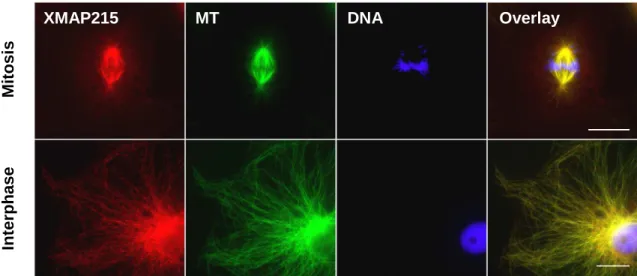
4.3 XMAP215
46
4.3.1 XMAP215 and tubulin dimmers
48
4.4 ch-TOG
49
4.5 XMAP215/ch-TOG mapping
50
V
5. Anti XMAP215/ch-TOG mcAB production
57
5.1 XMAP215 characterization
57
5.1.1 mcAB antibodies obtention
57
5.1.2 Cell Fixing and Immunofluorescence
58
5.2 mcAB Characterization
61
5.2.1 mcAB Specificity
61
5.2.1.1 mcAB specificity: egg extract obtention
61
5.2.1.2 mcAB specificity: XMAP215 depletion
63
5.2.1.3 mcAB Class
64
5.2.1.4 mcAB epitopes cartography
64
5.2.1.5 Fixed cells imaging
65
5.3 XMAP215 in vitro interaction
66
5.3.1 Co-sedimentation Assays
66
5.3.1.1 Tubulin/XMAP215-his7 interaction
66
5.3.1.2 Tubulin / XMAP215-his7 / mcAB
66
5.3.1.3 Microtubules/XMAP215-his7 / mcAB
67
5.4 XMAP215 5D12 in vivo behavior
68
5.4.1 5D12 mcAB labeling
68
5.4.2 Microscopy assay visualization of MTs/XMAP215
interaction: Total Internal Reflection Fluorescence
Microscopy (TIRF)
69
5.5 XMAP215/MT ultrastructural behaviour: Electron
microscopy
70
5.5.1 Negative staining
71
5.5.2 Specimen preparation and cryo-electron
microscopy
71
VI
5.6.1 Screening
72
5.6.2 Experimental approach
73
5.6.2.1 Molecules collection
73
5.6.2.2 Tubulin
73
5.6.2.3 Rhodamine conjugated to Tubulin
74
5.6.2.4 XMAP215-his7 Protein
75
5.6.2.5 Ni-NTA saturation by XMAP215-his7
75
5.6.3 MTs nucleation by XMAP215-his7 immobilized
on Ni-NTA beads
76
5.6.3.1 Principle and first part of the aster nucleation
test
76
5.6.4 Second round. Hits evaluation on the tubulin
Assembly
77
5.6.5 Third part: Final activity test for the positive
molecules
78
RESULTS
6. Anti-XMAP215 mcAB
79
6.1 Anti-XMAP215 mcAB production and
Characterization
79
6.1.1 Anti-XMAP215 mcAB production: Three
selected clones identifying the XMAP215/
ch-TOG cell localization
79
6.1.2 anti XMAP215 mcAB characterization
81
6.1.2.1 mcAB Specificity
81
6.1.2.2 mcABs staining activity on fixed cells
82
VII
6.1.2.5 mcAB technical data sheet
90
6.2 XMAP215/ch-TOG cellular localizations
92
6.2.1 Two cellular localizations for XMAP215/
ch-TOG
92
6.2.2 The +TIP XMAP215/ch-TOG and EB1
Colocalize
94
6.2.3 +TIP XMAP215 and other +TIPs, a hierarchy
95
6.2.4 XMAP215/ch-TOG is at the top of the +Tips
hierarchy. Our plus end complex hypothesis
97
6.3 +TIP XMAP215 does exist in vivo
100
6.4 XMAP215 is a travelling protein. From the tubulin
dimmer addition at the MT + TIP trough the
controlled depolymerization?
102
6.5 XMAP215 travels from the mitotic + TIP to the
lattice of interphasic MTs. Is XMAP215 a
multi-task protein?
104
6.6 XMAP215-MT interaction does exist in vitro.
Cosedimentation essay
106
6.7 XMAP215 and the MT ultrastructure
108
6.7.1 XMAP215 builds differentially the MT tip
108
6.7.2 Does XMAP215 builts the MT lattice?
115
6.8 XMAP215 inhibitors research
116
6.8.1 Screening first part: MTs Nucleation by
immobilized XMAP215-his7
118
6.8.2 Screening second part: Molecules effects on the
tubulin assembly
119
VIII
depolymerizing molecules
120
DISCUSSION
123
7.1 XMAP215/ch-TOG, a new +TIP protein
128
7.2 The N-terminal domain related activity
134
7.3 Our plus end hierarchy complex hypothesis
136
7.4 Microtubular XMAP215/ch-TOG: RNA trafficking
and other cellular activities.
Another protein-protein interaction?
141
7.5 Centrosomal XMAP215/chTOG and MT
Anchoring
144
7.6 ch-TOG, from the functional theory to the fact
147
theory
7.7 The XMAP215/ch-TOG future
153
CONCLUSION
157
BIBLIOGRAPHY
159
Appendix: Buffers and solutions
179
Table of solutions, buffers, and chemicals I
179
Table of solutions, buffers, and chemicals II
180
Table of solutions, buffers, and chemicals III
181
IX
1.The cell and its cycle
Figure 1 Overview of the cell cycle
8
Figure 2 Mitotic cycle and cytokinesis
11
2. Cytoskeleton
Figure 3 Interphasic and mitotic MT polymer organization
15
Figure 4 Microtubules structure
16
Figure 5 Polymerization dynamics in the GTP mechanism
17
Figure 6 MT dynamic instability model
18
Figure 7 Tubulin post-translational modifications
19
3. MAPS and the + TIPs
Figure 8 Described mechanism for the MT plus-end
localization
24
Figure 9 EB1 cell localization, structure and domain
organization
27
Figure 10 CLIP170 cell localization and structure
29
Figure 11 p150
Gluedcellular localization and domain
organization in the dynactin complex
31
Figure 12 Schematic +TIPs hierarchy
34
Figure 13 Protein kinases and tip traking
35
Figure 14 MT GTP-cap theoretical model
37
Figure 15 CLIP170 autoinhibition model
38
4. XMAP215/Dis1 family
X
Figure 18 TOG domain sub-classification
44
Figure 19 XMAP215 cellular localization
47
Figure 20 XMAP215 behaviour in vitro
49
Figure 21 XMAP215/ch-TOG schematic representation
51
Figure 22 XMAP215/ch-TOG domains described to bind
cyclin B1
53
5. Experimental procedures
Figure 23 Xenopus laevis Egg extract obtention
62
Figure 24 Mitotic spindle assembly Xenopus laevis egg
Extract
63
Figure 25 XMAP215 cloned fragments
65
Figure 26 Labelling of mcAB 5D12
69
Figure 27 Tubulin purification and quantification
74
Figure 28 Rhodamine labelled tubulin spectrum
75
Figure 29 Ni-NTA saturation by XMAP215-his7
76
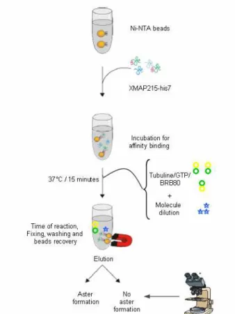
Figure 30 Microtubule nucleation test on magnetic beads
saturated by XMAP215-his 7
77
Results
Figure 31 Immunization evolution of the BALB/c mice
80
Figure 32 ch-TOG single staining in PHF
81
Figure 33 mcAB anti XMAP215/ch-TOG specificity
82
Figure 34 4B6 mcAB staining on PHF
83
Figure 35 5A6 mcAB staining on PHF
84
Figure 36 5D12 mcAB staining on PHF
85
XI
Figure 39 XMAP215 antigen cartography for the mcAB
88
Figure 40 4B6 mcAB technical data sheet
90
Figure 41 5D12 mcAB technical data sheet
91
Figure 42 Two different intracellular localizations for ch-TOG
in PHF
92
Figure 43 +TIP ch-TOG co-localizes with EB1
94
Figure 44 +TIPs in the PHF model: ch-TOG and EB1
95
Figure 45 +TIPs in the PHF model: CLIP170 and p150
Glued96
Figure 46 Analysis method of a « comet like » structure
97
Figure 47 +TIPs proteins hierarchy
98
Figure 48 Astral MTs +TIPs in the Egg extract are
marked by mcAB 5D12- Atto 488
101
Figure 49 +TIP XMAP215 presence is confirmed at the
+ TIPs of growing MTs in the mitotic egg extract
103
Figure 50 +TIP XMAP215 is part of the in vivo
depolymeryzation mechanism
104
Figure 51 XMAP215 travels on the tubulin polymer
105
Figure 52 Cosedimentation assays of XMAP215-his 7
using the mcAB 5D12 and 4B6
107
Figure 53 MT ends in cryo-EM
109
Figure 54 MT tips conformation at the cryo-electron
microscopy
111
Figure 55 Cryo-electron images of MT tips and their
trajectories
113
Figure 56 XMAP215 longitudinal binding model
114
Figure 57 XMAP215 organizes MT into ordered bundles
115
XII
Figure 60 Examples of the different effects of the molecules
on tubulin assembly
119
Figure 61 Dose activity test
120
Discussion
Figure 62 XMAP215/ch-TOG isoforms comparison
126
Figure 63 Different + TIPs XMAP215 activity models
132
Figure 64 Hypothetical model of the + TIP XMAP215/ch-TOG
tubulin dimmers addition
134
Figure 65 In vitro XMAP215 behaviour
138
Figure 66 +TIPs hierarchy theory
139
Figure 67 Hypothetical model of MT-XMAP215/ch-TOG
participation in complex protein formation
143
Figure 68 Model for XMAP215/ch-TOG activity in the
microtubular environment
144
Figure 69 A XMAP215 activity at the centrosome in M phase 147
Figure 70 Identified genes in chromosome 11p and their
possible implication in cancer
151
XIII
Table I
Eukaryotic cytoskeletal polymers
14
Table II
Properties of mamailian MAPs
22
Table III
Cellular localization for some of the proteins
of the MAP215/Dis1 family
45
Table IV
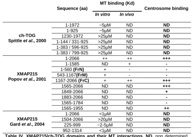
XMAP215/ch-TOG domains and their interactions 52
Table V
Immunization schedule of BALB/c mice for the
XMAP215 mcAB production
58
Table VI Cell lines used in this study
59
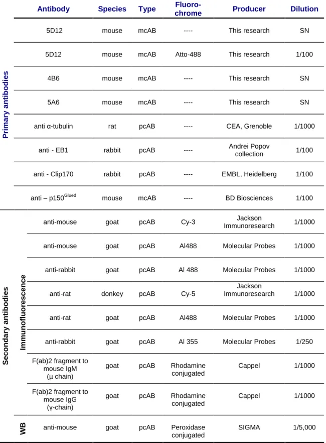
Table VII Primary and secondary antibodies used in the IF 60
Table VIII mcAB anti-XMAP215/ch-TOG in vitro
immunoreactivity
88
Table IX
mcAB anti-XMAP215/ch-TOG characterization
89
Table X
+ TIPs comets lengths
99
1
XMAP215/ch-TOG are members of an evolutionary conserved family of microtubule-associated proteins (MAPs), the XMAP215/Dis1 family. This family of proteins plays a key role in the regulation of the microtubule (MT) cytoskeleton, particularly during the cell division. In humans, ch-TOG is the colon-hepatic tumor overexpresed gene, a protein whose sequence was originally reported from blastic cells and from several forms of cancer. A few members of the XMAP215/ch-TOG family have been found to be present in different cell localizations, always MT-related, perhaps providing in this way a selected activity. However, the XMAP215/ch-TOG exact localization and activity has remained as a theory to be probed.
In this scientifical context, we developed a series of monoclonal antibodies (mcABs) that allow us to identify two different populations of the XMAP215/ Dis1 family of proteins. Confocal images of fixed cells revealed a first and well known XMAP215/ch-TOG population, a microtubular-XMAP215 (MT-XMAP215), co-localizing with microtubules (MTs) in interphase and mitotic spindle. A second localization was identified at the tips of growing MTs, placing XMAP215/ch-TOG as one more member of the known microtubule plus end tracking proteins (+TIPs). This second population was identified as the +TIP
XMAP215/ch-TOG.
The + TIP XMAP215 is the most distal +TIP protein at the tip of MTs. The + TIPs hierarchy was established comparing the XMAP215/ch-TOG localization with other well known + TIPs proteins such as EB1, CLIP170 and p150Glued. In the Xenopus laevis mitotic egg extract, the Total Internal Reflection Fluorescence (TIRF) In vivo images, identified a +TIP XMAP215 that is present at the tip of polymerizing and depolymerising MTs.
2
promote the formation of outwardly sheet structures at the tip of MTs, compatible with growing mechanisms in MTs.
Based in our results we propose a model where free XMAP215 is previously loaded with tubulin dimers to become a protofilament-like structure. This structure joins the MT tip using its C-terminal domain, not only adding the tubulin dimers but also perhaps participating in the MT sheet closure. The possibility that the protein participates in the MT depolymerization could be associated to a “controlled” depolymerization mechanism. Once the tubulin addition has taken place, the +TIP XMAP215 protein could evolve to a MT-XMAP215, the well know form of the protein that has been related to the RNA granules traffic.
3
Les protéines XMAP215/ch-TOG appartiennent à une famille de protéines associées aux microtubules (MAPs), dont la séquence génétique a été conservée tout au long de l’évolution, il s’agit de la famille XMAP215/Dis1. Cette famille joue un rôle dans la régulation du cytosquelette des microtubules (MT), en particulier pendant la division cellulaire. Chez l’humain, ch-TOG est la protéine surexprimée dans les tumeurs du colon et du foie. Certaines protéines XMAP215/ch-TOG ont été retrouvées dans différentes localisations cellulaires, toujours reliées aux MTs, à l’origine d’une activité spécifique. Cependant, la localisation exacte de XMAP215/ch-TOG ainsi que son activité restait à être déterminées.
Dans ce contexte scientifique, nous avons développé une série d’anticorps monoclonaux (mcAB) qui nous ont permis d’identifier deux populations différentes de XMAP215/ch-TOG. Les images de microscopie confocale des fibroblastes humaines primaires et de xenope ont montré une première localisation microtubulaire. C’est la colocalisation établit XMAP215-microtubulaire (MT-XMAP215) qui s’observe pendant l’interphase et pendant la mitose cellulaire. Une deuxième localisation a été identifiée sur le bout plus des MTs, donnant XMAP215/ch-TOG comme faisant parti de la famille des protéines de bout plus (+TIPs). Cette deuxième colocalisation a été identifiée comme +TIP XMAP215/ch-TOG.
La +TIP XMAP215 est la protéine la plus distale du bout des MTs. La hiérarchie a été établie en faisant la comparaison de la localisation de XMAP215/ch-TOG avec les protéines les plus connues du bout plus, telles qu’EB1, CLIP170 et p150Glued. Dans l’extrait mitotique de Xenopus laevis, les images obtenues in vivo par la microscopie de fluorescence par réflexion totale interne (TIRF) ont permis d’identifier une +TIP XMAP215 présente au bout des MTs qui polymérisent et dépolymérisent.
4
tubuline pure, XMAP215 induit la formation de structures au bout des MTs, cette activité est compatible avec les mécanismes de croissance des MTs.
Sur la base de nos résultats, nous proposons un modèle où XMAP215 sert de transporteur des dimères de tubuline en devenant une structure de type protofilament. Cette structure se lie au bout du MT en utilisant son domaine C-terminal, en rajoutant les dimères de tubuline et probablement en participant à la fermeture de la structure microtubulaire même. La protéine interviendrait donc dans la dépolymérisation et aurait un rôle dans le mécanisme de dépolymérisation contrôlée. Une fois que l’addition de tubuline a eu lieu, la +TIP XMAP215 pourrait évoluer en MT-XMAP215, forme la plus connue de la protéine associée au trafic des granules d’ARN.
5
GENERAL INTRODUCTION
Cell support, organization function and division rely on cell cytoskeleton. It is composed by microtubules (MTs), actin and intermediate filaments. MTs are dynamic and polar polymers that develop a complex rearrangement process going from the interphasic MT net until the mitotic spindle in the cell cycle. Once the cell enters mitosis, the interphasic MT network reorganizes and forms a bipolar mitotic spindle, the resulting structure is a well organized “mitotic machine”, able to separate the chromosomes between the two daughter cells.
The increased dynamics of MTs in mitosis is an essential prerequisite for spindle formation. These changes are executed by subtle modifications in the balance of the activities of MT stabilizing and destabilizing factors. This highly synchronized mechanism is the product of the coordinated activity of different classes of proteins including microtubule-associated proteins (MAPs) and molecular motors.
Among many others, a MAP that takes part in the MT dynamics is XMAP215. XMAP215 is the founding member of a highly conserved family of proteins, the XMAP215/Dis1 family of microtubule-associated proteins (MAPs), originally was identified in 1987 in the Xenopus laevis egg extract. Ch-TOG, the human homologue was described eight years later, sharing an 80% homology in their sequence. Both proteins promote MT assembly in vitro and localize to spindle MTs and centrosomes in vivo. XMAP215 in particular can affect the MT plus end.
Eventhough the XMAP215 activity at the MT +TIP has been predicted and its possible role in the conformation in the MT tip ultrastructure has been proposed, the XMAP215/ch-TOG functional activity remains to be probed or denied. Based on the different theories available, our research is focused on the establishment of the functional XMAP215/ch-TOG activity in vivo and in vitro.
6
experimental procedures, results and discussion.
The introduction concerns the first four chapters. The first one discusses the generalities of the cell cycle and its biological mechanism. The second chapter discusses the cytoskeleton nature, with particular reference to the MT structure, properties and activity in to the cellular context. A third chapter introduces and develops the +TIPS concept. In it, three of the most well known +TIPs proteins are described (EB1, CLIP170 and p150Glued) from their structure until reaching their potential (or predicted) cellular activity. A fourth chapter introduces the reader to the XMAP215/Dis1 family of proteins, focusing in particular on XMAP215 and ch-TOG.
The employed methods in this work are explained in to the fith chapter.The differents approaches go from the fixed cells imaging in the confocal microscopy domain, through physical principle of the TIRF until finally reaching the Cryo- electron microscopy analysis. This chapter is composed of six sub-chapters: the mcAB production and characterization (5.1), the XMAP215 / ch-TOG over-expression (5.2), the in vitro interaction tubulin or MT/XMAP215 (5.3), the XMAP215 in vivo interactions (5.4), the MT ultrastructure related to the XMAP215 effects (5.5) and finally the in vitro research for a XMAP215 inhibitor is described (5.6).
The sixth chapter list the results obtained through the experimentation phase. The interpretation is realized with reference to the cell localization, MT ultrastructure and XMAP215/ch-TOG predicted activity.
An seventh chapter addresses to the results discussion and to the different conclusions and theories that can be extracted from this work.
1. The cell cycle
In a eukaryotic cell, the cell cycle can be described as a fundamental task: the
copy and pass of the genetic
information to the next generation.
7
1. The cell and its cell cycle.
1.1 The cell.
The cell is the functional unit of life and is often described as the building block of life. It was discovered by Robert Hooke, an English natural philosopher (from Latin philosophia naturalis, the study of nature). Hooke published in 1665 Micrographia, a book that described microscopic observations. He coined the term cell, a word derived from the Latin “cellula”, which means small compartment.
In 1673 the Dutch microscopist Anthonine van Leeuwenhoek, known as the father of microbiology, described in his letters to the Royal society of London the single celled structures, today known as protist. Later in 1839, the Germans Mathias Schleiden (botanist) and Theodor Schwann (zoologist) elucidate the principle that plants and animals are made of cells, concluding that cells are a common unit of structure and development, funding the cell theory.
Rudolf Virchow completed the cell theory in 1858, publishing the epigram Omnis cellula e cellula (every cell originates from another existing cell like it), originating the concept of cell multiplication by cell division.
1.2 The cell cycle
The cell cycle is the mechanism by which a parent cell divides into two daughter cells. The cell division is a small part of a larger cell cycle. In eukaryotes this type of cell division is known as Mitosis.
The basic function of the cell division is to duplicate cellular DNA and to
segregate the copies in two identical daughter cells in an ordered process.
These processes define the two major phases of the cell cycle (Fig.1). DNA duplication occurs during S phase (Synthesis), which requires 10 -12 hours. After S phase, chromosome segregation and cell division occur in M Phase (Mitosis), which requires about 1 hour. G1 is the gap between M phase and S
8
phase, while G2 is the gap between S phase and M phase (Scholey et al., 2003).
Figure 1. Overview of the cell cycle.
The cell cycle is an ordered set of events in eukaryotic cells. They are designed: G1, S, G2 and M. G1 (GAP1) the cell grows and produce the enzymes needed for future DNA replication. S stage (Synthesis) involves the DNA replication and occurs until the cell DNA has doubled.
G2 (GAP2) protein synthesis occurs preparing the cell for mitotic events.
During the M stage (Mitosis) the nuclear and cytoplasmic division occurs producing two daughter cells. Mitosis is divided into 4 phases: Prophase, Metaphase, Anaphase and Telophase.
G1, S and G2 together are called Interphase. The two gap phases allow the cell to growth and monitor the internal and external environment to ensure that the conditions are suitable and preparations are complete before mitosis.
These cell cycle events are regulated by the activation of a complex family composed of cyclin-dependent kinases (CDK) and cyclin subunits, CDK1/cyclin B being the universal cell cycle regulator implicated in the G2/M transition (Le Breton et al., 2005).This mechanism is only possible using the highly synchronized cellular machine that divides the cell using cytoskeletal proteins as the key tools to accomplish mitosis (Fig.2a). MTs (mitotic spindle) and actin (contractile ring), molecular motors and MAPs coordinate their activities driving the cell through mitosis and cytokinesis.
9
1.3 Mitosis phases.
1.3.1 Interphase
Eventhoug the cell prepares itself for cell division during interphase, the interphase is not part of the mitosis mechanism. Interphase is divided into three phases: G1 (first gap), S (synthesis), and G2 (second gap). During these three phases the cells grows producing proteins and cytoplasmic organelles. The cell grows (G1), keeps growings duplicating the chromosomes (S), pepares for mitosis (G2), and finally divides (Mitosis).
1.3.2 Prophase
The genetic material is condensed into ordered structures, the chromosomes. During the S phase, the genetic material was duplicated, now the replicated chromosomes have two sister chromatids, bounded by the centromere.
The cell centrosome is located close to the nucleus and is made of a pair of centrioles, a centrosome is duplicated before the new mitosis. This structure nucleates MTs to form the spindle and the molecular motor proteins push each centrosome to opposite sides of the cell, forming microtubular asters.
1.3.3 Prometaphase
The nuclear envelope disassembles and MTs invade the nuclear space. Chromosomes keep condensing and the spindle MTs develops several different organizations: Astral, polar and kinetochore MTs. The Kinetochore MTs will be attached to proteic structures of each chromosome centromere.
Each chromosome forms two kinetochores at the centromere, one attached at each chromatid. When a MT connects to the kinetochore, some molecular motors activate, using energy from ATP to “crawl” up the MT to the originating centrosome. Is the molecular motor activity who added to the MT polymerization-depolymerization, providing the pulling force necessary to later separate the two sister chromatides.
10 1.3.4 Metaphase
As the prometaphase evolves, the highly condensed centrosomes of chromosomes arrive to the metaphase plate (equatorial plane), they are finally aligned. The resulting alignment is the consequence of the different pulling forces generated by the opposing kinetochores.
1.3.5 Anapahase
During the early anaphase, the proteins that bind sister chromatids together are cleaved, allowing them to separate. Doing this, each sister chromatid is pulled apart. The mechanistic method consists in a shortening of the kinetochore MTs, allowing the movement to the respective centrosomes.
In the late anaphase the non kinetochore MTs elongate, pulling the centrosomes (and the attached centrosomes) to the opposite poles of the cell. At the end of anaphase, the cell has separated successfully the two identical copies of the genetic information in two different groups.
1.3.6 Telophase
The nonkinetochore MTs continue to lengthen, elongating the cell even more. Corresponding sister chromosomes attach to each other and a new nuclear envelop is created from the “parent” fragments of the cells nuclear membrane. The chromosomes unfold back into chromatin.
1.3.7 Cytokinesis
Even though cytokinesis is not a phase of mitosis, but is a necessary process to complete cell division. The cleavage furrow containing the contractile ring at the metaphase plate level. Each daughter cell has a complete copy of the genome of its parent cell. The end of cytokinesis marks the end of the M-phase (Scholey et al., 2003, Cheeseman et al., 2008).
11
(a)
Figure 2. Mitotic cycle and cytokinesis. (a) During Interphase
(G1,S and G2), the cell is engaged in metabolic activity previous to mitosis. Prophase, the chromatin in the nucleus begins to condense and the duplicated centrosomes begin to migrate to opposite poles of the cell. Prometaphase, the nuclear membrane dissolves; chromosomes are attached by MTs through the kinetochore, moving the chromosomes to the cell equator. Methaphase, sister chromatids face opposite poles. Anaphase, sister chromatids moves to opposite poles of the cell. Telophase, chromatids arrive to opposite poles of the cell and nuclear envelopes reassemble around. The contractile ring creates the spindle mid-zone called the midbody. Finally, the furrow “seals” separating the daughter cells. (b) Mitosis general
vocabulary. Sister chromatids, kinetochores, spindle equator,
centromeres, poles
(b)
2. The Cell Cytoskeleton
The cytoskeleton is composed by actin filaments, intermediate filaments and MTs. Together with molecular motors they perform many biological activities including mitosis, cellular shape, cellular signalling, intracellular transport and cellular motility.
13
2. Cytoskeleton
The term cytoskeleton (cytosquelette) was first introduced in 1931 by the French embryologist Paul Wintrebert. There are three types of cell cytoskeletal polymers: actin filaments, MTs and intermediate filaments (Table I).
Actin was described for first time by Straub in 1943. In 1948 De Robertis and Schmitt described intra-axonal fibers, with a tubular shape. These structures were known years later as “microtubules” (Slautterback, 1963). Ishikawa and collaborators identified filaments possessing a calibre “intermediate” between MTs and actin filaments, thus the intermediate filaments concept was born in 1968 (Frixione, 2000).
Cytoskeletal polymers are in highly dynamic structures that posses a rapid reorganization mechanism. They are capable of polymerizing, depolymerising and moving within the cytoplasm only in a few seconds. MTs are implicated in movement, using polymerization for pushing and depolymerization by pulling (Desai and Mitchinson, 1997).
MTs are polar and linear polymers, built from 13 strands of α/β-tubulin heterodimers. Actin filaments (F-actin) are built by two strands of globular actin (G-actin) monomers. Intermediate filaments assemble through the association of antiparallel dimers into oligomers, generating apolar filaments that confer strength to the cell. Each one of these polymers can generate pushing and pulling forces as they grow and shrink by addition and loss of subunits from their ends. They also serve as tracks for motor proteins that use GTP or ATP hydrolysis to generate force and motility whereas intermediate filaments use accessory proteins, such as kinases and phosphatases (Scholey et al., 2003; Galjart et al., 2005).
In the interphasic cell, the cytoskeletal proteins generate multiple forces, at the piconewtons scale producing movements at the nanoscale. During cell
14
division they work as a unique machine, able to generate forces in the nanonewtons scale, moving the cellular components and rearranging the cell structure in a surface of many microns.
Polymer Actin filament Microtubule Intermediate filament Protein
subunit
Actin monomer Tubulin heterodimer Various proteins with
α -helical coiled-coil
Polymerization yes yes Probably
Bound nucleotide ATP GTP None Dynamic instability No Yes, dramatic No Track for motors Yes, 20 families of myosins
Yes, several dyneines and many families of
kinesins No Electron micrographs of polymers Fluorescence micrographs of cell polymers
Table I: Eukaryotic cytoskeletal polymers. Electron micrographs and 3D illustrations:
migration.worldpress.com. Fluorescence images showing PHF stained for each one of the cytoskeletal polymers. 10 µm scale bar. Modified from Pollard et al., 2003.
15
2.1 Microtubules.
The MTs are a key component of the cell cytoskeleton. They allow a variety of tasks in the different cell state. During interphase they are responsible for the cell shape, vesicle and mitochondria movement and cell signalling. In mitosis build the mitotic spindle that segregates the duplicated chromosomes, providing support to the organelle organization and also orienting the cell plane of cleavage (Desai and Mitchinson, 1997; Jordan and Wilson 2004).
As the cell enters mitosis, a radial array of long interphase MTs (Fig.3a) is disassembled and converted to a bipolar spindle composed of much shorter MTs (Fig.3b). This reorganization is accompanied by changes in MT assembly dynamics; entry into mitosis corresponds with a 10-fold increase in the rate of MT turnover (Cassimeris et al.,1999).
(b)
(a) (b)
(a) Figure 3. Interphasic and mitotic MT
polymer organization . The MT polymer
organization at interphase (a) and
mitosis (b) is a consequence of both MT
dynamics regulated by accessory proteins and additional organization provided by the actin cytoskeleton or motor proteins (Cassimeris et al.,1999). Scale bar 10µm
MTs are hollow polymers assembled from α/β-tubulin heterodimers. These heterodimers are arranged linearly into protofilaments; 13 protofilaments typically form the MT wall. The organization of heterodimers in the MT lattice is polarized, resulting in structural polarity and kinetic differences at the MT ends. The faster growing end is designated the plus end (+ end, close to the cell cortex) and has the β-tubulin subunit of each heterodimer exposed, whereas the slower growing end (from the centrosome) has an α-tubulin subunit exposed and is designated the minus end (- end,) (Fig.4) (Cassimeris et al.,1999; Siegrist et al., 2007).
16
Figure 4. Microtubules structure.
Typically a MT is composed of 13 individual protofilaments consisting of α/β -tubulin heterodimers, this organization forms a hollow tube of 25 nm diameter. An α/β-tubulin heterodimer measures 8 nm has an exchangeable GDP/GTP binding site on the β subunit. The MT is a polar structure where, the β-tubulin is
exposed at the growing end (+ end) and the α–tubulin is exposed at the slower
growing end (- end). A GTP- tubulin cap (orange) stabilizes MT growth by keeping individual protofilaments close to each other. (Gardner et al., 2008)
In interphase cells the MT polarity is a key issue, it permits the motor proteins the unidirectionally movement on the MT lattice executing their diverse functions. In this case they function as tracks for intracellular transport of vesicles and cellular organelle distribution (Dammermann et al., 2003 ; Siegrist et al., 2007). In interphasic cells the network shows a slow growing end (- end), localized at the cell centrosome. The faster growing end (+ end) is close to the cell cortex.
Growing MT + ends are composed of GTP-tubulin, which helps to stabilize them (more mature sections of MTs typically bind MAPs, which promotes stability). Is this region, where the tubulin dimers are exchangeable that is known as the GTP cap. Here the polymerization dynamics is created by the gain and lost of tubulin-GTP or tubulin-GDP and inorganic phosphate (Pi) at the MT end. Tubulin-bound GTP is hydrolyzed to tubulin GDP and Pi at the time that tubulin is added to the MT end. The tubulin-GDP remains non-dissociable and non-exchangeable from the MT tip until the tubulin subunit dissociates from the MT (Fig 5). The cycles of MT growth and collapse are termed “dynamic instability” (Jordan and Wilson 2004; Siegrist et al., 2007).
17
Figure 5. Polymerization dynamics in the cap associated mechanism.
GTP-tubulin is incorporated at polymerizing MT ends, the bound GTP is hydrolyzed and inorganic phosphate (Pi) is released. Thus, the MT lattice is basically composed of GDP-tubulin. Cassimeris et al.,1999.
2.1.1 The dynamic instability model.
The observation of MT assembly from purified preparations in vitro of tubulin has led to the description of dynamic instability. MTs exist in continuous alternation phases of growth (elongation) and shrinkage (shortening) with stochastic switches between these states (Fig.6) (Desai and Mitchinson, 1997; Shiina et al., 1999; Carvalho et al., 2003).
During the growth and shrinkage tubulin heterodimers are added or lost at the filaments ends. The switch from elongation to shortening is termed
catastrophe, and the switch from shortening to elongation, rescue (Cassimeris et al.,1999). MTs ends can also exhibit a “pause” state, showing no significant growth or shrinkage, behaviour often observed in living cells, but its functional significance is yet to be understood (Dammermann et al., 2003)
18
Figure 6. MT dynamic instability model.
MT dynamics length changes by dynamic instability during interphase and mitosis. (Cassimeris et al., 1999)
MTs within cells display similar behaviours, but the specific parameters of dynamic instability are influenced by a variety of MAPs that allow the cell to assemble and modify an extensive MT network (Morrison et al., 2006).
2.1.2 Tubulin post-translational modifications.
The α/β tubulin dimers are used to generate a variety of MT structures with organelle-specific properties and functions. This characteristic can be achieved in the post-translational modifications (PTMs) of the tubulin dimer (Fig.7a). These modifications may allow the recruitment of different protein complexes, regulating in this way the MT specific properties (Hammond et al., 2007).
One of the most studied tubulin post-translational modifications is the
tyrosination cycle of the α-tubulin. The carboxy-terminal tyrosine is removed
by a tubulin tyrosine carboxypeptidase (TTCP), giving origin to the Glu-tubulin. The addition of the tyrosine residue to the α-tubulin occurs on soluble tubulin heterodimers, a reaction catalyzed by tubulin tyrosine ligase (TTL). The resulting Glu-MTs seems to be stabilized by suppression of the dynamics at the
19
(a)
(b)
Figure 7. Tubulin post-translational modifications.
(a) Crystal structure representation
showing the different PTM on an α/β– tubulin dimer. Tyrosination/ Detyrosination cycle (Y) is considered
to be involved in cell differentiation.
Polyglutamylation (EEE) is proposed
to participate in the centriole maturation and MAPs interaction. Acetylation occurs on the lysine 40 (K40), it is supposed to participate in the regulation of cell motility and the binding of MAPs to MTs. Polyglycylation (GGG)
participates in Tethrahymena: axonemal organization, ciliary motility and cytokinesis. Phosphorylation (Ser 441/444 β–tubulin subunit), could
participate in neuronal differentiation.
(b) α–tubulin tyrosination cycle.
Detyrosination/tyrosination cycle of the
α-tubulin C-terminal tyrosine. The Tyr-tub from α-tubulin is removed by a tubulin tyrosine carboxypeptidase
(TTCP), producing a Glu-tubulin. The
tyrosine is restored by the action of the tubulin tyrosine ligase (TTL). (Modified from Westerman et al., 2003; Hammond et al.,2007)
Detyrosination/ tyrosination cycle recruits differentially two types of MAPs, molecular motors and microtubule plus-end tracking proteins (+TIPs). For example the motor protein Kinesin-1 binds preferentially to detyrosinated MTs. In contrast, tyrosinated MTs are involved in the recruitment of + TIP proteins that contain a CAP-Gly domain (e.g. CLIP170 and p150Glued). The basic region contained in the CAP-Gly domain is required for targeting to the acidic EEY/F sequence present at the C-terminus of the α-tubulin. Also, CAP-Gly domains and EEY/F motifs contribute to the binding interactions between +TIPs proteins. Thus, multiple interactions among tyrosinated α-tubulin, +TIPs and the dynein/dynactin complex contribute to MT-based functions during mitosis and interphase (Rosenbaum et al., 2000; Westermann et al., 2003; Hammond et al., 2007).
3. +TIPS
In living cells, a large number of
protein factors regulate the MT dynamic
instability and the MT based transport,
having profound effects on the shape of the MT network during cell division, motility and morphogenesis. Particularly, a group of proteins specifically and dynamically track the tips if growing MTs, they are known as
Microtubule plus-end-tracking proteins (+ TIPS).
21
3. MAPs and the Plus-end-tracking proteins (+ TIPS)
In vivo, the cellular MT dynamics is the product of a large variety of microtubule associated proteins (MAPs). These proteins play an important role in the MT dynamic instability either stabilizing or destabilizing the MT. MAPs function by targeting soluble, non polymerized tubulin subunits, the MT wall lattice and/or MT ends (Akhmanova and Steinmetz., 2008).
Among the different MAPs we found the ones who bind to the length of MTs, and others able to regulate the MT plus-end dynamics. The last ones are known as the microtubule plus-end-tracking proteins (+TIPs). Their functions include regulation of MT + end dynamics, providing the MT-to-cortex attachment necessary for vesicle delivery, force generation, and signalling for cortical cell polarity (Carvalho et al., 2003; Galjart et al., 2005).
The +TIPs are a highly diverse group of MT-associated proteins, found in species from yeast to humans. These include both MT-dependent motors and non-motor proteins (Table II). The particularity of + TIPs compared to other MT-associated proteins is their specific accumulation at the plus-ends of growing MTs, with a characteristic localization described as “comet-like” structures, coinciding at the end of polymerizing MTs (Akhmanova et al., 2005).
The +TIPs comprise a combinations of a limited set of evolutionary conserved modular binding domains, repeat sequences and linear motifs. An increasing number of proteins have been identified so far, a short list of + TIPs includes the end binding proteins EB1, EB2 and EB3; the cytoplasmic linker protein CLIP170 and CLIP 115; the dynein/dynactin MT motor complex, in particular the p150Glued subunit of dynactin, the adenomatous polyposis coli tumour suppressor protein, APC; the CLIP-associating proteins, CLASPs; and the Mitotic Centromere-Associated Kinesin, MCAK; among many others. The EB family, the CLIPs and P150Glued are found at growing MT ends and are the best
22 + Tip family Structural
domains Interaction partners among + TIPs Main Functions NON-MOTOR PROTEINS EB Family (EB1,EB2,EB3) CH, coiled coil, EBH, EEY/F CAP-Gly domains, basic/serine rich stretches)
Promotion MT growth and dynamicity
Catastrophe activity
Targeting of other + TIPs to MT ends EB proteins CLASPs CLIP170 P150Glued Stu2 XKCM1
MT rescue and stabilization Targeting of dynein to MT ends MT interaction with the cell cortex, kinetochores and vesicles. CAP-Gly family (CLIP 170 CLIP 115) p150Glued CAP-Gly, Coiled coil, basic serine rich, zinc finger, EEY/F (CLIP170)
Cytoplasmic dynein CLIP170
Binding dynein cargo
Regulation of dynein processivity
CLASPs
CLASP1 and 2
TOG-like, Basic serine rich
EB proteins CLIP170 CLIP115
MT rescue and stabilization MT interaction with kinetochores, cell cortex and Golgi
APC
Armadillo, SAMP repeats, coiled coil, basic serine rich
EB1 proteins XKCM1
MT stabilization, anti catastrophe activity
Interaction with the cell cortex and kinetochores MT-unrelated functions in Wnt signalling, etc XMAP215/Dis1 family ch-TOG TOG EB proteins XKCM1 MT stabilization, promotion of MT growth MT-cortex interactions
MICROTUBULE MOTOR PROTEINS Cytoplasmic dynein Dynein heavy chain AAA-ATPase, coiled coil Dynactin (p150Glued) LIS1
MT - end directed transport of macromolecular complexes and organelles, pulling MTs at the cortex.
EB proteins
Kinesin 13
MCAK
Kinesin motor, coiled coil, basic/ serine rich XMAP215, CLIP170, APC (Xenopus laevis) MT depolymerization, induction of catastrophes
Table II. + TIPs proteins according to their structural properties. Main families of Microtubule
plus-end tracking proteins (+TIPs). They are grouped according to their structural characteristics. Modified from Akhmanova and Steinmetz, 2008
23
candidates to be considered as the core component of the MT + TIP complexes (Mimori et al., 2005, Morrison et al., 2006, Siegrist et al., 2007).
3.1 Differential mechanism
The dynamic accumulation of + TIPs at the ends of polymerizing MTs is a poorly understood mechanism. There are three described mechanisms:
Treadmilling kinesin-mediated transport (Fig.8a); Hitchhiking on other proteins already localized at the plus-end (Fig.8b) and by direct binding to the MT plus-end itself by copolymerizing with tubulin (Fig.8c) (Wu et al., 2006; Siegrist et al., 2007; Slep and Vale, 2007; Dragestein et al., 2008).
Treadmilling (known also as motor driven) refers to the specific
association of + tips proteins at the new formed MT tip by a high affinity mechanism. In the case of CLIP170 the mechanism was described as a “riding on a plus-end directed motor” (Folker et al., 2005). They are coupled to be quickly released from the older part of the MT within a half-life of 1-3 seconds. This mechanism results in a transient concentration of proteins at the growing MT end (Komarova et al., 2005, Dragestein et al., 2008).
In contrast, Hitchhiking (or surfing) refers to a MT plus-end-directed transport through an association with microtubule-binding partners (other + TIPs). This interaction might be sufficient to target preferentially some proteins to MT ends (Komarova et al.,2005, Akhmanova and Steinmetz 2008).
The direct binding to the MT plus-end (also refers as end loading) mechanism is based on the in vitro interaction among +TIP proteins with tubulin dimers in solution. This result suggest a model in which +TIPs bind tubulin, once the +TIP protein has been charged, it joins the MT plus end and the co-assembling takes place. By itself it is a mechanism that provides physically additional interactions for tubulin dimers that finally join the MT tip (Folker et al., 2005; Slep et al., 2007).
24 (a ) T re a d m il li n g (b ) H it c h h ik in g (c ) D ir e c t b in d in g
Figure 8. Described mechanism for the MT plus-end localisation.
Three possible mechanisms that can explain the plus end tracking behaviour.
(a) Treadmilling, the plus end directed
motor drives the + TIP protein to the MT end.
(b) Hitchhiking, the + TIP protein
respond to an end-specific conformation or another +TIP protein.
(c) Direct binding, the + tip protein
binds directly to the MT + end and dissociates a few seconds later. The protein could be pre-loaded with tubulin dimers followed by a MT co-polymerization or by association of the free protein to the MT “cap recognition”, providing a “tubulin free” surface available for the dimers loading.
The + TIPs dissociation has been suggested to occur either by decreased affinity after incorporation into the mature MT lattice of by release due to phosphorylation (Carvalho et al., 2003; Folker et al., 2005; Slep and Vale, 2007)
Molecular motors (yellow), EB1
(blue), CLIP170 (pink), α/β-tubulin hetero dimer (green/orange).
Besides interacting with the MT ends, some + TIPs can bind to tubulin dimers and oligomers in vitro. This finding led to the idea that CLIPs reach MT ends by co-polymerizing with tubulin, and are then gradually released from the older lattice. Both EBs and CLIPs are dimers and seems to require at least two tubulin binding domains (calponin homology-CH or CAP-Gly respectively) to track MT ends, which suggest that the affinity of individual sites for MTs or
25
tubulin is relatively weak. The arrangement of these sites with respect to each other does not seem to be important because an artificial combination of a CH domain and a CAP-Gly domain is capable of plus-end tracking (Akhmanova and Steinmetz.,2008).
In the case of CLIP170, co-assembly with tubulin dimers or oligomers together with quick release from the MT has been proposed. Alternatively, proteins may have an increased affinity for a specific structure of the MT polymerizing end, such as tubulin sheets or the GTP cap (Akhmanova et al., 2005).
Another unresolved aspect of plus-end tracking is the mechanism of protein release from the older part of the MT lattice. Many + TIPs proteins have a high affinity for MTs and decorate the whole MTs in vitro, for CLIP170 it was shown that it dissociates from the MTs in vitro much slower than in vivo (Lansberggen et al., 2006). This preferential dissociation of the + TIPs from the older part of the MT could be the result of their phosphorylation by a MT-bound kinase. This theory remains not proved because the knowledge of + TIPs directed kinases remains fragmentary (Akhmanova et al., 2005).
The most characteristic +TIPs (CLIP170, p150Glued and EB1) share certain structure domains such as the CAP-Gly domain (CLIP170 and p150Glued) and the dimerization domain, common to the three of them (Galjart et al., 2005; Ligon et al., 2006)
3.2 EB1 (Ending Binding Protein 1)
EB family members are MAPs conserved in plants, yeast and mammals. In general all EB-like proteins share a common role in keeping the MT dynamic. In all species, EB1 homologues promote polymerization by increasing the MT rescue frequencies and decreasing the rate of depolymerization and the time MTs spend pausing (Lansbergen et al., 2006).
26
EB1 was first described as an adenomatous polyposis coli (APC) – interacting protein whose binding domain was affected by APC mutations implicated in colorectal cancer. EB1 contributes to the accumulation of APC at the MT ends, where EB1-APC complex can promote MT polymerization and stability.
The EB1 gene was predicted to encode a 268 amino acid protein (Su et al., 1995). Later, Morrison described the EB1 preference for the MT + TIP (1998). Further experiences showed the EB1 tendency for the elongating MTs, and appear to “track with the MT end”, since they are known as “tip tracking” (Fig.9a) (Vaughan et al., 2005).
3.2.1 EB1 Structure
EB proteins (EB1, EB2 and EB3) contain a highly conserved N- and C- terminal domains that are separated by a linker sequence (Fig.9b). The MT binding domain of EB1 is localized at the N-terminal region of the protein, whereas the C-terminal regions contributed to its localizations at the MT +TIP (the dynactin binding domain) and centrosome (Askham et al., 2002; Ligon et al., 2003).
EB proteins exert a growth promoting or stabilizing function in Xenopus laevis egg extracts (Su et al.,1995; Tirnauer et al., 2002). They form homodimeric complexes composed of two calponin homology (CH) domains (Gimona et al., 2002), essential for MT association (Hayashi and Ikura, 2003). The MT plus ends recognition is realized via its CH domain through its residues 55-102, the CH domain of EB1 contains a highly basic area that is predicted to form the interface to MT (Hayashi and Ikura, 2003; Sandblad et al., 2006).
27
(a)
(b)
Figure 9. EB1 cell localization and structure. (a), Immunostaining showing EB1 localization
on primary human fibroblast (PHF) at the tip of MTs. Cells are coimmunostained for MTs (green) and EB1 (red) Scale bar 5 µm. (b) EB family of proteins general domain structure. EB1 (268aa), EB2 (327aa) and EB3 (281). CH domain, Calponin homology domain; EBH, End-binding protein homology. Modified from Bu et al., 2003.
The EB1 CH domain is spherical and approximately half the size of a tubulin monomer. Mutagenesis suggests that several residues around one hemisphere contribute to tubulin binding (Slep et al., 2007). Previous studies have suggested that the locus of interaction is near the carboxyl terminus of α– tubulin (Honnappa et al., 2006).
The C terminal region of the EB proteins contains a coiled-coli domain, which mediates the parallel dimerization of EB monomers. The α-helical coiled-coil is a universal protein oligomerization motif that is frequently found in + TIPs (Akhmanova and Steinmetz., 2008)
3.2.2
EB1 Interactions
EB1 binds to and labels a subset of the total MT population, specifically, the tips of MTs undergoing elongation. Immunofluorescence microscopy images