RESEARCH OUTPUTS / RÉSULTATS DE RECHERCHE
Author(s) - Auteur(s) :
Publication date - Date de publication :
Permanent link - Permalien :
Rights / License - Licence de droit d’auteur :
Bibliothèque Universitaire Moretus Plantin
Dépôt Institutionnel - Portail de la Recherche
researchportal.unamur.be
University of Namur
Impact of functional inorganic nanotubes f-INTs-WS2 on hemolysis, platelet function
and coagulation
Laloy, Julie; Haguet, Hélène; Alpan, Lutfiye; Raichman, Daniel; Dogné, Jean-Michel;
Lellouche, Jean-Paul
Published in: Nano convergence DOI: 10.1186/s40580-018-0162-1 Publication date: 2018 Document VersionPublisher's PDF, also known as Version of record
Link to publication
Citation for pulished version (HARVARD):
Laloy, J, Haguet, H, Alpan, L, Raichman, D, Dogné, J-M & Lellouche, J-P 2018, 'Impact of functional inorganic nanotubes f-INTs-WS2 on hemolysis, platelet function and coagulation', Nano convergence, vol. 5, no. 1, 31. https://doi.org/10.1186/s40580-018-0162-1
General rights
Copyright and moral rights for the publications made accessible in the public portal are retained by the authors and/or other copyright owners and it is a condition of accessing publications that users recognise and abide by the legal requirements associated with these rights. • Users may download and print one copy of any publication from the public portal for the purpose of private study or research. • You may not further distribute the material or use it for any profit-making activity or commercial gain
• You may freely distribute the URL identifying the publication in the public portal ? Take down policy
If you believe that this document breaches copyright please contact us providing details, and we will remove access to the work immediately and investigate your claim.
Laloy et al. Nano Convergence (2018) 5:31 https://doi.org/10.1186/s40580-018-0162-1
RESEARCH
Impact of functional inorganic nanotubes
f-INTs-WS
2
on hemolysis, platelet function
and coagulation
Julie Laloy
1,2*, Hélène Haguet
2,3, Lutfiye Alpan
1,2, Daniel Raichman
4, Jean‑Michel Dogné
1,2and Jean‑Paul Lellouche
4*Abstract
Inorganic transition metal dichalcogenide nanostructures are interesting for several biomedical applications such as coating for medical devices (e.g. endodontic files, catheter stents) and reinforcement of scaffolds for tissue engineer‑ ing. However, their impact on human blood is unknown. A unique nanomaterial surface‑engineering chemical meth‑ odology was used to fabricate functional polyacidic polyCOOH inorganic nanotubes of tungsten disulfide towards covalent binding of any desired molecule/organic species via chemical activation/reactivity of this former polyCOOH shell. The impact of these nanotubes on hemolysis, platelet aggregation and blood coagulation has been assessed using spectrophotometric measurement, light transmission aggregometry and thrombin generation assays. The func‑ tionalized nanotubes do not induce hemolysis but decrease platelet aggregation and induce coagulation through intrinsic pathway activation. The functional nanotubes were found to be more thrombogenic than the non‑functional ones, suggesting lower hemocompatibility and increased thrombotic risk with functionalized tungsten disulfide nanotubes. These functionalized nanotubes should be used with caution in blood‑contacting devices.
Keywords: Functional tungsten disulfide nanotubes, Safety, Hemocompatibility, Thrombin generation
© The Author(s) 2018. This article is distributed under the terms of the Creative Commons Attribution 4.0 International License (http://creat iveco mmons .org/licen ses/by/4.0/), which permits unrestricted use, distribution, and reproduction in any medium, provided you give appropriate credit to the original author(s) and the source, provide a link to the Creative Commons license, and indicate if changes were made.
1 Introduction
Inorganic transition metal dichalcogenide (TMD) mate-rials, such as tungsten and molybdenum disulfides (WS2 and MoS2, respectively) are of significant inter-est to the scientific community because of their unique multi-layered structures and functional properties, with nano-sized fullerene-like (IF) particles tending to exhibit a different set of properties compared to the corresponding bulk forms. These metal dichalcogenide nanomaterials have emerged as one of the most promis-ing classes of nanomaterials since the discovery of car-bon nanotubes (CNTs) [1–8]. As with early researches in the field of CNTs, a wide number of potential appli-cations have been proposed and investigated including
areas such as energy storage [9], field effect transistors [10], nanocomposite coatings [11, 12], battery anodes [13], light-emitting diodes [14], self-lubricating medical devices [15], and high-performance nanoscale lubricants [16–23]. In addition, the outstanding shock absorb-ing ability of IFs-WS2 nanotubes holds a great potential for new impact and shock-resistant materials [24–26]. Composite hybrid materials formed by incorporating small amounts (less than 5% weight ratios) of such nano-sized inorganic fillers into any given polymer matrix are also of particular interest, showing improved mechani-cal properties, higher thermal properties, and improved performances as barriers to heat, moisture, and solvents [27–29] when compared to similar composites prepared with conventional fillers [28, 30]. Indeed, considerable research work has been conducted dealing with poly-mer-based nanocomposites that incorporate inorganic IFs-WS2 NPs into matrices of epoxy [30], polystyrene/ poly(methylmethacrylate) [28], poly(propylene fuma-rate) [29], nylon 12 [31], and poly(phenylene) sulphide
Open Access
*Correspondence: [email protected]; [email protected]
2 Department of Pharmacy, NARILIS, University of Namur, Namur, Belgium 4 Department of Chemistry & Institute of Nanotechnology &
Advanced Materials (BINA), Bar‑Ilan University, Max & Anna Web Street, 5290002 Ramat‑Gan, Israel
[32]. Due to the superior mechanical properties of cor-responding inorganic IFs-WS2 NPs, such as high stiffness and strength [33], ultrahigh-performance polymer nano-composites have been readily produced [34]. In addition, commercial performant lubricants are now presently available that include same inorganic IFs-WS2 NPs that impart unique tribological properties [35] to the corre-sponding final composite products. Although there are many potential applications in a wide variety of fields for such inorganic metal dichalcogenide IFs-WS2 NPs and inorganic INTs-WS2 nanotubes (INTs), novel develop-mental research has been strongly hampered analogously to early CNTs-based research. Indeed, these dichalcoge-nide nanomaterials are highly hydrophobic, thus quite insoluble in common organic/aqueous solvents, difficult to homogeneously disperse into most liquids and resins, while disclosing serious limited dual phase compatibility when admixed with common polymers.
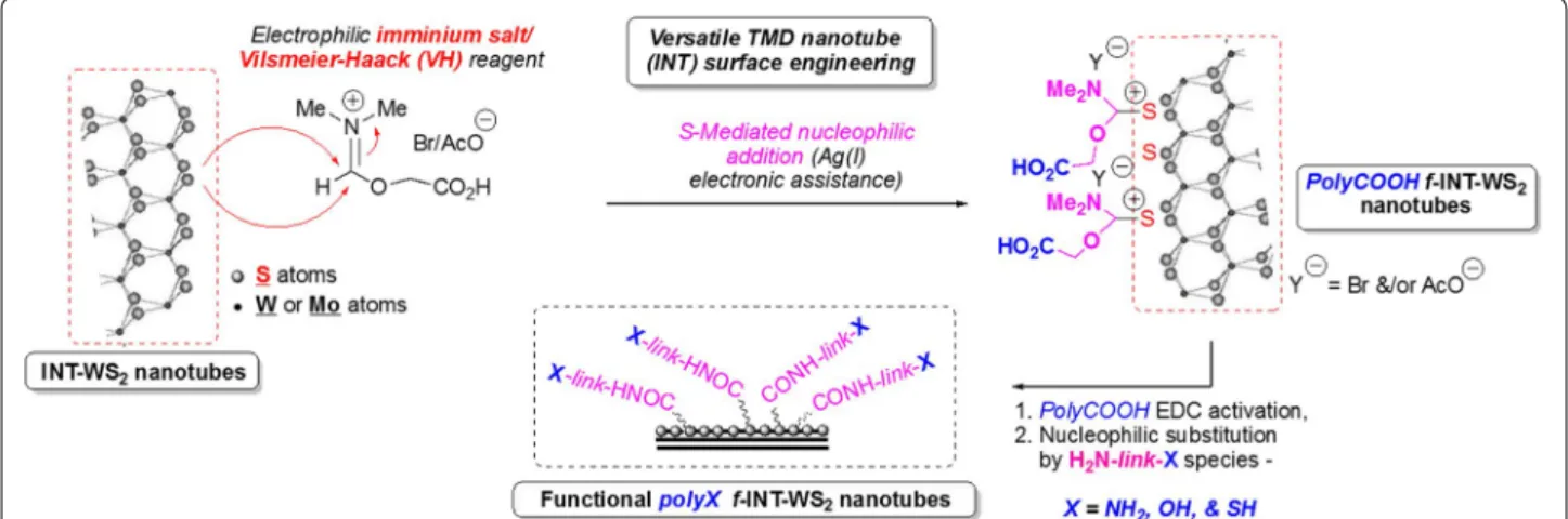
In this specific context, we recently developed a unique nanomaterial surface-engineering chemical methodology to fabricate covalently decorated functional polyacidic polyCOOH–INTs-WS2 using Vilsmeier–Haack (VH) complex chemistry/reactivity (polyCOOH shell decora-tion) [36]. This novel surface engineering method ena-bles effective covalent bonding of any desired molecule/ organic species via polyCOOH shell chemical activation/ reactivity that may improve and optimize any requested interfacial property of corresponding functional INTs-WS2 (f-INTs-WS2). This polycarboxylated shell can be readily exploited as an anchoring shell for subsequent second-step covalent attachment of a wide variety of organic molecules/polymers, including even other com-ponents such as NPs, for example, onto the functional nanotube surface. Therefore, a quite versatile simple organic activation chemistry (EDC•HCl activation of
polyCOOH shell/species) readily enables corresponding surface property tuning to match those requested for any contacting material (polymeric phases, solvents, etc.). Moreover and in this context, by employing appropriate bifunctional linkers such as those described in this study (obtainment of novel 2nd step polyNH2/polySH/polyOH shells, Fig. 1), the resulting chemically modified f-INT-WS2 can be covalently bound to an even wider variety of reactivity-complementing materials.
Recent progress in studies of this original novel class of inorganic nanomaterials suggests that they can be also impregnated into metallic coatings for medical admin-istration/application [37]. For example, it was demon-strated that the use of orthodontic wires coated with metallic films containing IFs-WS2 NPs in dentistry could significantly reduce the mechanical forces required for teeth realignment, thus preventing unnecessary excess forces that would lead to unacceptable teeth movement, longer treatment, and adverse damage to the roots of the teeth [10, 37, 38].
Since both IFs-WS2 NPs and INTs-WS2 are already commercially available in the market thus providing effective potentialities of incorporation/involvement towards innovative future medical applications, extensive research investigations concerning the overall biocom-patibility and toxicity of these inorganic materials need to be performed to ensure that they are safe for com-posite-based usage. Researches on the toxicity of TMD nanomaterials is still in its infancy with only a hand-ful of assessments performed on IFs-MoS2 and IFs-WS2 NPs. Preliminary results from in vivo toxicology tests of IFs-WS2 NPs showed no apparent toxic effects on mam-mals, suggesting its high biocompatibility [39]. In addi-tion, in vitro cytotoxicity examination of IFs-MoS2 NPs on three different human cell lines (i.e. CCC-ESF-1,
Fig. 1 Preparation of functional polyX (X: COOH, NH2, OH, SH) f‑INT‑WS2 inorganic nanotubes. The functional inorganic nanotubes were prepared
Page 3 of 10 Laloy et al. Nano Convergence (2018) 5:31
A549, and K562) revealed that they are nontoxic to cells after 48 h exposure [17]. However, at the present time, no experimental studies assessed the hemocompatibility of TMD materials. With the influx of research and pos-sible commercialization of TMDs in the future, it is vital to both initiate hemocompatibility studies of this group of nanomaterials and assess their impact on hemolysis, platelet functions, and blood coagulation [40].
In this special work, we characterized the hemocom-patibility of such different functional INTs-WS2 and assessed their impact on red blood cells, platelet aggrega-tion and blood coagulaaggrega-tion using human blood.
2 Methods
2.1 Materials
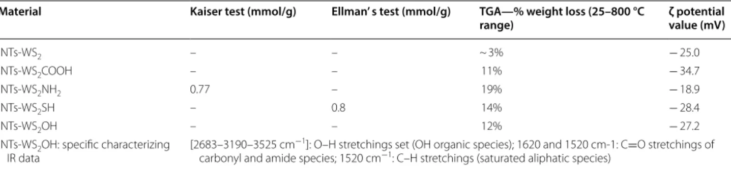
Non-functional INTs-WS2 have been bought from Nano-Materials Ltd. Company (Yavne, Israel). All reagents and solvents have been purchased from commercial sources and used without any further purification. Thermogravi-metric analyses (TGA) have been performed on a TA Q600-0348, model SDT Q600 (Thermofinnigan) device using a temperature profile of 25–800 °C at 10 °C/min under nitrogen flow (180 mL/min) with sample amounts of 5–15 mg. Infrared (IR) spectra were recorded on a Fourier transform infrared spectrometer Tensor 27 (Bruker) using attenuated total reflectance (ATR). Nano-material surface charges were evaluated by ξ potential measurements using a Zetasizer Nano-ZS device (Mal-vern Instruments Ltd., United Kingdom) in water (pH adjusted) at 25 °C and 150 V. Both VH-untreated starting and resulting VH-modified f-INTs-WS2 nanotubes have been also characterized using G2, FEI High Resolution transmission electron microscopy (TEM) (Tecnai). Dis-persions of INT-WS2 and f-INT-WS2 have been prepared with a low-power ElmaSonic S30 bath sonicator (Elma GmbH & Co., Deutschland). The chemically accessi-ble polyCOOH shell present on the surface of the poly-COOH f-INT-WS2 has been also quantified by both (i) Kaiser testing after shell derivatization using 1,3-diami-nopropane and (ii) Ellman’s one after subsequent similar shell derivatization using cysteamine.
2.2 Polycarboxylation of INT‑WS2—fabrication
of polyCOOH‑f‑INT‑WS2
To a solution of 2-bromoacetic acid (2-BrCH2COOH), (1.0 g, 7.19 mmol) in anhydrous dimethyl formamide (DMF, 3 mL) was added Ag(I)OAc (10.0 mg, 0.059 mmol) and dry INT-WS2 (200.0 mg). The mixture was heated in an oil bath to 80 °C and stirred over 2 days at the same temperature. After cooling to room temperature, the mixture was centrifuged (11,000 rpm, 5 min). The result-ing cleaned (EtOH, 5 washresult-ing cycles) solids were dried
under vacuum to obtain 190 mg of functional polyCOOH
f-INT-WS2.
2.3 Diamine coupling onto polyCOOH f‑INT‑WS2—
fabrication of polyNH2‑f‑INT‑WS2
To a solution of 1-ethyl-3-(3-dimethylaminopropyl)car-bodiimide (EDC, 20.0 mg, 4 mmol) in dichlorometh-ane (DCM, 12 mL) was added polyCOOH f-INT-WS2 (200.0 mg) and 4-dimethylaminopyridine (DMAP, 10.0 mg, 0.08 mmol). The mixture was stirred for 2 h at room temperature followed by addition of 1,3-diamino-propane (NH2–(CH2)3–NH2, 800 µL, 9.58 mmol) and stirring continued at room temperature overnight. The mixture was centrifuged (11,000 rpm, 5 min) and the supernatant discarded. The solids were worked up as described for former polyCOOH f-INT-WS2. The prod-uct contained 0.77 mmol NH2 groups/g of polyNH2
f-INT-WS2 as determined by Kaiser testing.
2.4 Cysteamine coupling onto polyCOOH f‑INT‑WS2—
fabrication of polySH‑f‑INT‑WS2
To a solution of EDC (3.0 g, 19.32 mmol) in DCM (40 mL) was added polyCOOH f-INT-WS2 (1.8 g). The suspension was stirred for 2 h at room temperature followed by addi-tion of cysteamine (NH2–(CH2)2–SH, 4.0 g, 51.85 mmol) and DMAP (20.0 mg, 0.16 mmol) and stirring continued for 2 days at room temperature. The mixture was centri-fuged (11,000 rpm, 5 min) and the supernatant discarded. The solids were worked up as described for former poly-COOH f-INT-WS2 to obtain 1.6 g of functional product. The product contained 0.8 mmol SH groups/g of polySH
f-INT-WS2, as determined by Ellman testing.
2.5 2‑Aminoethanol coupling onto polyCOOH f‑INT‑WS2—
Fabrication of polyOH‑f‑INT‑WS2
To a solution of EDC (3.0 g, 19.32 mmol) in DCM (40 mL) was added polyCOOH f-INT-WS2 (1.5 g). The suspension was stirred for 2 h at room temperature fol-lowed by addition of 2-aminoethanol (NH2–(CH2)2–OH, 4.0 mL, 64.71 mmol) and DMAP (20.0 mg, 0.16 mmol) and stirring continued for 2 days at room temperature. The mixture was centrifuged (11,000 rpm, 5 min) and the supernatant discarded. The solids were worked up as described for former polyCOOH f-INT-WS2 to obtain 1.3 g of functional product.
2.6 Preparation of human platelet‑rich plasma, platelet‑poor plasma, normal pooled plasma and washed red blood cells suspension
Human platelet rich plasma (PRP), platelet poor plasma (PPP), whole blood, washed red blood cell (RBC) suspen-sion and normal pool plasma (NPP) were prepared with blood from healthy volunteers who were free from any
medication for at least 2 weeks. Blood was collected by venipuncture into tubes containing buffered sodium rate (109 mM, nine parts blood to one part of sodium cit-rate solution) (BD Vacutainer®). The study protocol was in accordance with the Declaration of Helsinki and was approved by the Medical Ethical Committee of the CHU UCL Namur (Yvoir, Belgium).
PRP was carefully prepared by centrifugation at 200g of whole blood at room temperature for 10 min. The platelet count was adjusted to 300,000 platelets/μL and PRP was used immediately after preparation. Platelet free plasma used to adjust platelet concentration is obtained after centrifugation at 2000g in 10 min of the pellet at room temperature.
The preparation of washed RBC suspension was pre-pared by centrifugation of whole blood at 3000g over 5 min. The PPP is removed and used for interference assays. RBC are washed with physiological phosphate buffered saline (PBS, 6.7 mM phosphate, pH = 7.4) three times with intermediate centrifugation of 3000g over 5 min. RBC are then resuspended in PBS with the same volume as PBS removed.
For NPP, a total of 47 healthy individuals were included in the study. The exclusion criteria were thrombotic and/ or hemorrhagic events, antiplatelet and/or anticoagulant medication, pregnancy and uptake of drugs potentially affecting the platelet and/or coagulation factor functions during the 2 weeks prior to the blood drawn. A written informed consent was obtained from each donor. The study population displayed the following characteris-tics: 27 females and 20 males aged from 18 to 53 years (mean age = 25 years) with body mass index (BMI) rang-ing from 17.6 to 34.9 kg/m2 (mean BMI = 22.7 kg/m2). After collection of blood, the PPP was obtained from the supernatant fraction of the blood tubes after a double centrifugation for 15 min at 2000g at room temperature. It was immediately frozen at − 80 °C after centrifuga-tion. The NPP samples were thawed and kept at 37 °C just before use.
2.7 Hemolysis assays
Hemolysis assays were performed as previously described on the blood of one healthy donor [41]. Briefly, 15 μL of nanomaterial suspended in tyrode, tyrode (negative control) or triton X-100 (positive control) are added to 285 μL of whole blood or washed RBC (final NP concen-tration: 100 µg/mL). The suspension is incubated at room temperature on a shaking plate during 1 h. After the incubation time, the suspension is centrifuged at 10,000g over 5 min. Supernatant is read in a 96-well plate using a microplate scanning spectrophotometer XMark (Biorad, USA) at 550 nm. The percentage hemolysis was then cal-culated as:
For each term of the equation, the corresponding interference was subtracted. The interference corresponds to the same conditions except that the solution does not contain RBCs. Positive (triton X − 100 at 1%) and negative (Tyrode) controls induced 100% and 0% of hemolysis, respectively. The results were expressed as mean ± SD (n = 3).
2.8 Light transmission aggregometry
The impact of f-INTs-WS2 on induced platelet aggre-gation was studied using the chronometric aggregom-eter type 490-2D as previously reported [41]. Briefly, the reaction mixture for induced aggregation tests con-tained 213 or 233 μL of PRP at 300,000 platelets/μL, with respectively 25 μL of collagen (final concentration: 190 μg/mL, calf skin, Bio/Data corporation, USA) or 5 μL of arachidonic acid (AA, final concentration: 600 μM, Calbiochem, Germany) and 12.5 μL of NPs at final con-centration of 100 μg/mL. Inducers alone were also used before any experiment to check platelet reactivity. PPP was used as a reference. Data were collected with the chronolog two channel recorders at 405 nm connected to a computer.
2.9 Coagulation: calibrated thrombin generation test (cTGT)
The impact of non-functional and functional INTs-WS2 on coagulation was studied using the calibrated throm-bin generation test (cTGT) as previously reported [41]. For each experiment, a fresh mixture of fluorogenic sub-strate/calcium chloride buffered solution was prepared as follows: 2.6 mL of Fluo Buffer® (Thrombinoscope BV, The Netherlands) were mixed with 65 μL of Fluo substrate® (100 mM in DMSO, Thrombinoscope BV, The Nether-lands). PPP-Reagent, PPP-Reagent LOW, MP-Reagent and Thrombin Calibrator (Thrombinoscope BV, The Netherlands) are four inducers, giving final assay concen-trations of 5 pM tissue factor (TF) with 4 μM phospho-lipids (PL) and 16.7 mM CaCl2; 1 pM TF with 4 μM PL and 16.7 mM CaCl2; 4 μM PL and 16.7 mM CaCl2; and 620 nM α2- macroglobulin-thrombin complex, respec-tively. They are reconstituted with 1 mL distilled water according to the instructions provided by the manufac-turer. A calibration curve was simultaneously performed using the thrombin calibrator. The acquired data were automatically processed by the software, which provided thrombin activity curves and 3 parameters based on this curve: lagtime (minutes), peak concentration (nM) and endogenous thrombin potential (ETP, nM × minutes).
H (%) = ODsample−ODtyrode
ODTritonX−100 at 1%− ODtyrode
Page 5 of 10 Laloy et al. Nano Convergence (2018) 5:31
The INT/f-INTs suspensions were tested at final concen-trations from 5 to 500 μg/mL. Statistical analyses were conducted with an unpaired t-test using the GraphPad Prism software (GraphPad software, v 5.01, USA).
3 Results
3.1 Fabrication and characterization of f‑INTs‑WS2
Functional INTs-WS2 have been effectively fabricated using the two-step surface engineering methodology described in Fig. 1 below. First and as the first critical chemical modification methodology, a strongly electro-philic VH complex arising from DMF–BrCH2COOH reactivity has been generated in situ in the presence of starting INTs-WS2 to provide intermediate polyacidic functional polyCOOH f-INTs-WS2.
In a 2nd derivatization step, resulting chemically modi-fied polyCOOH f-INTs-WS2 nanotubes might be readily chemically activated (EDC activation) and reacted with bifunctional nucleophilic linkers of the type H2
N-link-X to provide corresponding functional polyN-link-X (polyNH2, polySH, polyOH) f-INTs-WS2 nanotubes. All these func-tional nanomaterials have been fully characterized by combined thermogravimetric analysis (TGA), spectro-scopic FT-IR/XPS, XRD, Kaiser (NH2 species quantifica-tion)/Ellman (SH species quantification) tests, HR-TEM and ζ potential values measurements (Table 1). All these characterization spectroscopy-based spectra/data and TEM/HR-TEM microphotographs including nanomate-rials are fully detailed in the corresponding Ref. [36].
3.2 Hemocompatibility
3.2.1 Red blood cells
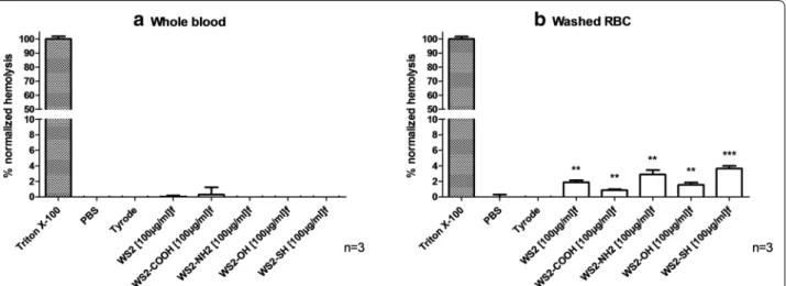
Absorbance spectrum of RBC suspension 10% (v/v) supernatant incubated with Triton X-100 1% (v/v) is measured. The interference of nanotubes within assay is determined at 550 nm. This interference was avoided by subtracting the OD550 nm of INTs-WS2/f-INTs-WS2 sus-pended in the vehicle from the measured OD550 nm at
the same concentration (data not shown). Measurement of absorbance at 550 nm in whole blood or washed RBC supernatant assesses the release of hemoglobin from lysis RBCs. Both non-functionalized and functionalized INTs-WS2/f-INTs-WS2 at 100 µg/mL did not induce hemoly-sis in whole blood (Fig. 2a) and in washed red blood cells (Fig. 2b) according to the ASTM E2524-08 standard (hemolysis ratio of all samples was below 5%) [42].
3.2.2 Platelet function
Second important parameter to be determined is the impact on platelet and in particular on platelet aggrega-tion. At 100 µg/mL, non-functionalized and function-alized INTs-WS2/f-INTs-WS2 significantly decreased platelet aggregation induced by AA (Fig. 3b). When collagen is the inductor, only polyCOOH-f-INTs-WS2 decreased significantly platelet aggregation (Fig. 3a).
3.2.3 Coagulation
Impact of f-INTs-WS2 on blood coagulation was assessed through cTGT. Non-functionalized and functionalized INTs-WS2/f-INTs-WS2 impact blood coagulation when the intrinsic pathway is triggered (Fig. 4). A procoagulant effect of these nanomaterials is observed with a decrease of lagtime and an increase of peak concentration and ETP (Table 2). Based on their procoagulant activity on the intrinsic pathway, INTs-WS2/f-INTs-WS2 can be clas-sified as follows: WS2-NH2 > WS2-OH > WS2-SH=WS2 -COOH > WS2. Experiments with coagulation initiated by the extrinsic and common pathways demonstrated no effect of f-INTs-WS2 at the exception of polyNH-f-INTs-WS2 which had a procoagulant effect when common pathway is triggered (data not shown).
4 Discussion
As quite novel inorganic multi-layered nanomaterials, hydrophobic non-functional INTs-WS2 nanotubes have been recently shown to be reactive towards a strongly
Table 1 Selected characterization (TGA) and functionality quantification data
INTs, inorganic nanotubes; TGA, thermogravimetric analysis—starting INTs-WS2 nanotubes are negatively charged (− 25.0 mV) due to known OH-based defects arising from industrial nanofabrication step
Material Kaiser test (mmol/g) Ellman’ s test (mmol/g) TGA—% weight loss (25–800 °C
range) ζ potential value (mV)
INTs‑WS2 – – ~ 3% − 25.0
INTs‑WS2COOH – – 11% − 34.7
INTs‑WS2NH2 0.77 – 19% − 18.9
INTs‑WS2SH – 0.8 14% − 28.4
INTs‑WS2OH – – 12% − 27.2
INTs‑WS2OH: specific characterizing
IR data [2683–3190–3525 cm
−1]: O–H stretchings set (OH organic species); 1620 and 1520 cm‑1: C=O stretchings of
electrophilic acidic VH complex arising from both DMF/ Br-CH2COOH reagents that enabled stable covalent nanotube surface chemical engineering/chemical modifi-cation by a corresponding polyCOOH shell (polyCOOH
f-INTs-WS2 nanotubes). Quite innovatively while using specific bifunctional linkers (Fig. 1), this polyacidic shell might be readily exploited via EDC activation for addi-tional surface engineering to get a wide variety of func-tional f-INTs-WS2 inorganic nanotubes, i.e., polyNH2/ polySH/polyOH f-INTs-WS2 nanotubes [36]. It must be noticed that this innovative covalent surface engineering enables the quite effective development of any requested appropriate interfacial surface feature (surface reac-tive functionality, surface hydrophobicity/hydrophilicity
Fig. 2 Impact of INTs‑WS2/f‑INTs‑WS2 on hemolysis after 1 h at 100 μg/mL. Experiments were performed on a whole blood and b washed RBC. Triton X‑100 1% and Tyrode buffer (v/v) were respectively used as positive and negative controls. Mean (%) ± SD, n = 3
Fig. 3 Effect of functionalized INTs‑WS2 at 100 µg/mL on platelet aggregation. Platelet aggregation was induced by (a) collagen or (b) AA. Tyrode
was used as a negative control. Results are expressed as % of response (Mean ± SD, n = 2–4)
Fig. 4 Thrombin activity profiles in the presence of
INTs‑WS2/f‑INTs‑WS2 at 100 µg/mL. Data represent the mean of three independent experiments
Page 7 of 10 Laloy et al. Nano Convergence (2018) 5:31
balance) when incorporated into any polymeric matrix for example.
Before being used in human, biocompatibility of blood contacting devices needs to be considered to detect potential deleterious effects. Cytotoxicity studies have been initiated with TMD nanomaterials and first results are encouraging. In vitro studies have been conducted in different cellular models and do not demonstrate WS2 nanotubes induced cytotoxicity [43, 44]. Teo Chng con-firmed this safety profile and demonstrates that WS2 is the least toxic of TMD nanomaterials [45]. In vivo studies in murine models confirmed the safety of these particles [46, 47]. In addition to cytotoxicity studies, hemocompat-ibility assays are also part of preclinical assessment of any biomedical device according to ISO-10993-4. Common hemocompatibility testing includes hemolysis, platelet function, and coagulation assays. The hemocompatibil-ity of TMD is to our knowledge currently unknown. For the first time, we are reporting here the impact of non-functional/functional INTs-WS2/f-INTs-WS2 on human blood. Additionally, physicochemical properties of nano-materials (e.g. NP shape, hydrophilicity, solubility, size, chemical composition) are linked to toxic outcomes. As a matter of direct consequence, it has been quite attrac-tive to determine, check, and eventually confirm how such versatile surface engineering functionalization shells might influence the hemocompatibility of corresponding surface-engineered INTs-WS2.
Hemolysis refers to the destruction of red blood cells inducing release and buildup of toxic red blood cell con-tent (i.e. hemoglobin), which may cause pocon-tential life-threatening conditions (e.g. hepatic and renal injuries). Because of their small size, nanomaterials bind red blood cells and could induce by this way hemolysis [48]. There-fore, assessment of hemolytic potential of all medical devices in contact with blood is required. We assessed the hemolytic potential of our nanomaterials using a spectro-photometric assay suitable to study of nanomaterials (i.e.
nanoparticle/nanotube interferences need to be ruled out) [49] and demonstrated that non-functionalized and functionalized INTs-WS2/f-INTs-WS2 do not impact hemolysis on human blood and washed red blood cells (i.e. results below the 5% threshold) in accordance to ISO-10993-4. Higher levels of hemolysis are reported in experiments with washed red blood cells compared to those performed in whole blood. This difference was previously reported with silver and silica nanoparti-cles and is possibly related to the adsorption of human plasma biomolecules on nanoparticles, which possibly affect their hemolytic potential [41, 50]. Our results are in accordance with prior studies, which demonstrated no hemolytic effect of other TMD nanomaterials (i.e. MoSe2 nanosheets) [51, 52]. Li et al. [53] demonstrated that coating of TiNi alloy with tungsten nanomaterial reduces hemolysis rate, which confirms the safety of such mate-rials toward red blood cells [54]. Our results are also in accordance to prior studies that indicate that nanomate-rials with anionic surface does not induce hemolysis [40]. The few effect of these nanotubes on red blood cells is reassuring for future biomedical applications.
Platelet function is also part of preclinical characteri-zation and is an important parameter to predict impact of nanomaterials on human blood clotting. Indeed, hemostasis is regulated by both plasmatic coagulation and platelet functions and alteration of platelet func-tions may lead to either bleeding or thrombosis [55]. Our study assessed platelet aggregation on human blood by light transmission aggregometry following activation by two different inducers, a suitable method to assess nanomaterial potential [56]. We demonstrate nonsig-nificant decrease of collagen-induced platelet aggre-gation by f-INTs-WS2 and also that same f-INTs-WS2 decrease platelet aggregation when induced by arachi-donic acid. To our best knowledge, no other investigated impact of such nanomaterials on platelet functions has been ever reported. Therefore, the mechanism by which
Table 2 Influence of INTs-WS2/f-INTs-WS2 at 100 µg/mL on thrombin generation parameters induced by the intrinsic
pathway
ETP, endogenous thrombin potential; NPP, normal pool plasma Data are expressed in percentage in comparison with control (PBS) (n = 3)
4 µMPL % Lagtime % Lagtime SD % ETP % ETP SD % Peak % Peak SD
NPP 100 9 100 9 100 11 Tyrode 81 1 110 4 112 6 WS2 71 5 107 3 102 3 WS2‑COOH 58 2 114 3 113 2 WS2‑NH2 48 3 130 5 150 19 WS2‑OH 59 3 119 6 122 10 WS2‑SH 58 2 119 1 131 6
f-INTs-WS2 induced decreased platelet aggregation is unknown. Potential hypothesis to explain this effect on platelets could be that these nanomaterials decrease agonist-induced activation. Additionally, the hydropho-bicity of functional groups might be implicated in the decreased platelet aggregation. Indeed, Elbert and Hub-bell have demonstrated that hydrophobic surfaces adsorb more proteins which might cause platelet adhesion and activation and therefore be responsible of blood clot [57]. This might explain why functionalization through addi-tion of highly hydrophilic COOH groups reduces colla-gen-induced platelet aggregation.
As foreign materials, biomedical devices can activate human blood coagulation and dysregulate hemostasis. Human blood coagulation is characterized by a cascade of sequential proteolytic reactions which can be initi-ated by two pathways, the intrinsic and extrinsic ones, that both converge to thrombin generation [55]. Because coagulation is dependent to thrombin, we studied the impact of our various nanotubes on human coagulation through a thrombin generation assay, a suitable method to assess nanomaterial impact on coagulation [58] com-pared to routine tests, which are insensitive for small changes [55]. An additional advantage of this test is that it is performed on human plasma, a protein-containing media which limits nanomaterial interference by their coating with physiological proteins [55]. We demonstrate that non-functional INTs-WS2 possess a procoagulant activity, which is accentuated by the functionalization feature of relating corresponding functional f-INTs-WS2 nanomaterials. This procoagulant effect is mediated by activation of the intrinsic pathway while INTs-WS2 do not affect the extrinsic pathway (data not shown). This is in line with data prior studies which indicate that nano-materials mainly activate coagulation through intrinsic pathway [55].
The mechanism by which f-INTs-WS2 induce coagulation is unknown. Numerous nanomaterial physicochemical properties are implicated in hemo-compatibility and nanomaterial surface is predominant because of its interactions with plasma proteins [59]. Zeta potential is an indicator of surface charge and has been already used to predict nanomaterial effects on human health [60]. Indeed, negatively charged surfaces are expected to be more thrombogenic because contact with anionic surface initiates physiological coagula-tion [61]. An hypothesis suggests that the procoagulant effect of some nanomaterials is the consequence of their binding capacity with coagulation factors which induce their activation [59]. Factor XII, a factor impli-cated in the intrinsic pathway, is of special interest and might undergo self-activation after interaction with an anionic surface [61]. Additionally, it was already
demonstrated that anionic carbon nanotubes effectively induce human coagulation through activation of the intrinsic pathway [55]. Therefore, the anionic proper-ties of our INTs-WS2 may explain their prothrombotic activity. Additionally, functionalization of our INTs-WS2 modifies surface properties and decreases zeta potential values, at the exception of NH2-INTs-WS2 [36]. Our study reports correlation between thrombotic potential of f-INTs-WS2 and their zeta potential, at the exception of NH2-INTs-WS2. However, surface charges are difficult to interpret because of binding of proteins on nanomaterial surface and because zeta potential was determined in protein-free media (i.e. in water) compared to coagulation testing performed in human plasma. Finally, it is interesting to highlight that in our study, TGA weight loss correlates with TGTc peak con-centration, with higher weight loss and procoagulant activity with NH2-INTs-WS2. TGA determines the amount of organic material bound to the f-INTs [36]. Therefore and together with their unique zwitterionic surface charge features (mixed positive ammonium/ NH3+ charges with negative OH-based defects), one might speculate that NH2-INTs-WS2 might better pro-mote and bind highest amounts of organic materials to more effectively induce coagulation by better binding coagulation factors.
Tungsten disulfide nanostructures possess interesting physicochemical properties and high load bearing properties implying new opportunities in medicine [47, 62]. Potential health applications include blood-contacting and invasive devices (e.g. medical device coating, drug delivery inorganic systems, reinforcement of scaffolds for tissue engineering) [32, 46]. Moreover and quite recently, same NH2-INTs-WS2 nanomaterials have been successfully derivatized by nanotube surface-localised C-quantum dots towards both (i) cancer cell fluorescence imaging/investigation, and (ii) quite effective photothermal cell killing capability (PTT therapy potentiality), [63] thus opening a quite attractive future field of PTT cancer therapy by such non-toxic inorganic nanotubes (nanoparticle theranostics) [64, 65]. Serious concerns exist about nanomaterial-induced coagulation disorders. Therefore, the analysis of nanomaterial toxic effects on human blood cells is quite mandatory. We demonstrated using in vitro models that INTs-WS2 decrease platelet aggregation and induce a procoagulant state that is heighten by both functionalization type and level of innovative functional nanotubes. This observed effect on coagulation can be either beneficial or adverse according to its applications Therefore, we recommend the use of the functionalized nanoparticles in applications that imply blood coagulation such as wound dressing.
Page 9 of 10 Laloy et al. Nano Convergence (2018) 5:31
Abbreviations
AA: arachidonic acid; ATR : attenuated total reflectance; BMI: body mass index; CNT: carbon nanotube; cTGT : calibrated thrombin generation test; DCM: dichloromethane; DMAP: 4‑dimetylaminopyridine; DMF: dimethyl formamide; EDC: 1‑ethyl‑3‑(3‑dimethylaminopropyl)carbodiimide; ETP : endogenous thrombin potential; f‑INT: functional inorganic nanotube; IF: fullerene‑like; IR: infrared; MbS2: molybdenum disulfide; NPP: normal pool plasma; PL: phospho‑
lipid; PPP: platelet‑poor plasma; PRP: platelet‑rich plasma; RBC: red blood cell; TEM: transmission electron microscopy; TF: tissue factor; TGA : thermogravi‑ metric analyses; TMD: transition metal dichalcogenide; VH: Vilsmeier–Haack; WS2: tungsten disulfide.
Authors’ contributions
JL and JPL designed the study. DR and JPL fabricated and characterized the nanomaterials. LA performed the hemocompatibility experiments. LA, JL and JPL analyzed and interpreted the data. JL, HH and JPL were major contributors in writing the manuscript. All authors read and approved the final manuscript.
Author details
1 Namur Nanosafety Centre, University of Namur, Rue de Bruxelles 61,
5000 Namur, Belgium. 2 Department of Pharmacy, NARILIS, University
of Namur, Namur, Belgium. 3 Department of Haematology Laboratory, Univer‑
sité catholique de Louvain, CHU UCL Namur, NARILIS, Yvoir, Belgium. 4 Depart‑
ment of Chemistry & Institute of Nanotechnology & Advanced Materials (BINA), Bar‑Ilan University, Max & Anna Web Street, 5290002 Ramat‑Gan, Israel.
Acknowledgements
Authors greatly acknowledge the Israel National Nanotechnology Initiative Focal Technology Area (FTA) organization for partial funding of this research,— FTA project “Inorganic Nanotubes: From Nanomechanics to Improved Nanocomposites” (Prof. Reshef Tenne, Weizmann Institute, FTA program coordinator).
Competing interests
The authors declare that they have no competing interests.
Availability of data and materials
The datasets used and/or analysed during the current study are available from the corresponding author on reasonable request.
Funding
This project has been partially funded by the Israel National Nanotechnology Initiative Focal Technology Area (FTA) organization (FTA project “Inorganic Nanotubes: From Nanomechanics to Improved Nanocomposites”).
Publisher’s Note
Springer Nature remains neutral with regard to jurisdictional claims in pub‑ lished maps and institutional affiliations.
Received: 15 June 2018 Accepted: 7 October 2018
References
1. S. Bertolazzi, M. Gobbi, Y. Zhao, C. Backes, P. Samorì, Molecular chemistry approaches for tuning the properties of two‑dimensional transition metal dichalcogenides. Chem. Soc. Rev. 47(17), 6845–6888 (2018) 2. Z. Cai, B. Liu, X. Zou, H.‑M. Cheng, Chemical vapor deposition growth and
applications of two‑dimensional materials and their heterostructures. Chem. Rev. 118(13), 6091–6133 (2018)
3. R. Dong, I. Kuljanishvili, Review article: progress in fabrication of transition metal dichalcogenides heterostructure systems. J. Vac. Sci. Technol. 35(3), 030803 (2017)
4. A. Eftekhari, Tungsten dichalcogenides (WS2, WSe2, and WTe2): materials chemistry and applications. J. Mater. Chem. 5(35), 18299–18325 (2017) 5. J. Ping, Z. Fan, M. Sindoro, Y. Ying, H. Zhang, Recent advances in sens‑
ing applications of two‑dimensional transition metal dichalcogenide nanosheets and their composites. Adv. Func. Mater. 27(19), 1605817 (2017)
6. M. Samadi, N. Sarikhani, M. Zirak, H. Zhang, H.‑L. Zhang, A.Z. Moshfegh, Group 6 transition metal dichalcogenide nanomaterials: synthesis, applications and future perspectives. Nanoscale Horizons. 3(2), 90–204 (2018)
7. J. Shi, M. Hong, Z. Zhang, Q. Ji, Y. Zhang, Physical properties and potential applications of two‑dimensional metallic transition metal dichalcogenides. Coord. Chem. Rev. 376, 1–19 (2018)
8. Y. Zhou, Z. Huang, R. Yang, J. Liu, Selection and screening of DNA aptamers for inorganic nanomaterials. Chemistry 24(11), 2525–2532 (2018)
9. M.F.L. De Volder, S.H. Tawfick, R.H. Baughman, A.J. Hart, Carbon nanotubes: present and future commercial applications. Science 339(6119), 535–539 (2013)
10. R. Tenne, M. Redlich, Recent progress in the research of inorganic fullerene‑like nanoparticles and inorganic nanotubes. Chem. Soc. Rev.
39(5), 1423–1434 (2010)
11. H.Y. Zhao, S.T. Oyama, E.D. Naeemi, Hydrogen storage using heterocyclic compounds: the hydrogenation of 2‑methylthiophene. Catal. Today
149(1–2), 172–184 (2010)
12. B. Radisavljevic, A. Radenovic, J. Brivio, V. Giacometti, A. Kis, Single‑layer MoS2 transistors. Nat. Nanotechnol. 6(3), 147–150 (2011)
13. T. Hübert, H. Hattermann, M. Griepentrog, Sol–gel‑derived nanocom‑ posite coatings filled with inorganic fullerene‑like WS2. J. Sol‑Gel. Sci. Technol. 51(3), 295–300 (2009)
14. T. Polcar, A. Nossa, M. Evaristo, A. Cavaleiro, Nanocomposite coatings of carbon‑based and transition metal dichalcogenides phases—a review. Rev. Adv. Mater. Sci. 15(2), 118–126 (2007)
15. C. Feng, L. Huang, Z. Guo, H. Liu, Synthesis of tungsten disulfide (WS2) nanoflakes for lithium ion battery application. Electrochem. Commun.
9(1), 119–122 (2007)
16. G.L. Frey, K.J. Reynolds, R.H. Friend, H. Cohen, Y. Feldman, Solution‑pro‑ cessed anodes from layer‑structure materials for high‑efficiency polymer light‑emitting diodes. J. Am. Chem. Soc. 125(19), 5998–6007 (2003) 17. A. Katz, M. Redlich, L. Rapoport, H.D. Wagner, R. Tenne, Self‑lubricating
coatings containing fullerene‑like WS2 nanoparticles for orthodontic wires and other possible medical applications. Tribol. Lett. 21(2), 135–139 (2006)
18. M. Ratoi, V.B. Niste, J. Walker, J. Zekonyte, Mechanism of action of WS2 lubricant nanoadditives in high‑pressure contacts. Tribol. Lett. 52(1), 81–91 (2013)
19. R. Greenberg, G. Halperin, I. Etsion, R. Tenne, The effect of WS2 nanoparti‑ cles on friction reduction in various lubrication regimes. Tribol. Lett. 17(2), 179–186 (2004)
20. O. Eidelman, H. Friedman, R. Rosentsveig, A. Moshkovith, V. Perfiliev, S.R. Cohen et al., Chromium‑rich coatings with Ws2 nanoparticles containing fullerene‑like structure. Nano 06(04), 313–324 (2011)
21. J.F. Wu, W.S. Zhai, G.F. Jie, Preparation and tribological properties of WS2 nanoparticles modified by trioctylamine. Proc. Inst. Mech. Eng. 223(4), 695–703 (2009)
22. F. Abate, V. D’Agostino, R. Di Giuda, A. Senatore, Tribological behaviour of MoS2 and inorganic fullerene‑like WS2 nanoparticles under boundary and mixed lubrication regimes. Tribology 4(2), 91–98 (2013)
23. L. Rapoport, Y. Bilik, Y. Feldman, M. Homyonfer, Hollow nanoparticles of WS2 as potential solid‑state lubricants. Nature 387(6635), 791 (1997) 24. L. Rapoport, Y. Feldman, M. Homyonfer, H. Cohen, J. Sloan, J.L. Hutchison
et al., Inorganic fullerene‑like material as additives to lubricants: struc‑ ture–function relationship. Wear 225, 975–982 (1999)
25. Y.Q. Zhu, T. Sekine, K.S. Brigatti, S. Firth, R. Tenne, R. Rosentsveig et al., Shock‑wave resistance of WS2 nanotubes. J. Am. Chem. Soc. 125(5), 1329–1333 (2003)
26. Y.Q. Zhu, T. Sekine, Y.H. Li, M.W. Fay, Y.M. Zhao, C.H. Patrick Poa et al., Shock‑ absorbing and failure mechanisms of WS2 and MoS2 nanoparticles with fullerene‑like structures under shock wave pressure. J. Am. Chem. Soc.
127(46), 16263–16272 (2005)
27. J. Cook, S. Rhyans, L. Roncase, G. Hobson, C. Luhrs, Microstructural study of IF‑WS2 failure modes. Inorganics 2(3), 377–395 (2014)
28. M. Naffakh, A. Díez‑Pascual, Thermoplastic polymer nanocomposites based on inorganic fullerene‑like nanoparticles and inorganic nanotubes. Inorganics 2(2), 291–312 (2014)
29. E. Zohar, S. Baruch, M. Shneider, H. Dodiuk, S. Kenig, R. Tenne et al., The effect of WS2 nanotubes on the properties of epoxy‑based nanocompos‑ ites. J. Adhes. Sci. Technol. 25(13), 1603–1617 (2012)
30. L. Chang, H. Yang, W. Fu, N. Yang, J. Chen, M. Li et al., Synthesis and thermal stability of W/WS2 inorganic fullerene‑like nanoparticles with core–shell structure. Mater. Res. Bull. 41(7), 1242–1248 (2006) 31. W. Zhang, S. Ge, Y. Wang, M.H. Rafailovich, O. Dhez, D.A. Winesett et al.,
Use of functionalized WS2 nanotubes to produce new polystyrene/poly‑ methylmethacrylate nanocomposites. Polymer 44(7), 2109–2115 (2003) 32. G. Lalwani, A.M. Henslee, B. Farshid, P. Parmar, L. Lin, Y.X. Qin et al., Tung‑
sten disulfide nanotubes reinforced biodegradable polymers for bone tissue engineering. Acta Biomater. 9(9), 8365–8373 (2013)
33. F. Xu, C. Yan, Y.T. Shyng, H. Chang, Y. Xia, Y. Zhu, Ultra‑toughened nylon 12 nanocomposites reinforced with IF‑WS2. Nanotechnology 25(32), 325701 (2014)
34. A. Díez‑Pascual, M. Naffakh, Inorganic nanoparticle‑modified poly(phenylene sulphide)/carbon fiber laminates: thermomechanical behaviour. Materials 6(8), 3171–3193 (2013)
35. O. Tevet, O. Goldbart, S.R. Cohen, R. Rosentsveig, R. Popovitz‑Biro, H.D. Wagner et al., Nanocompression of individual multilayered polyhedral nanoparticles. Nanotechnology 21(36), 365705 (2010)
36. D. Raichman, D.A. Strawser, J.‑P. Lellouche, Covalent functionalization/ polycarboxylation of tungsten disulfide inorganic nanotubes (INTs‑WS2). Nano Res. 8(5), 1454–1463 (2014)
37. Consumer products inventory 2014. http://www.nanot echpr oject .org/ cpi/searc h‑produ cts/?%20tit le=nanol ub
38. M. Naffakh, A.M. Díez‑Pascual, C. Marco, G.J. Ellis, M.A. Gómez‑Fatou, Opportunities and challenges in the use of inorganic fullerene‑like nano‑ particles to produce advanced polymer nanocomposites. Prog. Polym. Sci. 38(8), 1163–1231 (2013)
39. A.R. Adini, M. Redlich, R. Tenne, Medical applications of inorganic fullerene‑like nanoparticles. J. Mater. Chem. 21(39), 15121 (2011) 40. M.A. Dobrovolskaia, P. Aggarwal, J.B. Hall, S.E. McNeil, Preclinical studies
to understand nanoparticle interaction with the immune system and its potential effects on nanoparticle biodistribution. Mol. Pharm. 5(4), 487–495 (2008)
41. J. Laloy, V. Minet, L. Alpan, F. Mullier, S. Beken, O. Toussaint et al., Impact of silver nanoparticles on haemolysis, platelet function and coagulation. Nanobiomedicine 1, 4 (2014)
42. J. Choi, V. Reipa, V.M. Hitchins, P.L. Goering, R.A. Malinauskas, Physico‑ chemical characterization and in vitro hemolysis evaluation of silver nanoparticles. Toxicol. Sci. 123(1), 133–143 (2011)
43. M. Pardo, T. Shuster‑Meiseles, S. Levin‑Zaidman, A. Rudich, Y. Rudich, Low cytotoxicity of inorganic nanotubes and fullerene‑like nanostructures in human bronchial epithelial cells: relation to inflammatory gene induction and antioxidant response. Environ. Sci. Technol. 48(6), 3457–3466 (2014) 44. I. Corazzari, F.A. Deorsola, G. Gulino, E. Aldieri, S. Bensaid, F. Turci et al.,
Hazard assessment of W and Mo sulphide nanomaterials for automotive use. J. Nanoparticle Res. 16(5), 2401 (2014)
45. W.Z. Teo, E.L. Chng, Z. Sofer, M. Pumera, Cytotoxicity of exfoliated transition‑metal dichalcogenides (MoS2, WS2, and WSe2) is lower than that of graphene and its analogues. Chemistry 20(31), 9627–9632 (2014) 46. E.B. Goldman, A. Zak, R. Tenne, E. Kartvelishvily, S. Levin‑Zaidman, Y. Neu‑ mann et al., Biocompatibility of tungsten disulfide inorganic nanotubes and fullerene‑like nanoparticles with salivary gland cells. Tissue Eng.
21(5–6), 1013–1023 (2015)
47. Ganzleben C, Pelsy F, Hansen SF, Corden C, Grebot B, Sobey M. Review of environmental legislation for the regulatory control of nanomateri‑ als: final report. DG Environment of the European Commission Project Contract No 070307/2010/580540/SER/D (2011), pp. 1–244
48. T. Mocan, Hemolysis as expression of nanoparticles‑induced cytotoxicity in red blood cells. Biotechnol. Mol. Biol. Nanomed. 1(1), 7–12 (2013) 49. M.A. Dobrovolskaia, J.D. Clogston, B.W. Neun, J.B. Hall, A.K. Patri, S.E.
McNeil, Method for analysis of nanoparticle hemolytic properties in vitro. Nano Lett. 8(8), 2180–2187 (2008)
50. J. Shi, Y. Hedberg, M. Lundin, I. Odnevall Wallinder, H.L. Karlsson, L. Moller, Hemolytic properties of synthetic nano‑ and porous silica particles: the effect of surface properties and the protection by the plasma corona. Acta Biomater. 8(9), 3478–3490 (2012)
51. C. Zhong, X. Zhao, L. Wang, Y. Li, Y. Zhao, Facile synthesis of biocompat‑ ible MoSe2 nanoparticles for efficient targeted photothermal therapy of human lung cancer. RSC Adv. 7(12), 7382–7391 (2017)
52. S. Wang, K. Li, Y. Chen, H. Chen, M. Ma, J. Feng et al., Biocompatible PEGylated MoS2 nanosheets: controllable bottom‑up synthesis and highly efficient photothermal regression of tumor. Biomaterials 39, 206–217 (2015)
53. H. Li, Y. Zheng, Y.T. Pei, J.T.M. de Hosson, TiNi shape memory alloy coated with tungsten: a novel approach for biomedical applications. J. Mater. Sci. Mater. Med. 25(5), 1249–1255 (2014)
54. A.G. Oomen, P.M. Bos, T.F. Fernandes, K. Hund‑Rinke, D. Boraschi, H.J. Byrne et al., Concern‑driven integrated approaches to nanomaterial testing and assessment–report of the NanoSafety Cluster Working Group 10. Nanotoxicology 8(3), 334–348 (2014)
55. E. Frohlich, Action of Nanoparticles on Platelet Activation and Plasmatic Coagulation. Curr. Med. Chem. 23(5), 408–430 (2016)
56. J. Laloy, F. Mullier, L. Alpan, J. Mejia, S. Lucas, B. Chatelain et al., A compari‑ son of six major platelet functional tests to assess the impact of carbon nanomaterials on platelet function: a practical guide. Nanotoxicology
8(2), 220–232 (2014)
57. D.L. Elbert, J.A. Hubbell, Surface treatments of polymers for biocompat‑ ibility. Annu. Rev. Mater. Sci. 26(1), 365–394 (1996)
58. J. Laloy, S. Robert, C. Marbehant, F. Mullier, J. Mejia, J.P. Piret et al., Valida‑ tion of the calibrated thrombin generation test (cTGT) as the reference assay to evaluate the procoagulant activity of nanomaterials. Nanotoxi‑ cology 6(2), 213–232 (2012)
59. A.N. Ilinskaya, M.A. Dobrovolskaia, Nanoparticles and the blood coagula‑ tion system. Part II: safety concerns. Nanomedicine 8(6), 969–981 (2013) 60. Y. Zhang, M. Yang, N.G. Portney, D. Cui, G. Budak, E. Ozbay et al., Zeta
potential: a surface electrical characteristic to probe the interaction of nanoparticles with normal and cancer human breast epithelial cells. Biomed. Microdevices 10(2), 321–328 (2008)
61. M.B. Gorbet, M.V. Sefton, Biomaterial‑associated thrombosis: roles of coagulation factors, complement, platelets and leukocytes. Biomaterials
25(26), 5681–5703 (2004)
62. H. Wu, R. Yang, B. Song, Q. Han, J. Li, Y. Zhang et al., Biocompatible inor‑ ganic fullerene‑like molybdenum disulfide nanoparticles produced by pulsed laser ablation in water. ACS Nano 5(2), 1276–1281 (2011) 63. S. Nandi, S.K. Bhunia, L. Zeiri, M. Pour, I. Nachman, D. Raichman et al.,
Bifunctional carbon‑Dot‑WS2 nanorods for photothermal therapy and cell imaging. Chemistry 23(4), 963–969 (2017)
64. J.T. Rashkow, G. Lalwani, B. Sitharaman, In vitro bioactivity of one‑ and two‑dimensional nanoparticle‑incorporated bone tissue engineering scaffolds. Tissue Eng. 24(7–8), 641–652 (2018)
65. W. Wang, S. Liao, M. Liu, Q. Zhao, Y. Zhu, Polymer composites reinforced by nanotubes as scaffolds for tissue engineering. Int. J. Polym. Sci. 2014, 14 (2014)