HAL Id: tel-03139820
https://tel.archives-ouvertes.fr/tel-03139820
Submitted on 12 Feb 2021HAL is a multi-disciplinary open access archive for the deposit and dissemination of sci-entific research documents, whether they are pub-lished or not. The documents may come from teaching and research institutions in France or abroad, or from public or private research centers.
L’archive ouverte pluridisciplinaire HAL, est destinée au dépôt et à la diffusion de documents scientifiques de niveau recherche, publiés ou non, émanant des établissements d’enseignement et de recherche français ou étrangers, des laboratoires publics ou privés.
Frontal contributions to conscious visual perception
through causal manipulation of brain rhythms
Chloe Stengel
To cite this version:
Chloe Stengel. Frontal contributions to conscious visual perception through causal manipulation of brain rhythms. Neuroscience. Sorbonne Université, 2019. English. �NNT : 2019SORUS354�. �tel-03139820�
Thèse de doctorat de Sorbonne Université
École doctorale Cerveau Cognition Comportement
Spécialité : Neurosciences
Frontal contributions to conscious visual perception through
causal manipulation of brain rhythms
Chloé STENGEL
Dirigée par Antoni VALERO-CABRÉ, MD, PhD
Réalisée à l’Institut du Cerveau et de la Moelle épinière.
Membres du jury :
Dr. Til Ole BERGMANN Dr. Laura DUGUE
Dr. Vincenzo ROMEI Dr. Rachel SHERRARD Dr. Gregor THUT
Dr. Antoni VALERO-CABRÉ
Deutsches Resilienz Zentrum, Mainz Université Paris Descartes
Université de Bologne Sorbonne Université Université de Glasgow Sorbonne Université Examinateur Examinateur Rapporteur Représentante de SU Rapporteur Directeur de thèse
ACKNOWLEDGEMENTS
The work presented in this thesis is far too extensive to be the work of a single person. I would like to thank all who have contributed or who have been alongside me for this adventure.
First, I would like to thank Toni who welcomed me into his team 5 years ago. Thank you for the trust you placed in me and for all the opportunities to learn, to travel, to meet passionate researchers that I was given as your student. Your energy and enthusiasm for the research we have done together pushed me to work harder and better and I am proud of what we accomplished.
A heartfelt thank you as well to Marine, who was my first teacher when I first came into the lab and knew nothing about EEG or TMS. I hope you can see through the work of this thesis how far I have come since then and I do believe a large part of everything I now know came from you. You taught me to be rigorous in everything I do and you continue to be an example of this quality. I would also like to thank Julià and Tristan, who were always generous with their time and shared their expertise with me when I had technical questions. Thank you for the precious feedback you gave me throughout the years and the new ideas you contributed to take our results even further.
Thank you to Anna, Monica and Fanny also who I may not have gotten to work very closely with but who have followed my journey and were always here with a smile and an encouraging word or available for any help I needed.
I would like to thank Marisa Carrasco for inviting me into her lab in NYU for two months and for offering me a wonderful learning experience, both scientifically and personally. I would also like to thank all the members of her lab who were so welcoming to me and a particular thank you to Yong-Jun Lin for his critical feedback on our TMS-EEG data.
Thank you to Gregor Thut and Valentin Wyart for accepting to be part of the committee following me through the year of my PhD. Thank you for your attentive feedback.
I would also like to thank the members of the jury for my defense: Til Ole Bergmann, Laura Dugué, Vincenzo Romei, Rachel Sherrard and Gregor Thut for accepting to come review my work.
Thank you to the foundation Naturalia & Biologia for providing the funding which enabled me to present my work in international conferences and to visit a partnering lab in NYU.
Thank you to Clara, Chris and, a little bit later, Marcela, the little group of PhD students of the Frontlab. Thank you Clara for your generosity and your patience. You have helped me so
me as we both experienced the same difficulties and frustrations and we solved them together. Thank you Chris for your enthusiasm about science and research and for all the philosophical debates. Your passion for your work was an inspiration. Thank you Marcela for your kindness and your optimism. You are such a caring person and a single conversation with you is enough to make anyone feel better after a rough day.
A huge thank you to Hughes who has been a great friend to me ever since my very first year in the lab as a master student. Yes, you make very strange jokes sometimes but you are amazingly kind and you have always been there to cheer me on, believing in me and encouraging me to have ambition in my future as a researcher. I have faith that one day you will be able to start your own PhD and I am waiting for the day where I will be able to read your thesis!
Thank you to all the students or visiting researcher who passed through the lab throughout the years, Frederica, Juan, Rocio, Lydia, Justine, Valentin, Roberto, Jeremy, Angelina, Fanny, Patrick and many others. All of you brought life into the lab and I am very glad to have met each and every one of you. I must add a very special thank you to those of you who helped me during my very long experimental sessions, either as pilot subjects or as an additional set of hands to set up the EEG. Thank you for not minding getting EEG gel all over your hands and clothes and for hours spent in a damp basement with me!
And thank you to Adrien, who may only have joined the lab a few months before the end of my PhD, but who has been with me in the lab the whole long months of July and August while I was writing this thesis. It was a bonding experience!
Enfin, en français, je voudrais remercier mes parents, qui m’ont soutenue pendant toutes mes études mais qui se sont toujours assurés qu’avant la réussite académique j’étais surtout heureuse dans ce que je faisais. Une mention particulière pour ma mère et sa ‘pensée positive’ qu’elle a fait marcher en ma faveur plus d’une fois.
Merci à ma sœur qui m’a toujours encouragée à sortir de ma zone de confort, à vouloir plus et à ne pas avoir peur de saisir de nouvelles opportunités. Je n’aurais pas été aussi ambitieuse dans mes études sans toi.
Un énorme merci également à Mathilde et Léa qui ont une telle confiance en moi qu’elles m’ont offert alors que j’avais à peine 20 ans ma propre médaille de prix Nobel, en pâte à modeler, certes, mais dont je ne suis pas moins fière et qui trône toujours sur ma bibliothèque.
TABLE OF CONTENTS
ACKNOWLEDGEMENTS ...3 SUMMARY ...9 RÉSUMÉ (Français) ... 13 TABLE OF ABBREVIATIONS ... 17 INTRODUCTION ... 19I – Brain oscillations, local and network synchronization and orienting spatial attention ... 19
I.1 – Oscillations and synchronization in network communication and information transfer ... 20
I.2 – Network synchronization subtending visuo-spatial attention and visual perception ... 23
II – Manipulation of brain oscillations subtending attentional and visual behaviors ... 26
II.1 – Non-invasive stimulation techniques to manipulate brain oscillations and synchrony ... 27
II.1.1 – Rhythmic peripheral sensory stimulation for oscillatory entrainment... 27
II.1.2 – Transcranial brain stimulation technologies for oscillatory entrainment... 28
II.2 – Rhythmic Transcranial Magnetic Stimulation in attentional and visual behaviors ... 35
II.3 – Rhythmic Transcranial Magnetic Stimulation in neuropsychiatric rehabilitation ... 37
III – Neural noise, stochastic resonance and the modulation of visual perception ... 39
III.1 – Cognitive impairments associated to abnormal oscillations and synchrony... 40
III.2 – Stochastic Resonance Theory, modulation of neural coding and information processing 42 III.3 – Neural noise and Stochastic Resonance in the modulation of perception ... 45
REFERENCES ... 50
SPECIFIC AIMS ... 63
GENERAL METHODS ... 67
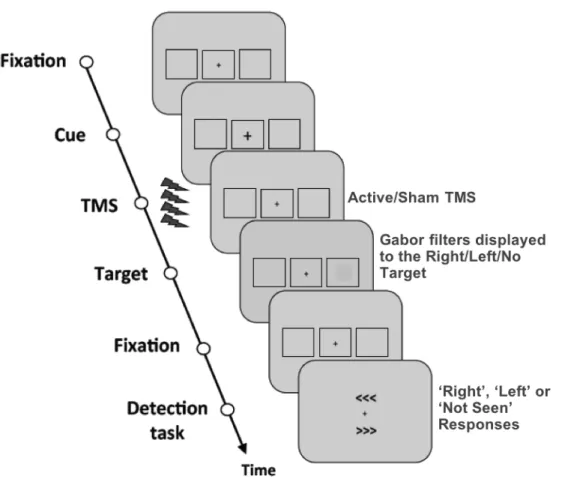
I – Behavioral paradigm to assess visual performance... 68
I.1 – Near-threshold lateralized visual detection paradigm ... 68
I.2 – Visual target properties, features and titration procedures ... 71
I.3 – Experimental blocks and session organization ... 74
I.4 – Subjective and objective measures of perception ... 75
II – Transcranial Magnetic Stimulation ... 80
II.1 – Stimulation parameters ... 81
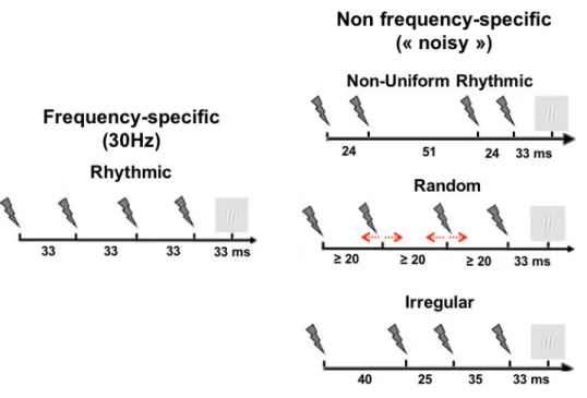
II.2 – Design of rhythmic and random TMS patterns ... 83
II.3 – Cortical target selection and MRI-based frameless neuronavigation ... 87
III – Concurrent TMS-EEG recordings of brain activity ... 88
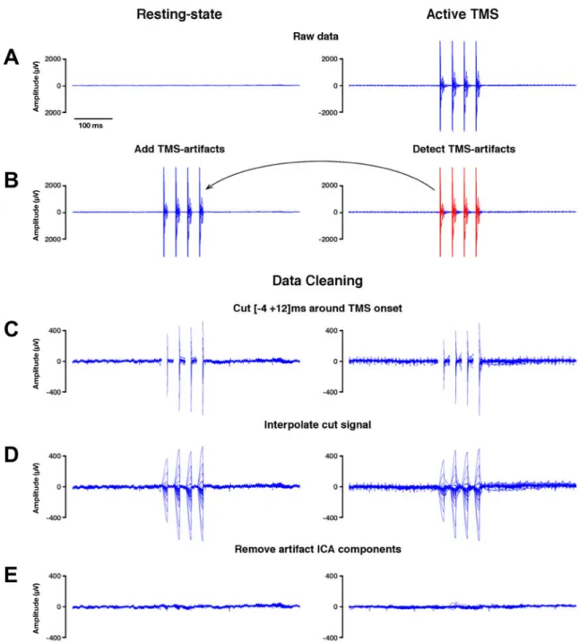
III.1 – Electromagnetic TMS-EEG artifact removal and data cleaning procedures ... 89
III.2 – Concurrent TMS-EEG recordings and EEG data pre-processing ... 93
III.3 – Control analysis on the TMS-EEG artifact removal and data cleaning procedures ... 94
III.4 – Outcome measures to assess the impact of TMS on oscillatory activity ... 98
III.4.1 – Outcome measures for local oscillatory activity... 98
III.4.2 – Outcome measures for inter-regional network synchronization ... 100
III.5 Outcome measures to quantify and characterize noise in EEG datasets ... 101
III.5.1. Measures to characterize noise in the time-frequency domain ... 102
III.5.2. Measures to characterize noise in the time domain ... 105
III.6 – Cluster-based permutation tests for the correction of multiple comparisons ... 107
REFERENCES ... 110
PROJECT 1: Causal role of high-beta oscillations in the right fronto-parietal network for conscious visual detection ... 119
I – Entrainment of local synchrony reveals a causal role for high-beta right frontal oscillations in human visual consciousness ... 119
II – Causal role of high-beta right fronto-parietal synchrony in the modulation of human conscious visual perception ... 135
PROJECT 2: Exploring unexpected contributions of left frontal neural noise to the modulation of conscious visual perception in the human brain: a combined TMS-EEG study ... 173
PROJECT 3: Non-specific effects of auditory stimulation generated by transcranial magnetic stimulation (TMS) on cortical oscillations and visual detection performances ... 227
GENERAL DISCUSSION ... 275
I - Summary of the main results ... 275
II – Frontal and fronto-parietal contributions to the modulation of visual perception ... 278
II.1 – Interhemispheric asymmetries in top-down systems for the facilitation of visual performance ... 279
II.3 – Modulating visuo-spatial attention and recording conscious visual perception ... 283
III- Pending questions and some future directions ... 284
III.1 – Towards an oscillatory model of attentional orienting and perceptual modulation ... 285
III.2 – Contributions of parietal and occipital cortices to conscious perception ... 288
IV- Further considerations ... 290
IV.1 – Unexpected impact of ‘control’ TMS patterns on EEG activity ... 291
IV.2 – Network impact and state dependency of frequency-tailored TMS effects ... 293
V- Conclusion and final remarks ... 296
SUMMARY
Two decades of studies on the role of oscillatory activity and network synchrony have provided extensive evidence supporting the contribution of these mechanisms to a large variety of cognitive processes and behaviors. In the domain of visuo-spatial attention, a process that mediates our ability to focus, select and extract relevant visual information from natural environments, theoretical and experimental evidence have suggested a role for high-beta phase synchrony, or the lack thereof, mediating top-down attentional influences on human conscious visual perception. Such contributions have proven to be site- and network-specific, hence calling for a systematic exploration of further coding contributions for fronto-parietal nodes in a bilaterally distributed network with bearing on orientation of attention and perception.
The studies included in the current doctoral dissertation used MRI neuronavigated Transcranial Magnetic Stimulation (TMS) in either rhythmic patterns designed to entrain high-beta oscillations or arrhythmic patterns designed to induce different levels of neural noise and desynchronization. TMS patterns were delivered trial-by-trial to the right and left Frontal Eye Fields (FEF) while participants carried out a visual detection task, in which they had to report the presence of lateralized near-threshold Gabors titrated at 50% visibility. In parallel, by means of concurrent scalp EEG recordings, we aimed to better understand the influence of entrained oscillations and noise patterns in the generation of frequency-specific synchrony, and ultimately assess the ability of the probed regions and TMS-coding patterns to modulate conscious access for near-threshold lateralized visual stimuli.
The INTRODUCTION of this dissertation summarizes the latest knowledge with regards to the role of oscillations, synchrony and neural noise in the coding, transfer and processing of information subtending the orienting of spatial attention and the modulation of visual perception. Complementarily, we also review the features and application of brain stimulation technologies, and in particular rhythmic TMS, to identify the relevant cortical regions and characterize the oscillation and synchronization/desynchronization-based coding mechanisms involved in enabling attentional orienting and the facilitation of conscious perception. The introduction is completed with a short section presenting the SPECIFIC AIMS, stating the underlying question pursued by the different studies of the dissertation, including their relevance, methodological approaches and a priori hypothesis and prediction for outcomes. A
detailed section of GENERAL METHODS presents, discusses critically and justifies the choice of behavioral paradigms, stimulation technologies patterns, experimental designs and EEG recording, data processing and measures employed in the three sets of studies included in the dissertation.
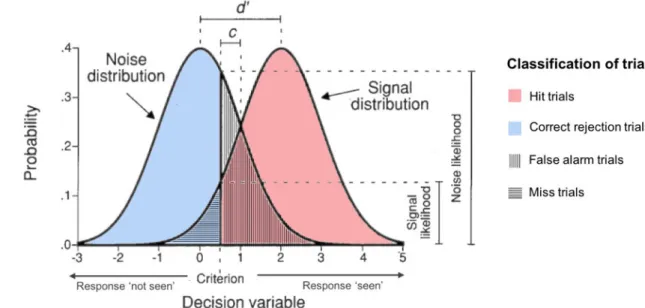
The RESULTS section integrates 3 different projects using each time the format of scientific papers. The two papers included in PROJECT 1 used concurrent rhythmic TMS-EEG approaches (high-beta 30 Hz rhythmic TMS vs random TMS 4 pulse bursts) to probe the contribution of the right FEF to conscious visual sensitivity (d’), as measured from the Signal Detection Theory. They showed that high-beta rhythmic TMS patterns increase local and inter-regional synchronization in a right lateralized fronto-parietal attentional network. This outcome supports a causal role for episodic high-beta oscillations entrained prior to target onset in the facilitation of conscious visual perception, likely via top-down attentional orienting mediated by the fronto-parietal dorsal attentional network. The paper in PROJECT 2 uses very similar TMS-EEG approaches, probing the role of the left FEF with TMS patterns similar to those used previously (high beta 30 Hz rhythmic TMS vs 3 different non-frequency specific TMS 4 pulse bursts: non-uniform rhythmic, irregular and random patterns) inducing different levels of local noise during task performance. Our data showed that, in this region, arrhythmic or irregular patterns of TMS increased neural noise locally and also throughout nodes of the bilateral dorsal attentional network. None of the tested patterns showed an impact on perceptual sensitivity (d’). Nonetheless, based on prior evidence collected in our lab for an improvement of visual sensitivity following arrhythmic TMS bursts, we provide preliminary evidence for a causal relationship between TMS-induced optimal levels of neural noise and enhancements of conscious visual perception. Finally, the paper presented in PROJECT 3 explored the impact of different patterns of TMS-generated sounds sharing a similar temporal structure with the electromagnetic patterns tested in prior study projects (30 Hz rhythmic sham TMS, random sham TMS 4 pulse bursts and single sham TMS pulse) on evoked and oscillatory EEG activity and also conscious visual perception correlates. None of the clicking sound patterns were able to impact visual sensitivity (d’) neither did they entrain frontal or fronto-parietal oscillations. Nonetheless, irrespective of TMS pattern type, stimulation phase-locked oscillations in central contacts and decreased response criterion (c), rendering participants less conservative when making perceptual decisions.
Taking all studies together, we CONCLUDE that oscillatory and phase-synchrony contributions to visual perception probed with causal methods were site-, network- and
pattern-(right FEF) high-beta oscillations and fronto-parietal synchronization to conscious visual perception. In a homotopic left fontal site (left FEF), we obtain preliminary evidence of ‘stochastic-resonance-like’ effects of graded neural noise levels facilitating visual perception, but further studies will be needed to better pinpoint this finding. Finally, at difference with active electromagnetic TMS, sham TMS-generated sounds in rhythmic high-beta patterns failed to entrain rhythmic activity or modulate visual sensitivity. Stimulation wise, concurrent TMS-EEG recordings demonstrated the ability of some active TMS patterns to modulate, during their delivery, oscillatory activity and inter-regional cortical synchrony, while other active TMS patterns proved able to modulate neural noise levels in a TMS pattern-dependent manner. In the GENERAL DISCUSSION we highlight further interpretations of these results in the wider context of the existing literature on the anatomical and physiological correlates of spatial attention and the top-down modulation of visual perception as well as the future technological advances in the field of non-invasive brain stimulation to manipulate oscillations and synchrony for fundamental and clinical research.
RÉSUMÉ (Français)
Deux décennies de recherche sur le rôle de l’activité oscillatoire et de la synchronisation des réseaux neuronaux ont fourni de nombreuses preuves de la contribution de ces mécanismes à une grande variété de processus cognitifs et de comportements. Dans le domaine de l'attention visuo-spatiale, qui est notre capacité à se focaliser, sélectionner et extraire des informations visuelles pertinentes dans notre environnement naturel, des preuves théoriques et expérimentales soutiennent le rôle de la synchronie des oscillations neurales à une fréquence beta-haute (ou de son absence) dans l’attention et la modulation de la perception visuelle consciente. De telles contributions se sont révélées spécifiques à des sites corticaux et à des réseaux neuronaux, appelant ainsi à l’exploration systématique des stratégies de codage des nœuds au sein d'un réseau fronto-pariétal bilatéral de l'attention et de la modulation de la perception consciente.
Les études incluses dans ce mémoire de thèse doctorale utilisent la Stimulation Magnétique Transcrânienne (SMT) sous la forme des rafales rythmiques conçues pour entraîner des oscillations beta-hautes ou arythmiques afin d'induire différents niveaux de bruit neural et de désynchronisation des rythmes cérébraux. Les rafales de SMT sont délivrées essai-par-essai sur les champs oculomoteurs frontaux (en anglais, FEF) des hémisphères droit et gauche, tandis que les participants effectuent une tâche de détection visuelle dans laquelle ils doivent détecter et localiser à droite ou à gauche la présence d’une cible visuelle au seuil de détection (c’est-à-dire, adapté en contraste à un taux de visibilité de 50%). En parallèle, au moyen d’enregistrements d’EEG de surface, nous avons cherché à mieux comprendre l’influence des oscillations ou du bruit neural entraînés par la SMT sur la génération de la synchronisation locale ou inter-régionale à une fréquence spécifique, et à évaluer la capacité des régions cérébrales étudiées et des rafales de SMT à moduler l’accès conscient des stimuli visuels latéralisés présentés au seuil de visibilité.
L’INTRODUCTION de ce mémoire résume l’état de l’art en ce qui concerne le rôle des oscillations, de la synchronie et du bruit neural dans le codage, le transfert et le traitement de l’information sous-tendant l’orientation de l’attention spatiale et la modulation de la perception visuelle. Par ailleurs, nous détaillons également les caractéristiques et l’application des technologies de stimulation cérébrale non-invasives, et en particulier de la SMT rythmique, pour identifier les régions corticales et caractériser les mécanismes de codage basés sur
l’activité oscillatoire et la synchronisation/désynchronisation des réseaux impliqués dans l’orientation de l’attention et la facilitation de la perception consciente. L'introduction est complétée par une courte section présentant les OBJECTIFS SPÉCIFIQUES, exposant les questions sous-jacentes poursuivies par les différentes études incluses dans ce mémoire, y compris leur pertinence, leurs approches méthodologiques et leurs hypothèses et prédictions à priori. Une section détaillée de MÉTHODES GÉNÉRALES présente, discute de manière critique et justifie le choix du paradigme comportemental, des technologies et motifs de stimulation, du design expérimental ainsi que de l’enregistrement, du traitement de données et des mesures d’analyse d’EEG utilisées dans les trois séries d’études incluses dans ce mémoire. La section RESULTATS intègre 3 projets différents présentés à chaque fois sous la forme d’un article scientifique. Les deux articles inclus dans le PROJET 1 utilisent des approches EEG-SMT rythmiques (rafales de SMT rythmique beta-hautes à 30 Hz versus rafales de SMT aléatoires de 4 impulsions : non-uniformes-rythmiques, irrégulières et aléatoires) afin d’explorer la contribution du FEF droit à la sensibilité visuelle consciente (d '), telle que mesurée par la Théorie de Détection du Signal. Les rafales de SMT rythmiques à une fréquence beta-haute augmentent la synchronisation locale et inter-régionale sur un réseau attentionnel fronto-pariétal latéralisé à droite. Ces résultats corroborent le rôle causal des oscillations épisodiques dans une bande de fréquence beta-haute entraînées avant l’apparition de la cible dans la facilitation de la perception visuelle consciente, probablement via des effets descendants de l’attention médiés par le réseau fronto-pariétal dorsal de l’orientation de l’attention. L’étude du PROJET 2 utilise des approches EEG-SMT très similaires à celles déjà mentionnées, pour explorer cette fois le rôle du FEF gauche, avec des rafales de SMT périodiques proches de celles utilisées précédemment (rafales de SMT rythmique à 30 Hz et 3 motifs de rafales de SMT non spécifiques en fréquence à 4 impulsions) induisant différents niveaux de bruit neural lors de l'exécution d'une tâche d’accès à la perception consciente. Nos données montrent que, sur cette région, des rafales non spécifiques en fréquence de SMT augmentent le bruit neural, localement et également tout au long des nœuds du réseau bilatéral de l’attention. Aucun des motifs de rafales de SMT délivrées n’a montré d’impact sur la sensibilité perceptuelle (d’). Néanmoins, selon des résultats antérieurs obtenus dans note laboratoire qui ont montré une amélioration de la sensibilité visuelle à la suite de rafales de SMT non spécifiques en fréquence, nous fournissons des preuves préliminaires d'une relation de cause-à-effet entre les niveaux optimaux de bruit neural induits par la SMT et les améliorations de la perception visuelle consciente. Enfin, l’étude présentée dans le PROJET 3 examine l’impact de différents types de sons
électromagnétiques testés dans les projets d’études précédentes (rafales SMT placebo rythmiques à 30 Hz, rafales SMT placebo aléatoires de 4 impulsions et des impulsions uniques de SMT placebo), sur l'activité EEG évoquée et oscillatoire, ainsi que sur la perception visuelle consciente. Les rafales sonores ne montrent pas d’impact sur la sensibilité visuelle (d’) ni aucuns signes électroencéphalographiques d’entrainement oscillatoire frontaux ou fronto-pariétaux. Néanmoins, quel que soit le type de rafale SMT placebo délivrées, elles ont abouti à une synchronisation en phase l’activité oscillatoire du cortex auditif et ont diminué le critère de réponse (c), engendrant des stratégies moins conservatrices lors de la prise de décisions perceptuelles.
Considérant l’ensemble de nos résultats, nous CONCLUONS que les contributions oscillatoires ou de la synchronie de réseau sur la perception visuelle consciente étudiées avec des méthodes causales sont dépendantes du site et réseau stimulé ainsi que de la structure temporelle de la rafale magnétique. À cet égard, notre approche SMT-EEG a attesté une influence potentielle des oscillations dans une bande de fréquence beta-haute au niveau du cortex frontal droit (FEF droit) et la synchronisation fronto-pariétale dans l’hémisphère droit sur la perception visuelle consciente. Dans la région homotope à gauche (FEF gauche), nous obtenons des preuves préliminaires d’effets présentant les mêmes propriétés que le phénomène de résonance stochastique, c’est-à-dire une facilitation de la perception visuelle par des niveaux de bruit graduels. Cependant, des études supplémentaires sont nécessaires pour identifier et confirmer les corrélats comportementaux de ce résultat. Enfin, à la différence des rafales de SMT beta-hautes actives, les rafales sonores rythmiques générées par la SMT placebo ne parviennent pas à entraîner d'activité neurale rythmique ni à moduler la sensibilité visuelle. En ce qui concerne la stimulation SMT, le couplage avec des enregistrements EEG nous a permis de démontrer la capacité de certaines rafales de SMT active à moduler l’activité corticale oscillatoire et sa synchronisation, tandis que d’autres motifs de rafales de SMT permettent la modulation du niveau de bruit neural. Dans la DISCUSSION GÉNÉRALE, nous présentons les interprétations de nos résultats dans le contexte plus large de la littérature existante sur les bases anatomiques et physiologiques de l'attention spatiale, la modulation de la perception visuelle consciente et les futurs développements technologiques dans le domaine de la stimulation cérébrale non invasive afin de manipuler les oscillations cérébrales et la synchronie à des fins expérimentales ou cliniques.
TABLE OF ABBREVIATIONS
ASSR Auditory Steady-State Response cTBS continuous Theta Burst Stimulation EEG ElectroEncephaloGraphy
EMG ElectroMyoGraphic ERP Event-Related Potential FEF Frontal Eye Field
ICA Independent Component Analysis IPS IntraParietal Sulcus
iTBS intermittent Theta Burst Stimulation ITC Inter-Trial Coherence
LFP Local Field Potential MEG MagnetoEncephaloGraphy MRI Magnetic Resonance Imaging MSE Multi-Scale Entropy
MT Middle Temporal visual area NIBS Non-Invasive Brain Stimulation PCA Principal Component Analysis PLV Phase-Locking Value
RMT Resting Motor Threshold
rTMS repetitive Transcranial Magnetic Stimulation SDT Signal Detection Theory
SE Sample Entropy
SLF Superior Longitudinal Fasciculus SR Stochastic Resonance
SSVEP Steady-State Visual Evoked Potential tACS transcranial Alternating Current Stimulation tCS transcranial Current Stimulation
tDCS transcranial Direct Current Stimulation TMS Transcranial Magnetic Stimulation tRNS transcranial Random Noise Stimulation
INTRODUCTION
I – Brain oscillations, local and network synchronization and orienting of
spatial attention
Electrophysiological recordings of neural activity at any scale, either electroencephalographic (EEG) activity from large neuronal assemblies (Berger, 1929), in vivo local field potentials produced by local neuronal clusters (Gray & Singer, 1989), or in vivo (Alonso & Llinás, 1989) and in vitro (Draguhn et al., 1998) single-cell voltage changes, reveal patterns of rhythmic activity which have been referred to as neural oscillations. This neurophysiological phenomenon is characterized by highly regular, repetitive and synchronous activity patterns, which ensure the precise timing of neuronal activity, and can operate in a wide range of frequencies across brain sites and neural circuits.
Oscillations were initially reported as particularly prominent during sleep or in situations in which consciousness was decreased and neural systems did not seem to be involved in a specific behavior (Steriade et al. 1994). For this reason, they were considered unrelated to cognitive processes, and their physiological and behavioral role was long ignored, or considered an irrelevant by-product or epiphenomenon bearing no role on human behaviors. Two decades ago however, neural oscillations started to be revisited with renewed interest and since then the number of studies addressing the role of oscillations in cognitive functions such as memory, attention or perception has skyrocketed (reviewed in Buzsáki & Draguhn, 2004).
Amongst cognitive functions that have been widely shown to be subtended by oscillatory activity is attentional orienting in space. Attention is the process by which we select information in our crowded environment (Desimone & Duncan, 1995). In spite of the high processing power of the human brain, our senses have a limited capacity to simultaneously uptake information from the inner and outer environment. To face the challenge of overcrowded environments, to which we are often exposed, attention acts as a selective filter that allows us to allocate resources to the most task-relevant stimuli, hence enhance the perception of important inputs and suppress the perception of irrelevant distractors.
Such core function of attention requires brain systems to be able to segment incoming inputs and selectively enhance the processing of some of them at the expense of others which
are suppressed. A decade ago, a framework was developed by which neural oscillations synchronized in phase across widely distributed neuronal assemblies connected by white matter pathways, could subtend these core mechanisms (Fries, 2009).
We will highlight the mechanisms by which neural oscillations could subtend the orientation of attention, then review empirical evidence supporting a link between this essential brain function and oscillatory activity and synchronization.
I.1 – Oscillations and synchronization in network communication and information transfer
Under normal conditions, the brain receives simultaneously a very high number of inputs from stimuli present in a visual scene. Each of these incoming stimuli will reach and activate neural assemblies in the early visual cortex. Converging input from several neuronal groups to common neuronal targets is a common neocortical connectivity motif (Jones & Powell, 1970), especially in the visual cortex (Salin et al., 1992), hence inputs from competing visual stimuli present in a given visual environment will converge on similar neural assemblies in higher order visual areas.
As a result of this organization of input patterns, high level neurons possess wide receptive fields (Gattass et al., 2005) and, at any moment in time, neuronal assemblies in higher order visual cortices can receive inputs generated by distinct objects and stimuli present in the visual field. These circuits assemblies cannot respond to several stimuli at the same time as it would give rise to a phenomenon that has been called the “curse of confusion through convergence” (Fries, 2009). To avoid this phenomenon, neural assemblies collecting the converging inputs from lower visual areas need to be able to segment the inputs into distinct visual stimuli to then be able to selectively respond to the inputs that correspond to visual stimuli relevant for the behavior at hand and ignore information from distractor stimuli.
Gamma-band oscillatory synchronization is a well-known mechanism to tackle the so called “binding problem”, a phenomenon by which a set of individual features are bound together to build a unified representation of an object (Singer & Gray, 1995). The phase-synchronization at gamma frequency between several neuronal assemblies serves to strengthen inter-regional communication and to create a dynamic network processing the same complex stimulus (Fries, 2005, 2009; Tallon-Baudry & Bertrand, 1999). In a seminal paper, Pascal Fries (2009) provides a detailed account on how gamma-band synchronization could help individual
neurons deal with the above-mentioned functional segmentation of inputs by means of two mechanisms: feedforward coincidence detection and input gain modulation.
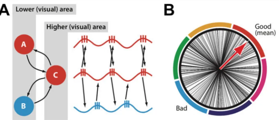
Consider two clusters of neurons entitled ‘A’ and ‘B’ both responding to competing stimuli in the receptive field of neurons in a cluster entitled as ‘C’. Through a convergence of connectivity, both A and B sustain structural synaptic connections with neurons in C (Fig. 1A, left). Hence when two competing stimuli are present in C’s receptive field, both A and B would fire and send an input to C. However, since A and B are involved in coding for competing stimuli, C is unable to respond to both inputs. Hence C has to be able to segment inputs from A and B in order to avoid confusion between the two.
As neurons in cluster A respond to a single object, through the binding by synchronization mechanism (Singer & Gray, 1995) they will locally synchronize, i.e. progressively phase-lock their activity at a gamma frequency. When all the neurons in A are synchronized, they will reach the phase of their oscillation cycle where the excitability of the neurons is maximal simultaneously and fire within a short time window. All the outputs from neuron group A will reach the dendrites of cluster C in close succession and, through summation of synaptic input, will have higher input gain, hence be more likely to depolarize neurons in cluster C. This phenomenon is called Feedforward Coincidence Detection and it will enable neurons in group A, when they are locally synchronized, to successfully depolarize C periodically when they send a volley of outputs at the excitable phase of their oscillation cycle, and hence, to progressively entrain neuron C to oscillate in a phase-locked manner with neurons in group A (Fig. 1A, right).
Once A and C are synchronized in phase a second mechanism comes into play to ensure that possible inputs from other groups of neurons cannot reach C. Indeed, once C is synchronized with A, its membrane potential oscillates at the same frequency, creating period of lower (i.e., more negative) transmembrane resting potential levels and therefore low excitability for neurons and periods of higher (i.e., more positive hence closer to firing threshold) transmembrane resting potential levels and high excitability. This input gain modulation provides a neuron in cluster C with the ability to selectively react either more strongly to inputs incoming in a phase-locked manner during high excitability phases or less strongly to inputs not synchronized to the rhythms of its oscillation hence coming randomly at any phase of its oscillation cycle (Fig. 1B). Such input gain mechanism would favor inputs coming from neural cluster A, which is synchronized with neurons of cluster C, while lowering the gain of inputs coming from cluster B not synchronized with the latter and therefore coming randomly at times of high or low excitability (Fig 1A, right).
Figure 1. Communication through coherence. (A) Feedforward Coincidence Detection mechanism. Neuronal clusters ‘A’ and ‘B’ have converging connections to neuronal cluster ‘C’. Through the
Feedforward Coincidence Detection mechanism, neurons in cluster A synchronize in phase with
neurons in cluster C. (B) Input Gain Modulation mechanism. Schematic representation of high (good) and low (bad) excitability phases for neuronal oscillators of cluster C. Neurons in cluster A are synchronized with neurons in cluster C so that inputs from neurons in A will always reach neurons in C during high excitability phases, ensuring a higher gain compared to inputs incoming from neurons in cluster B who reach neurons C at any phase. (Adapted from Fries, 2009).
This mechanism highlights the biased competition between neurons in group A and B. Both mechanisms of Feedforward Coincidence Detection, which progressively entrains C with the neuron group sending it the most synchronized inputs, and Input Gain Modulation, which renders the input gain in C favorable only to inputs from neurons groups synchronized with it, create a competition between A and B and only one of these competing neuron groups can successfully send inputs to C through a winner-take-all mechanism. An exclusive communication link is established between C and A, excluding inputs from all other competing neuronal groups connected to C. Biased competition, subtended by inter-neuronal gamma-band synchronization, is therefore a very effective model to explain the selectivity of attention, that is its ability to respond to a single set of features in a visual scene containing an infinite number of stimuli (Desimone & Duncan, 1995; Reynolds et al., 1999).
It is worth noticing that the emerging properties of selective attentional systems highlighted above emerge from a complex interplay between structural and functional connectivity. The former is essential to build, through learning and Hebbian rules, complex receptive fields in higher sensory cortices. The latter, articulated by means of inter-neuronal gamma-band synchronization, enables a dynamic segmentation of structural connections and a
I.2 – Network synchronization subtending visuo-spatial attention and visual perception
Empirical evidence developed in the last 20 years supports the theoretical framework presented above. In favor of the Feedforward Coincidence Detection mechanism, studies in animal models have shown enhanced local gamma synchronization in visual areas in response to attended stimuli (Fries et al. 2001; Bichot et al. 2005). There is also evidence supporting the enhancement of gamma or high beta (25-60 Hz) interregional synchronization between frontal, parietal and occipital areas either after the presentation of an attended target (Saalmann et al. 2007) or during the orientation of attention in space during a visual search task (Buschman & Miller, 2007). This evidence supports exclusive communication channels between different regions of the attentional network through phase synchronization during the orientation of attention. These findings in animal models have been replicated in healthy human with EEG/MEG recordings, showing the role of fronto-parietal synchronization at high-beta frequencies (15-40 Hz) during tasks manipulating attention (Gross et al., 2004; Phillips & Takeda, 2009) or conscious visual perception, a correlate of attentional orienting (Rodriguez et al. 1999; Hipp et al. 2011).
To further outline the importance of inter-regional synchronization at beta or gamma frequencies for cognitive activity, it should be noted that abnormal (enhanced or reduced) levels of neural synchronization have been shown to be relevant in many pathologies and neural disorders (Uhlhaas & Singer, 2006). In certain conditions, like in schizophrenia or autism, reduced gamma synchronization leads to deficits in object binding and perception (Grice et al., 2001; Uhlhaas et al., 2006). In other conditions, such as post-stroke neglect patients, it is an abnormally enhanced local beta synchronization that is detrimental for attentional orienting and perception (Rastelli et al., 2013).
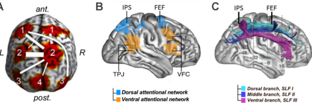
The highlighted evidence enables the characterization of a neural network for the orientation of attention and the top-down modulation of conscious visual perception as well as the relevant frequency bands allowing inter-regional synchronization in this network. The cortical regions of interest highlighted in most anatomical models for this cognitive functions of attention include the bilateral Frontal Eye Fields (FEF) and Intra Parietal Sulci (IPS) as well as visual areas in the occipital lobe like the medial-temporal region (MT) or the primary visual cortex (V1) (Buschman & Miller, 2007; Hipp et al., 2011; Saalmann et al., 2007) (Fig. 2A).
Figure 2. Grey and white matter components of the attentional orienting network. (A) Cortical regions synchronized at high-beta frequency during attentional orientating. In the frontal lobe (1), the Frontal Eye Fields (FEF); in the posterior parietal Lobe (2), the Intraparietal Sulcus (IPS); in the lateral occipito-temporal region (3), the middle temporal cortex (MT/V5); in the medial occipital region (4), the primary visual cortex (V1) (Adapted from Hipp et al. 2011). (B) Anatomical distribution of dorsal and ventral attention networks. (Adapted from Corbetta et Shulman, 2002). (C) Fronto-parietal structural white matter connections: the three branches (SLF I, II and III) of the Superior Longitudinal Fasciculus. (Adapted from Thiebaut de Schotten et al. 2011).
The FEF and IPS, as part of the dorsal attention network, have long been identified as crucial regions for orienting attention in space (Corbetta and Shulman 2002; Corbetta et al. 2008) (Fig. 2B). Damage in these two regions has been found to be crucial to explain the deficits and recovery of spatial attention orientating abilities in post stroke neglect patients (Corbetta et al., 2005). In addition to the isolated activity of both of these regions, anatomical connectivity between frontal and parietal areas of attentional networks, supported by the three branches of a white matter tract called the Superior Longitudinal Fasciculus (SLF I, II and III) (Fig. 2C), has been shown to subtend the deployment of spatial attention in healthy participants (Marshall et al., 2015; Thiebaut de Schotten et al., 2011). Moreover, the disconnection of this tract can lead to visuo-spatial attentional deficits in neglect patients (Bartolomeo et al. 2012; Thiebaut de Schotten et al. 2014).
With regards to the relevant synchronization frequency bands for attention, seminal and influential work by Buschman and Miller (2007) in non-human primates highlighted rhythmic activity in the high-beta to gamma range subtending different mechanisms tied to attentional orienting. More specifically, these authors reported a double dissociation with gamma oscillations (35-55 Hz) subtending exogenous attentional orienting (e.g. bottom-up or involuntary) whereas high-beta oscillations (22-34 Hz) underlay endogenous (e.g. top-down or
of frequencies (from 25 to 90 Hz) to the orientating of spatial attention (Bichot et al., 2005; Fries et al., 2001; Saalmann et al., 2007). Nonetheless, analogous follow-up studies in humans reported similar correlations for a narrower range of lower frequency bands (between 15 and 40 Hz), more consistent with high-beta than gamma activity (Gross et al., 2004; Hipp et al., 2011; Phillips & Takeda, 2009; Rodriguez et al., 1999).
Neural oscillations in lower frequencies bands have been associated to other processes at play in spatial attention. Local synchronization in the parietal and occipital cortex in the alpha band is thought to inhibit processing of distractor stimuli (Foxe & Snyder, 2011; Klimesch et al., 2007; Thut et al., 2006; Worden et al., 2000) and oscillations at alpha (Dugué et al., 2011; Mathewson et al., 2009, 2011) or theta frequency (Huang et al., 2015; Landau & Fries, 2012; Landau et al., 2015) could pace the rhythmic sampling of attention, alternating periods of concentration and periods of shifts of attention. Indeed, a multi-frequency model has been proposed, which integrates the roles of gamma, beta, alpha and theta oscillations in the orientation and reorientation of attention (Fiebelkorn & Kastner, 2019). However, none of these lower frequency oscillations have been associated to inter-regional synchronization and communication (the contributions of frequency bands outside of gamma and high-beta is discussed in more detail in the General Discussion).
All the evidence reviewed above lead to the conclusion that synchronization in the fronto-parietal dorsal attention network at a high-beta frequency is related to the orienting of visuo-spatial attention and conscious visual perception. However, the studies reviewed so far obtained their conclusions from correlations between LFPs or EEG recordings and performance outcomes in attentional and visual perceptual tasks. Consequently, the correlational nature of this evidence did not allow to establish any causal link between these two phenomena co-occurring in time, and could not rule out that cortical oscillations and interregional synchronization patterns were merely epiphenomena, holding no direct contribution to the neural coding subtending cognitive computations. It was only a decade ago, that a new attempt to push progress in this field explicitly advocated to move beyond correlations and called for a need of direct manipulation of rhythmic activity (either to temporally enhance, suppress or replace it) to unearth causal links between cortical oscillations and the modulation of attentional and visual behaviors.
II – Manipulation of brain oscillations subtending attentional and visual
behaviors
Traditional interventions to manipulate brain activity have required animals or human to be engaged in specific behavioral tasks, while relying on non-invasive technologies to record their neural activity (fMRI, surface EEG, MEG). Direct causal manipulations of cortical activity, by means of epidural/intracranially implanted electrodes to perturb brain activity, could exclusively be performed in animal models or in a very limited set of human patient populations (such as Parkinson’s, epilepsy, obsessive compulsive disorder, or brain tumor patients for which the implantation of epidural/intracranial electrodes is justified for diagnostic or therapeutic purposes).
In this context, the 21st century has seen the development of technological innovations
able to manipulate brain activity in humans without the need of invasive surgery. Currently, cortical rhythms can be entrained or manipulated experimentally by means of pulsed or fluctuating sensory stimuliwhich can influence activity along sensory pathways and reach the cortex. Alternatively, more recently, the field has seen the development of non-invasive transcranial brain stimulation techniques: Transcranial Magnetic Stimulation (TMS) and Transcranial Current Stimulation (tCS) using, respectively, electromagnetic pulses or electrical current delivered on the scalp that penetrates the skull and can reach the cortical surface to modulate neural activity.
As indicated above, direct electrical brain stimulation delivered through intracranial implanted electrodes in patients with medication resistant epilepsy (brief 5-10 second trains of 1, 50 or 60 Hz) to identify seizing foci or deep brain stimulation of the subthalamic nucleus in Parkinson’s patients (at high frequency, 90-180 Hz) to prevent tremor, bradykinesia or rigidity provide very interesting opportunities to causally explore the role of oscillations in healthy and pathological structures of unsound brains (Amengual et al., 2017; Cleary et al., 2012). Nonetheless, in spite of the high spatial and temporal precision and optimal signal-to-noise ratio for intracranial stimulation and recordings, implantation schemes are obviously guided by clinical criteria and hence show considerable variability across patients and provide a very sparse coverage of the cortex. Non-invasive stimulation methods enjoy much more flexibility to explore the same phenomena in a wide variety of cortical regions and patient or healthy subject populations.
II.1 – Non-invasive stimulation techniques to manipulate brain oscillations and synchrony
As summarized above, currently, two main non-invasive approaches have been used to entrain rhythmic activity in the human brain to improve cognition: (1) peripheral sensory stimulation, which uses auditory, visual or tactile sensory pulsed or oscillating patterns applied to peripheral receptors which are conveyed by bottom-up sensory pathways to influence brain systems and networks; (2) transcranial brain stimulation via magnetic pulses or electrical current fields targeting a cortical area or circuit directly to influence its activity patterns.
Each of these two approaches have strengths and limitations in terms of focality (spatial resolution), timing control (temporal resolution), safety, financial cost, ease of use and portability. The former uses a rather physiological stimulation source which can be made very selective by capitalizing on the modality-specific (somatotopic, tonotopic and retinotopic) organization of afferent receptors and pathways. Nonetheless, its effect depends on the integrity of afferent pathways and these can be modulated (hence dispersed in spatial precision and attenuated in intensity) at every synaptic step from the peripheral receptor to the receiving cortical systems and beyond. The latter can directly target any cortical region with a level of selectivity that depends on the spatial resolution of each technological approach. Nonetheless, focal approaches (TMS) deliver rather intense electrical currents which are far from physiological, whereas un-focal methods (tCS) often lack precision and intensity to produce convincing impact on neurophysiological activity.
Both types of technologies represent unique tools to probe causal links between local and network-mediated oscillatory synchronization on circumscribed anatomical locations and the behavioral effects that these patterns might subserve. For this reason, they have been widely used in the last decade and provided causal evidence for a functional role of cortical oscillations in coding for cognitive functions.
II.1.1 – Rhythmic peripheral sensory stimulation for oscillatory entrainment
Peripheral sensory stimulation is based on conveying rhythmic sensory patterns through the sensory pathways able to reach and influence the activity of cortical systems. Sensory stimuli (usually auditory, visual or less commonly tactile) that are either pulsed (a transient stimulus that is presented repeatedly) or continuously oscillating at a fixed specific frequency
can be easily applied to peripheral sensory receptors. Conveyed through afferent sensory pathways, they have been shown to entrain rhythmic activity in the brain within a frequency band which is dictated by the periodicity of incoming stimuli.
A quite common method for sensory entrainment is the use of a visual flicker, in which pulsed visual stimuli are rhythmically flashed while steady-state visual evoked potentials (SSVEPs) from brain systems at the frequency of the flicker (Srinivasan et al., 1999; Vialatte et al., 2010) are recorded via EEG recordings. Similar procedures have been translated to other sensory modalities, and auditory stimuli modulated in amplitude or frequency (Galambos et al. 1981; Picton et al. 2003) presented monoaurally or binaurally via headphones or patterns of rhythmic tactile stimulation applied to skin mechanoreceptors by means of pulsed electrical stimulation, air puffs or piezo-electrical tactile stimulation devices have been used (Nangini et al., 2006).
Entrainment through afferent sensory stimulation typically increases local and inter-regional synchronization at the stimulus frequency in a wide range of brain areas, not limited to the primary sensory cortices receiving afferent information, but distributed all over the cortex and extending up to frontal systems (Srinivasan et al. 1999; Srinivasan et al. 2006; Srinivasan et al. 2007). However, signals have to progress throughout a whole hierarchy of sensory pathways and synaptic steps before reaching specific cortical regions. Hence this approach cannot achieve high levels of spatial focality and entrains oscillations in a rather widely distributed network, including sub-cortical regions (Giraud et al., 2000). Moreover, once in primary sensory areas, to reach higher-level associative areas (e.g. frontal or prefrontal areas), input rhythms will need to progress across cortico-cortical relay pathways. Long and multi-synaptic afferent subcortical and cortico-cortical pathway (which can be influenced or modulated by other inputs) imply larger time delays and timing variability, making the phase and amplitude of oscillations entrained at destination uncertain or unstable.
II.1.2 – Transcranial brain stimulation technologies for oscillatory entrainment
In this specific context, non-invasive brain stimulation (NIBS) technologies able to directly deliver rhythmic activity to circumscribed regions in the brain and newly entrain oscillations or modulate ongoing rhythmic activity are called to become very useful tools in exploratory or therapeutic endeavors. These approaches induce electric currents directly in the cortex, by-passing sensory cortices, for the entrainment of specific patterns of cortical activity (see Polanía et al., 2018 and Valero-Cabré et al., 2017 for recent reviews). The two most widely
used NIBS techniques to date are Transcranial Current Stimulation (tCS) and Transcranial Magnetic Stimulation (TMS). These two techniques have very different modes of action and therefore their own set of advantages and limitations.
II.1.2.1 – Transcranial alternate current stimulation approaches (tACS)
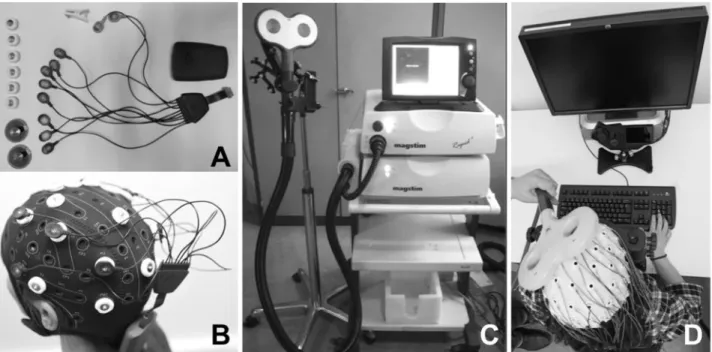
Transcranial Current Stimulation (tCS) is achieved by circulating a low intensity current (1-2 mA, ~0.06 mA/cm2) between at least two electrodes (an anode and a cathode) placed on
specific regions of the human scalp (Fig. 3A & B). A substantial portion of the circulating current is generally shunted through the scalp skin (Vöröslakos et al., 2018). Nonetheless part of it will penetrate across the different tissue layers between the skin and the cortical surface (i.e., bone outer and inner tables, and the cerebrospinal fluid cumulated in the epidural and subdural spaces) to reach the pia-mater and spread across rather large cortical areas located between both electrodes (Miranda et al. 2006).
The current gradients will polarize electrical charges in the extracellular space in a polarity dependent manner, shifting the resting membrane potential of exposed neurons closer (anodal stimulation) or away (cathodal stimulation) from their firing thresholds, hence increasing or decreasing their probability to generate an action potential when receiving physiological dendritic inputs of sufficient intensity.
If instead of a constant current (tCS modality know as transcranial direct current stimulation or tDCS), an alternating current (AC) is applied, the resting membrane potential and consequently the firing rate probability of neurons influenced by the current field will also fluctuate periodically, following the frequency of the AC signal. This specific modality of tCS is referred to as transcranial Alternating Current Stimulation (tACS) and has been used to non-invasively entrain oscillations in cortical regions (Fröhlich & McCormick, 2010; Herrmann et al., 2013; Merlet et al., 2013).
Although tCS devices delivering either tDCS or tACS are recognized as being portable and highly affordable compared to TMS (Fig. 3A & C), these technologies possess a rather poor spatial resolution. Given the diversity of possible electrode montages (particularly when density tCS approaches based on combination of several return electrodes in complex configurations are used) and interindividual differences in head anatomical features, it is not easy to predict how currents applied to the scalp will diffuse transcranially to reach the cortical surface. Indeed, it is generally accepted that induced brain currents will not remain restricted to cortical areas beneath the electrodes but will spread (Bikson et al., 2010; Datta et al., 2012).
Figure 3. Technical equipment for the delivery of Transcranial Current Stimulation (tCS) and Transcranial Magnetic Stimulation (TMS). (A) tCS is delivered through a small light and portable rechargeable battery system and controlled wirelessly from a computer or portable device. Current is conveyed by short and light physical wires to a montage (at least two, an anode and a cathode) of leads (either sponge contacts, or solid ferromagnetic leads, see both in the figure) placed in specific scalp locations. Systems also often integrate independent channels to record EEG signals. (B) The wireless tCS device is mounted directly on a lycra cap worn by participants while performing a task on a computer screen. A very mild current will flow between at least two electrodes (active and return) placed in separate locations of the scalp generating on the brain surface a large polarization gradient able to modulate the resting membrane potential of exposed neurons. The wireless control of tCS allows full head and possibly body motion. (C) TMS requires heavy non-portable equipment that charges current in a series of capacitors. From the central unit, accumulated current is then circulated through a stimulation coil (in the picture a double ¨butterfly¨ 70 mm coil) to generate a brief magnetic field, called a pulse, capable of penetrating through skull tissue layers and induce a focally distributed electrical current inside of the brain powerful enough to depolarize neurons. (D) The stimulation coil is placed lying flat on a subject’s head and held manually by an operator, or with help from a mechanic arm, while the subject is performing a task on a computer screen. TMS can be delivered in single pulse, short bursts (4 or 5 pulses) or long patterns of repetitive (rTMS) stimulation to modulate activity in a focal, targeted cortical region. Targeting is monitored throughout the session by means of an MRI-based neuronavigation system. Note in (D) TMS is delivered while EEG activity is being monitored through an independent equipment.
Moreover, some recent controversy debates if the standard and safe current intensities commonly used are high enough to reach critical current density levels in the cortex (> 0.5 V/m) able to shift transmembrane resting potentials and influence local excitability (Lafon et al., 2017; Vöröslakos et al., 2018; reviewed in Liu et al., 2018). Indeed, to reach meaningful current density levels, stimulation intensities should be of 4 to 6 mA, higher than the currently recommended stimulation intensities (Antal et al., 2017). Additionally, to induce a noticeable behavioral effect, tACS needs to be applied for relatively long periods of several minutes (Nitsche et al., 2008; Nitsche & Paulus, 2011), hence it lacks the temporal resolution to either entrain or modulate oscillatory activity at circumscribed time windows during task performance.
Regardless, thanks to its low cost, excellent safety profile, and ease of use, multichannel tACS is probably called to become the tool of choice to flexibly modulate local and interregional synchrony throughout cortical networks in humans. Nonetheless, currently, given the open debate on its potentially too low intensity, its known low spatial resolution and ineffectiveness to entrain episodic short lasting oscillations, tACS is not necessarily the most adapted technology to explore the causal role of cortical oscillations in well-defined anatomical regions at a specific time window during task performance.
II.1.2.2 – Rhythmic transcranial magnetic stimulation approaches (TMS)
TMS is currently the most established non-invasive technology used to activate clusters of neurons responsible for specific behaviors within a rather circumscribed cortical area (estimated ~12-15 mm radius) in healthy humans and patients.
TMS equipment consists in capacitators which charge and store electrical current, which is then briefly circulated (120 to 250 µs) through a stimulation coil (the most commonly used are figure-of-eight coils) made of two contiguous loops of copper wire encapsulated in butterfly shape protective case (Fig. 3C). Following the principles of electromagnetic induction discovered in 1831 by Michael Faraday, the circulation of the high-intensity current generates a brief and rapidly changing magnetic field, called a pulse, which distributes perpendicular to the surface of the TMS coil lying flat on the scalp. Thanks to the electromagnetic induction phenomenon, the magnetic field penetrates painlessly, and with very little distortion, the skull bone and the epidural and subdural spaces filled with CSF to reach the cortex under the coil and induce a current intracranially which will cause the depolarization of clusters of excitable neurons (Hallett, 2007; Kobayashi & Pascual-Leone, 2003) hosted within a focal area of 12-15 mm radius (see Valero-Cabré et al., 2005 for an estimation in animals models). To achieve its
effect, the TMS coil is placed on the scalp region most closely overlying a given cortical target (i.e. the one enabling the shortest straight path to cortical target) using a frameless stereotaxic MRI-based neuronavigation system customized to the anatomy of each healthy participant or patient (Fig. 3D).
Moreover, thanks to its excellent temporal resolution (Hallett, 2007), TMS allows single pulses or multi-pulse bursts arranged in a great variety of patterns to be used in online trial-by-trial designs to impact specific time windows during the performance of behavioral tasks (for recent reviews see Polanía et al., 2018 or Valero-Cabré et al., 2017). Likewise, long patterns of so called repetitive TMS (or rTMS) can induce, depending on stimulation parameters (essentially, stimulation frequency, pattern duration and number of pulses, magnetic field intensity and length of inter-burst intervals), excitatory or inhibitory offline modulations of neural activity and associated behaviors, which remain transiently active beyond the discontinuation of pulses.
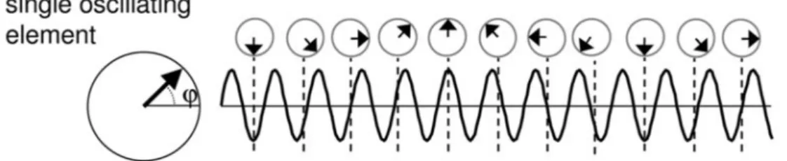
More interesting for the experimental work presented in this dissertation, either single pulses or, more efficiently, short episodes of the so-called rhythmic TMS (a modality of rTMS delivering short bursts of 4-5 regularly spaced TMS pulses) are being used to manipulate cortical oscillations within a targeted region. The first published precedent using TMS to manipulate ongoing oscillations used the ability of single isolated TMS pulses to phase-reset and synchronize local oscillators operating at the so called ‘natural frequency’ of the region. Such an approach has been applied to induce transient increases of oscillation amplitude in several cortical regions (Paus et al. 2001; Rosanova et al. 2009; Van Der Werf and Paus 2006). Some years thereafter, Thut and colleagues (2011a) put forward the notion that cortical populations of neurons consist in several oscillators, all fluctuating independently at an identical frequency but with a random phase (Fig. 4A). Given their rather natural desynchronized state in awake individuals, their summed spatio-temporal activity patterns tend to cancel off, and scalp EEG or MEG recordings prove unable to reveal clear signs of oscillations with a meaningful amplitude or increases of oscillatory power density in time-frequency analyses.
However, when rhythmic activity from different local oscillators is phase-locked, the amplitude of oscillatory activity at the level of the neuronal assembly increases by summation (instead of cancelling off) allowing the emergence of cortical oscillations visible in scalp EEG or MEG recordings. Single TMS pulses are the simplest stimulation pattern able to phase-lock ongoing un-synchronized oscillatory activity in local circuits. They act as an external force
Figure 4. Mechanisms of oscillatory entrainment by periodical TMS pulses. (A) Schematic drawing of an independent neural oscillator fluctuating naturally at the so called ‘natural frequency’. � labels the phase of the oscillation. (B) Schematic representation of three oscillators operating with a similar frequency. In physiological conditions these oscillators each have their own temporal dynamics and are not phase synchronized. A periodic external force, exerted by series of single TMS pulses (n=11 pulses in the figure) repeated rhythmically at a given frequency phase resets the cycles of the different units within each oscillator type, hence progressively phase-locking (i.e., synchronizing) their rhythms, making them fluctuate jointly. (C) As result of such a progressive synchronization of local oscillators, in-phase rhythms will sum up in time and space, increasing the so-called inter-trial coherence (ITC). Scalp EEG electrodes will record the emergence of cortical oscillations of higher amplitude hence showing higher levels of power density. (Figure extracted From Thut et al. 2011a).
that resets the phase of ongoing oscillators, transiently phase-locking their temporal dynamics, hence increasing for very few cycles, by amplitude summation, the power of the so called ‘intrinsic’ or ‘natural’ frequency at which these oscillators normally fluctuate (Fig. 4B). Although TMS pulses are often seen as alien perturbation phenomena that, by artificially depolarizing neurons, may interfere with their normal coding and behavioural contributions, in the context of the depolarisation of natural oscillators they can also be conceived as low energy stimuli able to enhance, in a specific cortical area, the power of frequency-specific oscillatory activity restricted to the ‘natural’ frequency of the stimulated area, hence respecting the ‘intrinsic’ rhythmic activity developed by local circuits.
The phase-reset and phase locking power of single pulses was confirmed experimentally (Rosanova et al., 2009). More specifically, single TMS pulses induced differential increases of oscillatory activity at specific frequency bands, depending on the stimulated cortical-region and similar to the most predominant rhythm at rest. Indeed, single pulses delivered over the occipital cortex (Brodmann Area 19) generated power increases around 11 Hz, in the alpha band which is well known for its role in visual processing, (Klimesch et al., 2007; Sauseng et al., 2005; Thut et al., 2006; Worden et al., 2000). TMS over posterior and superior parietal regions (Brodmann Area 7) selectively enhanced beta oscillatory activity with a peak at 20 Hz. Finally, frontal stimulation (Brodmann Area 6) induced broader-band effects increasing high-beta and gamma oscillatory activity around a 31Hz peak.
Although single pulses could be used to entrain natural oscillations, they are short-lasting and of low amplitude (Van Der Werf & Paus, 2006). Moreover entrainment is limited to the frequencies operating ‘naturally’, hence difficult to manipulate in exploratory or clinical applications. In an attempt to induce more robust oscillatory entrainment patterns by capitalizing on the phase-locking ability of TMS pulses, rhythmic TMS bursts aligning trains of pulses delivered at a frequency of choice have been developed. These consist in short bursts of pulses (usually 4-5) regularly spaced in time to emulate the periodicity of oscillators in the stimulated regions. As the inter-pulse interval of the burst is tailored to fit a full cycle of a local ‘intrinsic’ oscillator, each consecutive pulse in the burst will be delivered at the same phase of the oscillation we intend to entrain. The accrual of individual pulses within the burst will progressively phase-reset and phase-lock more and more oscillators, leading to a gradual build-up of a TMS entrained frequency in the targeted cortex (Fig. 4B and C).
Interleaving rhythmic TMS with EEG recordings, Thut et al. (2011b) were able to show the effect of 5 pulse TMS bursts delivered to the Intraparietal Sulcus (IPS) at a 10 Hz frequency