HAL Id: tel-00260987
https://tel.archives-ouvertes.fr/tel-00260987
Submitted on 6 Mar 2008HAL is a multi-disciplinary open access
archive for the deposit and dissemination of sci-entific research documents, whether they are pub-lished or not. The documents may come from teaching and research institutions in France or abroad, or from public or private research centers.
L’archive ouverte pluridisciplinaire HAL, est destinée au dépôt et à la diffusion de documents scientifiques de niveau recherche, publiés ou non, émanant des établissements d’enseignement et de recherche français ou étrangers, des laboratoires publics ou privés.
Biomechanical sensors from the macro to the nanoscale
-the way forward
Liviu Nicu
To cite this version:
Liviu Nicu. Biomechanical sensors from the macro to the nanoscale - the way forward. Micro and nanotechnologies/Microelectronics. Université Paul Sabatier - Toulouse III, 2008. �tel-00260987�
Habilitation à Diriger des Recherches
Préparée au Laboratoire d’Analyse et d’Architecture des Systèmes du
CNRS
En vue de l’obtention du Diplôme de l’Université Paul Sabatier de
Toulouse
Par Liviu NICU
Docteur de l’Université Paul Sabatier de Toulouse
Biomechanical sensors from the macro-to the nanoscale –
the way forward
Remerciements
Bien sûr, il est très difficile d’écrire cette partie. Je ne sais pas si c’est la même chose
pour tout le monde mais en ce qui me concerne, la crainte de ne pas être suffisamment
exhaustif me tétanise.
Avant toute chose, je voudrais remercier vivement les membres de mon jury qui ont
bien voulu dégager un peu de leur temps précieux pour venir juger l’ensemble du travail
accompli par mon équipe et moi-même depuis quelques années. Prof. Juergen Brugger de
l’EPFL, Dr. Harry Heinzelmann Vice-Président du CSEM Neuchatel, Prof. Karsten Haupt
de l’Université de Compiègne, Dr. Jean-Paul Leonetti du Centre d’études d’agents
Pathogènes et Biotechnologies pour la Santé de Montpellier, Dr. Francesc Perez-Murano du
CNM Barcelone, Prof. Robert Plana du LAAS, Prof. Olivier Bonnaud de l’Université de
Rennes, Dr. Lionel Buchaillot de l’IEMN Lille et Dr. Christian Bergaud, mon Directeur
de Travaux.
Par qui continuer ? Je viens de faire un tableau avec deux colonnes, celle intitulée
« qui ? » et celle « pourquoi ? ». La « qui ? », remplie naturellement, sans réfléchir, sans buter
sur les noms aurait dû me débloquer sur le « pourquoi ? ». C’est là où j’ai eu le sentiment de
refaire le même exercice d’écriture de remerciements qu’après ma thèse. Sept ans après…
grâce à qui, grâce à quoi je crois aujourd’hui encore en ce que je fais ? Et si c’était celui-là
mon fil conducteur dans l’écriture des remerciements ?
Grâce à qui ?
Tout d’abord, grâce à mes partenaires de travail de tous les jours : les séparer en
« doctorants », « permanents », « amis » ça rimerait à quoi, exactement ? Je ne suis
certainement pas tenu à refaire l’organigramme du LAAS « vu sous un angle personnel »,
enfin, je l’espère. Je me dois surtout de penser à tous ceux avec qui j’ai fait ce bout de chemin.
D’abord, sans compter, sans ordonner, venus entre une inspiration et une expiration,
instinctivement : Thierry, Christian, Cédric, Laurent, Thomas, Fabrice, Jean-Bernard, Daisuke.
Qui est qui, qui est quoi ? Mais quelle importance ça a ? Ils sont mes anges gardiens, ils l’ont
été à un moment ou à un autre, parfois en se rendant compte, parfois en passant sans se douter
de l’importance qu’ils ont à mes yeux. Avec certains j’ai construit, avec d’autres j’ai rêvé,
avec tous j’ai avancé, jour après jour ; ils m’ont chacun appris quelque chose.
Thierry la rigueur et l’excellence issues d’une terrible exigence envers soi,
Christian la ténacité et le sacrifice de tout au nom d’idées et idéaux partagés,
Cédric l’ambition en distillant persévérance et doutes,
Laurent le jeu des miroirs, où âme et cérébralité peuvent en faire un,
Thomas le pilier qui roule et compresse avec fausse naïveté,
Fabrice la droiture et l’honnêteté d’un caractère en acier trempé par des vies vécues en
une seule vie (au moins, on est deux à l’avoir éprouvé et ça lie, forcément !),
Jean-Bernard l’esprit des valeurs, des savoirs et de la présence inconditionnelle qui
rassure,
Daisuke l’efficacité absolue de la discrétion.
Nous avons toujours été libres de faire, refaire ou ne pas faire s’il n’y avait pas lieu de
faire. Cette liberté sans prix, avec son goût parfois salé par la peur de l’inconnu (appelant la
nécessité d’assumer ses responsabilités), parfois alcoolisé par l’ivresse d’instants d’égarement
de l’égo, cette liberté là je la dois à Christophe, dont la direction de groupe a été assumée avec
autant de tact que de fermeté. Je la dois également aux binômes directoriaux successifs, Mrs
Laprie-Martinez, Ghallab-Garcia(Munoz), Chatila-Sanchez qui ont œuvré au fonctionnement
irréprochable du LAAS. Eux seuls sachant l’ingratitude de la tâche, ça vaut la peine de
l’écrire au moins une fois et rendre ainsi le ressenti au niveau périphérique.
Cependant, comment faire autre chose que rêver de faire sans ce pas du rêve à la réalité
rendu possible par les services du LAAS ? Oh oui, pour les sceptiques, je pèse mes mots. Je
n’ai jamais eu la prétention de pouvoir m’en sortir tout seul et de toute façon, quelle douceur
que celle du confort d’une prise en charge là où les idées doivent prendre contours et formes ?
TEAM, 2I, Doc, Logistique, Gestion… pas d’organigramme, mais pas le droit à l’oubli, juste
le droit de dire Merci !, et la mention du rayon de lumière à odeur d’encre pour Christian B.
(il se reconnaîtra, bien qu’ils soient 2 à partager les mêmes initiales, l’autre ayant la mention
du cœur mais il le sait).
Une autre manière de « penser et c’est fait », dans un tout autre registre, celui de la
gestion de l’imprévu, la où le moindre détail peut prendre des proportions insensées, porte
tout naturellement ma reconnaissance envers l’efficacité et la gentillesse de Nicole, notre
secrétaire.
A mon collègue de bureau (Fuccio, le sicilien) avec qui je partage le quotidien (à en
rendre folles de jalousie nos épouses) en toutes les langues (ou presque, car l’italien
l’emporte), je dois également un clin d’œil affectif. J’ai également une pensée toute
particulière pour Childerick qui force mon admiration tant il est tenace et dont la force de
travail n’a d’égal que la contenance de son cœur.
Il paraît que les temps changent, il paraît que nuages se montrent de plus en plus
menaçants sur un univers professionnel qui m’a toujours été source de satisfactions… et si
mon port d’attache, mon phare dans la tempête était mon autre univers, ma famille ? Qui de
ceux qui prétendent me connaître sera étonné de le lire encore une fois ? Ma force, mon
moteur, mon jour après la nuit, mes enfants et mon épouse. A eux, je dois TOUT !
Table of Contents
PREAMBLE ... 1
1. FROM BIOSENSORS TO BIOM(N)EMS – THE WAY FORWARD ... 3
INTRODUCTION ... 3
DEFINITION OF THE BIOSENSOR ... 3
CLASSIFICATION OF BIOSENSORS ... 5
RECEPTORS ... 7
1.4.1 Catalytic receptors and related biosensing techniques ... 8
1.4.2 Affinity receptors and related biosensing techniques ... 8
1.4.3 DNA-based receptors and related biosensing techniques ... 10
1.4.4 Molecularly imprinted polymers and related biosensing techniques ... 11
IMMOBILIZATION TECHNIQUES ... 12
1.5.1 Adsorption to the biosensor’s surface ... 12
1.5.1.1 Gold ... 13
1.5.1.2 Glass and similar ... 14
1.5.1.3 Polymers ... 14
1.5.2 Entrapment methods ... 15
1.5.2.1 Physical entrapment behind membranes ... 15
1.5.2.2 Entrapment in hydrogels ... 16
1.5.2.3 Entrapment within conducting polymers ... 16
1.5.3 Covalent coupling ... 16
1.5.3.1 Coupling to carboxylic acids ... 17
1.5.3.2 Reactions involving thiols ... 17
1.5.3.3 Reactions involving amines ... 18
1.5.4 Other capture systems ... 18
1.5.4.1 Use of antibody-binding proteins... 18
1.5.4.2 Avidin/streptavidin capture systems ... 19
1.5.5 Spatial control of surface immobilization ... 19
1.5.5.1 Replication of patterns ... 20
1.5.5.2 Light direct immobilisation and patterning ... 20
1.5.5.3 Control of deposition by physical placement ... 21
1.5.6 Conclusion to immobilisation techniques issues ... 25
TRANSDUCTION TECHNIQUES ... 26
1.6.1 Definition of BioMEMS ... 26
1.6.2 Optical techniques ... 26
1.6.3 Electro(chemical) transduction ... 28
1.6.4 Mechanical detection ... 29
1.6.4.1 Piezoelectric excited acoustic waves in solids ... 30
1.6.4.2 Classification of acoustic piezoelectric resonators ... 31
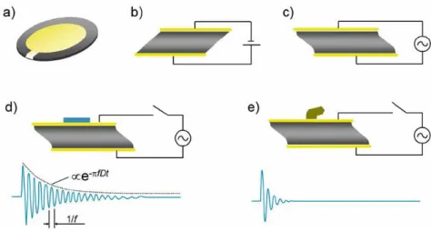
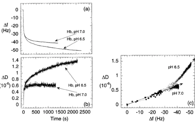
1.6.4.3 The quartz crystal microbalance with dissipation monitoring (QCM-D) ... 31
1.6.4.4 Acoustic micro(bio)sensors ... 34
1.6.4.5 One specific application: acoustic microsensors for pathogen agents detection ... 36
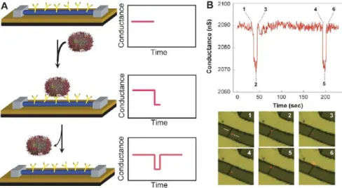
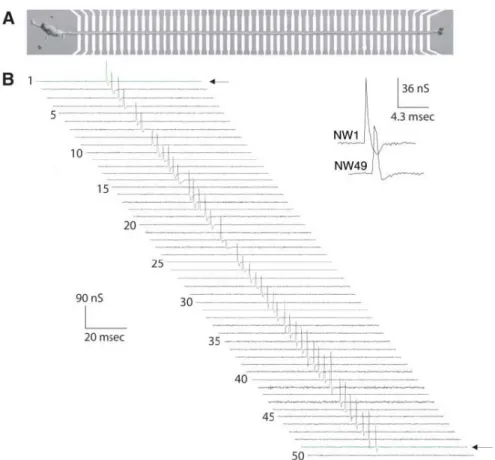
NANO(BIO)SENSORS: THE FUTURE? ... 38
THE CRUSADE FOR ULTIMATE PERFORMANCE – A SNAPSHOT OF THE PRESENT TIME AND PERSPECTIVE ... 41
COMPARING BIOSENSORS – IMPOSSIBLE MISSION? ... 43
2. RESEARCH ACTIVITIES SINCE 2003 ... 45
INTRODUCTION ... 45
BIOPLUME: A MEMS-BASED PICOLITER DROPLET DISPENSER FOR SURFACE PATTERNING ... 46
2.2.1 Previous work ... 46
2.2.2 Introduction ... 46
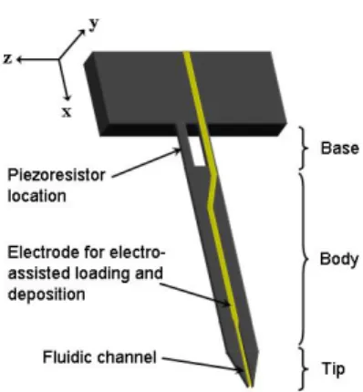
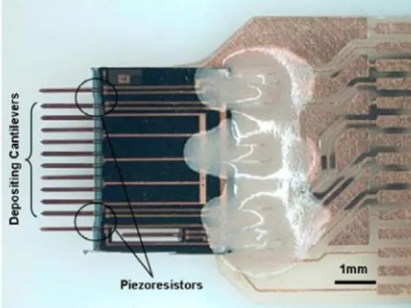
2.2.3 Integration of sensors and actuators ... 46
2.2.3.1 Force sensors ... 47
2.2.3.2 Electro-assisted loading and deposition methods ... 48
2.2.4 Design of the cantilevers ... 48
2.2.5 Fluidic requirements ... 49
2.2.6 Mechanical considerations ... 50
2.2.7 Electrical considerations ... 50
2.2.9 Piezoresistive cantilever measurement and characterizations ... 53
2.2.10 Automated spotter in closed-loop configuration ... 54
2.2.11 Loading and deposition tests ... 56
2.2.11.1 Influence of deposition parameters on the printed features... 56
2.2.11.2 Deposition of biological solutions ... 57
2.2.11.3 Loading tests using electrowetting ... 58
2.2.11.4 Local deposition of nanoparticles ... 59
2.2.11.5 Electrospotting ... 60
2.2.12 Conclusion ... 61
PIEZOELECTRIC MICROMEMBRANES: RESONANT MEMS FOR BIOLOGICAL AND CHEMICAL APPLICATIONS ... 64
2.3.1 Previous work ... 64
2.3.2 Motivation for continuing this work ... 64
2.3.3 Introduction to multilayered piezoelectric micromembranes286 ... 66
2.3.4 Piezoelectric micromembranes in Newtonian liquid media ... 69
2.3.4.1 Experimental set-up ... 69
2.3.4.2 Lamb’s model ... 70
2.3.4.3 Main results ... 71
2.3.5 Completing the MEMS: associated electronics ... 73
2.3.5.1 The concept ... 74
2.3.5.2 Mass sensitivity ... 75
2.3.5.3 Evaluation of the electronic scheme. Measurements in liquid media ... 77
2.3.6 Piezoelectric micromembranes for biological applications ... 78
2.3.6.1 Detection of streptavidin-gold nanoparticles interaction with biotinylated DNA. Dip-and-dry technique. .. 78
2.3.6.2 Real-time monitoring of antigen-antibody reactions. Flow-through experiments ... 82
COMPLETING THE PUZZLE:BIOPLUME AS A TOOL FOR BIOMEMS FUNCTIONALIZATION ... 85
CONCLUSION ... 88
3. PROSPECTIVE OUTLOOK ... 91
PRELIMINARY CONSIDERATIONS ... 91
CONTINUING THE MINIATURIZATION – TOWARDS ELECTRICAL-ASSISTED BIOCONCENTRATION ... 92
P
REAMBLELife is made of opportunities that have to be seized. So was my life so far.
I realize that the meaning of the words above could make the reader think that the author is someone born in the first half of the past century. Those who know me are aware of the fact that this is not exactly the case (even if sometimes I do feel like it is).
I will try to illustrate the different opportunities that occurred in my (professional) life, the private one being beyond the scope of this manuscript. In 1994, I chose to give up my entire Romanian-native life and start a completely new one in France, my adult-life I would say. Even if it happened for personal reasons, this was not without significant consequences on my future professional life. Indeed, this life change occurred when I was about to finish my Electrical Engineering Graduate Studies at the Politechnica University of Bucharest. I chose not to finish my studies there but to continue to Paul Sabatier University of Toulouse (UPS), in 1995. Let me call it “the First Opportunity”.
In 1997, while I was finishing a Master of Materials Sciences at UPS, I was recruited by the former Deputy Director of the Laboratory of Analysis and Architecture of Systems in Toulouse (Augustin Martinez) as PhD student in one of the laboratory’s research groups that at that time was named Silicon Microstructures and Microsystems (M2I). My PhD thesis started under the responsibility of a young researcher (Christian Bergaud) who was just arriving from Japan bringing the silicon cantilevers technology at LAAS after 2 years of post-doc. This was the Second Opportunity of my career: the PhD Director and the subject.
In 2001, I failed to be admitted either as a CNRS1 researcher or an Assistant Professor at the
National Institute of Applied Sciences in Toulouse and this was the Third Opportunity in my career. For those who are not familiar with the French system, to get a permanent position in the academic research, the candidates have to successfully succeed annually organized national competitions and I brilliantly failed in my two attempts. This was an opportunity though, as I immediately applied for an R&D Engineer position at Thales Avionics in Valence (France). I was hired at the Inertial Department in charge of the scientific part of the European Project Gyrosil that was just starting. I learnt a lot during my (almost) two years of industrial life. It was a short but extremely intense experience. The project was very ambitious and quite strategic at that time (June 2001) for the company: we had in charge the demonstration of a silicon-based microgyrometer prototype satisfying both military and civil specifications. Unfortunately, September 11 arrived with its significant economic impacts on the world markets, leading specifically to a nearly 20% cutback in air travel capacity, and severely exacerbating financial problems in the struggling world-wide airline industry. The effects of this event were felt with a certain inertia (word play is not intentional here!) in our Department as we have had enough time to submit 3 patents on a new gyrometer design. However, in the end, even if the first prototypes were successfully demonstrated, the project was placed on “stand-by” position after 18 months of hard work. I so realized how the market laws could impact on the life of a project, no matter how “strategic” it could be. I also realized that this would be a source of frustrations in my future life and I decided to knock again at the CNRS’ door.
In January 2003, I received my Researcher position in the Nanobiotechnology group, at LAAS. It thus became the Fourth Opportunity as I was back in my lab after a rich experience in the industrial world. The research project that I defended dealt with Microsystems for Biological Applications. Starting with that point, the opportunities took the form of projects (the European Project NaPa2) and of meeting extraordinary people (the European partners inside NaPa, the biologists from Montpellier,
1 CNRS stands for the National Center of Scientific Research in France
the chemists from Compiègne, my colleagues from LAAS and my team of PhD students and post-docs).
Almost 5 years after my reintegration at LAAS, I can say that I kept the same research direction that I have initially defended during the CNRS competition in 2002. The present manuscript retraces all the research work carried during those 5 years and shows how such a huge subject was divided in several complementary sub-projects with a common target that was the demonstration of resonant silicon-based MEMS sensors for biological applications taking into considerations all the bioMEMS-related aspects, from the surface functionalization (either global or localized), through the transduction (from the biological recognition event to the physically measurable signal) to the fluidics issues related to biological sensors.
Before digging into the manuscript, I would like to mention the last opportunity that I recently had. A few days before starting to write this dissertation I read by chance an article issued in the weekly Chemical and Engineering News (March 26, 2007, p.13) written by Prof. George M. Whitesides, an address that he presented at the American Chemical Society 233rd National Meeting in
Chicago (March 25-29, 2007), where he received the 2007 Priestley Medal for lifetime achievements and service to chemistry. In his article, Prof. Whitesides shares his point of view about the revolutions to arise in chemistry under the cover of Thomas Kuhn’s theories about the scientific revolutions. Without this article in mind, I would certainly never have written this manuscript in the “awareness state” in which I am today. Reading Prof. Whitesides’ article allowed me learning about Thomas Kuhn’s “The Structure of Scientific Revolutions” book that shed a new lucid light on my past scientific activities and on my future strategy. I finally understood what kind of science I was doing and this manuscript will be the basis of an introspective analysis based on several Kuhn’s assumptions. However, the manuscript’s structure is somehow “conventional”; it is divided in three chapters. In the first chapter, a state-of-the-art discussion upon the biosensors’ structure and classification highlights the main elements to be considered in any basic biosensor issue, i.e. the biological receptors topic, the biomolecules’ immobilization onto a substrate and the transduction techniques. To conclude this first chapter, a brief overview of the main nanodevices applied to the biology is given.
The second chapter contains the main results of my past 5 years research work dealing with silicon-based bio-microsensors. It is divided in three parts: the first one deals with microtools for local bio-functionalization of solid surfaces at the microscale (the so-called Bioplume system), the second part describes the microsensors developed for biological applications (from the mechanical structure to the biological application) and the third part presents promising applications yielded from the successful coupling between the Bioplume system and the microsensors described beforehand.
The final chapter is a more prospective one, from the biosensors field point-of-view (the limits of biosensing at the nanoscale is pointed out) and from a personal career point-of-view where a new research axis towards smart nano-electromechanical systems is proposed.
1. F
ROM BIOSENSORS TO BIO
M(N)EMS
–
THE WAY
FORWARD
Introduction
Before presenting an overview of biosensors, it is worth taking a closer look at Prof. Whitesides’ article3 cited previously. This will allow starting the discussion from a more general level.
Two theories of scientific revolutions are pointed out by the author. One is defended by Thomas Kuhn in its “The structure of Scientific Revolutions” book and it argues that scientific paradigm shifts occur when there is no way out, i.e. invention of new theories are brought about by the awareness of anomalies in existing ones. The transition between the worldview of Newtonian physics and the Einsteinian Relativistic worldview is an example. Moreover, Kuhn offers a dual vision of the science: the normal scientific research that is directed to “force nature into preformed and relatively inflexible boxes that paradigms are supplying” and discovery (or scientific revolution) that remains the basis of fundamental changes in thinking. On this first basis, it will turn out that all the scientific
work in the present manuscript can be hopefully qualified as “normal science”.
The second theory, supported by Peter Galison (Professor in History of Science and Physics at Harvard University), states that new experimental techniques enable scientific revolutions (in Kuhn’s sense of the word). Redefining this in Prof. Whitesides’ terms would lead to “new keys open new
doors”. One example is the role of scanning tunneling microscope in nanosciences; another one is the
surface plasmon resonance-based system that allows determining the affinity of two ligands meaning the measurement of their binding constants. With this second theory in mind, it will be shown that the
tools described within the next chapter of this manuscript can contribute to gain further insight into the biosensing issues.
Finally, Prof. Whitesides gives a series of examples of problems (or potential scientific revolutions) that would concern the scientific community many years from now. Among them (“The Cell and the Nature of Life”, “Energy, the Environment and Global Stewardship”, “The Molecular Basis of Sentience”), the molecular recognition in water and the design of drugs seems to be of primary importance. In the author’s opinion, the binding of a small molecule –a drug, ligand, substrate or transition state – to a protein is arguably the most fundamental molecular process in biology. Answers to fundamental questions in this area can without any doubt be conveniently brought by state-of-the-art biosensors, similar to those described in the following sections of this chapter.
Definition of the biosensor
There are numerous definitions of what a biosensor is, two of them will be given here: a
history-related and a general definition, for the sake of exhaustiveness.
In its brief but remarkable review of the biosensors field, Peter T. Kissinger4 goes back to the
early days of the biosensing (the 1960s and the 1970s) to pinpoint that a sensor seemed to always be a
probe of some sort because of systematic association to pH, ion selectivity or oxygen electrodes.
Following the old literature, biosensors are found as being called bioelectrodes or enzyme electrodes, or biocatalytic membrane electrodes5.
3 G. M . Whitesides, C&EN March 26, 13 (2007) 4 P. T. Kissinger, Biosens. Bioelectron. 20, 2512 (2005) 5 M. A. Arnold, M. E. Meyerhoff, Anal. Chem.56, 20R (1984)
More generally, according to International Union of Pure and Applied Chemistry (IUPAC) recommendations in 1999, a biosensor is a self-contained integrated receptor-transducer device,
which is capable of providing selective quantitative of semi-quantitative analytical information using a biological recognition element (Fig.1.1).
Figure 1.1: Schematic view of the general definition of a biosensor
The critical feature of the biosensor relates to the selectivity for the specific target analyte; this feature directly impacts on the specificity or how to maintain the selectivity in the presence of other, potentially interfering, species. The combination of these quality criteria with miniaturization, low
cost and essentially real-time measurements in various fields has generated intense commercial
interest.
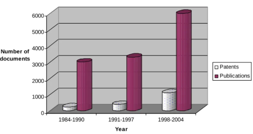
The last twenty years have witnessed an extraordinary growth in research on sensors in general and on biosensors in particular. As underlined by Collings and Caruso in their excellent review on biosensors advances6, “an intensively competitive research area is the result of the combined pressure
from the traditional well-springs of research and development – science push and market pull”7. A
measure of the rate of growth of interest in biosensors is quantified in Figure 1.2:
0 1000 2000 3000 4000 5000 6000 Number of documents 1984-1990 1991-1997 1998-2004 Year
Number of scientific publications and patents issued involving biosensors since 1984
Patents Publications
Figure 1.2: Overview of the rate of growth of activity involving biosensors since 19846,9
6 A. F. Collings and F. Caruso, Rep. Prog. Phys. 60, 1397 (1997)
7 P. T. Kissinger (in Ref 4) gives a « more provocative » vision of what motivates the biosensors interest, i. e. the triangle of “peer-review of science, funding agencies and politics”.
9 Fuji-Keizai USA, Inc., U.S. & Worldwide: Biosensor market, R&D, applications and commercial implication (2004), New York
In spite of all this, there is only one truly commercially successful biosensor and that is the blood glucose monitor. It is important to note that the glucose biosensor uses technology that was developed by Clark and Lyons well over 40 years ago and only recently has the world managed to grasp the true benefits of biosensors. Will commercial enterprises reap the benefits of the next generation of biosensors? In fact, it is suggested that between 80-90% of R&D activity in this area rarely results in a commercial product9. However, the observed growth in biosensor research increases
the probability of witnessing another success story in the next couple of decades. The future R&D outlook for biosensors looks positive despite very little market growth / progress over the past several years9.
As stated before, a biosensor is comprised of three essential components: the detector, which recognizes the biological stimulus; the transducer, which converts the stimulus to a useful, measurable, output; and the output system itself, which involves amplification, display etc. in an appropriate format. Because the biological detector or receptor is of primary importance (as it confers to the biosensor its specificity), receptors will be discussed in the next section of this chapter. The final component of the biosensor, the output system, is application or product-dependent, and while worthy of considerable effort as far as a finished product is concerned, is also beyond the scope of the first chapter.
It is the transduction process that completely falls into the physicist’s domain. Since the physics aspect is largely concerned with the transducer and the transducer-receptor interface, this will constitute the main focus of this chapter.
Classification of biosensors
Biosensors can be classified according to three schemes: (a) the receptor type, e.g. an immunosensor, (b) the physics of the transduction process, e. g. an amperometric sensor, or (c) the
application, e.g. a medical biosensor.
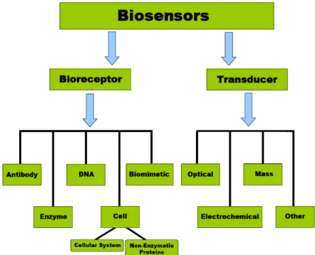
The biological recognition element or bioreceptor is the most crucial component of the
biosensor device. The bioreceptor is the key to specificity, and can be classified according to several different groups as shown in Figure 1.3. Generally, there are three principal classes of biosensors. The three groups are distinguished from one another by the nature of the process and in terms of their biochemical or biological component, e.g. biocatalytic (i.e., enzyme), immunological (i.e., antibody) and nucleic acid (i.e., DNA). It is important to note that some biosensors have been developed on the basis of biomimetic or cell bioreceptors10. However, these bioreceptors will not be discussed here.
Figure 1.3: Biosensors’ classification with respect to categories of bioreceptors and transduction
principles
Though the bioreceptors immobilisation techniques onto solid surfaces will be discussed apart in this chapter, this topic is encompassed by the biological recognition element issue and, in practical cases, both are of equal concern.
The transducer is another component of the biosensor, which plays an important role in the
detection process. A wide variety of transducer methods have been developed in the past decade; however, a recent literature review has shown that the most popular and common methods presently available are: a) electrochemical; b) optical; c) piezoelectric; d) thermal or calorimetric6, 11-13. To
produce a manuscript that is consistent with the main authors’ background (micromechanics), the focus will be narrowed to biosensors involving mainly piezoelectric transducers. For completeness sake, electrochemical and optical transduction principles in biosensing will be briefly reviewed across the first chapter when needed.
It is also important to note that these groups can be further divided into general categories: nonlabeled or label-free types, which are based on the direct measurement of a phenomena occurring during the biochemical reactions on a transducer surface; and labeled, which relies on the detection of a specific label. Research into ‘label-free’ biosensors continues to grow14; however “labelled” ones are more common and are extremely successful in a multitude of platforms.
From the application point of view, even if medical and clinical applications are the most lucrative and important avenues for biosensors, other areas like the environment, the industrial process monitoring and control or the defence require specific biosensing systems. Moreover, commercial biosensors can be divided into two categories on the basis of whether they are laboratory or portable/field devices. As no more market considerations will be highlighted in this chapter, a selection of the most significant commercial biosensors is made hereafter.
11-13 E. A. H. Hall, Biosensors (1990), Open University Press ; 12D. G. Buerk, Biosensors: Theory and
Applications (1993), Technomic Publishing Company; 13E. Gizeli and C. R. Lowe, Biomolecular Sensors (2002), Taylor&Francis
As pointed out before, the most successful handheld biosensor to date is the blood glucose monitor for people with diabetes, which is based on electrochemical transduction technology15.
Commercial blood-glucose meters are produced by many companies16. However, in terms of
laboratory-based instrumentation an optical detection system appears to be more commercially viable. Companies such as Affymetrix and Agilent have developed various commercial microarray optical detectors and scanners for genomic and proteomic analysis. Optical sensors that employ surface plasmon resonance (SPR) detection have also been successfully used in many laboratories and universities17. Hence, commercially available optical bench-size immunosensor systems such as BIAcore™ (Biacore AB, Uppsala, Sweden) and IAsys (Affinity Sensors, Cambridge, UK) have found their market in research laboratories for the detection and evaluation of biomolecular interactions, noting that these technologies are based on the principles of surface plasmon resonance.
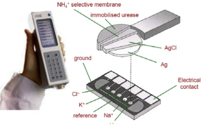
The development of disposable sensors in conjunction with handheld devices for point of care measurements has featured prominently. Microfabrication technology has played an important part in achieving miniaturised biosensors. Such technology has provided cheap mass-producible and easy-to-use / disposable sensor strips. Similarly, electrochemical methods have played a pivotal role in detecting the changes that occur during a biorecognition event, and the merging of microfabrication with electrochemical detection has enabled various handheld biosensor devices to be developed. In fact, i-STAT has developed the world’s first hand-held device for point-of-care clinical assay of blood (see Figure 1.4), noting that this biosensor array employs several electrochemical-based transduction methods (i.e., potentiometric, amperometric, conductometric).
Figure 1.4: The i-STAT multisensor for monitoring various blood electrolytes, gases and
metabolites18
The i-STAT Portable Clinical Analyser™ is a hand-held silicon-based multiple-analyte sensor array18, which is used to monitor various blood electrolytes (i.e., sodium, potassium, chloride, calcium,
pH), gases (i.e., carbon dioxide, oxygen) and molecules (i.e., urea, glucose, hematocrit). Oxford Biosensors has also developed a portable hand-held device (Multisense™) for cholesterol detection19.
The biosensor consists of disposable test strips (microelectrodes) and uses the electrochemical detection strategy.
Receptors
Identified beforehand as the most crucial component of a biosensor, the biological receptor is responsible for the selective recognition of the analyte to be detected thus generating the signal issued from the transducer and, ultimately, the sensitivity of the device. Three categories of biological
15 P. D’Orazio, Clin. Chim. Acta 334, 41 (2003) 16 A. P. T. Turner et al., Clin. Chem. 45, 1596 (1999)
17 R. L. Rich and D. G. Myszka, J. Mol. Recogn. 16, 351 (2003) 18 http://www.i-stat.com
receptors can be precisely pointed out, catalytic (as typified by enzymes), affinitive (of which the antibodies are the best-known example), and DNA-based detection.
Even if non-biological, the molecular imprinted polymers (MIPs) have to be highlighted here as an alternative to biological receptors. A specific section dedicated to MIPs will be included further in this section.
1.4.1 Catalytic receptors and related biosensing techniques
Enzymes are protein molecules, long chains of 20 different amino acids which are so structured as to confer a remarkable ability for catalysing specific reactions. This same structure also limits their functional stability. Compared to non-biological catalysts, enzymes are 1018-1013 more active and are capable of producing hundreds of thousands of molecules per second20.
Enzymes have the longest tradition in the field of biosensors. Since 1997, there have been over 2000 articles published in the literature on enzyme-based biosensors. These biosensors primarily rely on two operational mechanisms. They can be used as a bioreceptor based on their specific binding capabilities or according to their catalytic transformation of a species into a detectable form11,21. In
most reports in the literature they have been employed according to their catalytic activity10,22.
Horseradish peroxidase (HRP), alkaline phosphatase (AP) and glucose oxidase (GOD) are three enzymes that have been employed in most biosensor studies23,24.
The detection limit of these biosensors is mainly determined by the enzyme’s activity, which can be described by the Michaelis-Menten equation11. However, the major limitation of enzyme-based
biosensors is the stability of the enzyme, which depends on various conditions such as the temperature, pH, etc22,23. The ability to maintain enzyme activity for a long period of time still remains
a major obstacle23. Another issue that governs the success of an enzyme-based sensor depends
primarily on the contact between the enzyme and electrode surface21. Despite these pitfalls the
enzyme-based sensor is still the most commonly used biosensor, and this is largely due to the need for monitoring glucose in blood22. Some recent studies have shown that enzyme-based biosensors can be
used to detect very low levels (i.e., ~10−16 M) of pesticides25.
1.4.2 Affinity receptors and related biosensing techniques
The affinity class of receptors is more specific in the nature of the binding than the enzymes, with binding constants of 109-1012 M-1, but do not exhibit catalytic activity. This fact has consequences for sensing applications. Affinity receptors are more suited to “one-shot” detection rather than monitoring applications, since the binding is essentially irreversible. It is possible to break the binding complex, usually by changing the pH to acid values (1 to 2), but this tends to reduce the affinity and specificity of the receptor. The high binding constant also favours the selective detection of very small analyte quantities. Lowe et al.26 suggest that affinity receptors are suited to analyte concentrations of 10-6-10-9 M (note that concentrations as low as 10-15 M has recently been demonstrated, as it will be shown in the “nanodevices” further section), as compared with 10-3-10-6 M for catalytic systems. These authors also point out that the nature of the binding process is different, the receptor sites for catalytic binding “turning over”, whereas the binding sites of affinity receptors can be saturated.
20 M. Madou and M. J. Tierney, Appl. Biochem. Biotech. 41, 109 (1993) 21 J. Wang, Analytical Electrochemistry, 2nd ed. (2000), New York, Wiley-VCH 22 I. E. Tothill, Comp. Elec. Agr. 30, 205 (2001)
23,24 K. R. Rogers and M. Mascini, Field Anal. Chem. Technol. 2, 317 (1998); S. Laschi et al., Electroanal. 12, 1293 (2000)
25 K. A. Law and S. P. J. Higson, 8th World Congress on Biosensors (2004), Granada, Spain 26 C. R. Lowe et al., J. Chromat. 510, 347 (1990)
Of the affinity receptors concerned by biosensing applications (any proteins like transmembrane proteins, for instance), the immunoreceptors (antibody/antigen) are the dominant type. Antibodies are highly selective, chemical attractor molecules (glycoproteins) which are produced by mammalian immunological systems in response to the introduction of an antigen, a foreign molecule. An antigen is simply any substance (molecule, hormone, virus bacteria etc) which elicits antibody formation and reacts specifically with that antibody (antigenic reactivity or antigenicity). It should be noted that
immunogenicity is not an intrinsic property of the antigen but a relational property that depends on the
gene repertoire and regulatory mechanisms of the host being immunized and which has no meaning outside the context of the host13. For instance, human serum albumine (HSA) is an antigenic protein that is immunogenic in the mouse but not normally in human because of the regulatory mechanism known as immunological tolerance (the opposite being the starting point of autoimmune diseases).
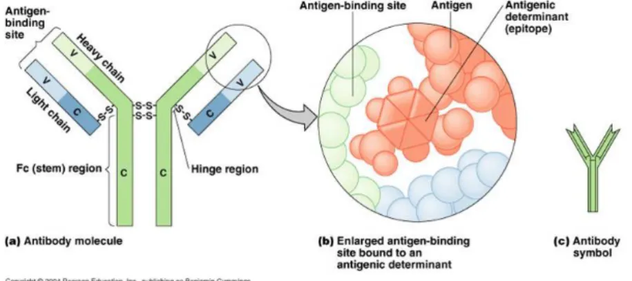
Since the antibody-antigen reaction is so specific, each can be utilized as a specific chemical detector for the other. Antibodies are typically represented schematically as Y-shaped structures (Figure 1.5).
Figure 1.5: Structure of an antibody
The antibody consists of two identical Fab (fragment antibody) portions hinged to an Fc (fragment crystallizable) part. The variable regions of the Fab fragments are where the amino acids are organized to produce a binding site for the specific antigen, two binding sites per antibody. The Fc fragment of the antibody does not combine with the antigen but contains carboxyl terminated amino acids which allow linkage to solid substrates (like transducers). The protein chains are, of course, not straight as represented in Figure 1.5 but are highly folded as revealed by the three-dimensional model of proteins determined by X-ray crystallography studies.
Immunoassays have become a standard tool in clinical chemistry, as they are highly sophisticated automated instruments used to analyze a number of samples in a short time frame27. It is
important to note that immunosensor technologies have been derived from the standard immunoassay approach27,28. A tracer either labels the occupied sites of the antibody or the free ones28.
Immunosensors can incorporate either the antigen or the antibody onto the sensor surface, although the latter approach has been used most often29. Optical and electrochemical detection methods are most
frequently used in immunosensors29 though since the recent commercialization of the Quartz Crystal Microbalance-Dissipative (QCM-D) system by Q-Sense company (Gotteborg, Sweden), the piezoelectric detection becomes a promising immunoassay technique30. A number of papers have been recently published on various immunosensors, noting that these have employed either an
27 M. C. Henion and D. Barcelo, Anal. Chim. Acta 362, 3 (1998) 28 B. Hock, , Anal. Chim. Acta 347, 177 (1997)
29 R. I. Stefan et al., Fresenius J. Anal. Chem. 366, 659 (2000) 30 C. Ayela et al., Biosens. Bioelectron. In press, (2007)
electrochemical31,32 or optical33-36 transduction method. Detection by electrochemical immunosensors
is generally achieved by using either electroactive labels or enzyme labelling22. A common challenge
facing immunosensors is that they are not completely reversible, so that only a single immunoassay can be performed12. Subsequently, some research efforts have been directed towards the development
of renewable antibody surfaces37.
1.4.3 DNA-based receptors and related biosensing techniques
In DNA sensors, the recognition is based on the formation of stable hydrogen bonds between the two nucleic acid strands. The bonding between nucleic acids takes place at regular (nucleotide) intervals along the length of the nucleic acid duplex38. The specificity of nucleic acid probes relies on the ability of different nucleotides to form bonds only with an appropriate counterpart. An important property of DNA is that the nucleic acid ligands can be denatured to reverse binding and then regenerated by controlling buffer-ion concentrations38. It is important to note that some workers have employed peptide nucleic acid as the biorecognition element10. The peptide nucleic acid is an artificial
oligo-amide that is capable of binding very strongly to complementary oligonucleotide sequences10.
Traditional techniques for DNA sequencing are based on the coupling of electrophoretic separations and radio-isotopic (32P) detection39. These methods are known to be labour intensive, time
consuming, high cost, hazardous, have disposal problems associated with radioactive waste, and are not well suited for routine and rapid environmental analysis38,39. Subsequently, various promising
alternative methods of DNA detection, which use a non-radioactive labelled probe, have been developed. The detection of specific DNA sequences provides the fundamental basis for detecting a wide variety of microbial and viral pathogens40. Several reviews have been published on the
development and application of DNA sensors for the testing of virus infections38-40, noting that viruses
appear to be almost uniquely DNA or RNA-composed within an outer coat or capsid of protein11. In
essence, the technology relies on the immobilisation of a short (20−40mer) synthetic oligomer [the single-stranded DNA (ssDNA)], whose sequence is complementary to the target of interest39.
Exposure of the sensor to a sample containing the target results in the formation of the hybrid on the surface, and various transduction methods (i.e., optical, electrochemical and piezoelectric) have been used to detect duplex formation39,41. Gooding (2002) revealed that relatively few DNA biosensor
studies have been carried out in real complex biological samples42.
Well over 700 papers have appeared in the literature, since 1997, on the development of nucleic acid biosensors. Almost all papers that have dealt with the DNA biosensor have used relatively short synthetic oligonucleotides for detecting target DNAs of about the same length43. Most reports have immobilised DNA in the form of a self-assembled monolayer onto a gold surface using thiol chemistry42-44. However, in some cases binding of the oligonucleotide probe to the sensing surface is achieved by using the biotin / avidin interaction45, immobilization technique that will be explain in Section 1.5.
31,32 W. E. Lee et al., Biosens. Bioelectron. 14, 795 (2000); 32S. Susmel et al., Biosens. Bioelectron. 18, 881 (2003)
33-36 A. W. Kusterbeck et al., Field Anal. Chem. Technol. 2, 341 (1998); 34M. J. Gomara et al., J. Immunol. Meth. 246, 13 (2000); 35S. Koch et al., Biosens. Bioelectron. 14, 779 (2000); 36V. Koubova et al., Sens. Act. Chem. B 74, 00 (2001)
37 G. A. Ganziani et al., Anal. Biochem. 325, 301 (2004) 38 D. Ivnitski et al., Bioens. Bioelectron. 14, 599 (1999) 39 J. Wang et al., Anal. Chim. Acta 347, 1 (1997) 40 M. Yang et al., Anal. Chim. Acta 346, 259 (1997)
41 M. Campas and I. Katakis, Trend. Anal. Chem. 23, 49 (2004) 42 J. J. Gooding, Electroanal. 14, 1147 (2002)
43 E. Palecek, Talanta 56, 809 (2002) 44 T. Peng et al., Electroanal. 14, 455 (2002) 45 K. Kukanskis et al., Anal. Biochem. 274, 7 (1999)
1.4.4 Molecularly imprinted polymers and related biosensing techniques
Even if from the early works dealing with biosensors this concept was likely to revolutionise “point-of-sample” analysis, biosensors development pace has been slow and only a very small proportion of the perceived market has been filled. Many of the key issues are directly related to the biological stability of the biological macromolecule at the heart of the biosensor. Thus, problems such as unpredictable shelf-life and stability of biological receptors, poor inter-batch reproducibility and availability, difficulties in incorporating biomolecules into sensor platforms, environmental intolerance (e.g. pH, temperature, ionic strength, organic solvents) and poor engineering characteristics have to be effectively addressed by alternative solutions.
Molecular imprinting is a technology that can potentially address some of the key issues listed above. It deals with the design and synthesis of biomimetic receptors (artificial macromolecules) capable of binding a target molecule with similar affinities and specificities to their natural counterparts46. In this technique, a target molecule (acting as a molecular template) is used to direct
the assembly of specific binders trapped in a polymer matrix formed by a polymerization step (Figure 1.6). Besides the simplicity in separating MIPs from the soluble template (that allows easily recovering and using them as artificial immobilized antibody, small molecules or enzyme mimic), it is a higher chemical and physical stability compared to biomacromolecules that make them very attractive for applications covering biochemical analysis, separation and catalysis.
Figure 1.6: Schematics of the molecular imprinting principle
Since 1993, the ISI Web of Knowledge47 Database registered more than 1100 publications
dealing with MIPs, taking roots in a report by Mosbach’s group on the development of a MIP-based immunoassay against theophylline and diapzepam48. Starting with that moment, MIPs have been used as substitutes for antibodies in radioimmunoassays (RIA) for drugs, showing strong binding to the target analytes (with dissociations constants from the nM to µM range) and cross-reactivity profiles similar to those of antibodies49,50. MIPs were later used to develop assay systems for other compounds as well, such as herbicides51,52 (such as 2,4-dichlorophenoxyacetic acid, mostly known as 2,4-D, which detection by means of silicon-based micromachined sensor is addressed in the second chapter of this manuscript).
Another aspect in assay development is the MIPs potential use in automated systems for unattended monitoring. In a recent paper53, Surugiu et al. described the design of a flow-injection
ELISA-type MIP assay using polymer microspheres-coated (using polyvinyl alcohol as glue) glass capillary. A photomultiplier tube (PMT) was used for 2,4-D analyte detection and concentrations ranging from 0.5 ng/mL-50 µg/mL (2.25 nM-225 µM) were detected, making the system one of the most sensitive MIP-based assays reported so far.
46 L. Ye and K. Haupt, Anal. Bioanal. Chem. 378, 1887 (2004) 47 http://portal.isiknowledge.com/
48 G. Vlatakis et al., Nature 361, 645 (1993)
49,50 L. I. Andresson et al., PNAS 92, 4788 (1995) ; O. Ramström et al., Chem. Biol. 3, 471 (1996)
51,52 M. T. Muldoon et al., J. Agric. Food Chem. 43, 1424 (1995) ; K. Haupt et al., Anal. Chem. 70, 628 (1998) 53 I. Surugiu et al., Anal. Chem. 73, 4388 (2001)
Though non-exhaustive, the before listed successful demonstrations of the use of MIPs for assays, sensors and very recently for drug development54, reveal the great potential of the technology
and amply justify our choice to dedicate a substantial effort in structuring MIPs at the microscale as well as integrating them with acoustic microsensors, as further highlighted in the second chapter.
Immobilization techniques
One key factor in the biosensors design is the development of immobilization technologies for stabilizing biomolecules and tethering them to surfaces6. The aim is to create a molecular recognition
interface that involves localisation and attachment of a molecular receptor (like those discussed in the previous section) near to or onto the transducer surface. Several approaches are available and the choice of the most adapted one to a particular application depends on various factors like the transduction principle, the nature of the biological receptor and the nature of the analyte to be detected. It will also depend on the way the biosensor will be used, and on its surface chemistry. The immobilization can be achieved in several ways:
• Adsorption of the biological receptor directly to the biosensor’s surface; • Physical entrapment near the biosensor’s surface (e. g. in a polymer layer); • Covalent coupling of the biological receptor directly to the biosensor’s surface; • Covalent coupling to a polymer layer on the biosensor’s surface;
• Use of a chemical/biochemical “capture system”.
Before entering into the details of each technique, one must note that the method chosen for immobilisation depends on the answers to several elementary issues to be addressed before adopting the final design of the biosensor:
• Will the sensor be regenerated and reused or just dedicated to “unique use” applications?
• What is the nature of the biological receptor (hydrophobic/hydrophilic, large/small)? • Is it sensitive to handling and, as a main concern, what are the storing constraints? The
biological receptors must be kept functional during immobilization and until the biological recognition takes place at the surface of the biosensor. Indeed, if a breakthrough could be achieved in immobilization technologies, in which the progressive loss of activity of the attached bioreceptors was eliminated, many of the present disadvantages of biosensors would be overcome.
• Is the development intended as a proof of principle or a research tool, or is it intended to be translated to commercial mass-production?
As stated by Andrew G. Mayes in Ref6, “arriving at the correct compromise for any particular
situation is a matter of knowledge, experience and careful consideration of the known parameters in relation to the types of issues outlined above”.
1.5.1 Adsorption to the biosensor’s surface
The following considerations will mostly concern the attachment of proteins onto a solid surface. This choice is deliberately made because the proteins attachment issue is so complex (due to the folded spatial structure of the proteins and to the tremendous arsenal of forces that a protein deploys while approaching and fixing to a solid surface) that the immobilization strategies in other receptor cases may be considered as a deviation of the protein-surface focus.
In the tailoring issue of a surface for proteins attachment, the physico-chemical properties of that surface have first to be considered. In general, proteins adsorb natively to hydrophobic surfaces; this phenomenon is often followed by a thermodynamically-driven unfolding of the protein structure, in order to increase the amount of hydrophobic polypeptide55 chain (usually from the protein interior) in
contact with the surface56. Though this lead to denaturation (and deactivation) for most proteins,
antibodies are rather resistant since they have a very rigid tertiary structure (see Section 1.4.2), which has evolved to stand the rigours of life in circulatory system.
The main concern regarding hydrophobic surfaces deals however with the non-specific binding issue. Indeed, strong adsorption means that any protein present in solution will tend to bind to the biosensor’s surface thus leading to high background signals that cannot be differentiated from the intended specific binding. Not only hydrophobic but also charged surfaces may non-specifically bind proteins due to ion-pair interactions between immobilised charges and ionised surface groups on the protein. However, charged surfaces may benefit to DNA fixing on positively charged membranes or to create exclusion barriers to prevent undesirable analytes reaching a transducer surface (particularly with electrochemical devices).
To prevent non-specific binding and denaturation of proteins from happening, surfaces most closely mimicking an aqueous solution (i.e. uncharged – so it does not ionise under any physiological conditions, rich in hydroxy groups – thus minimising the possibility of hydrophobic effect,
environment) have to be engineered. The best type of surface for (covalent) immobilization of a
protein is probably one containing a mixture of OH and oligo(ethylene glycol) chains57 – known for
their repellent effect regarding proteins – and just enough of another type of functional group to achieve the required degree of surface derivatisation.
So far the term “biosensor’s surface” has been used within a generic purpose; in the following sub-section several kinds of surfaces (gold, glass and similar, and polymers) will be described, though a very wide range of materials is used to fabricate transducers used in biosensors devices.
1.5.1.1 Gold
Gold is the most popular noble metal used in biosensors (either as an electrode material in electrochemical and acoustic sensors or as an integral part of most SPR sensor devices). Its main advantages yield from the properties listed below:
• Inert nature (it does not oxidise in air);
• Highly compatible with most semiconductor manufacturing processes;
• Quite hydrophilic when freshly evaporated, it rapidly becomes hydrophobic due to adsorption of various organic molecules;
• Suitable as a substrate for forming self-assembled monolayers (SAMs)58.
Thiols (R – SH), sulphides (R – S – R) and disulphides (R – S – S – R) all self assemble on gold. They are adsorbed directly from solution, usually in an alcohol. The solution is typically a few mM and self-assembly takes several hours (much less at higher concentration) to form a dense layer. The gold surface is simply immersed in the solution and left for a period of time before being removed and thoroughly washed (if a collective functionalization is suited). Dip-pen lithography technique59
may be used if nanoscale thiol-based features have to be “written” onto the gold surface (a specific section will be dedicated to the functionalization techniques further in this first chapter).
55 representative fragments (several amino-acids) of proteins 56 M. Wahlgren and T. Arnebrant, Trend. Biotechnol. 9, 201 (1991) 57 Z. Yang and H. Yu, Adv. Mater. 9, 427 (1997)
58 R. G. Nuzzo and D. L. Allara, J. Am. Chem. Soc. 105, 4481 (1983) – first publication reporting the phenomenon of self-assembly of thiols on gold
Thiols with long alkyl chains (n>10) assemble into dense monolayers with a 2D crystalline order. Maturation and reorganization to form this perfect self-assembled layer may take several hours (up to 24 hours) after the initial SAM deposition. Extensive characterizations have shown that the chains pack with a tilt angle of about 30° to the surface normal60.
Thiols deprotonate upon adsorption to create strong gold thiolate bond:
R – SH + Au → R – S – Au + e- + H+ (1.1)
These bonds show little tendency to dissociate under normal conditions. Moreover, the adsorbed molecules tend to remain in place and do not migrate around the surface.
Many different thiols with different chain lengths and functionality are available. This is due to the widespread interest in SAM technology, the rate of development in this area is thus constantly increasing.
1.5.1.2 Glass and similar
The common point of many glass, silica and metal oxides relies on the same general types of surface bonds as they usually contain a mixture of oxygen-bridged metal (or semiconductor) atoms and hydroxy groups. The latter make such surfaces quite hydrophilic and also provide them an opportunity to be modified using organosilanes reagents.
It has been shown in Section 1.5.1.1 that SAMs on gold are simple to prepare. Even though thiols have greater uniformity than silane monolayers, gold is not always suited for biosensor applications. Indeed, in most optical transducers (except SPR sensors), absorbance or fluorescence measurements are performed and in this case transparency of the substrate is mandatory. For such applications, glass or silica is often used, in the forms of fibres or optical waveguides. Silicon dioxide is exclusively used for micro-fabricated structures such as field-effect transistors and tin oxide is also popular since it is transparent, electrically conducting and compatible with semiconductor manufacturing technology.
Various chemical reactions may be used to couple chloro- or alkoxysilanes to the hydroxy groups on metal oxide surfaces. Silane derivatives are available with different terminal functional groups such as amine, thiol, epoxy, etc. These groups may be subsequently coupled to proteins or other receptors to biosensor’s surface.
The most common silanisation procedures involve exposing the thoroughly cleaned surface to the silane either in vapour phase or in an inert solvent such as toluene, although aqueuous methods are also available using water-soluble silanes such as 3-aminopropyl-triethoxysilane (APTES)61,62.
1.5.1.3 Polymers
Natural and artificial polymers are likely to become increasingly important in biosensors due to their low cost, optical properties and possibilities of processing options such as injection moulding, extrusion and thermal micro-moulding. Using such techniques allows producing complex shapes and surface structures (such as diffraction gratings) paving the way towards low-cost integrated devices with, for instance, combined liquid handling and optical components. This trend represented a real
60 H. O. Finklea, Electroanalytical chemistry: A Series of Advances, Vol. 19 (1996), A. J. Bard and I. Rubinstein (eds.), Marcel Dekker, New York
paradigm shift in the analytical systems area with the apparition of micro-scale total analysis systems63
(which may or may not contain sensing devices).
The most important natural polymers are carbohydrates, such as cellulose, agarose and dextran. These materials are very hydrophilic, due to the dominance of hydroxy groups which make them “protein friendly” non-denaturing materials. This means that there is very little adsorption (favourable to minimize non-specific binding) but needs the proteins be covalently coupled.
The properties of polymer surfaces intrinsically range from very hydrophobic to very hydrophilic. The strong adsorption of proteins to hydrophobic polystyrene surfaces was used in the development of radio-immunoassay and enzyme-linked immunoassay64. Many other polymers also adsorb strongly such as poly(L-lysine), used as intermediary layer in one of the biosensing applications described in the second chapter.
The large size and additive interactions of such molecules lead to almost irreversible binding under relatively mild aqueous conditions. Adsorption thus provides a convenient way to introduce functional groups such as amines onto polystyrene surfaces for subsequent covalent coupling65.
Similar approaches can also be used with many other polymers.
1.5.2 Entrapment methods
Physical or chemical entrapment of bioreceptors within semi-permeables membranes or polymers has attractive features as it deals with very simple and applicable system for a wide range of macromolecules, the using conditions being very mild, generally requiring no chemicals or procedures liable to cause denaturation or loss of viability. The main entrapment methods (physical entrapment behind membranes, entrapment in hydrogels, and entrapment within conducting polymers) will be briefly discussed in the followings.
1.5.2.1 Physical entrapment behind membranes
Physical entrapment deals with semi-permeable membranes, such as cellulose dialysis tubing, used to retain a solution containing the receptor molecules in a compartment adjacent to the transducer. Small molecules (in terms of molecular weight) are free to diffuse in and out of this compartment while the high molecular component is held insight. The first demonstration of a biosensor device based on an oxygen electrode66 used the physical entrapment approach, thus
representing an important breakthrough in the history of biosensors’ development. The technique is still quite used and is particularly suitable for cells and organelles although it can also be used for enzymes and antibodies.
For large bio-molecules such as enzymes and antibodies, dialysis tubing is often used as the semi-permeable membrane. The molecular weight cut-off (determined by the pores’ size) of such a membrane is typically around 5 – 10 kDa13 for globular molecules and thus completely retains typical
proteins while allowing relatively unhindered diffusion for small molecules. Many other types of membranes are also available, made from other materials and with different porosities, like polycarbonate and nylon.
A key limitation is the difficulty of transferring this approach to mass production, due to problems of sealing wet membranes over devices; thus while it remains a useful approach for laboratory demonstrations, it is unlikely to find its way into mass –produced sensors.
63 D. R. Reyes et al., Anal. Chem. 74, 2623 (2002) 64 R. C. Zangar et al., Exp. Rev. Prot. 3, 37 (2006)
65 I. V. Kalashnikova et al., J. Chromat. A 1144, 40 (2007) 66 L. C. Clark and C. Lyons, Ann. N. Y. Acad. Sci. 102, 29 (1962)
1.5.2.2 Entrapment in hydrogels
Enzymes and antibodies can be physically entrapped within the volume of a polymer hydrogel67,
rather than behind a membrane, while retaining substantial activity. In this technique polymers that can be dissolved in warm or hot water (while taking a “gel” form on cooling, due to hydrogen-bond formation) are used. The most used polymer is agarose, a polysaccharide obtained from marine algae. Due to its quite high porosity, agarose is most suitable for whole micro-organisms or organelles, rather than isolated proteins, since these would leach out from the gel’s structure.
Poly(vinyl alcohol) is an alternative used in case of proteins’ physical entrapment68. These can be mixed with the polymer solution and deposited on surfaces by printing, droplet deposition, etc. The film is then dried, entrapping the protein. When subsequently rehydrated, the polymer swells rapidly and the protein becomes biologically functional, but cannot diffuse out of the polymer.
In order to reduce the amount of proteins that can leach out of an entrapping hydrogel, it may be desirable to covalently link the protein to the growing polymer chains. In order to do so, it is often necessary to derivatise the protein so that it contains polymerisable functional groups that can be covalently incorporated into the growing polymer chains. The most common way to obtain this is to form acrylamide groups on a protein surface by reacting the protein with N-hydroxysuccinimide (NHS) reagent13.
1.5.2.3 Entrapment within conducting polymers
In some cases of biosensors based on electrochemical transduction principle, an electrically conducting polymer is used in the entrapment procedure. The conducting properties of the polymer may either be used as part of the transduction mechanism or to perform active grafting of biomolecules on metallic surfaces with a real-time control69. The most widely approach is by using
pyrrole which can be electropolymerised under relatively mild aqueous conditions at a potential that does not damage functional bioreceptors. This method has been used with a variety of enzymes, antibodies and DNA70,71 and ensures proper orientation and packing of biological receptors onto solid
surfaces. With regard to the fabrication of biochips, controlling the surface arrangement is mandatory since the sensitivity of the detection is dictated either by the amount of receptors immobilized or by their accessibility.
1.5.3 Covalent coupling
An alternative approach to the attachment of biomolecules to sensor surfaces is through covalent binding, where biomolecules have been immobilized on solid surfaces through the formation of defined linkages72,73. Covalent binding of biomolecules to the surface is a favoured method and
procedures resulting in minimal loss of biomolecule activity have been employed74-79.
Biomolecules such as enzymes and proteins have many functional groups present for covalent immobilization onto surfaces; these include amino-acid side chains (e.g. amino groups of lysine),
carboxyl groups (aspartate and glutamate), sulfhydryl groups (cysteine), phenolic, thiol and imidazole
67 K. F. O’Driscol, In Immobilised Enzymes Vol. XXXIV (1976), K. Mosbach (ed.), Academic Press, New York 68 M. T. Reetz, Adv. Mat. 9, 943 (1997)
69 T. Livache et al., Biosens. Bioelectron. 13, 629 (1998)
70,71 J. N. Barisci et al., Trends. Pol. Sci. 4, 307 (1997) ; T. Livache et al., Synth. Met. 121, 1443 (2001)
72,73 J. N. Lin et al., IEEE Trans. Biomed. Eng. 35, 466 (1988) ; J. I. Peterson and G. G. Vurek, Science 224, 123 (1984)
74-79 H. Muramatsu et al., Anal. Chim. Acta 188, 257 (1986) ; 75 M. Thompson et al., Anal. Chem. 58, 1206 (1986) ; 76 M. Thompson et al.,IEEE Trans. Ultrason. Ferroelec. Freq. Contr. UFFC-34, 127 (1987) ; 77 H. Muramatsu et al., Anal. Chem. 59, 2760 (1987) ; 78 E. Prusak-Sochaczewski and J. H. T. Loung, Anal. Lett. 23, 401 (1990); 79 G. J. Legget et al., Langmuir 9, 2356 (1993)
groups. This method has been employed to improve uniformity, density and distribution of the bound proteins, as well as reproducibility of the surfaces. Problems associated with instability, diffusion and aggregation, or inactivation of proteins which are common when biomolecules are trapped on sensor surfaces by polymer matrices, can also be overcome by using covalent immobilization. Moreover, covalent binding has the advantage that the biomolecule is generally strongly immobilized on the surface and therefore unlikely to detach from the surface during use. Reagents such as gluteraldehyde, carbodiimide, succinimide esters, maleinimides and periodate are extensively used for covalent immobilization, as it will be outlined in this section.
1.5.3.1 Coupling to carboxylic acids
Carboxylic acids on surfaces can be readily produced via SAMs, by oxidation through surface hydrolysis of olymers and via polymer grafting. As pointed out before, in most cases the soluble biomacromolecule to be detected has an available amine group; for instance, if an antibody is used, the majority of linkages will be made to surface amine groups (part of lysine amino-acid), even if a few surface tyrosine, cysteine or histidine residues are present (since the latter are usually much less abundant).
The most used immobilisation technique involving carboxylic acids is probably amide bond formation using carbodiimide reagents. This reaction has been studied extensively due to its importance in organic chemistry in general and in solid-phase peptide synthesis in particular80. the
reaction involves two stages; activation by the carbodiimide reagent to form an O-acyl isourea intermediate and nucleophilic displacement of the intermediate by the amine to form the final amide bond. This can be done either in organic solvents or under aqueous conditions. If in the former case the carboxylic acid is simply mixed with the amine in the presence of a mole equivalent amount of a suitable carbodiimide, things are a little more complex in the latter case where water soluble carbodiimides must be used (such as 1-ethyl-3-(3-dimethylaminopropyl)carbodiimide, mostly known as EDC). In order to increase the efficiency of the reaction, other reagents such as N-hydroxy succinimide (NHS) are used.
The EDC/NHS procedure has been popularised and extensively optimised by Biacore, since it is the most common method of coupling proteins to carboxymethyl-dextran SPR chips81 (typical
conditions being use of about 0.1 M EDC and NHS in a mild buffer82 at around pH 5-6).
Other immobilisation techniques by means of modification of carboxylic groups are available such as displacement of active esthers and mixed anhydride formation but their detailed description is far beyond the scope of this dissertation. For more details, readers are invited to refer to Ref 74-79.
1.5.3.2 Reactions involving thiols
Thiols groups are important in antibody coupling, due to the possibility of producing Fab fragments with free thiols82. They are generally coupled either y disulphide formation or by creating thioethers. Disulphides are relatively easily formed and have the unique property that, while stable under normal conditions, they can be readily cleaved and reformed via thiol-disulphide exchange. This offers the possibility of regenerating the biosensor’s surface by mild chemical stripping followed by reactivation and recoupling.
80 J. C. Sheehan and G. P. Hess, J. Am. Chem. Soc. 77, 1067 (1955) 81 B. Johnson and S. Löfas, Anal. Biochem. 198, 268 (1991)
82 Note that the nature of the buffer is important as it must not contain any traces of carboxylic acid that might interfere with the reaction.
82 A. Johnston and R. Thorpe, Immunochemistry in practice (1987), Blackwell Scientific Instrumentation Publications, Oxford