HAL Id: tel-02965506
https://tel.archives-ouvertes.fr/tel-02965506
Submitted on 13 Oct 2020
HAL is a multi-disciplinary open access
archive for the deposit and dissemination of sci-entific research documents, whether they are pub-lished or not. The documents may come from teaching and research institutions in France or abroad, or from public or private research centers.
L’archive ouverte pluridisciplinaire HAL, est destinée au dépôt et à la diffusion de documents scientifiques de niveau recherche, publiés ou non, émanant des établissements d’enseignement et de recherche français ou étrangers, des laboratoires publics ou privés.
between the bacterial actin homolog MreB and the Mur
enzymes which are involved in the synthesis of the
bacterial cell wall
Sandy Favini-Stabile
To cite this version:
Sandy Favini-Stabile. Biochemical and structural studies of the interactions between the bacterial actin homolog MreB and the Mur enzymes which are involved in the synthesis of the bacterial cell wall. Biomolecules [q-bio.BM]. Université de Grenoble, 2013. English. �NNT : 2013GRENV085�. �tel-02965506�
THÈSE
Pour obtenir le grade de
DOCTEUR DE L’UNIVERSITÉ DE GRENOBLE
Spécialité : Biologie Structurale
Arrêté ministériel : 7 août 2006
Présentée par
Sandy FAVINI-STABILE
Thèse dirigée par Andréa DESSEN
préparée au sein du Laboratoire Pathogénie Bactérienne dans l'École Doctorale Chimie Sciences du Vivant
Études biochimiques et
structurales des interactions
entre la protéine MreB,
homologue bactérien de
l'actine, et les enzymes Murs
impliquées dans le mécanisme
de formation de la paroi des
bactéries.
Thèse soutenue publiquement le 18 septembre 2013, devant le jury composé de :
Mme Paulette, CHARLIER
Directrice de recherche, Université de Liège, Rapporteur
Mr David ROPER
Associate Professor, University of Warwick, Rapporteur
Mr Jean-Pierre SIMORRE
Directeur de recherche, IBS, Grenoble, Président
Mme Andréa DESSEN
Abstract
Resistance to antibiotics is increasingly frequent, and therapy for patients infected by multi-resistant strains is more and more complicated and delicate, and sometimes even in-efficient. Therefore, there is an urgent need for novel antibiotics to stave off the resurgent threat of bacterial epidemics.
The relatively well-known mechanism of bacterial cell wall formation remains a path-way of prime interest in the search for therapeutic targets. Strikingly, recent studies have suggested that the biosynthesis of its main component, peptidoglycan, would involve macro-molecular protein-protein complexes. Particularly, Mur ligases were suggested to form a multipartite complex which would recruit the glycosyltransferase MurG and the bacterial actin homolog MreB as well. Interestingly, these enzymes are targetted by none of the antibiotics in clinical use.
The work carried out during this PhD on the Thermotoga maritima model showed by surface plasmon resonance and dot blot techniques that MurD, MurE, and MurF all recognize MurG and MreB, but not each other, whilst the two latter proteins interact. A crystallization screen allowed the determination of the crystallization fingerprints of single proteins and potential complexes, aiming for the crystal structure of one of the Mur complexes. Thanks to this screening, the structures of MurD, MurE, MurF were solved, suggesting that the conformational flexibility of their C-terminal domains could be involved in the formation and stability of complexes. In addition, one crystallization condition that could have isolated the MreB-MurF complex remains to be further investigated.
These results mark a further step in the characterization of the cytoplasmic peptidoglycan machinery, opening up towards novel therapeutic targets which would impair the integrity of the macromolecular complex.
Keywords: bacterial cell wall, peptidoglycan biosynthesis, Mur ligases, MurG, MreB, protein-protein interactions, surface plasmon resonance, X-ray crystallography, crystalliza-tion of protein-protein complexes, Thermotoga maritima, antibiotic targets.
Résumé
Les résistances aux antibiotiques sont de plus en plus fréquentes et la thérapie des pa-tients infectés par des souches multi-résistantes devient très complexe et délicate, voire dans certains cas inefficace. Il devient donc urgent de trouver des antibiotiques innovants et ainsi repousser la menace renaissante d’épidémie.
Pour ce faire, la biosynthèse du peptidoglycane – l’un des composants majeurs de la paroi des bactéries, est une cible qui a amplement fait ses preuves dans la lutte contre les infections bactériennes et reste d’intérêt thérapeutique. Des études récentes ont en effet suggéré que ce processus impliquerait des complexes macromoléculaires dont l’intégrité pourrait être per-turbée par de nouveaux antibiotiques. En particulier, il a été suggéré que les ligases Mur – qui participent à la synthèse de l’unité monomérique du peptidoglycane dans le cytoplasme, feraient partie d’un complexe multipartite recrutant probablement aussi la glycosyltrans-ferase MurG et la protéine du cytosquelette MreB. En outre, ces enzymes ne sont à l’heure actuelle la cible d’aucun antibiotique médical, malgré leur intérêt thérapeutique largement reconnu.
Les travaux réalisés lors de cette thèse ont permis de montrer par résonance plasmonique de surface et par "dot blot", que les ligases MurD, MurE, MurF interagissent toutes avec MurG et MreB, ces deux dernières formant elles-mêmes un complexe. En revanche, aucune interaction n’a été détectée entre les ligases. Un criblage de conditions de cristallogenèse a été effectué afin de déterminer l’empreinte cristallogénique et cristallographique des protéines seules ainsi que des complexes potentiels dans le but de déterminer la structure atomique de l’un des complexes étudiés. Grâce à ce criblage, la structure par diffraction aux rayons X des trois ligases a pu être résolue, suggérant que la flexibilité conformationnelle de leur domaine C-terminal pourrait être importante dans les interactions protéiques. En outre, une condition donnant naissance à des cristaux pouvant contenir le complexe MreB-MurF a pu être identifiée et son étude cristallographique est en cours.
Ces résultats marquent les premiers pas dans la caractérisation de la machinerie cy-toplasmique de la biosynthèse du peptidoglycane, ouvrant la porte à de nouvelles cibles thérapeutiques.
Mots-clés: paroi bactérienne, biosynthèse du peptidoglycane, Mur ligases, MurG, MreB, interactions protéine-protéine, résonance plasmonique de surface, cristallographie par diffrac-tion aux rayons X, cristallisadiffrac-tion de complexes protéiques, Thermotoga maritima, cibles antibiotiques.
Contents
Acknowledgments . . . 15
Preamble . . . 19
Abbreviations . . . 21
I Introduction
25
1 The bacterial cell wall 27 1.1 Functions and constitution . . . 271.2 Peptidoglycan . . . 29
1.2.1 An essential structural element . . . 29
1.2.2 Chemical composition . . . 29
1.2.3 Three-dimensional structure . . . 29
1.2.4 Variability between species . . . 30
2 Peptidoglycan biosynthesis 33 2.1 Cytoplasmic steps: towards the synthesis of Lipid II . . . 33
2.1.1 The very first precursor: UDP-N -acetylglucosamine . . . 33
2.1.2 Synthesis of UDPMurNAc by MurA and MurB enzymes . . . 34
2.1.3 Addition of a short peptide by Mur ligases . . . 34
2.1.4 Final steps of Lipid II synthesis . . . 35
2.2 Periplasmic steps: incorporation and polymerization . . . 35
2.3 Spatial organization of peptidoglycan biosynthesis . . . 35
2.3.1 MreB: a bacterial actin homolog with a strong morphogenetic role in rod-shaped bacteria . . . 35
2.3.2 Role of MreB in peptidoglycan biosynthesis . . . 38
3 Structural background 43 3.1 Crystal structures of Mur ligases . . . 43
3.1.1 N-terminal domain . . . 43
3.1.2 Central domain . . . 47
3.1.3 C-terminal domain . . . 48
3.2 Conformational changes and other specificities of Mur ligases . . . 50
3.2.1 Adaptation to a growing substrate . . . 50
3.2.2 Mechanism and conformational change . . . 50
3.2.3 Carbamoylation . . . 51 7
3.3 MurG . . . 51 3.3.1 Structure . . . 51 3.3.2 Substrate binding . . . 52 3.3.3 Membrane-association site . . . 53 3.4 MreB . . . 54 3.4.1 Crystal structure . . . 54 3.4.2 AMPPNP binding . . . 55 3.4.3 Membrane-binding site . . . 55 3.4.4 MreB-RodZ complex . . . 55
4 Goals of the project 59 Bibliography . . . 60
II Materials and Methods
67
5 Cloning 69 5.1 Cloning strategy . . . 69 5.1.1 Single genes . . . 69 5.1.2 Polycystronic forms . . . 69 5.2 Cloning procedure . . . 70 5.2.1 PCR . . . 705.2.2 Cloning into pCRBlunt . . . 70
5.2.3 Insertion of a Tev-cleavage site in pASK-IBA3C vector . . . 71
5.2.4 Cloning of single genes into expression vectors . . . 71
5.2.5 Cloning of murD, murE, and murF into pETDuetLIM1 . . . 73
6 Expression 75 6.1 Expression tests . . . 75
6.2 Optimized expression protocols for native proteins . . . 75
6.3 Over-expression of seleno-methionylated proteins . . . 76
6.3.1 Principle . . . 76
6.3.2 Protocol . . . 76
6.4 Co-expression . . . 76
7 Protein purification 77 7.1 Lysis and solubilization . . . 77
7.1.1 Solubilization tests . . . 77
7.1.2 Lysis and solubilization for purifications . . . 77
7.2 Membrane extraction and solubilization for MurG purification . . . 77
7.3 Affinity chromatographies . . . 78
7.3.1 Principle . . . 78
7.3.2 Experimental procedures . . . 79
7.4 Heat purifications . . . 79
CONTENTS 9
7.6 Tag cleavage . . . 80
7.7 Purification of Se-Met derivatives . . . 80
7.8 Storage . . . 80
8 Protein characterization 81 8.1 SDS-PAGE and Western Blotting . . . 81
8.2 Protein concentration . . . 81
8.3 Mass spectrometry . . . 81
8.4 Thermal Shift Assay . . . 82
8.4.1 Free energy of unfolding . . . 82
8.4.2 Monitoring unfolding of proteins upon temperature increase . . . 83
8.4.3 Melting temperature . . . 83 8.4.4 Sypro R orange . . . . 84 8.4.5 Protocol . . . 84 8.5 Electron Microscopy . . . 84 8.5.1 Principle . . . 84 8.5.2 Methodology . . . 85
8.6 N-terminal sequencing of MurG . . . 86
8.6.1 Principle . . . 86
8.6.2 Sample preparation and analysis . . . 86
9 Protein crystallization 87 9.1 Protein crystals . . . 87
9.2 Supersaturation, a requirement for crystallization . . . 88
9.2.1 Crystallization is a phase transition . . . 88
9.2.2 Supersaturation . . . 88
9.3 Nucleation . . . 89
9.3.1 Homogeneous nucleation . . . 89
9.3.2 Heterogeneous primary nucleation . . . 93
9.3.3 Secondary nucleation . . . 93 9.3.4 Conclusion . . . 94 9.4 Crystal growth . . . 94 9.4.1 Growth process . . . 94 9.4.2 Growth arrest . . . 95 9.5 Managing supersaturation . . . 95
9.5.1 From solubility to supersaturation: the vapour diffusion method . . . 95
9.5.2 Effect of pH . . . 96
9.5.3 Effect of temperature . . . 96
9.5.4 Kosmotropic and chaotropic effects . . . 96
9.5.5 Salting-in and salting-out effects . . . 97
9.5.6 Effect of organic solvents and polymers . . . 100
9.5.7 Effect of protein amino acid composition . . . 100
9.6 Crystals of protein-protein complexes . . . 100
9.6.2 Crystallization of protein-protein complexes . . . 100
9.6.3 Protein-protein interactions and crystal packing . . . 101
9.7 From theory to practice . . . 101
9.7.1 The paradox nucleation versus growth . . . 101
9.7.2 Finding a crystallization condition: high-throughput screens . . . 101
9.8 Experimental procedures . . . 102
9.8.1 Sample preparation . . . 102
9.8.2 High-Throughput Screening . . . 102
9.8.3 Optimized conditions for single proteins . . . 103
9.8.4 Towards the crystallization of a MreB-Mur ligase complex . . . 103
10 Crystallography 105 10.1 Protein crystals . . . 105
10.1.1 Unit cell, lattice, and asymmetric unit . . . 105
10.1.2 Symmetry and space groups . . . 107
10.2 X-ray scattering by protein crystals . . . 107
10.2.1 Properties of X-rays and scattering by crystals . . . 107
10.2.2 The theory of X-ray scattering . . . 108
10.2.3 Experimental procedures . . . 110
10.2.4 From intensities to electron density . . . 111
10.3 Diffraction conditions and indexation . . . 112
10.3.1 Laue conditions . . . 112
10.3.2 Miller indices and indexation . . . 113
10.4 The phase problem . . . 114
10.4.1 The Patterson function . . . 114
10.4.2 Molecular replacement . . . 115
10.4.3 Anomalous scattering . . . 117
10.5 Refinement . . . 124
10.5.1 Temperature factor and R-factors . . . 124
10.5.2 Refinement process . . . 125 10.6 Data processing . . . 126 11 Interactions 129 11.1 Dot-Blot assay . . . 129 11.2 Pull-down assay . . . 129 11.2.1 MreB-Mur ligases . . . 129 11.2.2 MreB-MurG . . . 130 11.2.3 MurG-Mur ligases . . . 130
11.3 Size Exclusion Chromatographies . . . 131
11.3.1 MurG-MreB, SuperDex200 . . . 131
11.3.2 MurG-MreB, Superose6 . . . 132
11.4 Cross-linking assay . . . 132
11.4.1 Principle . . . 132
CONTENTS 11
11.4.3 Glutaraldehyde cross-linking assays . . . 133
11.5 Native gels . . . 133
11.6 Surface Plasmon Resonance spectroscopy . . . 133
11.6.1 Principle of SPR . . . 133
11.6.2 From resonance angle to interaction assay . . . 138
11.6.3 Experimental aspects . . . 139
11.6.4 Experimental procedures . . . 141
Bibliography . . . 142
III Results
147
12 Purification and characterization of single proteins 149 12.1 MurC . . . 14912.1.1 Cloning, protein sequence, and predictions . . . 149
12.1.2 Expression and solubility tests . . . 149
12.1.3 Purification . . . 150
12.2 MurD . . . 150
12.2.1 Cloning, protein sequence, and predictions . . . 152
12.2.2 Expression and solubility tests . . . 152
12.2.3 Purification of native his-tagged MurD . . . 152
12.2.4 Purification of untagged MurD . . . 154
12.2.5 Purification of seleno-methionylated his-MurD . . . 154
12.2.6 Characterization . . . 154
12.3 MurE . . . 155
12.3.1 Cloning, protein sequence, and predictions . . . 156
12.3.2 Purification of native MurE . . . 156
12.3.3 Heat purification of MurE . . . 156
12.3.4 Purification of seleno-methionylated MurE . . . 157
12.3.5 Characterization . . . 157
12.4 MurF . . . 159
12.4.1 Cloning, protein sequence, and predictions . . . 159
12.4.2 Purification of native MurF . . . 159
12.4.3 Heat purification of MurF . . . 160
12.4.4 Characterization . . . 160
12.4.5 Purification of seleno-methionylated MurF . . . 161
12.5 MurG . . . 161
12.5.1 Cloning, protein sequence, and predictions . . . 161
12.5.2 Expression and solubility tests . . . 161
12.5.3 Purification . . . 162
12.5.4 Characterization . . . 164
12.6 MreB . . . 165
12.6.1 Cloning, protein sequence, and predictions . . . 165
12.6.3 Purification of seleno-methionylated MreB . . . 166
12.6.4 Characterization . . . 167
12.7 Automation of purifications . . . 167
13 Crystal structures of MurD, MurE, MurF, and MreB from T. maritima 169 13.1 Crystallization . . . 169 13.1.1 MurD . . . 169 13.1.2 MurE . . . 170 13.1.3 MurF . . . 172 13.1.4 MreB . . . 173 13.2 Structure solution . . . 175 13.2.1 Data processing . . . 175
13.2.2 Crystal structures of Mur ligases . . . 175
13.2.3 Crystal structure of MreB at 1.44 Å resolution . . . 180
14 Studies of interactions 183 14.1 Biochemical studies of Mur interactions . . . 183
14.1.1 Set-up of co-expression protocols . . . 183
14.1.2 Pull-down assays . . . 184
14.1.3 Gel filtration assays . . . 188
14.1.4 Cross-linking and native gels . . . 188
14.1.5 Dot-blot assays suggested that the Mur interaction network could be based on MurG and MreB . . . 189
14.2 Surface Plasmon Resonance assays . . . 191
14.2.1 Deciphering the interaction network of Mur ligases . . . 191
14.2.2 Interaction between MurG and MreB . . . 191
14.2.3 Self-interactions of MurG and MreB . . . 193
14.2.4 Negative conrols . . . 193
14.3 Towards the crystal structure of a MreB-Mur complex . . . 193
14.3.1 Crystallization trials of MreB:Mur complexes . . . 194
14.3.2 X-ray scattering . . . 195
14.3.3 Indexation . . . 196
14.3.4 Molecular replacement . . . 197
14.3.5 Crystallization of SeMet MreB-MurE mix . . . 197
14.3.6 Soaking with heavy atoms . . . 197
15 Discussion and future perspectives 199 15.1 Single proteins . . . 199
15.1.1 Mur ligases . . . 199
15.1.2 MurG . . . 201
15.1.3 MreB . . . 202
15.2 Towards the crystallization of a Mur complex . . . 202
15.2.1 Detection . . . 203
15.2.2 From detection to crystallization of a Mur complex . . . 203
CONTENTS 13
Appendices 209
A Vectors and strains 211
A.1 Vectors . . . 211
A.1.1 pCRBlunt . . . 211
A.1.2 The pET system . . . 211
A.1.3 Modified pETDuet vectors . . . 212
A.1.4 pASK-IBA3C vector . . . 213
A.2 Expression strains . . . 213
A.2.1 The BL21(DE3) strain . . . 213
A.2.2 Other expression strains . . . 214
B Mass spectrometry 217 B.1 Ionization techniques: MALDI and ESI . . . 217
B.1.1 MALDI . . . 217
B.1.2 ESI . . . 218
B.2 Time-of-flight (TOF) mass analyzer . . . 219
C Publication 223 Bibliography . . . 235
CONTENTS 15
Acknowledgements
My Ph.D. fellow at the Institute of Structural Biology has been a wonderful and enlight-ening experience. Clearly, the research project itself together with the scientific environment in Grenoble contributed to broaden my scientific and technical knowledge in a passionating field. But what made it an overwhelming adventure was all the gravitating tasks around as well, such as how to work in a group, give talks, set-up collaborations, write papers, stay focus on tens of things at once, open my eyes to all the knowledge and know-how available in the institute... and spend nights in front of scattering patterns until the birds start singing. I am indebted to many people for supporting my work, providing invaluable guidance, and making the time working on my Ph.D. an unforgettable experience.
First and foremost I would like to express my heartfelt gratitude to my advisor Andrea Dessen. Her enthusiasm was contagious and motivational to me and I admire her positive outlook. She was always supportive, inspirational, and encouraging in tough times, and has given me a great freedom in the way I wanted to tackle my research project. I hope that I could be as lively, generous, enthusiastic, and energetic as she is. I appreciate all her contri-butions of time, cooperation in allowing me to participate in associations, congresses, and all kinds of events, therefore making my Ph.D. experience productive, diverse, and stimulating. I am also thankful for the example she has provided as both a talented and hard-working researcher, and devoted mother as well. Above all, you made me feel a friend, which I ap-preciate from all my heart.
I am particularly indebted to Carlos, a knowledgeable expert in crystallography, with an amazing ability to manipulate 10 µm crystals. Carlos, known as ’the sample changer tamer’ of the ’fishing machine’, who fueled my fondness for data collection and crystallography. Carlos is so fast in structure solution that I felt guilty when I had too few crystals to pass during our beam times and I could not collect enough data sets for him to play with. When Carlos is refining, he looks like a kid playing video games. It was a real pleasure to work with you and see how you could explain crystallography formulas with your hands. I wish you all my best for the research project you are starting.
I have been very privileged to get to know and to collaborate with Nicole Thielens, who taught me everything about Surface Plasmon Resonance in biology. Always ready to help, Nicole assisted me in data analysis and interpretation very much, and was extensively in-volved in the preparation of my first paper.
Special thanks go to Viviana Job, ’the cloning queen’, who taught me, when I was a young Ph.D. student, great insights into molecular biology and protein purification, thus contributing to my good start. I am very grateful about her willingness to share her broad experience in protein purification. Your tremendous grasp of experimental issues had a great impact on me.
Other members of the Bacterial Pathogenicity Group have contributed immensely to my work through good advice and sometimes collaboration. I am particularly thankful to Tommaso Tosi, a great office mate who I would go to for answers to many questions. I am very much impressed by your technical know-how and deep understanding of cutting-edge methods. My deepest apologize for all the times I forgot to put the lab key back to the secret place. Special thanks go to Julia Nikolaidis, the self-taught specialist in membrane purifica-tion of the group, who shared with me some tips she picked up during her tens (hundreds?) of membrane purifications. As soon as I do not overuse your magic pipet, I am convinced it will become more productive in making membrane protein crystals. Best wishes to you as you finish up. Special thanks to Alex who helped me with bringing my very needed supplies from the other side of the world and contributed to the sportive atmosphere in the group, often talking about his weekly 30 km runs in the mountains.
Other past group members that I have had the pleasure to work with or alongside of are Karen Fulan-Discola (I was very much amazed by your ability to never give up, to never be discouraged by your very difficult projects); David Neves, the so friendly ’Junior Boss’ ; Munan Shaik, the ’structure-killer’, Steve Wong and his wife Jenn for sharing a few rock climbing afternoons with me, and Claire Gendrin for the very helpful discussions we had, her delicious cakes, and mountaineering trips as well; Alex Pflug for the enjoyable debates about life, Ph.D.s, politics, and so on. I am also indebted to the two undergraduate students I had the pleasure to work with, Emmanuel Hamelin and Florian Fabiani, who both worked very hard and learnt fast to become autonomous. Special thanks to Manu, who prepared a few nice crystals of MurF. It was a pleasure to welcome Damien Rondet for a voluntary intern-ship. Added to the promising results he obtained, his childish joviality contributed to a great working atmosphere in the lab. I am glad to have worked along side the numerous summer and rotation students who have come through the lab and provided a pleasant and produc-tive working atmosphere, especially Martina Hrast from Ljubljana who tested MurF activity. I had also the pleasure to work in collaboration with Andrej Perdih from the Laboratory for Biocomputing and Bioinformatics of the National Institute of Chemistry in Slovenia with inhibitors of Mur ligases.
I would like to acknowledge our group’s administrative assistant Chantal Robesson who helped me a lot in organizing my trips to workshops and symposiums and other very com-plicated administrative issues.
I thank the members of my PhD committee, who provided encouraging and construc-tive feedback. I very much appreciated Imre Berger enthusiasm and Jean-Pierre Simorre’s pragmatism. All this work would not have been possible without the contribution of the platforms of the Partnership for Structural Biology and their responsible persons who made significant contribution to this work, including the MP3 platform and highly professional Michel Thepaut, the crystallization platform directed by the so friendly and helpful Del-phine Blot, the HTX lab from EMBL which made more than 3,000 crystallization drops for my projects, the mass spectrometry platform and Luca Signor, the electron microscopy
CONTENTS 17 platform with Daphna Fenel. People at the Institute of Structural Biology are genuinely nice and helpful and I am glad to have interacted with many people who were not part of the Dessen group, including André Zapun who provided me with his modified pETDuet constructs.
I gratefully acknowledge the IRTELIS fellowship from CEA that made my Ph.D. work possible.
My time in Grenoble was made enjoyable in large part due to the many friends and groups that became a part of my life. I am grateful for time spent with mountaineering partners, especially Fabrice Rastello and our memorable trips into the mountains, Matthieu Battini for rock climbing introduction, and Lionel Imbert for noon joggings and countless chocolate cups. Furthermore, I also want to thank all other friends who put up with me through the whole Ph.D. process and helped me with personal and family challenges, in particular my two ’filles au pair’, Viktoriia Tovpeko and Lejla Ihracska, who took care of kids during beam time and endless experiments.
Lastly, I would like to thank my family for all their encouragement. My parents who raised me with a love of science and supported me in all my pursuits. Erwann who pretended listening to my experimental issues and complains about the lab. Though he met Andrea just once, he always had the same reply when I announced a good result about single proteins: "And what about complexes?"... My grand-mother who has always been showing interest in my research progress. Last but not least, I would like to thank my two children, Anna and Alan, who kindly let me sleep after synchrotron nights.
CONTENTS 19
Preamble
The work of this Ph.D. was performed in the Bacterial Pathogenesis Group of the Institute of Structural Biology1, under the supervision of Andréa Dessen and with the invaluable help
of Carlos Contreras-Martel and Viviana Job.
This thesis is intended to a public who possesses a general scientific background. Indeed, no extensive knowledge in crystallography nor biology is required. However basics about proteins, DNA, transcription and translation processes, optics, and electromagnetic waves are needed.
Attention was made to carefully explain every technique and experimental methods used in this work, and chapters were written to facilitate any potential partial reading of this thesis. In small letters are mentioned some theoretical points which are not necessary for a first reading, but which can be of interest for those who want to go deeper into details. Appendices give even more technical details. A quick overview of the thesis can be obtained by reading text boxes which contain the main explanations, results, and conclusions.
The essence of the work presented here was the object of a publication in Environmental microbiology, entitled ’MreB and MurG as scaffolds for the cytoplasmic steps of peptidogly-can biosynthesis’ and published in July 2013. Nevertheless, additional results are reported as well. Moreover, a review about resistance to antibiotics is being drafted in collaboration with Julia Nikolaidis and Andréa Dessen.
Abbreviations
3D: three-dimensional
β-OG: n-octyl-beta-D-glucopyranoside A2pm: meso-diaminopimelic acid
AHT: Anhydrotetracycline
AMPPCP: β,γ-methyleneadenosine 5’-triphosphate AMPPNP: Adenylyl-imidodiphosphate
A.U.: Absorbance units B. subtilis: Bacillus subtilis B. pertussis: Bordetella pertussis C. crescentus: Caulobacter crescentus CMC: Critical micelle concentration CV: crystal violet
C-ter: C-terminal
DDM: n-Dodecyl β-D-Maltopyranoside DNA: DeoxyriboNucleic Acid
dNTPs: DeoxyriboNucleotide Triphosphates dsDNA: Double stranded DNA
DTT: Dithiothreitol e.g.: exempli gratia
EDC: Ethyl(dimethylaminopropyl) carbodiimide EDTA: Ethylenediaminetetraacetic acid
EGS: Ethylene glycol bis(sulfosuccinimidylsuccinate) EM: Electron Microscopy
EMBL: European Molecular Biology Laboratory E. coli: Escherichia coli
ESI: ElectroSpray Ionisation FF: Fast-flow
FPLC: Fast protein liquid chromatography FT: Flow Through
GlcNAc: N -acetylglucosamine
H. influenzae: Haemophilus influenzae
HEPES: 4-(2-hydroxyethyl)-1-piperazineethanesulfonic acid HF: High-Fidelity
HP: High-performance
IBS: Institut de Biologie Structurale
IPTG: Isopropyl β-D-1-thiogalactopyranoside IWZ: Inner wall zone
LB: Lysogeny broth
LMW: Low molecular weight protein marker from GE Healthcare LPS: Lipopolysaccharide
LTA: Lipoteichoic acid M9: Minimum medium
MALDI: Matrix-Assisted Laser Desorption MES: 2-(N -morpholino)ethanesulfonic acid
MP3: Membrane Protein Purification Platform (IBS) MS: Mass Spectrometry
M. tuberculosis: Mycobacter tuberculosis N-ter: N-terminal
NaPi: Sodium Phosphate Buffer
NADPH: Nicotinamide adenine dinucleotide phosphate NEB: New England Bio labs
NHS: N-Hydroxysuccinimide OD: Optical density
O/N: Overnight OWZ: Outer wall zone
PBS: Phosphate Buffered Saline
PBS-T: Phosphate Buffered Saline supplemented with 0.05% Tween 20 PBP: Penicillin Binding Protein
PCR: Polymerase Chain Reaction PDB: Protein Data Bank
PG: Peptidoglycan pI: Isoelectric Point
PSB: Partnership for Structural Biology P. aeruginosa: Pseudomonas aeruginosa res: residue
RFU: Relative Fluorescence Units rpm: Revolutions per minute RT: Room Temperature
S. agalactae: Streptococcus agalactiae S. aureus: Staphylococcus aureus
SAD: Single-wavelength Anomalous Diffraction
SDS-PAGE: Sodium Dodecyl Sulfate PolyAcrylamide Gel Electrophoresis SEC: Size Exclusion Chromatography
SeMet: Seleno-methioninylated
SOC: Super Optimal broth medium with Catabolite repression T. maritima: Thermotoga maritima
ter: terminal
TIR: Total internal reflection
CONTENTS 23 Tris: Tris(hydroxymethyl)aminomethane
TSA: Thermal Shift Assay U: Unit
UDP: Uridine diphosphate
UDPGlcNAc: UDP-N -acetylglucosamine UDPMurNAc: UDP-N -acetylmuramic acid UMA: UDP-N -acetylmuramoyl-l-alanine WB: Western Blot
Part I
Introduction
Chapter 1
The bacterial cell wall
Many human illnesses, including pneumonia, tuberculosis, meningitis, and diverse skin and blood infections, are caused by bacterial pathogens. Unfortunately, current antibiotics are not able to fully control bacterial infections anymore, because of the emergence of an increas-ing number of multi-drug resistant strains, renewincreas-ing the threat of pandemics. Therefore, scientists have to explore new ways for developing antibacterials [1].
The bacterial cell wall remains a target of prime interest. Particularly, disrupting the assembly of peptidoglycan, the major structural component of the bacterial cell wall [2] [3], has been successfully exploited in developing most antibiotics in clinical use as most enzymes involved in this biosynthetic pathway are essential, well-conserved, and do not have any mammalian homolog [4]. Strikingly, while most antibiotics target the late steps of peptidoglycan synthesis, the earlier, cytosolic, steps are still underexploited and are the purpose of the work presented here.
This introduction first gives the basics about the role and composition of peptidoglycan, then describes its biosynthesis pathway, and lastly provides a state-of-the-art description of the structural knowledge of proteins involved in this pathway.
1.1
Functions and constitution
In contrast to human cells, the cell membrane of bacteria is surrounded by a cell wall which provides rigidity, impermeability to some compounds, and strength to counteract internal osmotic pressure [5] (see Figure 1.1).
The composition of the cell wall is at the root of the very common classification of bacteria between Gram-positive and Gram-negative species (see Figure 1.2), according to the Gram staining protocol1.
In Gram-positive bacteria, the cell wall is relatively thick (see Figure 1.2, left). Its major
1In this assay, cells are first heat fixed onto slides and incubated in a crystal violet (CV) aqueous solution, containing CV+and Cl−ions. The dye is taken up in similar amounts by all bacteria. Cells are subsequently treated with an I2-KI mixture containing iodine ions I− which will form a complex with CV+. This CV-I complex is captured within intact cells. Then, cells are washed briefly with 95% ethanol for destaining. Ethanol interacts with lipids and thus disrupts the outer membrane of Gram-negative cells. The peptidogly-can layer of Gram-negative cells is too thin to be impermeable to CV-I complexes, which will escape from the cells [7]. In contrast, the large CV–I complexes become trapped within Gram-positive cells due to the multilayered nature of its peptidoglycan [7].
Figure 1.1: Scheme of a common bacterium [6].
Figure 1.2: Composition of Gram-positive (Left) and Gram-negative (Right) bacterial cell walls. (Left) A Gram-positive bacterial cell wall is composed of a thick and multilayered peptidoglycan mesh outside of the cytoplasmic membrane. Hajipour and co-workers revealed the presence of two sublayers by cryo electron tomography technique: The inner wall zone (IWZ), and the outer wall zone (OWZ) which presents a higher electron density [8]. Teichoic acids are connected to and embedded in the peptidoglycan, and lipoteichoic acids (LTA) extend into the cytoplasmic membrane. (Right) A Gram-negative bacterial cell wall is composed of an outer membrane linked by lipoproteins to a thin peptidoglycan layer located within the periplasmic space that is formed between the outer and inner membranes. The outer membrane includes porins and lipopolysaccharide (LPS) molecules. Adapted from Hajipour et al., 2012 [9].
component (from 50% to 90%) is peptidoglycan. It harbors accessory polymers such as lipoteichoic acid2and/or teichuronic acid3which are covalently linked to peptidoglycan, and
does not contain any lipid, and often no protein. Interestingly, electron cryo-tomography revealed two layers: an inner wall zone (IWZ) of low-electron density, and an outer wall zone (OWZ) of high-electron density [10] [8].
2Lipoteichoic acid (LTA) is a phosphate-rich polymer whose structure varies between species and may contain long chains of ribitol or glycerol phosphate. LTA is anchored to the cell membrane via a glyceride. It acts as regulator of autolytic wall enzymes (muramidases) and possesses antigenic properties.
3Teichuronic acids are anionic, phosphate-rich polymers that play a role in the integrity of bacterial cell wall.
1.2. PEPTIDOGLYCAN 29 In Gram-negative bacteria, the cell envelope consists of a pair of membranes (inner and outer) with a thin, intermediate layer of peptidoglycan that could be multilayered, at least on some parts of the cell wall as reported by Vollmer et al. [11]. The outer membrane contains lipopolysaccharide (LPS)4 as well as lipids and proteins.
1.2
Peptidoglycan
Thus, peptidoglycan is a major component of the bacterial cell wall [5]. A brief state-of-the-art overview of its structure and composition is presented here.
1.2.1 An essential structural element
Peptidoglycan contributes to the main features of the cell wall by providing both rigidity in order to maintain a defined cell shape and preserve cell integrity against the osmotic pressure, and the necessary flexibility to adapt bacteria to the different cell shapes relative to various stages of cell cycle. Moreover, it is directly involved in the processes of cell growth and cell division and serves as a harbor for anchoring other cell envelope components such as proteins and teichoic acids [11] [4].
Peptidoglycan is essential to the survival of most bacteria, and any inhibition of its biosynthesis or its specific degradation during cell growth results in cell lysis [11], as observed for many antibacterial treatments.
1.2.2 Chemical composition
Although the detailed chemical composition varies in different species, peptidoglycan is ba-sically made of linear glycan strands interlinked by short peptides.
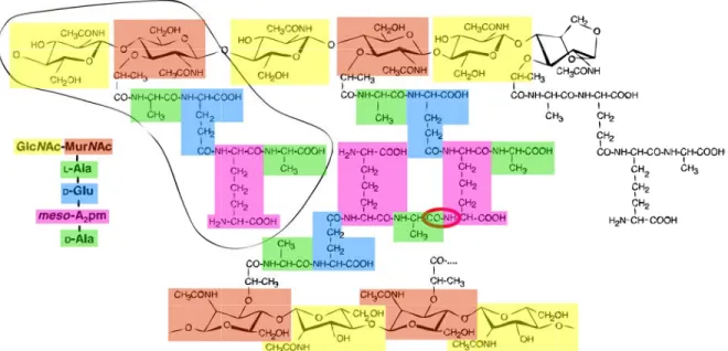
The glycan chains are composed of two alternating amino sugars, namely N -acetyl-glucosamine (GlcN Ac) and N -acetylmuramic acid (MurN Ac) [12]. The d-lactoyl group of each MurN Ac residue is substituted by a peptide stem whose composition depends on the species (see Section 1.2.4). Most often, the peptide is composed of l-Ala-γ-d-Glu-meso-A2pm (or l-Lys)-d-Ala-d-Ala (A2pm, 2,6-diaminopimelic acid) in nascent peptidoglycan,
the last d-Ala residue being lost in the mature macromolecule.
A proportion of these peptides are cross-linked either directly or through a second short peptide generally between the carboxyl group of d-Ala at position 4 and the amino group of the diamino acid at position 3. It is this cross-link that gives rise to the three-dimensional peptidoglycan polymer [12], with specific mechanical features. Figure 1.3 depicts an overall scheme of peptidoglycan.
1.2.3 Three-dimensional structure
On one hand, the large size, structural heterogeneity and flexibility of the peptidoglycan network prevent it from crystallizing and hence the three-dimensional crystal structure is
4LPS is the major component of the outer membrane of Gram-negative bacteria, contributing greatly to the structural integrity of the bacteria, and protecting the membrane from certain kinds of chemical attack. LPS induces a strong immune response.
Figure 1.3: Structure of the peptidoglycan of Escherichia coli. The glycan strands consist of alternating, β-(1,4)-linked GlcN Ac and MurN Ac residues, and are terminated by a 1,6-anhydroMurN Ac residue. The encircled part represents the basic disaccharide tetrapeptide subunit (monomer), which is also written with conventional abbreviations on the left-hand side. The middle part shows a cross-linked peptide, with the amide group connecting both peptide stems encircled in red. Adapted from Stenbak et al., 2004 [13].
inaccessible [10]. On another hand, glycan strands and peptides are too small to be visualized by conventional electron microscopy [10]. These are major impediments to the knowledge of precise molecular organization of peptidoglycan, and thus three-dimensional structure remains elusive [10].
However, several models have been proposed, based on chemical and biophysical data. One of them assumes that the peptides protrude helically from twisted glycan strands, with three or four disaccharides per turn [10].
1.2.4 Variability between species
Peptidoglycans differ by the composition of the stem peptide, the nature and extent of peptide cross-linking, and the length of glycan strands [10]. Remarkably, these differences do not seem to be obviously related to the Gram-positive or Gram-negative characteristics of the species.
Length of glycan strands
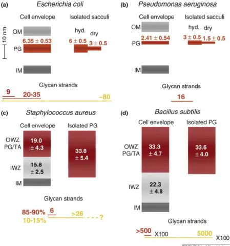
Strikingly, there are Gram-positive species with a thick cell wall with either short (S. au-reus) or long (B. subtilis) glycan strands. Similarly, there are Gram-negative species with either short (Helicobacter pylori) or long (Proteus morganii) glycan strands [11]. Therefore, it seems that there is no general correlation between the Gram-positive or Gram-negative nature of a species and the length of glycan strands in terms of 3D organization of pepti-doglycan itself [11]. Moreover, it has been reported that glycan-strand lengths depend on strain, growth state, and growth conditions [10]. Figure 1.4 summarizes average dimensions of cell envelope components.
1.2. PEPTIDOGLYCAN 31 Cryotomography data [14] and analyses of glycan-strand lengths in E. coli [10] [15] strongly suggest that glycan strands must be parallel to the cell membrane within the pep-tidoglycan layer.
Figure 1.4: Dimensions of cell envelope layers, isolated peptidoglycan and glycan strands in Gram-negative (a, b) and Gram-positive (c, d) species. The thickness of inner membrane (IM), peptidoglycan layer (PG), outer membrane (OM), inner wall zone (IWZ) and outer wall zone (OWZ) (with peptidoglycan (PG) and wall teichoic acid (TA)) are drawn to scale. Numbers indicate thickness with standard deviation in nm, which were measured either by atomic force microscopy (isolated sacculi in (a) and (b)] or by cryo-TEM (isolated sacculi in (c) and (d), and cell envelope layers in all panels). The length of the glycan strands is indicated in nm and drawn to scale (length of a disaccharide unit: 1.03 nm). Dark red line, length of abundant glycan strand; red line, average length of glycan strands; and yellow line, longest glycan strands. The glycan strands in B. subtilis are 100-fold longer than the length indicated by the lines shown on the figure. hyd., hydrated. From Vollmer and Seligman, 2010 [10].
Peptide composition
There is a high diversity in the composition and sequence of the peptidoglycan peptides from different species [10]. The variations of the peptide stem can be divided into two categories:
• Those due to the specificity of the enzymes responsible for its biosynthesis;
• Those occuring at a later step of the biosynthesis, often at the level of Lipid II - the peptidoglycan monomer, before its incorporation into the 3D layer [11].
Particularly, peptidoglycan from the Gram-negative thermophilic bacterium Thermotoga maritima has been reported to be made of a L-lysine or D-lysine at the third position of the peptide chain, while most Gram-negative bacteria such as E. coli display a
meso-diaminopimelic acid (A2pm) at this position [16] [17] [11]. Furthermore, the peptide
compo-sition may vary with growth conditions [10]. Peptide cross-linking
The most common peptide cross-linking is the 3-4 cross-linkage described above (Section 1.2.2). But other kinds of cross-links can be observed as well, such as the 3-3 cross-linkage, often seen in β-lactam-resistant strains [11]. Moreover, the size of the optional interpeptide bridge ranges from one to seven amino-acid residues, and its composition varies substantially between species [11].
Besides the diversity in the nature of cross-linking, there is a considerable variation in its degree. Indeed, while connections between only two peptides prevail in E. coli and B. subtilis, multimeric peptides with up to 20 connected peptides exist in S. aureus [11] [10]. Globally, it seems that peptidoglycans from Gram-positive bacteria are more cross-linked than Gram-negative peptidoglycans.
The peptidoglycan layer is a specific component of bacteria responsible for the main mechanical features of the bacterial cell wall, and therefore essential to most species. Basically made of glycan strands cross-linked by short peptides, its composition displays a very high variability between species. However, the pathway of its biosynthesis is very well conserved within the bacterial world, and is addressed in the next chapter.
Chapter 2
Peptidoglycan biosynthesis
Biosynthesis of peptidoglycan is a multi-step process involving a series of enzymes and struc-tural proteins (Figure 2.1), which can be described into two main steps. First, the monomer of peptidoglycan also known as Lipid II1, is synthesized in the cytoplasm and incorporated
in the membrane. Then, at the membrane level, Lipid II is flipped to the periplasm and subsequently transferred into the growing peptidoglycan layer where it is finally linked to the existing peptidoglycan network with the contribution of Penicillin Binding Proteins (PBPs) [5].
This chapter focuses on the synthesis of Lipid II, gives the basics of Lipid II polymeriza-tion and cross-linking into the peptidoglycan layer, and finally provides the current knowledge about the spatial organization of the peptidoglycan biosynthesis process which involves the bacterial actin homolog MreB.
2.1
Cytoplasmic steps: towards the synthesis of Lipid II
First of all, the MurA and MurB enzymes catalyze the synthesis of UDP-N -acetylmuramic acid (UDPMurNAc) from UDP-N -acetylglucosamine [18] [5]. Then, MurC, MurD, MurE, and MurF, which are ATP-dependent amino acid ligases, sequentially catalyze the addition steps of a short polypeptide chain to UDPMurNAc [5]. These first six steps occurring in the cytoplasm are followed by two steps taking place on the cytoplasmic side of the membrane where the final unit of peptidoglycan, known as Lipid II, is synthesized with the help of the transmembrane enzyme MraY and the glycosyltransferase MurG [5]. Based on Figure 2.1, this section describes into greater details the successive steps of Lipid II biosynthesis.
2.1.1 The very first precursor: UDP-N -acetylglucosamine
The basic precursor of peptidoglycan biosynthesis is a nucleotide sugar (see Figure 2.1) nat-urally synthesized through the hexosamine biosynthesis pathway from glucose, glucosamine, and uridine.2
1Lipid II is composed of a sugar moiety, a lipid, plus a peptide chain.
2The hexosamine pathway is used by a lot of glycosyltransferases to transfer N-acetylglucosamine residues to substrates in a high number of biological processes [19].
Figure 2.1: Schematic representation of the essential steps of peptidoglycan synthesis. The different domains in Mur ligases are shown in distinct colors (N-terminal, red; central domain, orange; C-terminal, green). Structures shown in the figure include MurA (1NAW), MurB (1MBT), MurC (1GQQ), MurD (2JFH), MurE (2WTZ), MurF (1GG4), and MurG (1F0K). The undecaprenyl phosphate carrier lipid is indicated in pink. From Mattei et al., 2010 [18].
2.1.2 Synthesis of UDPMurNAc by MurA and MurB enzymes
First, MurA catalyzes the addition of a phosphoenolpyruvate to the hydroxyl at carbon 3 of
the glucosamine ring of UDP-N -acetylglucosamine to form enolpyruvyl-UDP-N -acetylglucosamine3.
Subsequently, MurB catalyzes the reduction of the pyruvyl moiety to a lactyl group by NADPH, giving UDP-N -acetylmuramic acid (UDPMurNAc)4.
2.1.3 Addition of a short peptide by Mur ligases
A short polypeptide chain is then added to the sugar molecule UDPMurNAc through suc-cessive steps by four similar ATP-dependent amide bond ligases [5]. As described in the pre-vious chapter, in most species the peptide is composed of the following residues: l-alanine, d-glutamate, meso-diaminopimelate, d-alanine and d-alanine [5].
• MurC is an UDP-N -acetylmuramoyl:l-alanine ligase
• MurD is an UDP-N -acetylmuramoyl-l-alanine:d-glutamate ligase
• MurE is an UDP-N -acetylmuramoyl-l-alanine-d-glutamate:meso-diaminopimelate lig-ase
• MurF is an UDP-N -acetylmuramoyl-l-alanine-d-glutamate-meso-diaminopimelate:d-alanyl-d-alanine ligase [5].
3Thus, MurA is an UDP-N -acetylglucosamine enolpyruvyl transferase. 4Thus MurB is a UDP-N -acetylpyruvylglucosamine reductase.
2.2. PERIPLASMIC STEPS: INCORPORATION AND POLYMERIZATION 35
2.1.4 Final steps of Lipid II synthesis
The two last steps occur at the surface of the cytoplasmic membrane and are catalyzed by the transmembrane protein MraY and the membrane-bound glycosyltransferase MurG.
• The previously synthesized UDP-MurN Ac-peptide is attached to a membrane-bound lipid carrier molecule, undecaprenol-phosphate5, catalyzed by the integral membrane
protein MraY [20], to give Lipid I.
• The glycosyltransferase MurG transfers N -acetylglucosamine (GlcNAc) to Lipid I, forming Lipid II, the monomer of the final peptidoglycan polymer [5].
2.2
Periplasmic steps: incorporation and polymerization
Lipid II is translocated across the membrane, most likely with the contribution of RodA/FtsW in order to be incorporated into the growing peptidoglycan layer by the formation of glyco-sidic linkages between the disaccharide units, cross-linking of the peptide tails, and cleavage from the lipid carrier, facilitated by transpeptidases, endopeptidases, and penicillin-binding proteins (PBPs), the latter being the main targets of antibiotics in clinical use [5] [21].
2.3
Spatial organization of peptidoglycan biosynthesis
As seen in the previous section, the pathway of peptidoglycan biosynthesis is very well known. Indeed, most steps correspond to a known protein which has been extensively studied for most cases. However, how all these steps are spatially organized within the cell remains un-clear. Nevertheless, an increasing number of interactions between peptidoglycan biosynthesis actors have been described over the past decade, suggesting the existence of multipartite pro-tein complexes. This section focuses on the clues supporting the peptidoglycan machinery hypothesis, and introduces the bacterial actin homolog MreB which is thought to play a key organizational role in the process.
2.3.1 MreB: a bacterial actin homolog with a strong morphogenetic
role in rod-shaped bacteria
The mreB gene
In many organisms, mreB is part of the mre (murein cluster e) operon, one of the major operons involved in cell-shape determination in bacteria [22].
While most often Gram-negative species have only a single copy of mreB, Gram-positive organisms may have several mreB-like genes [23]. Such paralogues and orthologues could be specialized in different aspects of morphogenesis [23]. Interestingly, mreB genes are absent from most bacteria displaying spherical shapes [23].
Though mreB seems to be essential, the lethality or loss of its functionality can be suppressed either by overexpressing cell division proteins or reducing the rate of cell growth [24], or by growth in increased levels of Mg2+ [25] regarding B. subtilis.
The MreB protein: polymerization and membrane binding
Most in vitro work on MreB has been performed on the Thermotoga maritima protein, re-vealing that MreB presents two important and functional properties, namely polymerization and membrane binding.
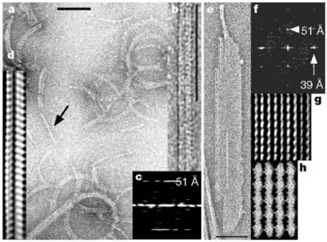
Most of studied MreB homologs were found to be capable of polymerization. For in-stance, MreB from T. maritima forms long multilayered sheets of interwoven filaments in the presence of either ATP or GTP in vitro [26] [22]. The longitudinal repeats in MreB filaments resemble that of actin [22], although a few differences have been described between the superstructures of filaments, and between polymerization kinetics as well [22] [27] [28]. Recent biochemical and functional studies on B. subtilis [29], Chlamydiaceae [30], and E. coli [31] underlined the high diversity of MreB homologues. For instance, B. subtilis MreB does not require nucleotide to polymerize, and its kinetic behavior is much different from that of T. maritima [32]; while work from Bean and Amann [33] suggested that most in vivo polymerized MreB could be in an ADP-bound state.
Figure 2.2: MreB polymers. Typical view of MreB filaments (a), double filaments (b, d: filtered image, c: diffraction image), and MreB sheets (e, g: filtered image, f: diffraction image) in different buffer conditions. In MreB sheets, the polymer is about 160 Åwide, which suggests four single protofilaments in total (each 40 Å). The longitudinal repeat is 51 Å, the lateral spacing is 39 Å. Scale bars, 100 nm. (h) The protofilaments found in the crystals of MreB fit well with the filtered image in (g). From van den Ent et al., 2001 [22].
Salje et al. showed that MreB from both T. maritima and E. coli bind directly to cell membranes [34] and provided a structural analysis (see next Chapter). Interestingly, they revealed that membrane-binding activity in E. coli is essential for the function of MreB in cell shape determination [34]. Their work suggests that membrane binding of MreB would orient the protofilament along the membrane surface [34].
2.3. SPATIAL ORGANIZATION OF PEPTIDOGLYCAN BIOSYNTHESIS 37 The role of MreB in cell-shape determination
The in vivo properties of MreB have been mainly characterized in B. subtilis, Caulobacter crescentus, and E. coli.
A number of depletion or functionality loss experiments have shown that MreB is required for cell shape in most non-spherical bacteria [35]. Figure 2.3 is an example of the aftermath of mreB depletion in C. crescentus: cells lose their regular shape and start lysing.
Figure 2.3: MreB depletion results in defects in cell shape. Cells of C. crescentus containing a xylose-inducible allele of mreB and a deletion in the wild-type copy of the mreB gene were grown in the presence of xylose, washed three times in inducer-free medium and resuspended in fresh medium lacking inducer. Phase-contrast images were obtained before washing (0 h) and after 2, 5 and 10 h of incubation without inducer, as indicated. Loss of MreB results in an abundance of lemon-shaped cells, some of which have membrane blebs indicating a loss of cell wall integrity (cells labelled ‘a’). Less abundant is the presence of other cells with defects in cell division (cells labelled ‘b’). These cells possess a constriction at the mid-cell, as would occur if they arrested at the pre-divisional stage. From Figge et al. [36].
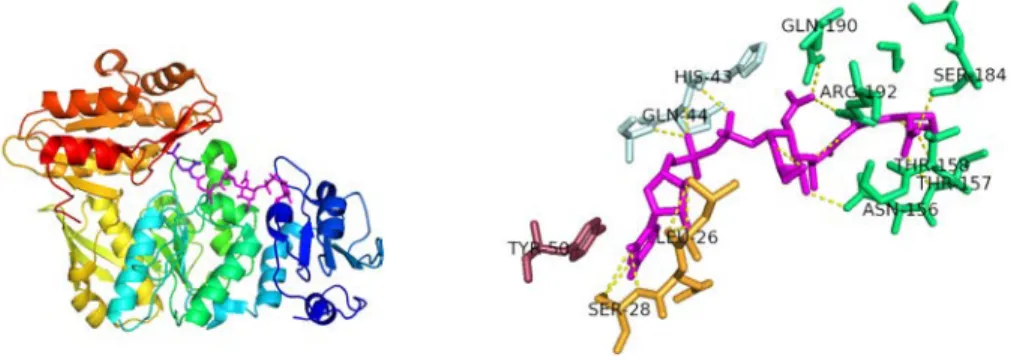
In addition to depletion assays, MreB was shown by immunofluorescence to colocalize with RodZ. This bitopic inner membrane protein has been widely reported to be required for cell shape determination as well [37] [38], though a few studies suggested that it is not always the case [39]. The interaction was further confirmed by work of van den Ent et al. [35] in 2010, who solved the crystal structure of MreB in complex with the cytosolic part of RodZ and provided in vitro data supporting the evidence of a functional significance of the interaction [35] (see Chapter 3).
MreB and cell-cycle
The prevailing model for bacterial growth predicts two spatially specialized pathways that control growth in rod-shaped bacteria: one for elongation in which MreB is strongly involved, and the other one for division that would be MreB independent [40] [41].
In support, conventional microscopy experiments in B. subtilis, E. coli, P. aeruginosa, and Rhodobacter have characterized both helical (elongation phase), and medial (division phase) MreB distributions [42] [43] [44] [29] as illustrated by Figure 2.4. In addition, Daniel
and Errington showed that, in B. subtilis, synthesis of the cylindrical part of the cell wall occurs in a helical manner that is reminiscent of the helical structure observed for Mbl filaments (a MreB homolog) [23]. Therefore, localization of MreB is believed to be regulated during the cell cycle [36] [45], presenting a helix-like (or patches, see below) pattern during elongation and a medial distribution during division.
Figure 2.4: Cellular localization of MreB labeled with GFP or YFP. (A to C) E. coli MreB localizes into extended coils (A), intertwined double helices (B), and band-like structures (C). (D) C. crescentus MreB localizes into a band-like structure at the division site in pre-divisional cells [41].
MreB pattern during elongation
However, the exact pattern of MreB during elongation is still under controversy. Indeed, MreB was thought to build a large, rigid, membrane-associated, helical scaffold within the cell which was suggested by conventional and confocal microscopy assays (see Figure 2.4 A and B). By contrast, two recent reports suggested that in B. subtilis MreB polymers could be composed of short, dynamic filaments as illustrated in Figure 2.5. In addition, the helical MreB superstructure in E. coli was reported to be an artifact of the N-terminal YFP tag [46].
This theory is supported by two main studies:
• TIRF6 experiments, in which Dominguez et al. could not see any MreB helix near or along the surface of the inner membrane during exponential growth of B. subtilis cells. Instead, the authors detected discrete MreB-patches that moved processively along peripheral tracks perpendicular to the cell axis and that colocalized with other morphogenetic factors [48].
• electron cryo-tomography studies of six different rod-shaped bacterial species at macro-molecular resolution could not distinguish any long (> 80 nm), membrane-associated helical filaments encircling cells [49].
2.3.2 Role of MreB in peptidoglycan biosynthesis
The implication of MreB in peptidoglycan biosynthesis was first suggested by microscopy experiments revealing co-localization patterns with murein enzymes. These data have been completed by biochemical results which will be discussed in this section. In addition, it has
6TIRF (Total internal reflection fluorescence microscopy) is a high-resolution technique able to capture the surface of one side of a cell and useful for visualizing activities at the membrane
2.3. SPATIAL ORGANIZATION OF PEPTIDOGLYCAN BIOSYNTHESIS 39
Figure 2.5: Schematic representation of MreB ultrastructure and movement seen by two different microscopic techniques. Both panels depict MreB (dark orange) coupled with the peptidoglycan (PG) elongation machinery (purple), which collectively represents cell wall synthetic enzymes and cell shape determining proteins, including, but not limited to, MreC, MreD, RodA, RodZ, PBPs and PG cytosolic synthetic enzymes. (a) Represents what is seen using deconvolution fluorescence microscopy, where a stack of images taken through the cell body depicts a helically structured MreB. (b) By contrast, TIRF microscopy found both MreB and a selection of several PG elongation proteins that move in short patches (b) as opposed to long helical filaments (a). Schematic representations are not drawn to scale [47].
been reported that inhibition of MreB with the small molecule A22 in Caulobacter leads to shortened cell wall glycan strands [50], indicating that MreB has a general influence on peptidoglycan assembly.
MreB as a bridge between the cytoplasmic and periplasmic steps
MreB, MreC, MreD, along with the cell wall assembly proteins PBP2 and RodA, all involved in late stages of peptidoglycan biosynthesis, have been proposed to form a complex [51] [48]. Therefore, the cytoplasmic MreB filaments may orchestrate peptidoglycan biosynthesis from the cytoplasm through its direct or indirect interactions with transmembrane or periplasmic proteins [52].
In addition, MreB was shown to interact with RodZ (see above). For instance, microscopy assays observed that RodZ was associated to MreB in space and time and marked future sites of peptidoglycan synthesis [37] in Caulobacter crescentus, suggesting an additional link through the bacterial cytoskeleton and future sites of peptidoglycan incorporation.
MreB could recruit cytoplasmic cell wall synthesizing proteins
White et al. found that in C. crescentus MreB cables are required for the organization of several other cytosolic murein biosynthetic enzymes such as MraY, MurB, MurC, MurE and MurF [53], each of these proteins adopting a subcellular pattern of localization comparable to MurG. These microscopy data strongly suggest the existence of cytoskeletal-dependent interactions among cytoplasmic actors of peptidoglycan biosynthesis [53] [54].
This has been supported by co-pelletting assays showing interactions between MreB and MurF [30], and bacterial two-hybrid system experiments, finding that MurG interacts with MreB in both C. crescentus [53] and Chlamydia [30].
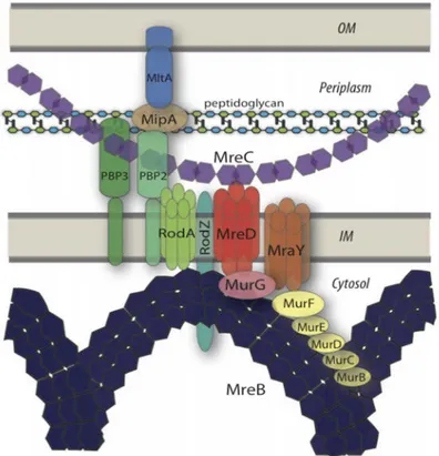
known or suggested interactions among bacterial morphogenetic proteins and cell wall syn-thesizing enzymes (Figure 2.6). They propose that MreB would organize the cytoplasmic steps by interacting with a number of murein-synthesizing proteins such as Mur enzymes, MraY, and the RodZ and RodA morphogenetic proteins as well. On the periplasmic side, MreC woud position a peptidoglycan assembly complex consisting of PBPs and lytic enzymes (i.e. MltA, MipA). The integral membrane protein MreD would contribute to connection of both cytoplasmic and periplasmic complexes through interactions with MreB, MreD, and transmembrane proteins [53].
Figure 2.6: Model depicting the peptidoglycan biosynthesis machinery. Schematic view of inter-actions within peptidoglycan biosynthesis pathway, focusing on the crucial role of Mre proteins in spatially organizing other actors of peptidoglycan biosynthesis [53].
Exact role of MreB in peptidoglycan biosynthesis
All these data suggest the existence of an interaction between MreB polymers and the cell-wall synthetic machinery. However, the exact role of the bacterial actin homolog remains unclear, and two theories have been proposed to date.
The first model, based on the MreB helix mentioned above and helical incorporation of newly synthesized peptidoglycan monomers, postulates that helical MreB cables direct cell wall growth by appropriately positioning the peptidoglycan elongation machinery [55]. The second model, based on the recent studies revealing patches rather than a helical superstruc-ture, proposes that MreB-patches, that contain sidewall elongation enzymes as well, would be effectively dragged along by peptidoglycan synthesis itself [55] [48] [56].
However, both models see MreB as an ’anchor’, either to target peptidoglycan biosyn-thesis (first model) or to constraint the movement of the peptidoglycan synbiosyn-thesis machinery
2.3. SPATIAL ORGANIZATION OF PEPTIDOGLYCAN BIOSYNTHESIS 41 (second model), ensuring the completion of peptidoglycan synthesis at a given site [55] and limiting diffusion of intermediates.
Nevertheless, direct evidence of interactions of MreB with peptidoglycan synthesis en-zymes is still lacking, mainly because most of these enen-zymes are transmembrane proteins difficult to work with. But recent studies have suggested that MreB could also recruit cy-toplasmic actors, therefore opening up the way towards the structural characterization of a cytoplasmic part of the peptidoglycan machinery.
The pathway of peptidoglycan biosynthesis involves a series of en-zymes. Among them, the Mur ligases participate to the formation of UDPMurNAc-peptide in the cytoplasm. Then, at the membrane level, a lipid career and an additional disaccharide are added by MraY and MurG enzymes, forming Lipid II, the basic block of peptidoglycan polymers. The spatial organization of these steps has not been elucidated yet. However, the bacterial actin homolog MreB seems to act as an anchor at the mem-brane surface for a multipartite complex which would restrict the diffusion of enzymes and intermediates, resulting in an optimized biosynthesis.
Chapter 3
Structural background
Direct and molecular evidence of the existence of the cytoplasmic peptidoglycan synthesis machinery is still lacking. Notably, no structure of any complex has been solved to date between either of the morphogenetic or murein synthesizing proteins. However, single actors of peptidoglycan biosynthetic machinery have been thoroughly analyzed from a structural point of view, as summarized in this chapter for Mur ligases, MurG, and MreB.
3.1
Crystal structures of Mur ligases
The crystal structures of all four of these similar ATP-dependent amino-acid ligases are known in different species in apo form or in complex with substrate, product, ADP, or in-hibitors (see Table 3.1). Together with kinetics studies, these data allow a good understand-ing of their substrate specificity and their catalytic mechanisms [5] in which conformational changes might be important for activity.
Since all four Mur ligases share structural similarities, the structural description [5] [57] presented here is based on MurC from Haemophilus influenzae (PDB code 1P3D), for which structures of each of the apo, substrate-bound, and product-bound forms are known. Num-bering of amino-acid residues thus refers to H. influenzae MurC, unless mentioned. Any relevant similarity or difference seen in the other three enzymes will be mentioned.
Mur ligases consist of three α/β-sheet domains formed from contiguous segments in the amino acid sequence: the N-terminal domain, the central domain, and the C-terminal domain (Figure 3.1).
3.1.1 N-terminal domain
Description
In all Mur ligases, the N-terminal domain is about 100 amino acids in length. This domain shows the highest degree of structural and sequence diversity among the three domains of Mur ligases and is mainly responsible for binding the growing peptidoglycan precursor [5].
The N-terminal domain of MurC from H. influenzae contains a common Rossmann-type 43
Protein Species Form Resolution Publication PDB MurC T. maritima apo 2.3 Å Spraggon et al., 2004 1J6U
MurC E. coli Mg2+ 2.5 Å Deva et al., 2006 2F00
MurC H. influenzae apo 3.1 Å Skarzynski et al., - 1GQQ MurC H. influenzae product.AMPPNP.Mn2+ 1.7 Å Mol et al., 2003 1P3D MurC H. influenzae apo.AMPPCP.Mg2+ 1.8 Å Hu et al., 2003 1GQY MurC H. influenzae substrate.Mn2+ 1.85 Å Mol et al., 2003 1P31 MurC Y. pestis apo.AMP 2.25 Å Halavaty et al., - 4HV4
MurD E. coli apo 2.4 Å Bertrand et al., 2000 1E0D
MurD E. coli substrate 1.9 Å Bertrand et al., 2000 1EEH MurD E. coli substrate.Mg2+ 1.7 Å Bertrand et al., 1999 2UAG MurD E. coli substrate.ADP 1.52 Å Kotnik et al., 2007 2JFG MurD E. coli inhibitor.aa 2.2 Å Humljan et al., 2008 2VTE MurD S. agalactiae substrate.ADP 1.5 Å Stein et al., - 3LK7 MurE M. tuberculosis substrate.Mg2+ 3.0 Å Basavannacharya et al., 2010 2WTZ MurE E. coli product.Cl− 2 Å Gordon et al., 2001 1E8C
MurF E. coli apo 2.3 Å Yan et al., 2000 1GG4
MurF S. pneumoniae inhibitor 2.8 Å Longenecker et al., 2005 2AM1
Table 3.1: Main structures of Mur ligases, in apo form or in complex with substrate, product, ATP analog, and/or amino-acid. Structures solved in complex with inhibitors are not men-tioned here, unless no other structure is known for this enzyme. aa: incoming amino acid (d-ala for MurD); ADP: adeno-sine diphosphate; ANP: phosphoaminophosphonic acid-adenylate ester; AMP: adenoadeno-sine monophosphate; AMPPCP: β,γ-methyleneadenosine 5’-triphosphate; AMPPNP: Adenylyl-imidodiphosphate; MurC substrate: UDP-N -acetylmuramic acid; MurD substrate: UDP-N -acetylmuramoyl-l-alanine; MurE substrate: UDP-N -acetylmuramoyl-l-alanine-d-glutamate; MurE product: UDP-N -acetylmuramoyl-l-alanine-d-glutamate-meso-diaminopimelate.
Figure 3.1: MurC from H. influenzae. Cartoon representation of MurC. Domain 1 (blue): residues 11-118; domain 2 (yellow): residues 119-324; domain 3 (red): residues 325-473. PDB entry: 1P3D.
α/β fold1, consisting of a five-stranded parallel β-sheet flanked by four alternating α-helices [57]. This conserved topology is present in MurD as well. Indeed, the first two enzymes in the pathway share an essentially identical structure for domain 1 and both contain a variant of the GxGxxG fingerprint motif typical of dinucleotide binding domains [5] which is within a largely conserved region (Figure 3.2).
By contrast, although MurE and MurF also have an α/β fold for this domain, the
topol-1The Rossmann fold is a general nucleotide-binding structural motif. The most common structure with two repeats is composed of six parallel β-strands linked to two pairs of α-helices in the topological order β − α − β − α − β. Each Rossmann fold can bind one nucleotide [58].
3.1. CRYSTAL STRUCTURES OF MUR LIGASES 45 ogy is much different and they display a mixed β-sheet [5] (Figure 3.3). In addition, no equivalent glycine-rich motif is found in the N-terminal domains of either of MurE and MurF enzymes.
Figure 3.2: Alignment of the 60 first residues of E. coli (Ec) MurC with H. influenzae (Hi) and T. maritima (Tm) MurCs. The consensus GxGxxG motif (black box) for dinucleotide binding domains is very well conserved among different species. In MurC, the motif is GIGGxGM.
Figure 3.3: Domains 1 of Mur ligases from E. coli in apo forms. Left: superposition of the domains 1 of MurC (red) and MurD (green). Right: superposition of the domains 1 of MurE (grey) and MurF (yellow) from E. coli. In purple and red: the main loop involved in substrate binding. PDB codes: 2F00 (MurC), 1E0D (MurD), 18EC (MurE), 1GG4 (MurF).
Substrate binding by MurC and MurD.
The N-terminal domain binds the nucleotide moiety of the UDPMurNAc substrate, with a number of interactions [5] [57] (see Figure 3.4):
• The uracil ring is sandwiched between two hydrophobic loops (β2-α2 and β4-α4) which form a hydrophobic pocket where residues Ile 50 and Ile 87 pack the uridine.
• A conserved histidine residue (His 70) from the β3-α3 loop anchors the ring by hydrogen bonding.
• The glycine-rich loop (Gly 25 to Met 31) between β1 and α1 contacts the phosphate groups of the UDP. Together with a conserved serine residue (Ser84), this forms the diphosphate binding pocket.
• A conserved aspartate (Asp 49) from the C-terminus of strand β2 forms hydrogen bonds with the ribose hydroxyl groups.
Though the muramic acid moiety makes no interaction with the protein, the lactyl side-chain extends towards the catalytic centre and interacts with a Mg2+ ion [5].
The substrate binding site in MurD is very similar to that of MurC [5]. Nevertheless, MurD does not have the conserved histidine residue but instead uses a conserved threonine from its β2-α2 loop to anchor the uracil ring through a hydrogen bond [5] [59].
Figure 3.4: Interactions of H. influenzae MurC with its UDPMurNAc substrate. Left: overall view of MurC in complex with UDPMurNAc substrate (pink). Rainbow representation from N-terminus (blue) to C-terminus
(red). Spheres: Mg2+. Right: hydrogen-bonding interactions from domain 1 MurC residues (yellow) to the UDPMurNAc (red)
are shown. From Mol et al., 2003 [57].
Substrate binding by MurE and MurF
In contrast, MurE binds its substrate in a very different way from MurC and MurD [5] (see Figure 3.5). Indeed, a long loop between strand β2 and helix α2, extending towards domain 3, lies along the uridine group and makes hydrogen bonding interactions with the diphosphate moiety [5]. As in MurC, the uracil ring is tightly anchored, but the loop and residues involved are different (Tyr50 and β1-β2 loop) [5] (Figure 3.5).
While in MurC and MurD the substrate makes no interaction with domain 2, in MurE all of the other interactions with the UDP substrate are provided by the central domain [5].
Figure 3.5: Interaction of M. tuberculosis MurE with its UDPMurNAc substrate. Hydrogen-bonding interactions from domain 1 MurE residues to the UDPMurNAc (pink) are shown. Blue: interacting residues from β2-α2 loop; yellow: interacting residues from β1-β2 loop; green: interacting residues from domain 2. PDB code: 2WTZ.
Although no structure of MurF in complex with its substrate is available yet, the su-perposition of domain 1 from apo MurF onto MurE (see Figure 3.3) shows that the same long loop is present and that the residues involved in UDP binding are essentially conserved, suggesting that a similar strategy is used for substrate binding by MurF [5] [1].


![Figure 3.16: Structure of MreB in complex with AMPPNP. From van den Ent et al., 2010 [35].](https://thumb-eu.123doks.com/thumbv2/123doknet/12883670.370173/55.918.286.611.758.1041/figure-structure-mreb-complex-amppnp-van-den-ent.webp)


