HAL Id: hal-02417294
https://hal.archives-ouvertes.fr/hal-02417294
Submitted on 14 Jan 2020
HAL is a multi-disciplinary open access
archive for the deposit and dissemination of
sci-entific research documents, whether they are
pub-lished or not. The documents may come from
teaching and research institutions in France or
abroad, or from public or private research centers.
L’archive ouverte pluridisciplinaire HAL, est
destinée au dépôt et à la diffusion de documents
scientifiques de niveau recherche, publiés ou non,
émanant des établissements d’enseignement et de
recherche français ou étrangers, des laboratoires
publics ou privés.
UVA-irradiated DNA: photosensitization or
photoisomerization?
Thierry Douki
To cite this version:
Thierry Douki. Pyrimidine (6-4) pyrimidone photoproducts in UVA-irradiated DNA:
photosensitiza-tion or photoisomerizaphotosensitiza-tion?. ChemPhotoChem, Wiley, 2020, 4, pp.294-299. �10.1002/cptc.201900280�.
�hal-02417294�
Pyrimidine (6-4) pyrimidone photoproducts in UVA-irradiated
DNA: photosensitization or photoisomerization?
Thierry Douki*
[a]Abstract: Formation of pyrimidine dimers in DNA is a major initiating
event in the induction of skin cancer. Model experiments suggest that, upon absorption of UVA, one type of dimers induced by UVB, the pyrimidine (6-4) pyrimidone photoproducts, photosensitizes the formation of mutagenic cyclobutane pyrimidine dimers by triplet -triplet energy transfer (TTET). We investigated whether this photoreaction actually took place when 64PP were located within a DNA duplex rather than added as external sensitizers like in available data. Our results show that this process is not detectable in DNA and double-stranded oligonucleotides exposed to a combination of UVB and UVA. TTET could only be observed, as a very minor photoreaction, in a short single-stranded oligonucleotide bearing a 64PP. It may be concluded that 64PP-mediated TTET does not significantly contribute to UV-induced DNA damage. In contrast, the photoisomerization of 64PP into their Dewar valence isomers is very efficient.
Introduction
Pyrimidine (6-4) pyrimidone photoproducts (64PP) is a class of DNA damage that, together with cyclobutane pyrimidine dimers (CPD), is responsible for a large fraction of the deleterious effects of solar UV radiation, in particular induction of skin cancer[1].
These photoproducts result from cycloaddition reactions between adjacent pyrimidine bases, thymine (T) and cytosine (C), within DNA and are mostly produced by UVB radiation (280-320 nm). UVA (320-380 nm) produces low amounts of CPD by direct
absorption [2,3,4,5,6] but more importantly induces the
photoisomerization of the pyrimidone ring of 64PP into a Dewar valence isomer (Dewar) [7,8,9,10,11]. Another interesting property of
UVA is its involvement in the photosensitized formation of CPD. Indeed, UVA excited chromophores with an energy of excited triplet state higher than that of DNA may undergo triplet-triplet energy transfer (TTET) and subsequent formation of CPD [12,13,14].
Thymine, especially at TT sites [15], is the major target of this
process because it exhibits the lowest triplet energy level among DNA components [16,17].
It was recently shown that the energy of the triplet excited state of pyrimidone rings was larger than that of T. Consequently, excitation of the pyrimidone ring of a 64PP could perform TTET to
an adjacent T [18,19]. Accordingly, formation of CPD in plasmid
DNA exposed to UVA in the presence of 64PP model compounds in solution led to the formation CPD. In addition, calculations on DNA strand where one base was substituted by a pyrimidone moiety suggested that 64PP-mediated TTET upon UVA irradiation, referred to below as “64PP-TTET”, is possible in a duplex environment [20]. Last, it was recently reported that
benzophenone covalently linked to a DNA strand was able to
carry out long distance TTET and formation of CPD [21].
64PP-TTET to an adjacent TT doublet within a DNA strand appears thus as an interesting hypothesis since it would lead to the formation of damaged sites bearing a 64PP and a CPD next to each other (Scheme 1). This would be similar to the cluster damage produced by ionizing radiation which are proposed to be highly mutagenic and lethal [22].
The actual occurrence of 64PP-TTET within double-stranded DNA remains yet to be experimentally documented. It should also be determined whether 64PP-TTET is more efficient that photoisomerization of 64PP into Dewar. The reported value of the quantum yield for the latter reaction ranges between 0.02 and 0.008 [9,10]. The intersystem crossing quantum yield of the
pyrimidone ring of 64PP is 0.86 [19] and that of the formation of a
CPD from the triplet excited state of thymine is 0.04 [23]. The
efficiency of TTET between excited 64PP and thymine is not known. However, even if it was 1, the quantum yields for Dewar formation and photosensitization of CPD would be similar. It is thus not expected that 64PP-TTET is the overwhelming process. Consequently, we undertook the present work to establish whether or not 64PP mediates TTET in DNA duplexes upon exposure to UVA. For this purpose, we exposed DNA and oligonucleotides to sources of UV radiation emitting both UVB and UVA. Preliminary kinetics modeling showed 64PP-TTET led to a positive deviation from linearity as the dose increases (Scheme S1). We thus tried to detect this signature under different experimental conditions. Additional experiments were performed with a purified short oligonucleotide bearing a single 64PP at a specific position.
Scheme 1. Photochemical processes triggered by absorption of UVA by a UVB
UVB UVA 2: TTET 1: isomerization (1) (2) 64PP 64PP CPD Dewar [a] Dr. T. Douki
Univ. Grenoble Alpes, CEA, CNRS, IRIG, SyMMES, F-38000 Grenoble
17 avenue des Martyrs, 38054 Grenoble Cedex 9, France E-mail: thierry.douki@cea.fr
Supporting information for this article is given via a link at the end of the document.
induced 64PP. Intramolecular electrocyclization results in a Dewar valence isomer (1) while TTET to adjacent thymine leads to formation of CPD (2).
Results and Discussion
Searching for 64PP-TTET in isolated genomic DNA
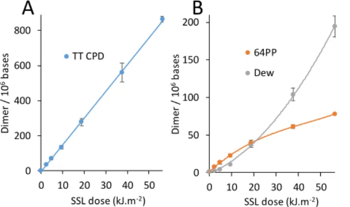
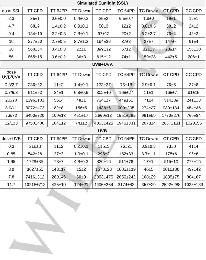
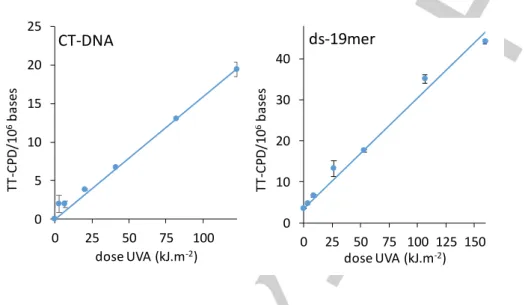
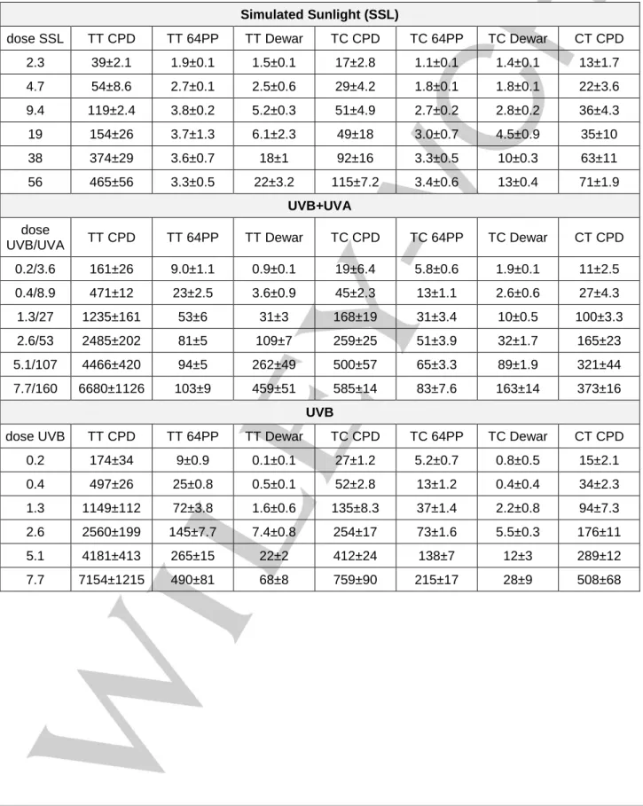
In a first experiment, we irradiated purified genomic calf thymus DNA (CT-DNA) with the light emitted by a solar simulator. Simulated sunlight (SSL) contains roughly 5% UVB and 95% UVA. All possible pyrimidine dimers were quantified by HPLC coupled to tandem mass spectrometry (Table S1) [24]. TT CPD (Figure 1A),
as well as other CPD, was produced in a linear way with respect to the dose. This contrasted with Dewar, the formation of which became more efficient at the expense of 64PP when the dose increased (Figure 1B). The latter feature reflects the efficient photoisomerization of 64PP. The remarkable similarity between the dose course formation of photodamage in CT-DNA exposed to SSL reported in Figure 1 and the kinetic predictions in the absence of 64PP-TTET reported on Scheme S1 shows that the latter process in not a major one under these experimental conditions.
Figure 1: Formation of pyrimidine dimers in CT-DNA exposed to simulated sunlight. A) TT CPD and B) 64PP and Dew. For the two latter photoproducts, values for damage at TT and TC sites were combined. Aliquot fractions of the irradiated solution were collected after increasing periods of time. The experiments were repeated twice and each sample analyzed twice. Results are means ± standard deviations.
In order to better control the respective effects of UVB and UVA, we then exposed CT-DNA to a more complex irradiation set-up. The sample was placed between UVB and UVA lamps that could be operated independently. The ratio between the intensity of the UVA and the UVB sources was approximately 10. HPLC-MS/MS analyses (Table S1) showed that the formation of TT CPD was linear with respect to the dose upon exposure to UVB, irrespective of the addition of UVA radiation (Figure 2A). The yields determined under the two conditions were not statistically significantly different (p=0.13). It may be added that exposure to UVA alone was found to induce CPD (Figure S1) but in a three orders of magnitude lower yield than UVB (Table S2). This observation, which is in agreement with published data [2,3,4,5,6],
rules out a major contribution of CPD directly induced by UVA in
our experiments. We also determined the proportion of 64PP that underwent photoconversion into their Dewar valence isomer. The value was much higher when irradiation was performed with UVB and UVA than with UVB alone (Figure 2B). It may be concluded that, like with SSL, exposure to the combination of UVB+UVA did not induce detectable 64PP-TTET in CT-DNA. Rather, UVA efficiently induces photoisomerization of 64PP.
Figure 2: Formation of pyrimidine dimers in CT-DNA exposed to UVB in the
presence of the absence of UVA in a 10 times larger intensity. Aliquot fractions of the irradiated solution were collected after increasing periods of time. The experiments were repeated twice and each sample analyzed twice. A) dose dependent formation of TT CPD. Results are means ± standard deviations. B) Proportion of 64PP converted into their Dewar valence isomers expressed in %. For the calculation, photoproducts arising from TT and TC sequences were combined. The proportion of Dewar was the ratio between the amount of Dewar divided by the combined amounts of Dewar and 64PP.
64PP-TTET in double-stranded pyrimidine tracks
Formation of TT CPD by 64PP-TTET can only take place in runs of 4 pyrimidines. Two are necessary to yield a 64PP and 2 to absorb the triplet energy of the excited pyrimidone and produce CPD. However, the probability of finding a T in such a context in CT-DNA with 42% G:C base pairs can easily be calculated to be only 0.7 % for TTTT and 0.5 % for TCTT. To better detect possible induction of 64PP-TTET in such pyrimidine runs, we studied the photolysis of a 19 base pairs double-stranded oligonucleotide bearing both a TTTT and a TCTT track. The sequence of the pyrimidine track containing strand was CATCTTACATTTTAC and that of the complementary strand was GTAAAATGTAAGATG. All dimeric photoproducts were quantified by HPLC-MS/MS (Table S3). CPD were detected only in their cis,syn and not their
trans,syn form, which shows the stability of the duplex under the
experimental conditions[25,26]. Like for CT-DNA, exposure to SSL
induced a linear dose dependent formation of TT CPD (Figure 3A). A linear formation was also observed when irradiations were performed with the source emitting UVB, UVA or both. Interestingly, the ratio between the levels of TT CPD in the oligonucleotide exposed to either UVB or UVB+UVA was around 1 at all time-points (Figure 3B), with an average value of 1.0±0.1. This observation shows that the formation of TT CPD does not deviate from linearity in the presence of UVA radiation. In addition, the difference between the yields of formation of CPD (Table S2) 0 200 400 600 800 0 10 20 30 40 50 D imer / 10 6b as es SSL dose (kJ.m-2) TT CPD 0 50 100 150 200 0 10 20 30 40 50 D imer / 10 6b as es SSL dose (kJ.m-2) 64PP Dew
A
B
0 15 30 45 60 0.0 0.3 0.7 2.0 3.9 7.8 11.7 % D ew ar UVB dose (kJ.m-2) UVB+UVA UVBA
B
0 2500 5000 7500 10000 0 4 8 12 TT -C PD / 10 6b as es UVB dose (kJ.m-2) UVB+UVA UVBin the presence or the absence of UVA was not statistically significant (p=0.24). It may be concluded that UVA does not favor the formation of TT CPD by 64PP-TTET in the oligonucleotide.
Figure 3: Formation of TT CPD in a 19 mer double-stranded oligonucleotide. A)
dose dependent formation upon exposure to simulated sunlight. B) ratio between the levels of TT CPD measured in oligonucleotides exposed to UVB in the presence or the absence of UVA applied in a 20 times larger intensity.
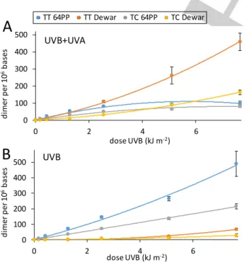
In contrast to 64PP-TTET, exposure of the oligonucleotide to a combination of UVB and UVA either with the solar simulator (Figure S2) or with the 2 lamps system induced significant photoisomerization of 64PP (Figure 4). This is shown by the larger yield of Dewar than of 64PP with SSL and UVB+UVA than with UVB alone. The yield of photoisomerization was 60% in the samples exposed to the largest dose of UVB+UVA whereas it was 10% with UVB. Exposure to UVA alone led to the formation of TT CPD in a 3 orders of magnitude lower yield than with UVB (Figure S1 and table S2).
Figure 4: Formation of 64PP and Dewars at TC and TT sites in the 19 mer
double-stranded nucleotide exposed to UVB in A) the presence or B) the absence of UVA. Aliquot fractions of the irradiated solution were collected after
increasing periods of time. The experiments were repeated twice and each sample analyzed twice. Results are expressed in photoproducts per million bases (mean ± standard deviation).
Pyrimidone mediated TTET in the vicinity of a purified 64PP
Following the same idea of using shorter models in order to better detect putative 64PP-TTET, we investigated the UVA photolysis of the TCTTA oligonucleotide where the TC doublet was photochemically converted into its 64PP (T(64)CTTA). An adenine was added on the 3’-end of the TT doublet in order to favor the formation of the CPD [27,28,29]. We chose to study TC
64PP rather than TT 64PP because the former is more frequently produced than the latter in isolated and cellular DNA [6,25,30].
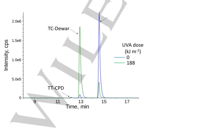
T(64)CTTA, prepared by UVC photolysis of TCTTA and isolated with a 95% purity, was exposed to increasing doses of UVA. Evolution of the absorption spectrum during irradiation showed that photoisomerization of the 64PP took place since the 316 nm absorption, typical of TC 64PP [31], was lost (Figure 5A). In
contrast, the absorption of normal bases at 263 nm decreased only by 2%, suggesting that only minute amounts of CPD or other non UV absorbing lesions were produced. This was confirmed on the differential absorption spectrum (Figure 5B). The very efficient photoisomerization of TC 64PP was also confirmed by HPLC-MS/MS analysis (Figure S3). Quantitative results obtained by the latter technique showed that the formation of TT CPD was 5500 times less efficient than that of TC Dewar. The rate constants calculated from a dose course study for the formation of TC Dewar and TT CPD were 1.7±0.1 10-3 and 3.3±0.4 10-7
kJ m-2 per oligonucleotide, respectively.
Figure 5: UVA photolysis of T(64)CTTA. A) UV absorption spectroscopy shows
the decreases of the 316 nm band resulting from the photoconversion of 64PP into Dewar. B) Differential spectrum corresponding to the difference between the initial solution of T(64)CTTA and that after 120 min of exposure to UVA radiation.
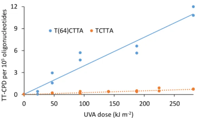
For comparative purposes, TCTTA was irradiated under similar conditions of concentration and UVA dose as T(64)ATTA. The yield of formation for TT CPD in the unmodified oligonucleotide was 2.4 ± 0.4 10-8 kJ m-2 per oligonucleotide. The difference by a
factor 13 between the yields formation of TT CPD in T(64)CTTA and TCTTA (Figure 6) was statistically significant (p<0.001). The bulk of these results show that the presence of a 64PP on the 0 200 400 0 10 20 30 40 50 D imer / 10 6b as es SSL dose (kJ.m-2) TT CPD 0.0 0.2 0.4 0.6 0.8 1.0 0.2 0.4 1.3 2.6 5.1 7.7 R at io in T T -C PD y iel d UVB dose (kJ.m-2) UVB+UVA / UVB
A
B
0 100 200 300 400 500 0 2 4 6 d ime r p er 1 0 6b as es dose UVB (kJ m-2)UVB+UVA
0 100 200 300 400 500 0 2 4 6 d im e r p e r 1 0 6b a se s dose UVB (kJ m-2)UVB
TT 64PP TT Dewar TC 64PP TC DewarA
B
-0.04 -0.03 -0.02 -0.01 0.00 0.01 0.02 230 260 290 320 350 Da b s wavelength (nm) 0.0 0.1 0.2 0.3 0.4 230 270 310 350 A b so rp ti o n wavelength (nm) UVA dose (kJ.m-2) 0 8 23 47 94 188A
B
235 nm
316 nm
5’-end of a TT doublet does favor the formation of CPD, mostly likely by 64PP TETT. However, this process is much less efficient than the photoisomerization of the 64PP into Dewar isomers.
Figure 6: Formation of TT CPD in the 5 mer oligonucleotide TCTTA used either
unmodified or after conversion of the 5’-end TC doublet into its 64PP..
Conclusions
In summary, our results show that 64PP-TTET does not significantly impact the yield of formation of TT CPD in double-stranded systems upon co-exposure to UVB and UVA in proportions similar to those found in sunlight. Significant contribution of this photochemical process could be observed neither in CT-DNA nor in an oligonucleotide bearing two pyrimidine tetrads. In contrast, as already observed both in isolated and cellular DNA, co-exposure to UVB and UVA leads to efficient conversion of 64PP into their Dewar valence isomers
[4,7,32,33,34,35,36]. These observations suggested that 64PP-TTET
was at the best a minor process. Accordingly, evidence for the occurrence of this process could only be obtained in a short single stranded oligonucleotide bearing a TC 64PP at the 5’-end of a TT sequence. The presence of the 64PP increased the yield of formation of TT CPD when compared to unmodified TC doublet. However, even under these conditions, the yield of 64PP-TTET remained between 3 and 4 orders of magnitude smaller than that of the photoisomerization of 64PP. It remains to be determined whether the single stranded form of the substrate used in this experiment reflects the photochemistry within a double helix. Indeed, orientation of the photosensitizer with respect to bases was found to play a key role in TTET [37] and the increased rigidity
of double-stranded DNA may affect its yield. Altogether, the role of “Trojan horse” proposed to be played by 64PP upon exposure to UV radiation is unlikely to be predominant even in pyrimidine rich sequences. Other DNA lesions, mostly of oxidative origin like 5-formyluracil, exhibit a residual absorption in the UVA range and have been proposed to play a similar role [38,39]. Further
experiments in DNA duplexes are mandatory to establish whether this interesting photochemistry takes place or not.
Experimental Section
Chemicals
Sodium chloride, Tris, EDTA, zinc chloride, and triethylammonium acetate were purchased from Sigma Aldrich (Saint Quentin Falavier, France). Acetonitrile was HPLC-MS quality (Merck, Darmstadt, Germany). Nuclease P1, alkaline phosphatase, phosphodiesterase I and phosphodiesterase II were from Sigma. Calf thymus DNA (Type I, Sigma) was purified by extensive dialysis in pure Milli Q water. Oligonucleotides (HPLC purified grade) were purchased from Eurogentec (Angers, France) and used as received. Hybridation of the two 19-mers (CATCTTACATTTTAC and GTAAAATGTAAGATG) was performed in solution in buffer (Tris HCl 10 mM, EDTA 0.5 mM, NaCl 50 mM). A solution containing the two strands at a concentration of 0.2 µM each was maintained at 60°C for 15 min and slowly cooled down to 25°C over 3 hours. The temperature was then decreased to 10°C by steps of 5°C kept for 5 min. Samples were then stored at 4°C.
UV sources and irradiation protocols.
The solar simulator was a LS1000 Solar Simulator (Solar Light Company, Glenside, PA), which emitted wavelengths in the 290-400 nm range. The radiation intensity was 4.7 kJ m-2 min-1. Samples (2.5 mL) of either DNA
(50 µg mL-1, 50 mM NaCl) or double-stranded oligonucleotide (0.2 µM in
hybridization buffer) were placed under the beam in a 3 mL spectrophotometer quartz cell place in ice. Irradiation were performed under magnetic stirring and 350 µL aliquot fractions were collected after increasing period. Experiments were duplicated. For co exposure to UVB and UVA, the sample was placed in a 3 mL quartz cell under magnetic stirring at 10 cm of a vertical 2×15 W UVA lamp (T 15.L tubes emitting between 310 and 400 nm with a maximum at 365 nm, Vilber-Lourmat) and 50 cm of a 15W UVB lamp (T 15.M tube emitting between 280 and 370 nm with a peak at 312 nm, Vilber-Lourmat) also placed vertically. Temperature was maintained at 25°C by a cooled airflow provided by a fan. The radiation emitted by the UVA lamp were filtered by a cut-off filter at 320 nm (WG20, Schott). The intensities of the UVA and UVB radiation reaching the CT-DNA samples were 1.37 and 0.13 kJ m-2 min-1, respectively. In the
experiments involving the double-stranded oligonucleotide, the corresponding values were 1.78 and 0.085 kJ m-2 min-1. Experiments were
performed with either the UVB lamp, the UVA lamp or both turned on. In all cases, aliquot fractions of 350 µL were collected after increasing periods of irradiation and kept at 4°C until use. Photolysis of the TCTTA pentamer was performed with a 2x15 W UVC lamp with the sample (0.2 mg mL-1 in
10 mL) place in a 3.5 cm diameter petri dish. Irradiation was performed for 30 min under magnetic stirring. UVA irradiation of T(64)CTTA and TTCTA by UVA was performed in solution in 1 mL of water. The intensity was 1.54 kJ m-2 min-1. Aliquot fractions of 50 µL were collected after increasing
periods.
Post irradiation treatment of the samples.
After irradiation, CT-DNA was precipitated from the 350 µL aliquots by addition of 35 µL of a 4 M NaCl solution and 875 µL cold ethanol. The samples were centrifuged for 5 min at 8000xg. The supernatant was discarded and the pellet was rinsed with 500 µL 70% cold ethanol and further by cold ethanol. Samples were stored at -20°C until use. Irradiated double-stranded oligonucleotides were desalted by size exclusion chromatography on NAP 5 column (GE Healthcare) according to the manufacturer’s instructions. The columns were equilibrated with water. The 350 µL samples were applied to the top of the column and 150 µL of water was added. After entering of the liquid in the column, 820 µL of water was added and the eluate was collected. The solution were then frozen and freeze dried.
0 3 6 9 12 0 50 100 150 200 250 TT -C P D p e r 1 0 5o lig o n u cl eo ti des UVA dose (kJ m-2) T(64)CTTA TCTTA
Isolation of T(64)CTTA.
Following UVC irradiation of TCTTA, T(64)CTTA was isolated by reverse HPLC on a 250×4 mm Nucleosil column particle size 5 µm (Macherey Nagel). A gradient of acetonitrile (ACN) in 5 mM triethylammonium acetate (TEAA) was used. The initial percentage of ACN was 2%. It rose to 8% within 30 min and then to 38 % within the next 8 min. A first purification was made with the elution monitored at 320 nm. The two mains detected products, exhibiting respective retention times of 26 and 29 min, were collected. HPLC-MS/MS analysis (vide supra) revealed that the first fraction contained mostly TT 64PP while the second one contained mostly TC 64PP. The products were purified a second time with the elution monitored at 260 nm in order to eliminate all possible contaminants. A UV absorption spectrum recorded on a Cary 60 UV Vis spectrophotometer (Agilent Technologies) showed that the fastest eluting product exhibited a 325 nm maximal absorption as expected for TT 64PP under the form of a dinucleoside monophosphate [40]. The corresponding value for the second
compound was 316 nm, in agreement with the properties of TC 64PP [31].
HPLC-MS/MS analyses allowed us to identify the latter photoproduct as T(64)CTTA obtained with 95% purity. The collected fraction also contained 2% TC Dew, 2%TT 64PP and 0.2% TT CPD.
Quantification of pyrimidine dimers.
Samples of either CT-DNA, double-stranded oligonucleotides or pentamer were hydrolyzed in solution in 50 µL of water by in two steps. The first one (pH 6, 2h, 37°C) involved DNase II, phosphodiesterase II and nuclease P1. The pH was then raised to 8 by addition of Tris buffer. Alkaline phosphatase and phosphodiesterase I were added and the sample incubated for 2h at 37°C. This procedure releases normal bases as nucleosides and pyrimidine dimers as dinucleoside monophosphates [24].
In a final step, diluted HCl was added to adjust the pH to 6. The samples were centrifuged and the liquid phase transferred into HPLC injection vials. Samples were then injected in a HPLC system (ExionLC, SCIEX) connected to a triple quadrupolar mass spectrometer (QTrap 6500+, SCIEX) used in negative electrospray ionization mode. HPLC separations were performed on a reverse phase column (Uptisphere ODB 150×2 mm ID, 3 µm particle size, Interchim, France). A gradient at 0.2 mL min-1 from
0 to 20% acetonitrile in 2 mM triethylammonium acetate was used. The mass spectrometer was operated in the “multiple reaction monitoring” mode. All possible TT, TC, CT and CC CPD, 64PP and Dewar were individually quantified by monitoring a dozen of specific transitions [24]. As
previously reported [25,30], 64PP and Dewar at CT and CC sites were very
minor products and their level was below the detection limit. Normal nucleosides were quantified by a diode array UV detector placed ahead of the mass spectrometer. Quantitative determination of the detected amount of all analytes was performed by external calibrations obtained by repeated injections of authentic standards.
Statistical analyses.
Comparison of the yields of photoproducts was based on the determination of the coefficients of the linear regression of the level of damage with respect to the dose. In order to compare data obtained under different conditions, the student parameter t was calculated with the equation (1) using the value of the slopes and the standard errors.
(1)
The parameter t was then used to retrieve the probability p in a student distribution with n1+n2-4 degrees of freedom, with n1 and n2 being the
number of samples analyzed to determine the two slopes.
Keywords: DNA photochemistry • photosensitization • intramolecular reaction • DNA damage
[1] J. Cadet, T. Douki, Photochem. Photobiol. Sci. 2018, 17, 1816-1841. [2] S. E. Freeman, H. Hacham, R. W. Gange, D. J. Maytum, J. C. Sutherland,
B. M. Sutherland, Proc. Natl. Acad. Sci. USA 1989, 86, 5605-5609. [3] C. Kielbassa, L. Roza, B. Epe, Carcinogenesis 1997, 18, 811-816. [4] D. Perdiz, P. Grof, M. Mezzina, O. Nikaido, E. Moustacchi, E. Sage, J.
Biol. Chem. 2000, 275, 26732-26742.
[5] T. Douki, A. Reynaud-Angelin, J. Cadet, E. Sage, Biochemistry 2003, 42, 9221-9226.
[6] S. Mouret, C. Baudouin, M. Charveron, A. Favier, J. Cadet, T. Douki,
Proc. Natl. Acad. Sci. USA 2006, 103, 13765-13770.
[7] J.-S. Taylor, M. P. Cohrs, J. Am. Chem. Soc. 1987, 109, 2834-2835. [8] J.-S. Taylor, H.-L. Lu, J. J. Kotyk, Photochem. Photobiol. 1990, 51,
161-167.
[9] D. G. E. Lemaire, B. P. Ruzsicska, Photochem. Photobiol. 1993, 57, 755-769.
[10] K. Haiser, B. P. Fingerhut, K. Heil, A. Glas, T. T. Herzog, B. M. Pilles, W. J. Schreier, W. Zinth, R. de Vivie-Riedle, T. Carell, Angew. Chem. Int. Ed.
2012, 51, 408-411.
[11] T. Douki, E. Sage, Photochem. Photobiol. Sci. 2015, 15, 24-30. [12] V. Lhiaubet-Vallet, F. Bosca, M. A. Miranda, Photochem. Photobiol. 2009,
85, 861-868.
[13] M. C. Cuquerella, V. Lhiaubet-Vallet, F. Bosca, M. A. Miranda, Chem.
Sci. 2011, 2, 1219-1232.
[14] M. C. Cuquerella, V. Lhiaubet-Vallet, J. Cadet, M. A. Miranda, Accounts
Chem. Res. 2012, 45, 1558-1570.
[15] T. Douki, I. Berard, A. Wack, S. Andra, Chem. Eur. J. 2014, 20, 5787-5794.
[16] F. Bosca, V. Lhiaubet-Vallet, M. C. Cuquerella, J. V. Castell, M. A. Miranda, J. Am. Chem. Soc. 2006, 128, 6318-6319.
[17] V. Lhiaubet-Vallet, M. C. Cuquerella, J. V. Castell, F. Bosca, M. A. Miranda, J. Phys. Chem. B 2007, 111, 7409-7414.
[18] V. Vendrell-Criado, G. M. Rodriguez-Muniz, M. C. Cuquerella, V. Lhiaubet-Vallet, M. A. Miranda, Angew. Chem. Int. Ed. 2013, 52, 6476-6479.
[19] V. Vendrell-Criado, G. M. Rodriguez-Muniz, V. Lhiaubet-Vallet, M. C. Cuquerella, M. A. Miranda, ChemPhysChem 2016, 17, 1979-1982. [20] E. Bignon, H. Gattuso, C. Morell, E. Dumont, A. Monari, Chem. Eur. J.
2015, 21, 11509-11516.
[21] L. Antusch, N. Gass, H. A. Wagenknecht, Angew. Chem. Int. Ed. 2017,
56, 1385-1389.
[22] E. Sage, N. Shikazono, Free Radic. Biol. Med. 2017, 107, 125-135. [23] L. Liu, B. M. Pilles, J. Gontcharov, D. B. Bucher, W. Zinth, J. Phys. Chem.
B 2016, 120, 292-298.
[24] T. Douki, Photochem. Photobiol. Sci. 2013, 12, 1286-1302. [25] T. Douki, J. Photochem. Photobiol. B: Biol. 2006, 82, 45-52. [26] M. H. Patrick, D. M. Gray, Photochem. Photobiol. 1976, 24, 507-513. [27] V. J. Cannistraro, J. S. Taylor, J. Mol. Biol. 2009, 392, 1145-1157. [28] Z. Pan, M. Hariharan, J. D. Arkin, A. S. Jalilov, M. McCullagh, G. C.
Schatz, F. D. Lewis, J. Am. Chem. Soc. 2011, 133, 20793-20798. [29] M. R. Holman, T. Ito, S. E. Rokita, J. Am. Chem. Soc. 2007, 129, 6-7. [30] T. Douki, J. Cadet, Biochemistry 2001, 40, 2495-2501.
[31] W. A. Franklin, P. W. Doetsch, W. A. Haseltine, Nucleic Acids Res. 1985,
13, 5317-5325.
[32] P. H. Clingen, C. F. Arlett, L. Roza, T. Mori, O. Nikaido, M. H. L. Green,
Cancer Res. 1995, 55, 2245-2248.
[33] X. S. Qin, S. M. Zhang, M. Zarkovic, Y. Nakatsuru, S. Shimizu, Y. Yamazaki, H. Oda, O. Nikaido, T. Ishikawa, Japanese Journal of Cancer
Research 1996, 87, 685-690.
[34] J. Cadet, E. Sage, T. Douki, Mutat. Res. 2005, 571, 3-17.
[35] J. A. Meador, A. J. Baldwin, J. D. Pakulski, W. H. Jeffrey, D. L. Mitchell, T. Douki, Environm. Microbiol. 2014, 16, 1808-1820.
[36] T. Douki, Photochem. Photobiol. 2016, 92, 587-594.
[37] M. C. Cuquerella, V. Lhiaubet-Vallet, M. A. Miranda, F. Bosca, Phys.
Chem. Chem. Phys. 2017, 19, 4951-4955.
[38] I. Aparici-Espert, G. Garcia-Lainez, I. Andreu, M. A. Miranda, V. Lhiaubet-Vallet, ACS Chem Biol 2018, 13, 542-547.
[39] A. Frances-Monerris, C. Hognon, M. A. Miranda, V. Lhiaubet-Vallet, A. Monari, Phys. Chem. Chem. Phys. 2018, 20, 25666-25675.
[40] H. E. Johns, M. L. Pearson, J. C. LeBlanc, C. W. Helleiner, J. Mol. Biol.
Entry for the Table of Contents
FULL PAPER
Text for Table of Contents: The DNA damage pyrimidine (6-4) pyrimidone
photoproduct has been proposed to photosensitize the formation of cyclobutane pyrimidine dimer. We show that this process is several orders of magnitude less efficient that the well documented formation of Dewar valence isomers
Thierry Douki* Page 1 – Page 5 Pyrimidine (6-4) pyrimidone photoproducts in UVA-irradiated DNA: photosensitization or photoisomerization?
Triplet-triplet energy transfer
Photoisomerization