HAL Id: hal-01211574
https://hal.archives-ouvertes.fr/hal-01211574
Submitted on 26 Apr 2016
HAL is a multi-disciplinary open access
archive for the deposit and dissemination of
sci-entific research documents, whether they are
pub-lished or not. The documents may come from
teaching and research institutions in France or
abroad, or from public or private research centers.
L’archive ouverte pluridisciplinaire HAL, est
destinée au dépôt et à la diffusion de documents
scientifiques de niveau recherche, publiés ou non,
émanant des établissements d’enseignement et de
recherche français ou étrangers, des laboratoires
publics ou privés.
Distributed under a Creative Commons Attribution - NonCommercial - NoDerivatives| 4.0
International License
Systemic embolism: a serious complication after cardiac
transplantation avoidable by bicaval technique
Alberto Riberi, Pierre Ambrosi, Gilbert Habib, Bernard Kreitmann, John
Yao, Jean Gaudart, Olivier Ghez, Dominique Metras
To cite this version:
Alberto Riberi, Pierre Ambrosi, Gilbert Habib, Bernard Kreitmann, John Yao, et al.. Systemic
embolism: a serious complication after cardiac transplantation avoidable by bicaval technique.
Eu-ropean Journal of Cardio-Thoracic Surgery, Oxford University Press (OUP), 2001, 19, pp.307-312.
�10.1016/S1010-7940(00)00653-9�. �hal-01211574�
Systemic embolism: a serious complication after cardiac transplantation
avoidable by bicaval technique
q
Alberto Riberi
a,*, Pierre Ambrosi
b, Gilbert Habib
b, Bernard Kreitmann
a, John G. Yao
a,
Jean Gaudart
c, Olivier Ghez
a, Dominique Metras
aaDepartment of Cardiovascular Surgery, Children's Hospital la Timone, Rue de l'ArmeÂe d'Afrique, 13385 Marseille, Cedex 5, France bDepartment of Cardiology, La Timone Hospital, Rue de l'ArmeÂe d'Afrique, 13385 Marseille, Cedex 5, France
cDepartment of Medical Information, La Timone Hospital, Rue de l'ArmeÂe d'Afrique, 13385 Marseille, Cedex 5, France
Received 8 September 1999; received in revised form 14 November 2000; accepted 11 December 2000
Abstract
Objective: Systemic embolism is a serious complication after classical orthotopic transplantation, presumably originating from enlarged left atrium. We speci®cally studied this problem after classical and modi®ed bicaval transplantation. Methods: Between December 1985 and March 1999 we consecutively performed 72 classical and 106 modi®ed heart transplantation. Modi®cation included bicaval anastomosis and recipient left atrium maximal reduction. Mean age was 47 years. All the patients received an antiplatelet therapy and were routinely followed. When clinical signs of systemic embolism were present, a neurological evaluation and transesophageal echocardiography were done. Sixty matched patients (30 of each group) had comparative transesophageal echocardiography study, at least 6 months after transplantation. Results: Perioperative mortality was 17.4%. Mean follow-up was 6.8 2 1 /47 years. All patients were in sinus rhythm. Among 147 survivors, 11 patients who underwent classical transplantation had a systemic embolism, 1 month to 12 years after transplantation, 15.3%, (11/72). Two limb ischemia and one mesenteric ischemia (needing surgery), seven strokes (one death, two permanent neurological de®cit). There was no systemic embolism in the modi®ed technique group (P 0:013). Left atrial comparative transesophageal echocardiography study showed a
larger left atrial surface in classical transplantation. 33 ^ 4 cm2versus 20 ^ 3 cm2in a modi®ed technique, P 0:01. Spontaneous echo
contrast was present in 56% of classical technique group associated with atrial thrombosis in nine patients, there were no atrial thrombosis in modi®ed technique group and spontaneous echocontrast was present in 0.5% (P ,0:001). Conclusion: The occurrence of systemic embolism, left atrial spontaneous echocontrast and thrombosis when using classical technique, and the absence of these complications with the bicaval technique justi®ed the use of this method. Our experience with atrial thrombosis and spontaneous echocontrast rises the question of anticoagulation in classical transplantation. q 2001 Elsevier Science B.V. All rights reserved.
Keywords: Cardiac transplantation; Atrial; Thrombosis; Systemic embolism
1. Introduction
Systemic embolism is a serious complication or classical orthotopic heart transplantation. The frequency of intracar-diac thrombi after carintracar-diac transplantation is probably under-estimated. The incidence of reported cardiac embolism after cardiac transplantation varies between 2 and 15% [1]. It is accepted that most patients with intracardiac thrombi are symptom free. However, several cases of systemic embo-lism in patients undergoing a classical heart transplantation (CHT) have been reported [2±6]. The reasons for formation of intracardiac thrombi after classical heart transplantation
are presumably the enlarged left atria, (resulting for addition of donor and recipient atria), the non-contractile portion of recipient atrium and the asynchrony between the donor and the recipient atria which encourages stasis [5,6].
The purpose of this study is to report our experience with systemic embolism and to analyze the risk factors for atrial thrombosis and systemic embolism after classical and modi-®ed bicaval heart transplantation (BCHT).
2. Materials and Method 2.1. Patients
Between December 1985 and April 1999 we consecu-tively performed 72 classical heart transplantation (CHT) and subsequently 106 modi®ed BCHT. Mean age was
1010-7940/01/$ - see front matter q 2001 Elsevier Science B.V. All rights reserved. PII: S1010-7940(00)00653-9
www.elsevier.com/locate/ejcts
qPresented at the 13th Annual Meeting of the European Association for
Cardio-thoracic Surgery, Glasgow, Scotland, UK, September 5±8, 1999. * Corresponding author. Tel.: 133-491-386676; fax: 133-491-386704.
46.6 ^ 19 years (0.8±71). The three staff members of the surgical team carried out all the operations and have used the same technique either in CHT or in BCHT. Indication for transplantation was in 47 ischemic cardiomyopathy, 123 dilated cardiomyopathy, nine congenital cardiomyopathy, and four valvular cardiomyopathy. Three patients had an history of ischemic stroke 1 month, 1 and 10 years before the cardiac transplantation.
All patients had before inscription in the waiting list a complete check-up looking for contraindication, in particu-lar peripheral vascuparticu-lar status, which was assessed by echo-doppler and angiography when necessary.
When transplanted patients presented symptoms and signs of systemic embolism, a transesophageal echocardio-graphy (TEE), a vascular echodoppler and a brain computed tomography (CT) scan were systematically done. Preopera-tive data are detailed in Table 1.
2.2. Surgical technique in recipients
Modi®cations of the classical heart transplantation tech-nique have consisted in: total right atria resection, reduction of the left atria to a minimal cuff containing the four pulmonary veins and everting suture between the recipient pulmonary veins and the donor left atrium
2.3. Surgical technique in donors
Modi®cations of cardiac harvesting consisted in dissec-tion and division of the superior vena cava just below the Pyrogoff con¯uent.
When lungs were also harvested the heart was separated from the lungs by division of the pulmonary artery 1 cm below the bifurcation and reduction of the left atrium, in order to keep a cuff of atrial tissue around the right and left pulmonary veins.
2.4. Follow-up
All survivors (147 patients) were followed-up in our insti-tution. A double immunosupression including Cya and
ster-oid was administrated in the ®rst two years following the transplantation, and then steroids were stopped and substi-tuted by Azathioprine.
All patients received an antiplatelet treatment (aspirin), which was stopped if poorly tolerated. In patients who had peripheral embolism an oral anticoagulant treatment was administrated. Surveillance consisted in routine physical examination, chest X-rays, surface EKG, yearly left and right cardiac catheterization with coronary angiography and myocardial biopsy. Transthoracic echocardiography was usually utilized for evaluate cardiac function and detect rejection. Transesophageal echocardiography was done in 60 matched patients (30 CHT and 30 BCHT) 6 months after transplantation, and a comparative study was undertaken. Subsequently TEE was performed routinely in all patients.
2.5. Statistical methods
Univariate analysis (Chi-square or Student t-test) and multivariate analysis (Cox test) were used.
To calculate the risk of event and the survival rate the Kaplan±Meier method was utilized. A P value equal or less than 0.05 was considered statistically signi®cant (Software S.P.S.S.).
3. Results
Since the beginning of our experience in December 1985, 178 patients underwent a cardiac transplantation in our insti-tution.
Seventy-two had a classical heart transplantation (group I) and 106 a modi®ed total bicaval transplantation (group II). There was no signi®cantly difference between the two groups concerning sex ratio, pre-operative diagnosis, func-tional NYHA class, and pulmonary vascular resistance. Mean age was slightly greater in group II (48 years versus 44 years), and there were more patients over 60 years of age in group II, 40.2 versus 19.8% in group I (P 0:02), due to wider acceptance of older patients.
There was no complication due to the surgical technique. The aortic cross-clamping time in the recipient (cardiac excision and donor heart implantation) was longer in group II (87.2 ^ 12 versus 65.4 ^ 15, P 0.001). The donor ischemic time was also longer in group II (87.2 ^ 53 versus 210 ^ 67 MN P 0:01). The increased ischemic time was partially due to the surgical technique, but also to harvesting carried out far from our center which were more frequent in group II, 50 versus 24% in group I (P 0:003). Perioperative mortality (30 days) was 17.4% (32/178) in the whole group, without signi®cantly difference between the two groups, 15.7% in group I and 18.4% in group II (P 0:1).
The follow-up period was longer in group I, than in group II 8.70 years (^4.50) versus 5.90 years (^3.28) P 0:001. The actuarial survival rate was 76.9,70.59 and 66.19% at 3,6
A. Riberi et al. / European Journal of Cardio-thoracic Surgery 19 (2001) 307±312 308 Table 1 Pre-operative data (n 178) Age 46.6 ^ 19 years (7 month±71 years) Sex Male 145 Female 33 Pre-operative diagnosis Dilated 123 Ischemic 47 Congenital 9 Valvular 4
Atherosclerosis of the carotid arteries 28 Atrial ®brilation 5 Pre-operative strokea 3
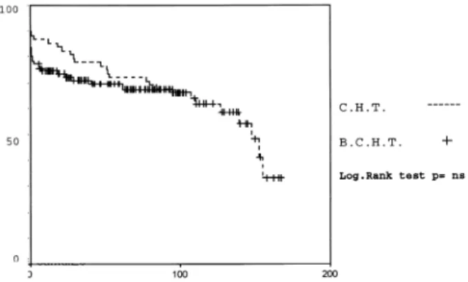
and 9 years for the classical technique and 71.01, 69.47 and 67.43% for bicaval, without difference between the two groups (P n.s.) (Fig. 1).
Among 82.5% perioperative (1 month) survivors 11 patients of group I (15.2%) had a systemic embolism 21 days to 12 years after operation. There were one mesenteric ischemia, two lower limb ischemia, and eight strokes. Three patients needed a surgical treatment: one a bowel resection and two a surgical embolectomy of the lower limb. One patient die due to a stroke, two had permanent neurological de®cit (one hemiplegia, one monoplegia) and another had a cerebral hemorrhage due to the anticoagulant treatment given after a stroke. Left atrial thrombosis was shown at TEE in 81.8% of these patients (9/11) The atrial thrombi were located in four cases in the left atrial appendage, in two cases on the atrio-atrial suture line and in the others three in the recipient left atrial wall, close to the suture line. None of the patient who underwent a BCHT had arterial embolism or atrial thrombosis at TEE (P 0:013) (Fig. 2).
The vascular echo-doppler showed that 28 patients had vascular lesions of the supraaortics trunks before the opera-tion. Among these, 26 had diffuse atherosclerosis lesions without evidence of stenosis and did not need any medical
or surgical treatment. Two patients had a stenotic lesion of the carotid artery one of them needed a percutaneus dilata-tion of the lesion with stenting. The other presented a mild carotid stenosis and an antiplatelet treatment was admini-strated. Eight patients who had diffuse atherosclerosis lesions of supraortics trunks underwent a classical cardiac transplantation and 3 of them had an episode of systemic embolism (istroke two limb ischemia).
Among the three patients who had a stroke before the transplantation, two underwent a cardiac transplantation with the modi®ed technique and the other with the classical technique. None of these patients presented any evidence of systemic embolism along their follow-up. None of the patients transplanted (group I and group II) presented a right sided (right atrium, ventricle or pulmonary artery) thrombosis or embolism.
Patient's characteristics are detailed in Table 2. All trans-planted patients were in long term sinus rhythm, the post operative surface EKG showed no major difference between the two groups except a double atrial activity in patients who had a CHT (14 versus 0) P 0:003.
The occurrence of acute rejection episodes was no signif-icantly different in patients who had a peripheral embolism than in patients free from embolism (1.8 ^ 2 versus 2.1 ^ 3) P 0:1. Chronic rejection de®ned by coronary vascular lesions at yearly coronary angiograms were presents in 19 patients. None of these patients bad any evidence of systemic embolism.
Sixty matched patients (30 of each group) had a compara-tive transthoracic and transeophageal echocardiography study, at least 6 months after surgery, which showed a major incidence of mild mitral incompetence in group, I 66 versus 33% in group II, (P 0:03), a major incidence of left atria thrombosis, 12.5% (9/72) group I versus 0 in group II, (P 0:02). Moreover a larger surface of the left atria in CHT than in BCHT (33 ^ 4 cm2versus 20 ^ 3 cm2) P 0:01, and an increased incidence of spontaneous echo-contrast in group I than in group II (56 ^ 0.5%) P 0:001 were founded. Nine patients had a concomitant lung harvesting in donors, four of group I and ®ve of group II. The measurement of the left atrium surface reveled also in these cases signi®cantly larger atria in patients operated with CHT 29 ^ 5 cm2 versus 21 ^ 21 cm2 in the BCHT (P 0:03).
The univariate and multivariate analysis of risk factors: age peripheral vascular status, the absence of antiplatelet treatment, the impaired left diastolic function, (de®ned as an end diastolic left ventricular pressure greater than 17 mmHg at cardiac catheterization), the decreased left systolic function, (de®ned as an ejection fraction inferior to 50% at echocardiography and or at cardiac angiocardiogram), atrial ®brillation or sinus rhythm before surgery, and surgical technique, recognized the surgical technique as the only factor favoring peripheral embolism. In contrast the altered diastolic function and the absence of antiplatelet treatment were risk factor for death (Table 3).
Fig. 1. Actuarial survival curve after CHT and BCHT Kaplan±Meier method.
Fig. 2. Univariate analysis (Kaplan±Meier). Actuarial risk of systemic embolism after cardiac transplantation with CHT and BCHT.
4. Discussion
Systemic embolism is a serious complication after classi-cal cardiac transplantation The incidence of left atrial thrombi in patients who underwent a CHT is unknown since the majority of these patients are asymptomatic [3,4] and intracardiac thrombi are only detected by TEE or thor-acic CT scan [1±3,6±8].
The physiopathology of intracardiac thrombi formation in patients undergoing CHT is not completely understood, although several factors may be involved:
² Alteration in hemostatic mechanism has been shown to exist. An increase in platelet aggregation has been observed which is due to the inhibited endothelial pros-tacylcine synthesis mediated by cyclosporine. This abnormality in platelet aggregation is resistant to conven-tional antiplatelet treatment [9]. In our series of patients, there was no difference in the incidence of atrial throm-bosis and peripheral embolism between patients with or without antiplatelet treatment. On the other hand rejec-tion deplete the endothelial thrombomodulin and the antithrombin III which results in an increased thrombo-genic incidence [10]. In our experience there were no
difference in rejection incidence between the group of patients having peripheral embolism and the free of systemic embolism group.
² The resulting enlarged left atrium after CHT favors blood stasis and thrombi formation. The presence of sponta-neous echocontrast has been observed in a high percen-tage of patients undergoing a CHT [1,2,5±8,11,12]. ² In our series all patients who had an episode of peripheral
embolism had underwent a classical cardiac transplanta-tion, and in this group of patients an enlarged left atrium and an increased incidence of spontaneous echocontast (SEC) were observed by TEE when compared to BCHT group. Others technical factors were found to predispose a patient to forming thrombi, because the TEE showed thrombotic formation along the atrial suture line [2,11]. This was also reported after lung transplantation on the cuff of left atrium [12,13]. In all patients undergoing a CHT the suture line was visualized and in two of then an intracardiac thrombus was observed at TEE. None of patients who underwent a BCHT had an apparent suture line at TEE. This is probably due to the everting suture technique used in the anastomosis between the recipients pulmonary veins and the donor left atrium. The impaired diastolic and systolic function may be linked to the formation of intracardiac thrombi [3,7,14]. In our series two patients among the 11 having a systemic embolism had an impaired diastolic function. The systolic function was normal in all patients of this group.
² The asynchronous contraction of the recipient and donor atrial components as well as the presence of arrhythmia induce blood stasis and may result in an increasing thrombogenic propensity [3,7,11,15,16]. Three patients who had a stroke in our series presented a double atrial activity and this was observed in 46.6% of patients undergoing a CHT. Atrial ®brillation in the recipient atrial component has been reported as favoring atrial thrombosis [1,15]. None of the patients having an atrial thrombosis and/or a systemic embolism had a ®brillation of the recipient component of the atria in our experience.
A. Riberi et al. / European Journal of Cardio-thoracic Surgery 19 (2001) 307±312 310
Table 2
Patients having a systemic embolism, after cardiac transplantation (n 11)
Patients No. Age (years) Clinical signs Time to embolism Surgical techniquea Evolution
1 30 Limb ischemia 21 days CHT Surgical embolectomy
2 48.5 Stroke 28 days CHT No sequel
3 51.8 Stroke 3 months CHT Permanent hemiplegia
4 38.9 Mesenteric ischemia 3 years CHT Surgical bowel resect ion
5 60.08 Stroke 5 years CHT No sequel
6 41.5 Stroke 6 years CHT Cerebral hemorrageb
7 42.0 Stroke 7 years CHT Monoplegia
8 55.8 Stroke 8 years CHT Death
9 69.3 Stroke 9 years CHT No sequel
10 54.2 Limb ischemia 10 years CHT Surgical embolectomy
11 56 Stroke 12 years CHT No sequel
a None of patients undergoing BCHT had a systemic embolism. bCerebral haematoma due to the anticoagulant treatment.
Table 3
Multivariate analysis (Cox test)
Variant Free of embolism Embolism Pa
Age (years) 46.3 ^ 17 48.4 ^ 11 1
Carotid disease 25 3 0.9
No anti platelets treatment 35 2 0.3 Impaired left diastolic function 32 2 1 Decreased left systolic function 12 0 1 Atrial ®brillation before surgery 5 0 1 Surgical technique
CHT 61 11 0.04
BCHT 106 0
Moreover we don't have observed arrhythmia among our cohort of patients.
² The peripheral vascular satus of patients undergoing heart transplantation may also predispose to arterial thrombosis or embolism. In our cohort of patients eight among the CHT group presented a diffuse atherosclerosis of the supraaortics trunks, three of them had an episode of arter-ial embolism and demonstration of a left atrarter-ial thrombi, which suggest that the arterial embolism was probably due to the left atrial thrombus. In the CHT group only two patients among eleven presenting an arterial embolism were free of left atrial thrombosis at TEE study and did not presented any evidence of peripheral vascular disease at the echodoppler study. Another possible important factor in thrombo-embolic events is the duration of the follow-up period. Incidence of thrombo-embolic compli-cations increases with time [17]. In our series 54.5% (6/11) of episodes of arterial embolism occurred in the early or mid term follow up and 45.5% (5/11) in the late follow-up period, and this represents a limitation in our study since the two compared series are consecutive and the mean follow-up for the bicaval group is shorter than in classical technique group 5.90 years versus 8.70 years, respectively.
5. Conclusion
Atrial thrombosis and systemic embolism after heart transplantation are probably underestimated, and more studies are necessary to shed light on its actual incidence. All the conditions favoring blood stasis and left atrial throm-bosis are found after CHT, in contrast BCHT seems to decrease considerably this problem, without increasing the perioperative mortality and morbidity and this only fact justi®es the use of this technique. Our experience of left atrial thrombosis, and frequent left atrial echocontrast rises the question of the anticoagulation in patients who had undergone a CHT.
References
[1] Derumeaux G, Habib G, Mouton Schleifer D. Standard orthotopic heart transplantation versus total orthotopic heart transplantation. A transesophageal echocardiography study of the incidence of left atrial thrombosis. Circulation 1995;9:196±201.
[2] Derumeaux G, Mouton Schleifer D, Redonnet M. FreÂquence des thrombus intra-auriculaires deÂtecteÂs par l'eÂchocardiographie transoe-sophagienne chez les transplanteÂs cardiaques. Arch Mal Coeur 1994;87:1459±1465.
[3] Fernandez-Gonzalez A, Llorens R, Herreros J. Intracardiac thrombi after orthotopic heart transplantation: clinical signi®cance and etiolo-gic factors. J Heart Lung Transplant 1994;13:236±240.
[4] Rahman A, Deiraniya A, Campbell C. Thrombotic complications after heart transplantation. J Heart Lung Transplant 1994;13:1146± 1147.
[5] Bouchart F, Derumeaux G, Mouton-Schleifer D. Conventional and
total orthotopic cardiac transplantation: a comparative clinical and echocardiography study. Eur J Cardio-thorac Surg 1997;12:555±559. [6] Metras D, Kreitmann B, Riberi A. Etude comparative de la transplan-tation cardiaque orthotopique standard et totale. Arc Mal Coeur 1997;90:27±34.
[7] Polanco G, Jafri SM, Alam M. Transesophageal echocardiographic ®ndings in patients with orthotopic heart transplantation. Chest 1992;101:599±602.
[8] Ishizuka N, Nakaura K, Fujita Y. Transesophageal echocardiography ®ndings in patients after heart transplantation. J Cardiol 1997;29:163± 170.
[9] De Lorgeril M, Dureau G, Boissonnat P. Increased platelet aggrega-tion after heart transplantaaggrega-tion: in¯uence of Aspirin. J Heart Lung Transplant 1991;10:600±603.
[10] Labarrere C, Pitts D, Halbrook H. Natural anticoagulant pathway in normal and transplanted human hearts. J Heart Lung Transplant 1992;11:342±347.
[11] Angermann C, Spes C, Tammen A. Anatomic characteristics and valvular function of the transplanted heart: transthoracic versus trans-esophageal echocardiographic ®ndings. J Heart Transplant 1990;9:331±338.
[12] Schmid C, Gulba DC, Heublein B. Systemic recombinant tissue plas-minogen activator lysis for left atrial thrombus formation after single lung retransplantation. Ann Thorac Surg 1992;53:338±340. [13] Stang MR, Hinderliter AL, Gott KK. Atrial anastomotic thrombus
causes neurologic de®cits in a lung transplant recipient. Transplanta-tion 1996;62:693±695.
[14] Clark VL, Levine TB. Thrombus causing fatal left ventricular out¯ow tract obstruction in a heart transplant patient. Cathet Cardiovasc Diagn 1992;25:132±134.
[15] Torrecilla EG, Garcia-Fernandez MA, San Roman D. Left atrial spon-taneous echocardiographic contrast after heart transplantation. Am J Cardiol 1992;69:817±818.
[16] Adair JC, Call GK, O'Connell JB. Cerebrovascular syndromes following cardiac transplantation. Neurology 1992;42:819±823. [17] Forrat R, Ferrera R, Boissonnat P, Adeleine P, Dureau G, Ninet J,
Lorgeril M. High prevalence of thromboembolic complications in heart transplantation. Transplantation 1996(61):757±762.
Appendix A. Conference discussion
Dr P. Totaro (Brescia, Italy): I did a similar study when I was working in Northern General Hospital in Shef®eld. We found a similar incidence of atrial thrombosis in patients without any sign of peripheral embolism. We treated them with oral anticoagulation and they recovered very well and their thrombosis disappeared.
So my question is, does your incidence regard overall population or only the patients with clinical signs of peripheral embolism?
And the second one is, how did you treat the patient with the left atrial thrombosis?
Dr Riberi: For all patients with left arterial thrombosis with an antic-oagulant treatment by an oral ± this is your question?
Dr Totaro: Yes. Did you give just oral anticoagulant, or did you give intravenous heparin for some time?
Dr Riberi: In the beginning we induced the treatment with heparin in vein and then we go on oral anticoagulant treatment with it.
I'm sorry, I didn't understand the ®rst question.
Dr Totaro: Does your incidence of left atrial thrombosis regard all the patients with a heart transplant or only the patients with clinical signs of peripheral embolism?
Dr Riberi: Forty percent of patients with classical heart transplantation in our population have a left atrial thrombosis.
Dr Totaro: I mean we found 14% of incidence of left atrial thrombosis just as a routine surveillance of echocardiographic in asymptomatic patients.
Dr Totaro: Is yours the same? Dr Riberi: Yes.
Dr C. Yankah (Berlin, Germany): You may have a peripheral thrombosis or thromboembolic episodes without having atrial thrombosis. These patients sometimes have preoperatively heparin therapy. And we have some experience in some certain patients who developed heparin-induced thrombocytopenia and also caused some peripheral thromboembolitic episodes, even as well as also the cerebral. Have you observed or diagnosed heparin-induced thrombocytopenia (HIT type II). Perhaps these two patients were HIT patients.
Dr Riberi: No, we have never found these kinds of complications with heparin.
Dr A. Haverich (Hannover, Germany): The bicaval technique in and by
itself does not in¯uence thromboembolism from the left atrium. Therefore the title of the presentation is somewhat misleading.
On the other hand, if you also change the technique of suturing the left atrium, making it much smaller, that has de®nitely an in¯uence. Our cardi-ologists always tell us to occlude the left atrial appendage on the donor heart. Did you change that as well when changing your technique between the ®rst and the second group of your study?
Dr Riberi: We sutured the left appendage ± this is your question? Dr Haverich: Yes. And did you occlude the left atrial appendage? Dr Riberi: Yes, always.
Dr Haverich: With both techniques?
Dr Riberi: No. We began with the bicaval techniques to do it. A. Riberi et al. / European Journal of Cardio-thoracic Surgery 19 (2001) 307±312