Missense mutations in COL8A2, the gene encoding the α2 chain of type VIII collagen, cause two forms of corneal endothelial dystrophy
10
0
0
Texte intégral
(2) 2416 Human Molecular Genetics, 2001, Vol. 10, No. 21. FECD is the commonest primary disorder of the corneal endothelium. Epidemiological data regarding its incidence or prevalence are unavailable. An indirect measure of the impact of the disease comes from surveys of clinical indications for corneal transplantation which rank FECD as one of the commonest indications for corneal transplantation (up to 19%) performed in developed countries (6–10). The high prevalence of this dystrophy co-existing with cataract in an older age group means that it is a risk factor for the requirement for penetrating keratoplasty (PK) subsequent to cataract surgery. Symptoms of painful visual loss result from corneal decompensation. Signs may be present from the fourth decade of life onwards with the development in the central cornea of focal wart-like guttata arising from DM, which is thickened by abnormal collagenous deposition. There is reduced endothelial function and cell density as well as cellular pleomorphism (11). FECD is usually a sporadic condition but familial highly penetrant forms showing autosomal dominant inheritance are also recognized (12–14). PPCD is a rare bilateral corneal endothelial dystrophy that is inherited in an autosomal dominant manner. The clinical features usually present earlier than FECD, being from birth onwards. The condition is characterized by formation of blister-like lesions within the corneal endothelium or by regions of endothelial basement membrane thickening with associated corneal oedema. There is replacement of the normal amitotic endothelial cells by epithelial-like cells (15) that possess abundant intermediate filaments, desmosomes and microvilli (16). The endothelium becomes multilayered and the abnormally proliferating cells may extend outwards from the cornea over the trabecular meshwork to cause glaucoma. In this regard, PPCD resembles iridocorneal endothelial (ICE) syndrome, a unilateral condition which is also associated with abnormal endothelial proliferation (17). Molecular data on the endothelial dystrophies are limited: a single family with PPCD was linked to a 30 cM region of chromosome 20q. Dominant CHED (CHED1) was then mapped to a 2.7 cM region within this interval, suggesting either that there is a cluster of genes in this region or that there is allelic heterogeneity (18,19). In addition an autosomal recessive form of CHED (CHED2) maps to 20p (20). In a multigenerational family with an early-onset form of FECD we demonstrated linkage to a 6–7 cM region of 1p34.3–p32 containing the COL8A2 gene which encodes the α2 chain of type VIII collagen. Through analysis of the coding sequence of COL8A2 we defined mutations in both familial and sporadic FECD as well as in a family with PPCD. This is the first description of the molecular basis of any of the corneal endothelial dystrophies or of mutations in type VIII collagen in association with human disease. RESULTS Clinical details Members of a three-generation family (FECDPed1, Fig. 1) from the north-east of England, with early-onset FECD (i.e. third and fourth decades), were independently examined by three ophthalmologists (S.Biswas, G.C.M.Black and B.Noble). There was clear evidence of male to male transmission and an absence of affected offspring from unaffected. individuals. Amongst the affected individuals from family FECDPed1, diagnosis was made between the ages of 21 and 48 years. Ungrafted individuals had visual acuities between 6/4 and 6/60. Individuals in their fourth to fifth decades had the most advanced grade of disease with coalescence of areas of epithelial oedema forming sub-epithelial bullae. The youngest affected individual examined was pre-symptomatic in his third decade with 1–2 mm of grouped central corneal guttata. Five out of 12 individuals from the family had undergone corneal transplantation. The histopathological findings confirmed a diagnosis of FECD. Two individuals from pedigree FECDPed2 (Fig. 2) were examined and found to have clinical evidence of FECD. The proband had undergone bilateral corneal transplantation by his sixth decade with histopathological evidence of FECD. The daughter, whose symptoms began in her third decade, had undergone a unilateral corneal transplant. The other eye had advanced changes consistent with FECD with confluent central guttata and sub-epithelial bullae. DNA was available on six other affected family members all of who had been diagnosed between the third and fifth decades. Two individuals from family PPCDPed1 were diagnosed with PPCD. Both individuals had already undergone bilateral PK for corneal decompensation secondary to endothelial dystrophy. This was performed on the proband in her third decade and on her father in his sixth decade. No other family members were available for examination. Ultrastructural analysis Ultrastructural analysis in affected individuals from family FECDPed1 demonstrated characteristic secretion of an abnormal posterior collagenous layer (PCL) between the endothelium and a thickened DM (Fig. 3). The PCL was organized into a hexagonal matrix resembling that normally formed by type VIII collagen (Fig. 3). In both affected individuals from family PPCDPed1, ultrastructural analysis of corneal material removed at PK revealed endothelial cell multilayering with desmosomal intercellular attachments and surface cells with short microvilli consistent with epithelial-like metaplasia of the endothelial cells. This confirmed a diagnosis of PPCD in this family. Linkage analysis and sequencing in family FECDPed1 Fifteen members of family FECDPed1, covering three generations, in which disease status was confidently assigned, were selected for linkage analysis. The only unaffected individuals included in the analysis were in their fifth decade. Exclusion analysis was performed using microsatellite markers D20S48 and D20S417 coupled with the PPCD and CHED 1 loci (19). Using two-point linkage analysis no significant linkage was obtained with either marker (LOD score less than –7). Thereafter, a genome-wide search was undertaken using 382 markers covering all 22 autosomes. High LOD scores were obtained for markers D1S2830 and D17S938 [Zmax = 3.72, recombinant fraction (θ) = 0.0, and Zmax = 3.78, θ = 0.0 respectively]. Further genotyping, using microsatellite markers close to D17S938, revealed no shared haplotype amongst all the affected members to within 50 kb of marker D17S938, implying that this marker was linked by chance alone..
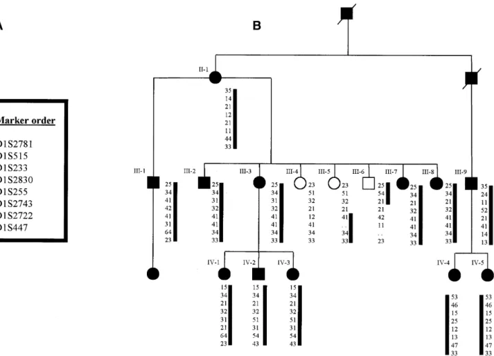
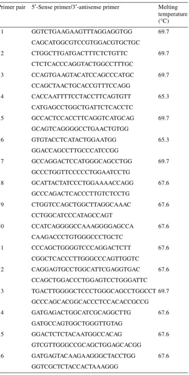
(3) Human Molecular Genetics, 2001, Vol. 10, No. 21 2417. Figure 1. Pedigree of family FECDPed1 with autosomal dominant FECD showing haplotype from chromosome 1p34.3–p32. Multiple informative recombination events in individuals III-5 and III-6 delineate the critical region.. Having demonstrated significant linkage to a region of chromosome 1, refinement of the critical region was undertaken using further microsatellite markers (Fig. 1). Haplotype analysis of the pedigree shows unaffected individual III-5 to be recombinant distal to marker D1S255 and a recombination is present in unaffected individual III-6 proximal to marker D1S233. These two individuals are aged 45 (III-5) and 53 years (III-6) and have entirely normal corneal examination. In addition they each have three children aged between 23 and 34 years, all of whom have normal corneal examinations; the majority of affected individuals within this family had abnormal corneal endothelia by age 20–25 years. These recombinants define a 6–7 cM critical interval that locates a putative disease gene to chromosome 1p34.3–p32. The COL8A2 gene, which encodes the α2 chain of type VIII collagen, lies within this region and represented a strong candidate gene in this family. The gene is fully encompassed within the genomic clone RP4-665N4 which has been fully sequenced (GenBank accession no. AL138787). The 2112 bp coding sequence is contained within two exons separated by an intron of 562 bp (Fig. 4A and B). These were sequenced in all family members revealing a C→A transversion at position 1364 from the beginning of the coding sequence (Fig. 5A and B). This results in a codon 455 glutamine to lysine substitution. The. mutation was present in all 13 affected individuals tested and no unaffected members of the family and was not present in 488 ethnically matched control chromosomes. COL8A2 mutation analysis in patients with FECD and PPCD Sixteen overlapping primer pairs spanning the coding region of COL8A2 were designed to amplify genomic DNA from a panel of affected individuals including 115 unrelated patients with FECD and 15 with PPCD (Table 1). Eight further probands carried missense changes that were not identified within ethnically matched normal control populations and are presumed to be pathogenic alterations (Table 2). The gln455lys mutation was detected in all affected individuals in three further families with endothelial dystrophies. Two (FECDPed2 and FECDPed3) had early-onset FECD. FECDPed2 is a large Australian kindred in whom the mutation was present in 8/8 affected individuals separated by 21 meioses (Fig. 2). The third family (PPCDPed1) consists of two affected individuals in whom ultrastructural analysis demonstrates the pathological endothelial changes of PPCD. An Arg155Gln mutation was demonstrated by SSCP in a family of five affected individuals from which DNA was.
(4) 2418 Human Molecular Genetics, 2001, Vol. 10, No. 21. Figure 2. Pedigree of early-onset FECD family FECDPed2. Asterisks denote individuals, from whom DNA had been obtained, who carry C1364A transversion.. Figure 3. (A) Transmission electron micrograph showing posterior corneal stroma, Descemet’s membrane and endothelium of an affected individual from FECDPed1. Descemet’s membrane is thickened, with a normal ABZ and a pathological posterior collagenous layer which contains a banded layer anteriorly (PCLB) and a disorganized fibrillar collagenous (PCLF) layer posteriorly. There are no guttata in this field. Endothelial cells (En) are vacuolated. (Scale bar = 2.4 µ m.) (B) Confocal microscopy image of corneal endothelium from a patient from family FECDPed1. Dark areas demarcate guttata (arrows). There is increased polymegathism and polymorphism of the endothelial cells typical of FECD. (Scale bar = 15 µm.) (C) Higher magnification of patient shown in Figure 1A showing PCL with a disorganized arrangement of wide-spaced collagen, which has an internode distance of ∼110 nm. (Scale bar = 750 nm.) (D) En face view of wide-spaced collagen fibrils from patient shown in Figure 1A showing the hexagonal, ‘six-spoked’ assembly characteristic of type VIII collagen. (Scale bar = 200 nm.). available on 3/5. All three carried the mutation, which was not present in 184 control chromosomes. This change was also found in a further two unrelated patients with sporadic FECD. Two changes, Arg434His and Arg304Gln, were identified by sequencing in sporadic patients as SSCP of the fragments,. which are highly GC rich, was unsatisfactory; neither was present in 150 control chromosomes. To define whether these substituted residues are conserved we sequenced the murine cDNA and compared this with the human and bovine sequences. Overall we confirmed that there.
(5) Human Molecular Genetics, 2001, Vol. 10, No. 21 2419. Table 1. Primer pairs for COL8A2 mutation screening Primer pair. 1. 5′-Sense primer/3′-antisense primer. GGTCTGAAGAAGTTTAGGAGGTGG. Table 2. COL8A2 coding sequence variants Melting temperature (°C). Presumed pathogenic variants. 69.7. CAGCATGGCGTCCGTGGACGTGCTGC 2. CTGGCTTGATGACTTTCTCTGTTC. 69.7. Sequence change/position. Individuals identified. Arg155Gln cgg→cag. FECD, sporadic (2) FECD, familial (1). Arg304Gln cgg→cag. FECD, sporadic (1). Arg434His cgc→cac. FECD, sporadic (1). Gln455Lys cag→aag. FECD, familial (1) PPCD, familial (1). CTCTCACCCAGGTACTGGCCTTTGC 3. CCAGTGAAGTACATCCAGCCCATGC. 69.7. Silent. CCAGCTAACTGCACCGTTTCCAGG 4. CACCAATTTTCCTACCTTCAGTGTT. Non-pathogenic variants. 65.3. CATGAGCCTGGCTGATTCTCACCTC 5. GCCACTCCACCTTCAGGTCATGCAG. 69.7. GCAGTCAGGGGCCTGAACTGTGG 6. GTGTACCTCATACTGGAATGG. 65.3. Missense. Ala35Ala gcg→gca. Normal controls. Gly495Gly ggg→gga. Normal controls. Pro486Pro ccg→cca. Normal controls + affected individuals. Pro586Pro ccc→cct. Normal controls + affected individuals. Gly3Arg ccc→acc. Normal controls + affected individuals. Gly357Arg ggg→ggc. FECD, familial (1)a. Pro575Leu ccg→ctg. FECD, familial (1)a. Thr645Ile acc→atc. Normal controls. GGACCAGCCTTGCCCATCCGG 7. GCCAGGACTCCATGGGCAGCCTGG. 69.7. GCCCTGGTTCCCCCTGGAATCCTG 8. GCATTACTATCCCTGGAAAACCAGG. 67.6. GCCCAGACTCACCCTTGTCTCCTG 9. CTGGTCCAGCTGGCTTAGGCAAAC. 67.6. aNot found in normal controls but did not cosegregate with disease in all affected family members.. CCTGGCATCCCATAGCCAGT 10. CCATCAGGGGCCAAAGGGGAGCCA. 67.6. CAAGACCCTGTGGGCCCTGCTC 11. CCCAGCTGGGGTCCCAGGACTCTT. 67.6. CGGCTCACCCTTGGGCCCAGTTGGTC 12. CAGGAGTGCCTGGCATTCGAGGTGAC. 67.6. CCAGCTGGACCCTGGAGTCCTGGGATTC 13. TGACTTGGGGCTCCCTGGGCAGCCTGGCCT 69.7 GCCCAGCACGGCACCCTCCACACCGCCG. 14. GATGAGACTGGCATCGCAGGCTTG. 67.6. GATGCCAGTGGCTGGGTTGTAG 15. GGACTCTCTACAATGGCCACAG. 67.6. GTCGTTGGGCCGCAGCTGGAGCACGG 16. GATGAGTACAAGAAGGGCTACCTGG. 67.6. GGTCGCTCTACCACTAAAGGG. is >90% sequence identity in mouse, human and bovine triple helical domains (data not shown) (21). All of these mutated residues lie within the triple helical domain and are conserved in all three species. In addition, a number of other sequence changes were detected amongst both affected individuals and normal controls (Table 2). Of these eight changes, four were silent and four missense. Of the latter, the two within the triple helical domain (Gly357Arg, Pro575Leu) were only detected amongst familial FECD patients and were not found amongst the unaffected controls tested (488 and 186 chromosomes, respectively). However, these cannot be presumed to be pathogenic. as they were not found in all affected individuals within these families and therefore did not fully co-segregate with the disease phenotype. Haplotype analysis of families carrying Gln455Lys mutation A close haplotype was constructed using five markers around COL8A2 for the four Gln455Lys families (Fig. 4B). Two FECD families (FECDPed1 and FECDPed3) and the PPCD family originate from Northern England and share a common haplotype across the region, suggesting the presence of a common founder mutation within these three families. In contrast, the Australian family FECDPed2 did not share this haplotype. This analysis included two separate SNPs within the intron interrupting the coding sequence (Table 3) which lie 1–1.5 kb from the putative mutation; this strongly suggests that FECDPed2 carries an identical but independent mutation. DISCUSSION A positional candidate strategy was employed to identify the defective gene in a family (FECDPed1) with a highly penetrant, early-onset form of FECD. Genetic mapping in this family revealed linkage of FECD to a 6–7 cM interval of 1p32.3–p34.3. In family FECDPed1 symptoms commenced during the third and fourth decades, with clinical signs of disease detectable earlier, overcoming the difficulties of analysing lateonset diseases such as FECD, where families of more than two generations are scarce and assignment of disease status in younger individuals is uncertain..
(6) 2420 Human Molecular Genetics, 2001, Vol. 10, No. 21. Table 3. Haplotype analysis of families carrying gln455lys (C1364A) mutation Marker. Calculated distance from COL8A2 (kb) FECDPed1 (UK). FECDPed2 (Australian). FECDPed3 (UK). PPCDPed4 (UK). AL139286 (GT)n. 140. 2. 3. 2. 2. COL8A2 SNP1. Within intron. 1. 2. 1. 1. COL8A2 SNP2. Within intron. 2. 1. 2. 2. AL138901 (CTTT)n. 100. 3. 4. 3. 3. D1S2729. 500–800. 5. 4. 5. 5. The COL8A2 gene had been localized to this segment of 1p by in situ hybridization (22) and was confirmed to lie within the critical region. The gene encodes the α2 (VIII) collagen chain, which is highly expressed in human corneal DM (23) and in addition is abnormally deposited in the corneas in both FECD and PPCD. On these grounds, the gene represented a biologically highly attractive candidate gene for FECD. Sequencing of the gene in members of this family (FECDPed1) revealed that affected members had a single nucleotide substitution at position 1364 of the major coding sequence of COL8A2. This results in a gln455lys missense mutation that alters the amino acid at position X of the Gly–X–Y repeat within the triple helical domain of α2 (VIII) collagen. Mutations in the X position within the Gly–X–Y repeat have also been described amongst mutations of COL2A1 that cause Stickler syndrome (24). In the case of the Gln455Lys substitution the change is from the non-charged, evolutionarily conserved glutamine for a negatively charged lysine close to the C-terminus. Lysine has a potential role in the formation of intermolecular bonds and, in turn, may alter the tertiary structure of the protein. We speculate that this disrupts the stability of supramolecular assembly (25). The Gln455Lys mutation was demonstrated in a further two multigenerational families with an inherited early-onset form of FECD as well as in a family PPCDPed1. Identical mutations associated with different phenotypes could represent the influence of genetic modifiers; other examples amongst ocular disorders include cerulean and Coppock cataracts (CRYBB2), Stargardt disease and dominant macular atrophy (ELOVL4) and Rieger and Peters’ anomaly (PITX2) (26–28). A close haplotype constructed around COL8A2 for the four families suggests that while the mutation is identical it has arisen independently in at least two of the families. It remains possible that this does represent a single mutational event and, if so, reduces the maximum size of the common inherited fragment to <100 kb, suggesting an old ancestral mutation. In addition, three further missense substitutions (Arg155Gln, Arg434His and Arg304Gln) within the triple helical domain were defined both in familial and sporadic forms of classical FECD. Overall, we found COL8A2 mutations in 9/116 (8%) of FECD probands. This suggests the presence of genetic heterogeneity in this condition, which was confirmed by linkage exclusion of the COL8A2 region in other multigenerational families with FECD. Type VIII collagen is a member of the short-chain collagenlike family of proteins that also includes type X collagen with which it shares many structural similarities (29). It comprises two α-chains, α1 (VIII) and α2 (VIII), that exist in vivo as hetero- or homotrimers (30–32). Type VIII collagen is a major. Figure 4. (A) Translated coding sequence of the human COL8A2 gene. (B; opposite.) Top, genomic region around COL8A2 gene showing positions of markers used for haplotype analysis of families carrying gln455lys mutation: positions of sequenced BACs are indicated. Bottom, genomic structure of COL8A2 including positions of NC1, NC2 and triple helical domains. Positions of presumed pathogenic mutations in the triple helical domain are marked..
(7) Human Molecular Genetics, 2001, Vol. 10, No. 21 2421. component of the hexagonal lattice of DM which normally forms a highly ordered array in the anterior banded zone (ABZ) of this tissue (22,33,34). The role of type VIII collagen in DM is uncertain. It may provide a structural function, with the hexagonal matrix acting to resist compression. However, its function in association with vascular endothelium indicates a role in determining cell phenotype while developmental studies suggest that type VIII collagen is important in cell differentiation: it is possible that it plays a similar role in DM (34). Therefore, our findings are in support of the hypothesis that both FECD and PPCD are disorders of endothelial cell terminal differentiation since it is these cells which secrete type VIII collagen. Furthermore, they suggest that changes in the interaction between the endothelium and DM can be the primary aetiological event and implicate the α2 (VIII) chain of type VIII collagen in this interaction. By inference, the COL8A1 gene, which encodes the α1 (VIII) chain, is therefore an attractive candidate gene not only as a causative gene for FECD but also as a modifier locus amongst patients with COL8A2 mutations. In FECD, type VIII collagen has been identified by immunolabelling techniques to comprise a large part of the abnormally secreted posterior collagenous layer of DM, where it forms the endothelial guttata characteristic of this disorder (5). A similar abnormally banded PCL is observed in some cases of PPCD (4). Ultrastructural analysis of the posterior region of DM from an affected individual from family FECDPed1 demonstrates the presence of this collagen in a thickened PCL, between the endothelium and a thickened DM (Fig. 2). The secretion of an abnormal PCL containing type VIII collagen has also been demonstrated in aphakic bullous keratopathy and the ICE. syndromes (3,15,35) suggesting that alteration of its expression and deposition may be relevant to the pathological responses of the endothelium to ageing and trauma. Moreover, as type VIII collagen is a component of other endothelial basement membranes, including those of vascular endothelia, understanding the interaction of the corneal endothelium with both DM and type VIII collagen may provide a broader insight into endothelial cell maintenance and ageing. MATERIALS AND METHODS Clinical examination Members of a three-generation family (FECDPed1, Fig. 1) from the north-east of England, with early-onset FECD (i.e. third and fourth decades), were independently examined by three ophthalmologists (S.Biswas, G.C.M.Black and B.Noble). Individuals were considered affected if they had typical signs and symptoms of FECD or had already undergone PK with histopathological confirmation of the diagnosis. Individuals <20 years of age were excluded, as none of those examined had signs of disease, and accurate definition of disease status in asymptomatic carriers could not be guaranteed. None of the individuals examined had a significant past history of ocular disease, inflammation or trauma to account for the appearance of central corneal guttata. All examining ophthalmologists were masked to genotype data at the time of examination. A further 115 unrelated familial and simplex cases of FECD and 15 probands with PPCD were identified either clinically or from histopathological evidence supporting a diagnosis of corneal endothelial dystrophy. After informed consent, DNA.
(8) 2422 Human Molecular Genetics, 2001, Vol. 10, No. 21. Figure 5. (A) HindIII restriction digest designed to detect the C1364A transversion. Lanes I and II, affected individuals from FECDPed1; lanes III–VII, controls. The presence of the transversion creates a restriction site resulting in two bands. (B) Direct sequencing of the major coding exon of COL8A2: (i) the heterozygous C1364A transversion in the antisense strand (arrow) in an affected individual from FECDPed1; (ii) the corresponding antisense strand sequence in an unaffected individual from FECDPed1.. was extracted from these individuals and families to compile panels for mutation screening. Ultrastructural analysis Tissue was dissected (segments <1 mm3), immersed in 2.5% glutaraldehyde in 0.1 M sodium cacodylate buffer and postfixed in 1% osmium tetroxide in 0.05 M sodium cacodylate buffer for 1 h. The tissue was dehydrated through a graded ethanol series and embedded araldite resin. Resin-embedded blocks were cut into semi-thin (1 µm) and thin (65 nm) sections. Semi-thin sections were stained with toluidine blue, thin sections with uranyl acetate and lead citrate. Thin sections were placed onto nickel grids and transmission electron microscopy carried out using a JEOL 1010 instrument. Linkage analysis After informed consent and ethical approval, blood samples were taken from 13 affected and four unaffected members of the family. Genomic DNA was extracted from lymphocytes (36). Amplification of markers D20S48 and D20S417 by polymerase chain reaction (PCR) was performed by using 100 ng of genomic DNA, 2 µl (0.2 µM) of forward and reverse primers, 3 µl of dH2O, 10 µl of PCR Reddymix™ master mix [0.75 mM each dNTPs, 67 mM Tris–HCl (pH 8.0 at 25°C), 16 mM (NH4)2SO 4, 3.7 mM MgCl2, 0.085 mg/ml BSA, 0.5 U Taq polymerase (ABgene; ABgene, Epsom, Surrey, UK)]. PCR amplification was performed on a dry block thermal cycler. Samples were denatured at 95°C for 5 min and the following cycling conditions were applied: 32 cycles at 94°C for 45 s, 60°C for 45 s, 72°C for 45 s. Final extension was at. 72°C for 5 min. Amplified products were phenol-extracted and loaded onto 8% polyacrylamide gel. Electrophoresis was carried out at 400 V for 3 h and products were silver-stained according to standard methods. A genome-wide search was carried out using a set of 382 fluorescent-labelled PCR primers (ABI Prism™ Linkage mapping set version 2; PE Applied Biosystems, Perkin-Elmer Corp., Foster City, CA) to amplify microsatellite loci, with ∼10 cM spacing, across the genome. A 15 µl PCR reaction mix comprised 100 ng of patients’ genomic DNA, 1 µl of primer mix, 1.5 µl of 10× GeneAmp PCR buffer II, 1.5 µl of GeneAmp dNTP mix (2.5 mM), 0.12 µl of AmpliTaq Gold™ DNA polymerase (5 U/µl), 1.5 µl of MgCl2 (25 mM) and 3.8 µl of dH 2O. DNA from CEPH individual 1347-02 acted as positive control; dH2O acted as negative control. The PCR conditions for amplification were as per the manufacturer’s instruction for the GeneAmp PCR system 9600 thermal cycler (PE Applied Biosystems). Two microlitres of pooled PCR product was mixed with 2 µl of size standard [GeneScan 400 HD (ROX); PE Applied Biosystems] and 24 µl of de-ionized formamide (Amresco Solon, Solon, OH). After denaturing at 95°C for 2 min and placing on ice, 2 µl of this mix was injected into a polymer (GeneScan STR POP4D)-containing capillary and analysed on an ABI prism 310 Genetic Analyser using GeneScan software. In addition, several other markers, ascertained from the Généthon linkage map database, were analysed using the PCR conditions described above: D1S2781, D1S513, D1S233, D1S2676, D1S2832, D1S2729, D1S496, D1S2830, D1S255, D1S2743, D1S2722 and D1S447. These markers spanned a 20 cM region around the ABI-prism marker D1S234, wherein a LOD score of 2.03 was obtained. The genotype data obtained from the family were entered into a PC using CYRILLIC 2.0 software for data storage and retrieval. These data were exported into the LINKAGE program for linkage analysis and LOD scores were calculated using MLINK (37) and using allele frequency data available from the genome database (GDB). DNA sequencing Primers were designed from the major coding region of COL8A2, whose sequence was elucidated from a bacterial artificial chromosome clone containing the gene sequence (Table 1). These were used to amplify the gene in two affected and two unaffected individuals and one control individual and later to confirm the sequence in all members of FECDPed1. Sterile water was substituted for genomic DNA as a negative control. Thirty microlitre PCR reactions were set up using 100 ng of genomic DNA, 3 µl (0.3 µM) of forward and reverse primers, 15 µl of Reddymix™ master mix and 4.5 µl of dH2O. PCR reactions were carried out on a dry block thermal cycler. Reaction mixtures were denatured at 95°C for 5 min, then 30 cycles at 94°C for 45 s, X°C for 45 s (where ‘X’ is the optimized annealing temperature for each primer pair shown in Table 1) and 72°C for 45 s. Final extension was performed at 72°C for 10 min. The amplified products were cleaned using 2 U shrimp alkaline phosphatase (1 U/µl) (USB Corporation, Cleveland, OH) and 4 U exonuclease I (10 U/µl) (USB) on a thermal cycler at 37°C for 38 min, 85°C for 20 min and 20°C for 2 min. A 10 µl sequencing reaction comprised 5 µl of cleaned PCR product, 0.7 µl of forward or reverse primer, 1 µl.
(9) Human Molecular Genetics, 2001, Vol. 10, No. 21 2423. of Thermo sequenase™ II mix A (Amersham Pharmacia Biotech Inc., Buckingham, UK), 1 µl of Thermo sequenase™ II mix B and 2.3 µl of dH2O. Sequencing amplification was performed under the following conditions: 94°C for 30 s for one cycle, 30 cycles at 95°C for 20 s, 55°C for 20 s, 60°C for 1 min. Precipitation of DNA was performed using 1 µl of sodium acetate (1.5 M)/EDTA buffer (250 mM) (Amersham Pharmacia Biotech Corp.) and absolute alcohol. DNA samples were denatured with formamide loading dye and run on an acrylamide gel. Sequencing was carried out on an ABI-377 analyser (PE Applied Biosystems) and the results analysed using GeneScan software. ACKNOWLEDGEMENTS The authors thank Heidi Haines and Kelly Kopp for their excellent technical assistance, and Gregory S.H.Ogawa for identifying one of the families who participated in this study. G.B. is a Wellcome Trust Clinician Scientist Fellow (reference 51390/Z). S.B. was supported by Manchester Royal Eye Hospital research endowments. R.P. is supported by Action Research. We would like to acknowledge support from the Swiss National Science Foundation (32-053750.98), the National Eye Institute of the United States (EY11543) and the families who donated samples to make this work possible. REFERENCES 1. Waring, G.O., Bourne, W.M., Edelhauser, H.F. and Kenyon, K.R. (1982) The corneal endothelium. Normal and pathologic structure and function. Ophthalmology, 89, 531–590. 2. Klintworth, G. (1999) Advances in the molecular genetics of corneal dystrophies. Am. J. Ophthalmol., 128, 747–754. 3. Bahn, C.F., Falls, H.F., Varley, B.S., Meyer, R.F., Edelhauser, H.F. and Bourne, W.M. (1984) Classification of corneal endothelial disorders based on neural crest origin. Ophthalmology, 91, 558–563. 4. McCartney, A.C. and Kirkness, C.M. (1988) Comparison between posterior polymorphous dystrophy and congenital hereditary endothelial dystrophy of the cornea. Eye, 2, 63–70. 5. Levy, S.G., Moss, J., Sawada, H., Dopping-Hepenstal, P.J. and McCartney, A.C. (1996) The composition of wide-spaced collagen in normal and diseased Descemet’s membrane. Curr. Eye Res., 15, 45–52. 6. Lang, G.K. and Naumann, G.O.H. (1987) The frequency of corneal dystrophies requiring keratoplasty in Europe and the USA. Cornea, 6, 209–211. 7. Mohamadi, P., McDonnell, J.M., Irvine, J.A., McDonnell, P.J., Rao, N. and Smith, R.E. (1989) Changing indications for penetrating keratoplasty, 1984–1988. Am. J. Ophthalmol., 107, 550–552. 8. Brady, S.E., Rapuano, C.J., Arentsen, J.J., Cohen, E.J. and Laibson, P.R. (1989) Clinical indications for procedures associated with penetrating keratoplasty, 1983–1988. Am. J. Ophthalmol., 108, 118–122. 9. Ramsay, A.S., Lee, W.R. and Mohammed, A. (1997) Changing indications for penetrating keratoplasty in the west of Scotland from 1970 to 1995. Eye, 11, 357–360. 10. Alldredge, O.C. and Krachmer, J.H. (1981) Clinical types of corneal transplant rejection. Their manifestations, frequency, preoperative correlates, and treatment. Arch. Ophthalmol., 99, 599–604. 11. Bigar, F. (1982) Specular microscopy of the corneal endothelium. Optical solutions and clinical results. Dev. Ophthalmol., 6, 1–94. 12. Krachmer, J.H., Purcell, J.J.,Jr, Young, C.W. and Bucher, K.D. (1978) Corneal endothelial dystrophy. A study of 64 families. Arch. Ophthalmol., 96, 2036–2039. 13. Rosenblum, P., Stark, W.J., Maumenee, I.H., Hirst, L.W. and Maumenee, A.E. (1980) Hereditary Fuchs’ dystrophy. Am. J. Ophthalmol., 90, 455–462. 14. Magovern, M., Beauchamp, G.R., McTigue, J.W., Fine, B.S. and Baumiller, R.C. (1979) Inheritance of Fuchs’ combined dystrophy. Ophthalmology, 86, 1897–1923.. 15. Levy, S.G., Moss, J., Noble, B.A. and McCartney, A.C. (1996) Early onset posterior polymorphous dystrophy. Arch. Ophthalmol., 114, 1265–1268. 16. Ross, J.R., Foulks, G.N., Sanfillipo, F.P. and Howell, D.N. (1995) Immunohistochemical analysis of the pathogenesis of posterior polymorphous dystrophy. Arch. Ophthalmol., 113, 340–345. 17. Howell, D.N., Damms, T., Burchette, J.L.,Jr and Green, W.R. (1997) Endothelial metaplasia in the iridocorneal endothelial syndrome. Invest. Ophthalmol. Vis. Sci., 38, 1896–1901. 18. Heon, E., Mathers, W.D., Alward, W.L., Weisenthal, R.W., Sunden, S.L., Fishbaugh, J.A., Taylor, C.M., Krachmer, J.H., Sheffield, V.C. and Stone, E.M. (1995) Linkage of posterior polymorphous corneal dystrophy to 20q11. Hum. Mol. Genet., 4, 485–488. 19. Toma, N.M., Ebenezer, N.D., Inglehearn, C.F., Plant, C., Ficker, L.A. and Bhattacharya, S.S. (1995) Linkage of congenital hereditary endothelial dystrophy to chromosome 20. Hum. Mol. Genet., 4, 2395–2398. 20. Hand, C.K., Harmon, D.L., Kennedy, S.M., FitzSimon, J.S., Collum, L.M. and Parfrey, N.A. (1999) Localization of the gene for autosomal recessive congenital hereditary endothelial dystrophy (CHED2) to chromosome 20 by homozygosity mapping. Genomics, 61, 1–4. 21. Mann, K., Jander, R., Korsching, E., Kuhn, K. and Rauterberg, J. (1990) The primary structure of a triple helical domain of collagen type VIII from bovine Descemet’s membrane. FEBS Lett., 273, 168–172. 22. Muragaki, Y., Jacenko, O., Apte, S., Mattei, M.-G., Ninomiya, Y. and Olsen, B.R. (1991) The a2 (VIII) collagen gene. A novel member of the short chain collagen family located on the human chromosome 1. J. Biol. Chem., 266, 7721–7727. 23. Kapoor, R., Sakai, L.Y., Funk, S., Roux, E., Bornstein, P. and Sage, E.H. (1988) Type VIII collagen has a restricted distribution in specialised extracellular matrices. J. Cell Biol., 107, 721–730. 24. Richards, A.J., Baguley, D.M., Yates, J.R., Lane, C., Nicol, M., Harper, P.S., Scott, J.D. and Snead, M.P. (2000) Variation in the vitreous phenotype of Stickler syndrome can be caused by different amino acid substitutions in the X position of the type II collagen Gly-X-Y triple helix. Am. J. Hum. Genet., 67, 1083–1094. 25. Sawada, H., Konomi, H. and Hirosawa, K. (1990) Characterisation of collagen in the hexagonal lattice of Descemet’s membrane: its relation to type VIII collagen. J. Cell Biol., 110, 219–227. 26. Doward, W., Perveen, R., Lloyd, I.C., Ridgway, A.E.A., Wilson, L. and Black, G.C. (1999) A mutation in the RIEG1 gene associated with Peters’ anomaly. J. Med. Genet., 36, 152–155. 27. Zhang, K., Kniazeva, M., Han, M., Li, W., Yu, Z., Yang, Z., Li, Y., Metzker, M.L., Allikmets, R., Zack, D.J. et al. (2001) A 5-bp deletion in ELOVL4 is associated with two related forms of autosomal dominant macular dystrophy. Nat. Genet., 27, 89–93. 28. Gill, D., Klose, R., Munier, F.L., McFadden, M., Priston, M., Billingsley, G., Ducrey, N., Schorderet, D.F. and Heon, E. (2000) Genetic heterogeneity of the Coppock-like cataract: a mutation in CRYBB2 on chromosome 22q11.2. Invest. Ophthalmol. Vis. Sci., 41, 159–165. 29. Yamaguchi, N., Benya, P.D., van der Rest, M. and Ninomiya, Y. (1989) The cloning and sequencing of a1 (VIII) collagen cDNAs demonstrate that type VIII collagen is a short chain collagen and contains triple-helical and carboxyl-terminal non-triple helical domains similar to those of type X collagen. J. Biol. Chem., 264, 16022–16029. 30. Greenhill, N.S., Ruger, B.M., Hasan, Q. and Davis, P. (2000) The a1 (VIII) and a2 (VIII) collagen chains form two distinct homotrimeric proteins in vivo. Matrix Biol., 19, 19–28. 31. Illidge, C., Kielty, C.M. and Shuttleworth, C.A. (2001) Collagen VIII can form stable homotrimers and heterotrimers. Int. J. Biochem. Cell Biol., 31, 521–529. 32. Illidge, C., Kielty, C. and Shuttleworth, A. (1998) The a1 (VIII) and a2 (VIII) chains of type VIII collagen can form stable homotrimeric molecules. J. Biol. Chem., 273, 22091–22095. 33. Sage, H., Treub, B. and Bornstein, P. (1983) Biosynthetic and structural properties of endothelial cell type VIII collagen. J. Biol. Chem., 258, 13391–13401. 34. Shuttleworth, C.A. (1997) Type VIII collagen. Int. J. Biochem. Cell Biol., 29, 1145–1148. 35. Kenney, M.C. and Chwa, M. (1990) Abnormal extracellular matrix in corneas with pseudophakic bullous keratopathy. Cornea, 9, 115–121. 36. Sambrook, J., Fritsch, E.F. and Maniatis, T. (1989) Molecular Cloning: A Laboratory Manual, 2nd edn. Cold Spring Harbor Laboratory Press, Cold Spring Harbor, NY. 37. Lathrop, G.M., Lalouel, J.M., Julier, C. and Ott, J. (1984) Strategies for multipoint linkage analysis in humans. Proc. Natl Acad. Sci. USA, 81, 3443–3446..
(10) 2424 Human Molecular Genetics, 2001, Vol. 10, No. 21.
(11)
Figure


+2

Documents relatifs