Bacillus subtilis bacteriophage SPP1 hexameric DNA helicase, G40P, interacts with forked DNA
10
0
0
Texte intégral
(2) Nucleic Acids Research, 2002, Vol. 30, No. 11 2281. M13mp18 RF (dsDNA) and viral ssDNA were prepared as described (11). The labelling of DNA ends was performed as described (11). Labelled oligonucleotide with the appropriate length was gel purified. The linearised and labelled M13mp18 ssDNA was separated from the oligonucleotides generated by heating and further purification through a Sephadex G-100 column. The DNA helicase substrate, consisting of a 50-nt long ssDNA segment, of which 30 nt are complementary to viral M13mp18 at position 6230–6259 and the remaining 20 nt are unpaired (3′-tail DNA), has been reported previously (12). The sequence of the oligonucleotides used in the binding studies was: 51 nt (5′-ATCGATGTCTCTCTAGACAGCACGAGCCCTAACGCCAGAATTCGGCAGCGT-3′); 50 nt (5′-AGAGGATCCCCGGGTACCGAGCTCGAATTCCATTAGTACCAGTATCGACA-3′); 21 nt (5′-GACAATATTAGTTTTGTTATC-3′); 18 nt (5′-GGCCTTCCTGTAGCCAGC-3′); 16 nt (5′-AAGAAATCGAATCGGA-3′). The 39/16 double-arm substrate consists of a 39-bp duplex and a double arm with 16-nt long poly(dA) ssDNA segments (forked molecule), it is made by annealing 55-nt top (5′-GCTTGCATGCCTGCAGGTCGACTCTAGAGGATCCCCGGGAAAAAAAAAAAAAAAA-3′) and 55-nt bottom (5′-AAAAAAAAAAAAAAAACCCGGGGATCCTCTAGAGTCGACCTGCAGGCATGCAAGC-3′). The 39/25 forked molecule is similar to 39/16, but both ssDNA arms consist of a 25 poly(dA)-nt segment instead of a 16 poly(dA)-nt segment. The 39/16 or 39/25 5′ single-arm substrates were made by annealing a 39-nt top (5′-GCTTGCATGCCTGCAGGTCGACTCTAGAGGATCCCCGGG-3′) with the complementary 55- or 64-nt bottom oligonucleotides, to generate a 39-bp duplex and a 5′ single-arm fork containing 16- or 25 poly(dA)-nt, respectively. The 39/16 or 39/25 3′ single-arm substrates were made by annealing a 39-nt bottom (5′-CCCGGGGATCCTCTAGAGTCGACCGCAGGCATGCAAGC-3′) with the complementary 55- or 64-nt top oligonucleotides, to generate a 39-bp duplex and a 3′ single-arm fork containing 16- and 25 poly(dA)-nt, respectively. The 39-bp duplex substrate was made by annealing oligonucleotides 39-nt top and 39-nt bottom, and gel purified. The cold dNTPs, rNTPs, ATPγS, AMP-PNP, the [ 32P]dNTPs (3000 Ci/mmol) and [ 32P]rNTPs (3000 Ci/mmol) and poly(dA), poly(dC) and poly(dI)(dC) were purchased from Roche Diagnostics, Amersham Corp. and Sigma Chemical Co., respectively.. predominantly with an apparent molecular mass of ~300 kDa, whereas the purification in the absence of the metal ion renders a mixed population of ~80% hexamers, and two minor forms of ~4% of dodecamers and ~16% of monomers. Unless stated otherwise, we are characterising the activity of hexameric G40P. Analysis of the first 10 N-terminal residues of pure G40P was in full agreement with the amino acid sequence deduced from the nucleotide sequence of gene 40. The protein concentration was determined by UV absorbance, using the molar extinction coefficient at 280 nm of 48 970 M –1 cm–1 as described previously (13) and is expressed in moles of hexamers. ATPase activity measurement Standard reactions were incubated for 10 min at 37°C in buffer B [50 mM Tris–HCl pH 7.5, 5 % (v/v) glycerol, 50 µg/ml BSA, 8 mM MgCl2, 1 mM DTT, 50 mM NaCl] with 10 nM of G40P in a volume of 25 µl. The ATP concentration used was 1 mM, containing 10 nM of [γ-32P]ATP (1*:100 000). The effector ssDNA used is indicated in each experiment. ATPase activity was determined by measuring the amount of phosphate set free upon hydrolysis as described previously (12). dNTP or rNTP hydrolysis reactions (25 µl) were performed using a constant amount of 150 µM poly(dT)30 and G40P (10 nM) and increasing amounts of dNTP (with a constant ratio of 1*:100 000) or rNTP during 10 min at 37°C in buffer B. Products were separated by chromatography on polyethylenimine-cellulose as described previously (14), and the formation of [γ-32P]dNDP or [α-32P]rNDP quantified by using the Molecular Imager (MI) and Molecular Analyst (MA) software package version 2.1 (Bio-Rad). All reactions were performed in duplicate. Helicase activity measurement The reaction was incubated for 15 min at 30°C in buffer B containing 2 mM of the different dNTPs or rNTPs with 10 nM of G40P in a 20 µl volume as described previously (4). The reaction was stopped by addition of 5 µl of stopping solution [100 mM EDTA, 2% (w/v) SDS in DNA loading buffer] and subsequently separated using a 10% (w/v) non-denaturing polyacryamide gel electrophoresis (ndPAGE). Gels were run and dried prior to autoradiography. The substrate and resulting product were quantified by using the MI and MA software package as described above.. Protein purification. Measurements of G40P–DNA complexes. G40P was purified from E.coli BL21(DE3) plysS cells expressing the protein from plasmid pBT323 as follows: the cell paste was resuspended in buffer A [50 mM Tris–HCl pH 7.5, 10 mM MgCl2, 0.5 mM PMSF, 5% (v/v) glycerol] containing 100 mM NaCl and lysed by ultrasonication (20 pulses of 10 s) at 100 W. The cell debris and the DNA were removed by polyethylenimine precipitation. G40P was precipitated by addition of solid ammonium sulfate to 60% saturation. The precipitated protein was resuspended in buffer A, dialysed and loaded onto a DEAE-Sepharose column equilibrated with buffer A containing 25 mM NaCl. G40P was eluted using a salt gradient, from 100 to 140 mM NaCl in buffer A. G40P was loaded onto a Q-Sepharose column, and eluted from the column by a gradient from 120 to 250 mM NaCl in buffer A. G40P purified and stored in the presence of Mg2+ elutes. G40P–DNA complexes were measured by electrophoretic mobility shift assay (EMSA) as described previously (3). γ- 32P-labelled substrate (dsDNA, ssDNA segments or single- or double-arm molecules) (2 nM) was incubated with increasing concentrations of G40P in buffer B containing 1 or 3 mM AMP-PNP or 2 mM of the different NTPs or dNTPs during 10 min at 37°C. The protein–DNA complexes were separated on 8% (w/v) ndPAGE in buffer Tris–glycine pH 8.5 (25 mM Tris, 300 mM glycine and 2 mM MgCl2) containing 20 µM of AMP-PNP or the corresponding NTP or dNTP both, in the gel and buffer, as described previously (3). Exonuclease protection assays The protection of a forked molecule by G40P (12.5 or 25 nM), in the presence or the absence of 1 mM AMP-PNP, against.
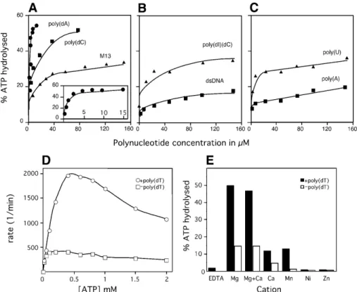
(3) 2282 Nucleic Acids Research, 2002, Vol. 30, No. 11. exonuclease attack was measured as described previously (15). The 39/16 or 39/25 [γ- 32P]-forked substrate, labelled at the 5′ end (top strand), or 39/16 or 39/25 [α-32P]-forked substrate labelled at the 3′ end (bottom strand) were incubated with increasing concentrations of G40P in buffer B with or without 1 mM AMP-PNP, during 15 min at 20°C in a volume of 15 µl. ExoVII (0.2 U) was then added and the incubation was continued for another 15 min. The reaction was stopped by addition of 10 µl of formamide solution containing 20 mM EDTA, heated for 5 min at 90°C and the products were separated using 10% (w/v) denaturing (d)PAGE run in TBE. The gels were dried, analysed by autoradiography and quantified as described above. [γ-32P]poly(dA) was used as a molecular weight marker. The 39/16 or 39/25 [γ-32P]-forked substrate, labelled at the 5′ end (bottom strand), was incubated with increasing concentrations of G40P in buffer B with or without 1 mM AMP-PNP, during 15 min at 20°C in a volume of 15 µl. ExoIII (1 U) was then added and the incubation was continued for another 15 min. The reaction was stopped as indicated for the ExoVII assays and the products were separated using 15% (w/v) dPAGE run in TBE. The 39/16 or 39/25 [α-32P]-forked substrate, labelled at the 3′ end (top strand), was incubated with increasing concentrations of G40P in buffer B with or without AMP-PNP, during 15 min at 20°C in a volume of 15 µl. ExoVI (1 U) was then added and the incubation was continued for another 15 min. The reaction was stopped and the products were separated and quantified as indicated for the ExoIII assays. KMnO4 footprinting assay The protection of a forked molecule by G40P (12.5 or 25 nM) against KMnO 4 attack, in the presence or the absence of 1 mM AMP-PNP, was measured as described previously (11). The 39/16 or 39/25 [γ-32P]-forked substrate, labelled at the 5′-end (top or bottom strand), was incubated with increasing concentrations of G40P in buffer B with or without AMP-PNP, during 15 min at 20°C in a volume of 15 µl. The samples were then treated with 1 mM KMnO 4 for 30 s at 37°C, the DNA was precipitated in presence of 1 µg of tRNA and cleaved with piperidine. The samples were separated using 15% (w/v) dPAGE run in TBE, and the gels dried and analysed by autoradiography. Electron microscopy Viral M13mp18 (0.2 nM) was incubated in buffer C [20 mM Triethanolamine–HCl pH 7.0, 8 mM magnesium acetate, 50 mM NaCl, 1 mM AMP-PNP, glycerol 8% (v/v)] with G40P (50 nM) for 15 min at 37°C in a 10 µl reaction. Complexes were then fixed with 0.15% (w/v) glutaraldehyde and negative stained with uranyl acetate (16). RESULTS AND DISCUSSION The ATPase activity of G40P is modulated by the presence of different effectors G40P purified from E.coli cells was >99% pure as judged by SDS–PAGE followed by Coomassie Blue staining and quantitative sequence analysis (data not shown). Purified G40P is free of the E.coli DnaB DNA helicase. The separation of G40P from. DnaB was achieved by a DEAE chromatographic step. G40P elutes at ∼100–140 mM NaCl, whereas DnaB elutes at 300–400 mM NaCl (data not shown). The effect of different DNA substrates on G40P NTP hydrolysis was analysed. G40P was able to hydrolyse ATP to ADP and Pi, and this activity was stimulated up to 5-fold in the presence of artificial ssDNA substrates such as poly(dA), poly(dC) (Fig. 1A) or unstructured 21-nt ssDNA (see below), whereas the stimulation of the ATPase activity was only increased ∼3-fold when the ssDNA substrate has a self-annealing potential (e.g. viral M13mp18 ssDNA), or when dsDNA and RNA were used (Fig. 1A–C). The kinetics of ATP hydrolysis Km values of G40P for ATP in the presence or the absence of poly(dT) 30 was analysed. In the absence of ssDNA, the ATPase activity increased to reach a plateau at very low ATP concentrations and then decreased slightly at higher ATP concentrations. In the presence of ssDNA, the ATPase activity increased with increasing amounts of ATP up to 0.4–0.8 mM and then decreased at higher ATP concentrations (Fig. 1D). In the presence of 0.4 mM ATP the presence of ssDNA increased ATP hydrolysis up to 5-fold. Michaelis–Menten kinetics has been observed for E.coli DnaB, or T7 gp4 helicase (reviewed in 6), but increasing concentrations of ATP have been shown to inhibit the ATPase activity of the only two replicative helicases of Gram-positive bacteria characterised so far, namely Bacillus stearothermophilus DnaB (17) and G40P (Fig. 1D). ATP hydrolysis occurs in the presence of Mg2+, to a lesser extent in presence of Mn2+, and Ca2+, but ATP hydrolysis drops to background levels in the presence of Ni2+ or Zn2+, or when EDTA was added (Fig. 1E). The activity of G40P is modulated by the different nucleotide cofactors Although most hexameric helicases do not show a strict nucleotide preference and specificity, as they hydrolyse a number of rNTPs or dNTPs (reviewed in 6,18,19), their nucleotide requirements for the DNA unwinding activity may be more strict (6,18,19), and a comparison of the effect of nucleotides on the three activities that hexameric helicases show has not been done yet. Therefore, the effect of nucleotides on G40P NTP hydrolysis, ssDNA binding and DNA unwinding activity was examined. The NTPase activity of G40P (10 nM) in the presence of poly(dT) 30 and increasing concentrations of NTPs was measured. The G40P NTPase activity increased with increasing amounts of NTP up to 0.4–0.8 mM and then decreased at higher NTP concentrations (Fig. 2A). G40P hydrolyses ATP (at 0.5 mM) with highest efficiency, and the rate of hydrolysis of the other NTPs (at 0.5 mM NTP) is 76 (GTP), 70 (CTP) and 69% (UTP) of the rate of ATP hydrolysis (Fig. 2A). The apparent binding constant (Kapp) of G40P for a naked 50-nt ssDNA segment observed in the presence of 2 mM CTP was 120 nM. The G40P binding affinity to a 50-nt ssDNA was reduced 2.5-, 3- and 5-fold in the presence of ATP, GTP and UTP, respectively (Fig. 2B). To test the DNA helicase activity of G40P (10 nM) in the presence of the dNTPs or rNTPs, M13mp18 ssDNA annealed to a 30-nt segment with a 20-nt non-complementary tail (3′-tailed substrate) was incubated with G40P and, after deproteinisation, the unwound products were visualised by 10% ndPAGE. G40P.
(4) Nucleic Acids Research, 2002, Vol. 30, No. 11 2283. Figure 1. ATPase activity of G40P. (A, B and C) The effect of polynucleotide effector concentration on G40P ATPase activity is shown. Reactions were as described in the Materials and Methods. ATPase activity of G40P is presented as turnover in percent versus concentration of effector nucleotides. (A) ssDNA as effector. The results for poly(dA) are shown enlarged in the small window; (B and C) dsDNA and RNA as effector, respectively. (D) ATPase activity in the presence (circles) and absence (squares) of ssDNA as a function of ATP concentration. (E) Effect of different cations on the ATPase activity of G40P. Filled squares or bars denote results in the presence of ssDNA, and empty squares or bars indicate results in absence of ssDNA.. preferentially unwinds the 50-nt ssDNA segment in the presence of ATP after 15 min of incubation. When the ATP was replaced by GTP, UTP or CTP, under the same conditions, 62, 20 and 42% of the unwinding observed with ATP, respectively, was observed (Fig. 2C). The unwinding activity of G40P was not observed in the presence of the four dNTPs. It is probable, therefore, that although G40P is able to hydrolyse the four rNTPs (at low NTP concentrations) with similar efficiency, only CTP, GTP and ATP are optimal for binding and hydrolysis, but the unwinding activity is maximal only with ATP, and with GTP to a lesser extent. G40P hydrolyses ATP and GTP binds ssDNA with higher efficiency in the presence of CTP, but unwinds DNA preferentially in the presence of ATP and GTP. Escherichia coli DnaB can bind and hydrolyse all rNTPs with similar affinity and efficiency (20), whereas T4 gp41 and T7 gp4 show some preference for purines. T4 gp41 hydrolyses GTP, dGTP, ATP and dATP, but only ATP and GTP support helicase activity (21,22), and T7 gp4 hydrolyses ATP, dATP, dTTP and dGTP, but unwinds DNA in the presence of dTTP, and to some extent with dATP and ATP (23,24). G40P preferentially binds unstructured ssDNA Previously, the Kapp of G40P for naked linear viral [α-32P]M13mp18 ssDNA or M13mp18 dsDNA, in the presence or absence of saturating amounts of ATP or its poorly hydrolysable analogue (AMP-PNP), was determined by filter binding assay following the amount of complex formation as a function of G40P. concentration (25). The affinity of G40P for dsDNA was shown to be independent of nucleotide and ~20-fold lower than for ssDNA (25). To measure the specificity of binding, a series of unrelated short [γ-32P]ssDNA segments (2 nM) have been used in EMSA in the presence or the absence of a nucleotide cofactor. In the absence of a nucleotide cofactor, the Kapp of G40P for a 51-nt ssDNA was estimated to be ~990 nM, and upon addition of ATP, binding of G40P to ssDNA is increased ~3.3-fold (Fig. 3A). When ATP was replaced by non-hydrolysable ATP analogues (1 mM AMP-PNP or ATPγS) the affinity of G40P for ssDNA increased ~25-fold (Kapp ~12 nM) (Fig. 3A). It is probable, therefore, that in the presence of ATP G40P binds and releases the ssDNA substrate, whereas in the presence of AMP-PNP stable binding to ssDNA is mainly observed (Fig. 3A). Similar results were obtained when an unrelated 50-nt long ssDNA segment or M13 viral ssDNA (25) were used. The affinity of G40P for a series of unrelated short DNA segments (16–21-nt ssDNA) with no potential DNA pairing is greatly enhanced (G40P–ssDNA complex with a Kapp of ~1.5 nM) when compared with the affinity to the 51- or 50-nt ssDNA segments (G40P–ssDNA complex, Kapp ~13 nM) (Fig. 3A). However, when we used a 55-nt segment consisting of 39 nt of random sequence with low self-annealing potential followed by a poly(dA) 16 nt long, we observed that G40P binds to it with similar affinity as to the 16-nt long molecule (see below). These results suggest that G40P preferentially binds to unstructured ssDNA and in a sequence-independent manner..
(5) 2284 Nucleic Acids Research, 2002, Vol. 30, No. 11. Figure 2. Effect of ribonucleotides on the G40P activities. (A) NTPase activity of G40P in the presence of ssDNA as a function of NTP concentration. (B) Kapp of G40P for a 50-nt ssDNA in the presence of 2 mM NTP. (C) DNA unwinding activity of G40P in the presence of the 2 mM NTP. The symbols + and – denote the presence or absence of G40P. H denotes heated susbtrate. The data represent the average of three independent experiments. Experimental details are described in the Materials and Methods.. Previously, it has been shown that the DnaB enzyme binds ~20 ± 3-nt ssDNA that were predicted to be inside the central hole of the hexameric helicase (26). The DnaB binding site is bipartite with ~10-nt ssDNA defining the strong binding site to the C-terminal part and ~10-nt ssDNA a weak binding to the N-terminal half of the enzyme (27). To learn how many G40P hexamers bind to a ssDNA segment, a 21- or a 51-nt ssDNA (3 nM) segment was incubated with increasing amounts of G40P and the protein–ssDNA complexes separated by ndPAGE. G40P forms one type of complex (I) with the 21-nt ssDNA segment even at high G40P protein concentrations, but two types of complexes (I and II) were observed with the 51-nt ssDNA segment (Fig. 3B). Under equilibrium conditions, in the presence of AMP-PNP, there is approximately one G40P molecule per 21-nt ssDNA segment. It is probable, therefore,. that in the presence of AMP-PNP, there is one G40P molecule bound to ssDNA in complex I and two G40P molecules in complex II. The binding of two G40P molecules to the same ssDNA segment does not seem to be cooperative, because only when all the ssDNA is saturated with one helicase are type II complexes formed. To understand whether the affinity of G40P for a replication fork structure is similar to the affinity of G40P for naked ssDNA (i.e. whether G40P also interacts with an adjacent dsDNA region), the affinity of G40P for a single-arm substrate [a 39-bp duplex with either a 16- or a 25-nt single-arm poly(dA) at the 5′ end (39/16 5′ single-arm and 39/25 5′ single arm), or 3′ end (39/16 3′ single arm or 39/25 3′ single arm)] or a forked substrate [a 39-bp duplex with a double arm of 16-nt or 25-nt poly(dA) (39/16 forked substrate or 39/25 forked substrate)], in the presence of 1 mM AMP-PNP, was assayed. The affinity of G40P for the 39/16 5′ single arm or 39/16 forked substrates is ~8-fold lower, when compared with the unstructured 55-nt ssDNA (Fig. 3C). The affinity of G40P for the 39/25 5′ single arm or 39/25 forked substrates is ~2.5-fold higher than that for the 39/16 5′ single arm and forked substrates (Fig. 3C) but still >3-fold lower than to the unstructured 55-nt ssDNA. It is probable that the binding site of G40P for the 5′-tail ssDNA must be somehow longer than 25 nt. Alternatively, the duplex region of the forked molecule affects the binding to ssDNA. In this respect G40P is different from DnaB. Here, the duplex part of the fork does not significantly affect the binding of the enzyme to the ssDNA arm (28). G40P forms only one type of complex with the 39/16 singlearm substrates (Fig. 3D). The affinity of G40P to the 39/16 3′ single-arm substrate was 9-fold lower, compared with the affinity of G40P for the 39/16 5′ single-arm substrate (Fig. 3C and D), indicating that the duplex region affects the positioning of G40P on the single-arm substrate. These results are consistent with the different binding affinity observed by fluorescence for E.coli DnaB with 5′ or 3′ single-arm substrates. DnaB accommodates a hexamer in the 20-nt long 3′ tail while another hexamer is associated, in opposite orientation, with the 5′ tail of the fork (28), suggesting that polarity in the binding may be a common feature of all replicative hexameric helicases. G40P preferentially binds to the 5′-tail ssDNA and interacts with duplex DNA DNA unwinding by the catalytic action of a replicative helicase, with an interconverting population of hexameric rings with a C6 and C3 symmetry, requires two ssDNA tails next to the duplex region and involves unidirectional translocation and base pair separation (reviewed in 6,18,19). Several general models (helix destabilising, rolling circle, wedge and torsional) for disruption of the forces that stabilise the duplex have been proposed (reviewed in 6,18,19). In the helix-destabilising model the helicase would bind to the unwinding junction, interact with the duplex DNA, but an interaction with the 3′-ssDNA arm is not obvious. In a type of rolling circle model, the hexamer encircles the 5′ tail and interacts with the 3′ tail and the duplex DNA. In the wedge and torsion models, the hexameric helicase does not interact with the duplex DNA. In the wedge variant the hexameric helicase interacts with the separated strand at the unwinding junction and the excluded 3′ tail does not have a specific interaction.
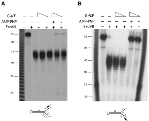
(6) Nucleic Acids Research, 2002, Vol. 30, No. 11 2285. Figure 3. G40P–DNA complex formation. (A) Kapp of G40P for 51- or 50-nt [ γ-32P]ssDNA (2 nM) in the absence or the presence of ATP or AMP-PNP measured by EMSA. The Kapp of G40P for 21-, 18- and 16-nt [γ-32P]ssDNA segments, in the presence of AMP-PNP, were also measured. (B) EMSA of G40P with a 21-nt [γ-32P]ssDNA (G40P concentrations doubling from 0.25 to 32 nM) and with a 51-nt [γ-32P]ssDNA segment (G40P concentrations doubling from 3 to 384 nM) were assayed. The two types of complexes obtained are denoted by I and II, and FD denotes the ssDNA in the absence of G40P. (C) Kapp of G40P for [γ-32P]-single-arm or [γ-32P]-forked substrates in the presence of AMP-PNP, measured by EMSA, are shown. All Kapp data represent the average of three independent experiments. The poly(dA) 16 and poly(dA)25 regions are denoted as an oblique line. (D) EMSA of G40P with a 5′ single-arm substrate (G40P concentrations doubling from 6 to 50 nM) and with 3′ single-arm substrate (G40P concentrations doubling from 25 to 200 nM). The complexes obtained are denoted by I and FD denotes the protein-free DNA.. with the hexameric helicase, whereas in the torsion variant of the model, the hexameric helicase interacts with both tails of the fork. Previously, it has been shown that the optimal rate of unwinding of forked DNA of hexameric helicases requires a DNA varying from ~8 to 35 nt at the 5′ tail and ~15 to ~30 nt at the 3′ tail (20,29,30). DNA binding affinity experiments suggest that G40P has an ~8-fold higher preference for 5′ single-arm substrates than for a 3′ single-arm substrates. To address with which DNA strand G40P interacts, we have performed nuclease protection studies using either ExoIII, ExoVI or ExoVII in the presence or absence of a non-hydrolysable ATP analogue (e.g. AMP-PNP or ATPγS) and an artificial 39/16 or 39/25 forked substrate with a 16- or 25-nt long tails. As the same results are obtained when 1 mM AMP-PNP or 1 mM ATPγS, or when the 39/16 or 39/25 forked substrate were used, only the former conditions are shown. The protection of the DNA ends by the binding of G40P (12.5 and 25 nM), in the presence or absence of 1 mM AMP-PNP, were performed and the products separated by dPAGE. Protection at the 3′-ssDNA tail from digestion by ExoVII was analysed by labelling the 5′ end (top strand, denoted by an asterisk in. Fig. 4A) of the duplex. ExoVII catalyses exonucleolytic cleavage of ssDNA in either 5′ → 3′ or 3′ → 5′ to yield 5′phosphomononucleotides. In the presence of AMP-PNP, G40P bound to a single region of the forked substrate fails to protect >90% of the 3′-tailed DNA segments from ExoVII digestion rendering a 44 ± 2-nt long segment (Fig. 4A). Such effect is independent of the addition of saturating amounts of G40P (25 nM, Kapp ~11 nM). G40P in the presence of AMP-PNP only protects a very small fraction of the 3′-tailed DNA segment from ExoVII degradation. This is consistent with the fact that at these G40P concentrations, there is little binding of G40P to the 3′ arm (Kapp 110 nM; see Fig. 3C). Furthermore, we could rule out a low ‘strength’ of the ExoVII when attacking the 3′ arm, because when a G40P mutant was assayed, the 3′-arm substrate is fully protected from ExoVII degradation (data not shown). The protection at the 5′-ssDNA tail was then analysed by labelling the 3′ end (bottom strand, denoted by an asterisk in Fig. 4B) of the duplex. In the absence of AMP-PNP, there is no binding of G40P to ssDNA hence the fork is sensitive to ExoVII digestion rendering a 44 ± 2-nt long labelled segment, whereas in the presence AMP-PNP G40P fully protects the.
(7) 2286 Nucleic Acids Research, 2002, Vol. 30, No. 11. Figure 4. G40P protection of the ssDNA arms of a forked molecule. G40P (12.5 and 25 nM) was incubated with 2 nM of double-arm substrate labelled at the 5′ end (A) or at the 3′ end (B) of the duplex in the presence or in the absence of 1 mM AMP-PNP. After 15 min incubation at 37°C, ExoVII (0.2 U) was added and the incubation was continued for 15 min at 37°C. The digestions were stopped and samples separated by 10% dPAGE and analysed by autoradiography. G40P encircling the 5′ tail of the 39/16 forked substrate is depicted not at scale. The symbols + and – denote the presence or absence of the indicated product. The asterisk denotes the labelled end. The arrow indicates the direction of exonucleolytic digestion. [ γ-32P]poly(dA) is used as a 1-nt molecular weight ladder.. ssDNA segment of exonucleolytic attack (Fig. 4B). Similar results were obtained with the 39-bp duplex containing a 25-nt long [poly(dA)] double arm, indicating that although the affinity of G40P for forked molecules having short (16 nt) tails is ~2-fold lower (see Fig. 3C), it is positioned in the same manner in the 39/16 and in the 39/25 forked molecules. It is likely therefore that G40P binds ssDNA with a 5′ → 3′ orientation with respect to the polarity of the ssDNA sugar–phosphate backbone. This is consistent with the differences in Kapp observed for a single-arm substrate with a 5′ or 3′ tail (Fig. 3C), with the findings of EM studies for the T7 gp4 helicase (31), or with fluorescence studies for DnaB (27). We show that at the concentrations used, G40P interacts with the 5′ tail, but is unable to encircle or even interact with the 3′-tail region of a forked DNA substrate. This is consistent with the observation that the Kapp is similar for the 5′ single-arm substrate and for the forked substrate, but ~9-fold lower for the for the 3′ single-arm substrate (Fig. 3C). It is also consistent with other indirect data described, such as that the Thermus aquaticus DnaB unwinds forked-duplex DNA with a similar rate when the 3′ tail has a reversed polarity (32), or as that streptavidin, bound to the 3′ end of the duplex, fully substitutes for the 3′-ssDNA tail in T7 gp4 helicase activity (30). The protection at the 5′ dsDNA from digestion by ExoVI was analysed by labelling the 3′-ssDNA end (top strand denoted by an asterisk in Fig. 5A). ExoVI catalyses in a stepwise non-processive reaction removal of 5′-phosphomononucleotides from the 5′ termini of dsDNA. In the absence of AMP-PNP,. no protection of the duplex was observed, because digestion with ExoVI renders a 26 ± 3-nt long labelled segment in the presence or absence of G40P (Fig. 5A). Perhaps due to poor stability of the short duplex (15), complete digestion by ExoVI was not observed and ~10 bp from the duplex became resistant to degradation. In the presence of AMP-PNP, a labelled 32 ± 2-nt segment, indicative of a G40P protection of the duplex ~15 bp away from the junction was observed, which could not be degraded by using the double amount of ExoVI (Fig. 5A). The protection at the 3′ dsDNA from digestion by ExoIII was then analysed by labelling the 5′-ssDNA end (bottom strand, Fig. 5B). ExoIII catalyses the stepwise removal of 5′-phosphomononucleotides from the 3′-OH termini of blunt or 3′ recessed dsDNA, but 3′ overhangs >4 nt are protected from ExoIII digestion. In the absence of AMP-PNP, protection of ExoIII digestion renders 24 ± 1-nt long labelled segment (Fig. 5B). A complete digestion of the duplex by ExoIII was not observed and ~8 bp became resistant to degradation. This has also been observed by other authors (15) and suggests again a poor stability of the duplex. In the presence of AMP-PNP, a labelled segment of ~25 ± 1 nt indicative of a protection of the duplex ~9 bp away from the junction by G40P was observed, which could not be degraded by using the double amount of ExoIII (Fig. 5B). The dependence of the protection caused by G40P in the duplex region on AMP-PNP indicated that G40P did not bind directly to the dsDNA, since binding to dsDNA is independent of AMP-PNP (25). We might envisage that the protection of the duplex is due to initial binding of G40P to.
(8) Nucleic Acids Research, 2002, Vol. 30, No. 11 2287. Figure 5. G40P protection of the duplex region of the 39/16 forked molecule. G40P (12.5 and 25 nM) was incubated with 2 nM of forked molecule labelled at the 3′ (A) or at the 5′ end (B) of the ssDNA tail, in the presence or in the absence of 1 mM AMP-PNP. After 15 min incubation a 37°C, ExoVI (1 U) (A) or ExoIII (1 U) (B) was added and the incubation was continued for 15 min at 37°C. The digestions were stopped and samples separated by 15% dPAGE and analysed by autoradiography. G40P encircling the 5′ tail of the 39/16 forked substrate is depicted not at scale. The symbols + and – denote the presence or absence of the indicated product. The asterisk denotes the labelled end. The arrow indicates the direction of exonucleolytic digestion. [γ-32P]poly(dA) is used as a 1-nt molecular weight ladder.. ssDNA and then to the adjacent duplex junction. Alternatively, a destabilisation of the duplex is due to initial binding of G40P to ssDNA with a subsequent ‘breathing’ of the ss/dsDNA junction by distorting the structure of the encircled tail as proposed previously by Yu et al. (33). To discriminate between these two options we performed KMnO4 footprinting on both strands using the 39/16 or 39/25 forked molecule, labelled at the 5′ end. The KMnO 4 footprinting pattern in the presence of both AMP-PNP and G40P (25 nM) is indistinguishable from that in the absence of G40P, hence we favoured the model in which G40P interacts asymmetrically with the ‘fork DNA’, is bound to the 5′ tail, and contacts the duplex region of both strands. No breathing of the ss/dsDNA junction could be observed by KMnO4 footprinting by using a forked substrate having an AT-rich segment at the junction region (data not shown). G40P seems to make a more extent contact with the duplex region of the strand opposite to where G40P is bound. Furthermore,. G40P does not seem to interact with the 3′ tail. These results are not consistent with the rolling circle, wedge and torsional model of DNA unwinding (6). The G40P central channel binds ssDNA Hexameric replicative DNA helicases have a central channel with a diameter of 25–45 Å that could accommodate a dsDNA or a ssDNA (reviewed in 6,18,19). No G40P protection on the 3′-ssDNA arm and full protection on the 5′-ssDNA arm (Fig. 4) suggests that the 5′-end ssDNA might have entered the central channel of G40P whereas the 3′-end ssDNA might be excluded. We cannot exclude a wrapping of the 5′-ssDNA arm around G40P from our studies, but we find it unlikely that a small 16-nt long ssDNA tail can be occluded from exonuclease digestion by wrapping around a G40P monomer. To address whether the ssDNA occupies the central channel of G40P, electron microscopic analysis was performed. Protein-free.
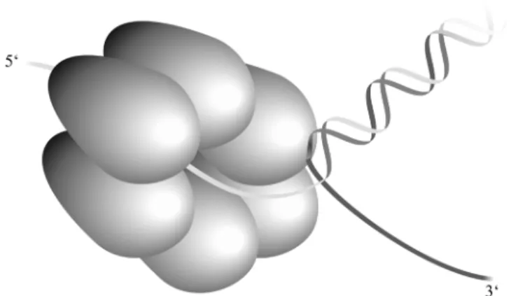
(9) 2288 Nucleic Acids Research, 2002, Vol. 30, No. 11. Figure 7. Proposed model of unwinding of DNA by G40P. G40P binds ssDNA in a 5 ′ → 3′ polarity through the inner channel, contacting the 5 ′-ssDNA arm, whereas there is no interaction with the 3 ′ arm which is fully excluded. G40P interacts with dsDNA at the junction. Protein conformational changes and ATP hydrolysis would lead to base pair separation. Then, the hexamer would translocate and bind to the newly separated stretch of the strand.. Figure 6. Electron micrographs of G40P bound to viral M13mp18 DNA in the presence of AMP-PNP. ssM13mp18 DNA (0.2 nM) was incubated, in buffer C, with G40P (50 nM) during 15 min at 37°C, and the complexes were visualised by EM. The regions of ssDNA entering the G40P inner channel are denoted by arrows (a–l). Three controls of ssM13mp18 in the absence of G40P are shown in (m–o).. ssDNA is at the visualisation limit of electron microscopy, but the addition of single-stranded DNA binding (SSB) protein to visualise the ssDNA will obscure the view in our case, because SSB exert a negative effect in G40P–AMP-PNP interaction with ssDNA (4). The thin filament of ssDNA might bind along the circumference or in the centre of the G40P hexameric ring. Binding along the circumference would require tangential approach of ssDNA to the G40P ring, whereas the binding in the centre would result in an approach that is along the radius of the G40P ring. In the images, where the circular ssDNA could be visualised, a perpendicular approach of ssDNA to the circumference of the G40P–AMP-PNP hexameric ring is observed (Fig. 6). These results support the proposal that ssDNA binding is in the centre of the G40P ring. This is consistent with other indirect observations reported for DnaB, T4 gp41 and T7 gp4, which. suggest that replicative helicases bind ssDNA through the central channel (reviewed in 6,19), and argue against a wrapping of the ssDNA around G40P. Although the precise mechanism of DNA unwinding is not known, we assume that the different G40P conformational states and the coupling of ssDNA binding and ATP hydrolysis may be the driving force to melt the duplex DNA and propel the helicase forward along the ssDNA strand. This is consistent with the fact that we did not observe any difference in the KMnO4 footprint with G40P in the presence of the non-hydrolysable ATP analogue AMP-PNP. As illustrated in Figure 7, on an artificial substrate, G40P binds to the 5′ arm of a forked molecule and encircles it in an NTP-dependent fashion while it fully excludes the 3′ arm. G40P makes a contact with both strands of the dsDNA at the junction, and this contact is more extended with the strand to which G40P is not bound. At least 5 nt are protected in this strand from ExoVI attack, whereas only 1 nt is protected from ExoIII digestion in the dsDNA region adjacent to the 5′-ssDNA arm to which G40P is bound. The fact that (i) only one of the six subunits of ATP-activated helicase appears to be in close contact with the ssDNA (27,28, our unpublished results) and that (ii) the helicase dissociates from the DNA upon ATP hydrolysis, favours a mechanism in which a monomer participates in the ssDNA binding and release. The contact with dsDNA could arise as the consequence of the fact that the ring is specifically positioned just at the junction. Only the hydrolysis of the NTP produces helicase movement and unwinding of the DNA in a 5′ → 3′ direction. ACKNOWLEDGEMENTS This research was partially supported by grants BMC2000-0548 and SAF2001-4339 from MCyT-DGI and QLK2-CT-2000-00634 from the European Union to J.C.A., 31-58841.99 from Swiss National Science Foundation to J. Dubochet and A.S. and from Human Frontier Science Program to A.S. We are very grateful.
(10) Nucleic Acids Research, 2002, Vol. 30, No. 11 2289. to T. A. Trautner and J. Dubochet for their interest in this project. REFERENCES 1. Pedré,X., Weise,F., Chai,S., Lüder,G. and Alonso,J.C. (1994) Characterization of cis and trans acting elements required for the initiation of DNA replication in the Bacillus subtilis bacteriophage SPP1. J. Mol. Biol., 236, 1324–1340. 2. Missich,R., Weise,F., Chai,S., Lurz,R., Pedré,X. and Alonso,J.C. (1997) The replisome organizer (G38P) of Bacillus subtilis bacteriophage SPP1 forms specialized nucleoprotein complexes with two discrete distant regions of the SPP1 genome. J. Mol. Biol., 270, 50–64. 3. Ayora,S., Stasiak,A. and Alonso,J.C. (1999) The Bacillus subtilis bacteriophage SPP1 G39P delivers and activates the G40P DNA helicase upon interacting with the G38P-bound replication origin. J. Mol. Biol., 288, 71–85. 4. Ayora,S., Langer,U. and Alonso,J.C. (1998) Bacillus subtilis DnaG primase stabilises the bacteriophage SPP1 G40P helicase-ssDNA complex. FEBS Lett., 439, 59–62. 5. Bárcena,M., San Martin,C., Weise,F., Ayora,S., Alonso,J.C. and Carazo,J.M. (1998) Polymorphic quaternary organization of the Bacillus subtilis bacteriophage SPP1 replicative helicase (G40P). J. Mol. Biol., 283, 809–819. 6. Patel,S.S. and Picha,K.M. (2000) Structure and function of hexameric helicases. Annu. Rev. Biochem., 69, 651–697. 7. San Martín,M.C., Stamford,N.P., Dammevora,N., Dixon,N.E. and Carazo,J.M. (1995) A structural model for the Escherichia coli DnaB helicase based on electron microscopy data. J. Struct. Biol., 114, 167–176. 8. Yu,X., Jezewska,M.J., Bujalowski,W. and Egelman,E.H. (1996) The hexameric E.coli DnaB helicase can exist in different quaternary states. J. Mol. Biol., 259, 7–14. 9. Yanisch-Perron,C., Vieira,J. and Messing,J. (1985) Improved M13 phage cloning vectors and host strains: nucleotide sequences of the M13mp18 and pUC19 vectors. Gene, 33, 103–119. 10. Studier,F.W. (1991) Use of bacteriophage T7 lysozyme to improve an inducible T7 expression system. J. Mol. Biol., 219, 37–44. 11. Sambrook,J., Fritsch,E.F. and Maniatis,T. (1989) Molecular Cloning. A Laboratory Manual, Vol. 1–3. Cold Spring Harbor Laboratory Press, Cold Spring Harbor, NY. 12. Ayora,S., Rojo,F., Ogasawara,N., Nakai,S. and Alonso,J.C. (1996) The Mfd protein of Bacillus subtilis 168 is involved in both transcription-coupled DNA repair and DNA recombination. J. Mol. Biol., 256, 301–318. 13. Gill,S.C. and von Hippel,P.H. (1989) Calculation of protein extinction coefficient from amino acid sequence data. Anal. Biochem., 182, 319–326. 14. Scott,J.F., Eisenberg,S., Bertsch,L. and Kornberg,A. (1977) A mechanism of duplex DNA replication revealed by enzymatic studies of phage phi X174: catalytic strand separation in advance of replication. Proc. Natl Acad. Sci. USA, 74, 193–197. 15. Soultanas,P., Dillingham,M.S., Wiley,P., Webb,M.R. and Wigley,D.B. (2000) Uncoupling DNA translocation and helicase activity in PcrA: direct evidence for an active mechanism. EMBO J., 19, 3799–3810.. 16. Sogo,J., Stasiak,A., DeBernardin,W., Losa,R. and Koller,Th. (1987) Binding of proteins to nucleic acids as studied by electron microscopy. In Sommerville,J. and Scheer,U. (eds), Electron Microscopy in Molecular Biology. IRL Press, Oxford, UK, pp. 61–79. 17. Bird,L.E., Pan,H., Soultanas,P. and Wigley,D.B. (2000) Mapping protein–protein interactions within a stable complex of DNA primase and DnaB helicase from Bacillus stearothermophilus. Biochemistry, 39, 171–182. 18. Lohman,T.M. and Bjornson,K.P. (1996) Mechanisms of helicase-catalyzed DNA unwinding. Annu. Rev. Biochem., 65, 169–214. 19. Soultanas,P. and Wigley,D.B. (2001) Unwinding the ‘Gordian knot’ of helicase action. Trends Biochem. Sci., 26, 47–54. 20. Arai,K.I and Kornberg,A. (1981) Mechanism of dnaB protein action. Allosteric role of ATP in the alteration of DNA structure by dnaB protein in priming replication. J. Biol. Chem., 256, 5260–5266 21. Liu,C.C. and Alberts,B.M. (1981) Characterization of the DNAdependent GTPase activity of T4 gene 41 protein, an essential component of the T4 bacteriophage DNA replication apparatus. J. Biol. Chem., 256, 2813–2820. 22. Venkatesan,M., Silver,L.L. and Nossal,N.G. (1982) Bacteriophage T4 gene 41 protein, required for the synthesis of RNA primers, is also a DNA helicase. J. Biol. Chem., 257, 12426–12434. 23. Matson,S.W. and Richardson,C.C. (1983) DNA-dependent nucleoside 5′-triphosphatase activity of the gene 4 protein of bacteriophage T7. J. Biol. Chem., 258, 14009–14016. 24. Hingorani,M.M. and Patel,S.S. (1993) Interactions of bacteriophage T7 DNA primase/helicase protein with single-stranded and double-stranded DNAs. Biochemistry, 32, 12478–12487. 25. Weise,F. (1997) Biochemische Charakterisierung der Initiation der DNA-Replikation des Bacillus subtilis-Bakteriophagen SPP1. Dissertation thesis, Freie Universität Berlin, Germany. 26. Bujalowski,W. and Jezewska,M.J. (1995) Interactions of Escherichia coli primary replicative helicase DnaB protein with single-stranded DNA. The nucleic acid does not wrap around the protein hexamer. Biochemistry, 34, 8513–8519. 27. Jezewska,M.J., Rajendran,S. and Bujalowski,W. (1998) Functional and structural heterogeneity of the DNA binding of the Escherichia coli primary replicative helicase DnaB protein. J. Biol. Chem., 273, 9058–9069. 28. Jezewska,M.J., Rajendran,S. and Bujalowski,W. (1998) Complex of Escherichia coli primary replicative helicase DnaB protein with a replication fork. Recognition and structure. Biochemistry, 37, 3116–3136. 29. Ahnert,P. and Patel,S. (1997) Asymmetric interactions of hexameric bacteriophage T7 DNA helicase with the 50 and 30-tails of the forked DNA substrate. J. Biol. Chem., 272, 32267–32273. 30. Hacker,K.J. and Johnson,K.A. (1997) A hexameric helicase encircles one DNA strand and excludes the other during DNA unwinding. Biochemistry, 36, 14080–14087. 31. Egelman,E.H., Yu,X., Wild,R., Hingorani,M. and Patel,S.S. (1995) Bacteriophage T7 helicase/primase proteins form rings around single-stranded DNA that suggest a general structure for hexameric helicases. Proc. Natl Acad. Sci. USA, 92, 3869–3873. 32. Kaplan,D.L. (2000) The 3′-tail of a forked-duplex sterically determines whether one or two DNA strands pass through the central channel of a replication-fork helicase. J. Mol. Biol., 301, 285–299. 33. Yu,X., Hingorani,M.M., Patel,S.S. and Egelman,E.H. (1996) DNA is bound within the central hole to one or two of the six subunits of the T7 DNA helicase. Nature Struct. Biol., 3, 740–743..
(11)
Figure


+3

Documents relatifs
dé dire tasse radis tarte train doux. rapide tortue douche étoile crevette domino
Ce changement de règle a eu un impact à la hausse sur le nombre de demandeurs d’emploi inscrits à Pôle emploi à la fin janvier 2013 de +21 100 pour la catégorie A et de +24 000
En France métropolitaine, au mois de novembre 2012, le nombre de demandeurs d’emploi inscrits en catégorie A et ayant un droit payable au Revenu de solidarité active (RSA) est de
The nearly complete skeleton, unearthed from 160-million-year-old mudstone deposits in northwestern China’s Junggar Basin, extends the fossil record of alvarezsauroids back in time by
The remaining terms are surface integrals over the outer bound- ary (normal direction taken as pointing outward): the second line is the contribution of total pressure,
To this end, the following approach is adopted: port- Hamiltonian formulation of the complete system, model order reduction of the infinite dimensional component (beam b),
L’archive ouverte pluridisciplinaire HAL, est destinée au dépôt et à la diffusion de documents scientifiques de niveau recherche, publiés ou non, émanant des
