IUPAC Technical Report
John A. Robinson* and Kerstin Moehle
Structural aspects of molecular recognition
in the immune system. Part II: Pattern
recognition receptors
Abstract: The vertebrate immune system uses pattern recognition receptors (PRRs) to detect a large variety
of molecular signatures (pathogen-associated molecular patterns, PAMPs) from a broad range of different invading pathogens. The PAMPs range in size from relatively small molecules, to others of intermediate size such as bacterial lipopolysaccharide, lipopeptides, and oligosaccharides, to macromolecules such as viral DNA, RNA, and pathogen-derived proteins such as flagellin. Underlying this functional diversity of PRRs is a surprisingly small number of structurally distinct protein folds that include leucine-rich repeats in Toll-like receptors (TLRs) and NOD-like receptors (NLRs), the DExH box helicase domain in RIG-like receptors (RLRs), and C-type lectin domains (CTLDs) in the C-type lectins. Following PAMP recognition by the PRRs, down-stream signaling pathways activate the innate immune system to respond to invading pathogenic organisms. The resulting stimulatory response is also vital for a balanced adaptive immune response to the pathogen, mediated by circulating antibodies and/or cytotoxic T cells. However, an aberrant stimulation of the innate immune system can also lead to excessive inflammatory and toxic stress responses. Exciting opportunities are now arising for the design of small synthetic molecules that bind to PRRs and influence downstream signal-ing pathways. Such molecules can be useful tools to modulate immune responses, for example, as adjuvants to stimulate adaptive immune responses to a vaccine, or as therapeutic agents to dampen aberrant immune responses, such as inflammation. The design of agonists or antagonists of PRRs can now benefit from a surge in knowledge of the 3D structures of PRRs, many in complexes with their natural ligands. This review article describes recent progress in structural studies of PRRs (TLRs, NLRs, CTLs, and RLRs), which is required for an understanding of how they specifically recognize structurally diverse “foreign” PAMPs amongst a back-ground of other “self” molecules, sometimes closely related in structure, that are present in the human body.
Keywords: immune system; IUPAC Chemistry and Human Health Division; NOD-like receptors (NLRs);
pathogen- associated molecular patterns (PAMPs); pattern recognition receptors (PRRs); RIG-like receptors (RLRs); Toll-like receptors (TLRs).
DOI 10.1515/pac-2013-1026
Received October 22, 2013; accepted April 17, 2014
Article note: Sponsoring body: IUPAC Chemistry and Human Health Division; see more details on p. 1533. Part I is DOI 10.1515/pac-2013-1020
*Corresponding author: John A. Robinson, Department of Chemistry, University of Zurich, Winterthurerstrasse 190, 8057 Zurich, Switzerland, e-mail: [email protected]
Kerstin Moehle: Department of Chemistry, University of Zurich, Winterthurerstrasse 190, 8057 Zurich, Switzerland © 2014 IUPAC & De Gruyter
CONTENTS
1. INTRODUCTION TO PATTERN RECOGNITION RECEPTORS ... 1484
1.1 Toll-like receptors (TLRs) ...1486
1.2 C-type lectin receptors (CLRs) ... 1487
1.3 NOD-like receptors (NLRs) ...1488
1.4 RIG-like helicase receptors (RLRs) ...1489
2. STRUCTURES OF TOLL-LIKE RECEPTORS ... 1490
2.1 Overview ...1490
2.2 Structures of TLR1/2 and 2/6 ... 1492
2.2.1 The human TLR1/2 heterodimer ...1494
2.2.2 The mouse TLR2 monomer ... 1495
2.2.3 The mouse TLR2/6 heterodimer ... 1497
2.2.4 Applications of TLR1/2 and TLR2/6 agonists ...1498
2.2.5 Accessory proteins involved in TLR2 signaling ...1499
2.3 Structures of TLR3...1499
2.4 Structures of TLR4 ... 1501
2.4.1 The human MD-2/lipid IVa complex ...1503
2.4.2 The human TLR4/MD-2 with bound Eritoran ...1504
2.4.3 The human TLR4/MD-2/LPS complex ...1504
2.4.4. The mouse TLR4/MD-2/LPS and lipid IVa complexes ... 1507
2.5 Structures of TLR5 ...1508
2.6 TLRs 7, 8, and 9 ... 1510
2.6.1 Structure of TLR8 ...1512
2.7 TLRs 10, 11, 12, and 13 ...1513
3. STRUCTURES OF C-TYPE LECTIN RECEPTORS ...1514
3.1 Structures of DC-SIGN and DC-SIGNR ...1514
3.2 Mannose-binding lectins ...1519
3.2.1 Mannose-binding proteins... 1520
3.2.2 The mannose receptor ...1521
4. STRUCTURES OF NOD-LIKE RECEPTORS ...1523
4.1 RNA-binding domain of NLRX1 ... 1523
4.2 Pyrin domains and CARDs ... 1524
5. STRUCTURES OF RIG-LIKE RECEPTORS ...1524
5.1 RNA recognition by RIG-I ... 1524
5.2 RNA recognition by MDA5 ... 1527
6. NOTE ADDED IN PROOF ...1529
7. ABBREVIATIONS ...1532
8. MEMBERSHIP OF SPONSORING BODY ...1533
9. ACKNOWLEDGMENTS ...1533
10. REFERENCES ...1533
1 Introduction to pattern recognition receptors
The immune system has evolved to protect organisms as diverse as plants, flies, and animals against infec-tion by invading microorganisms. A critical first step in achieving this protecinfec-tion is to quickly recognize when infectious agents are present; the immune system must distinguish what is foreign (or “non-self”) from what is normally present in the body (i.e., “self”). It is the main function of the innate immune system to provide this rapid surveillance, using germ-line encoded proteins called pattern recognition receptors (PRRs) [1–4]. PRRs function by recognizing “danger signals” and then initiating an appropriate innate immune response.
Danger signals can be derived from both foreign pathogens as well as endogenous sources, such as lysed cells. The PRRs comprise four main families of proteins, some membrane-bound and others soluble cytoplasmic proteins that recognize molecules (or molecular patterns) associated with microbes – so-called pathogen-associated molecular patterns (PAMPs). The activation of PRRs by binding to PAMPs initiates intra-cellular signaling cascades and altered patterns of gene expression that lead to a rapid first line defense against the invading microorganism, as well as promoting activation and maturation (more slowly – over days to weeks) of the adaptive immune system. The adaptive immune response is mediated mainly by B and T cells. Signals from the innate immune system drive the selective expansion and activation of B- and T-cell populations with specificity for the infectious agent, optimized through cycles of somatic mutation and selection (Fig. 1) [5]. The main effector mechanisms of adaptive immunity include the production of antibodies by B cells (to act as blocking antibodies, or as opsonins for complement- and phagocyte-mediated killing), and the killing of infected host cells by cytotoxic T cells. In addition, activation of adaptive immunity results in the production of memory B and T cells, which can provide long-term specific protection (e.g., over years to a whole lifetime) against subsequent infections by a pathogen bearing the same antigens [6, 7].
Adherence of microbes to the surface of cells of the innate immune system, mediated through binding of PAMPs to cell surface PRRs, actively promotes ingestion of the microbe and entrapment and digestion within phagosomes. On the other hand, soluble cytoplasmic PRRs act as sensors for intracellular pathogens. Through PAMP–PRR interactions, innate immune cells are activated and respond by producing inflammatory cytokines and chemokines that trigger responses from other cell types. Furthermore, activation of the innate immune system leads to deployment of a range of killing factors to attack invading pathogens. These include bactericidal enzymes, toxic chemicals such as reactive oxygen and nitrogen species, and antimicrobial pep-tides (e.g., defensins). The detection of certain PAMPs may also activate the complement system, a cascade of triggered enzyme reactions that can lead to tagging (or opsonization) of the microbe surface with the comple-ment protein C3b, which marks it out for destruction by other components of the immune system. Finally, PRR–PAMP interactions stimulate cells of the adaptive immune system, including antigen-presenting cells
Fig. 1 The innate immune system operates largely in peripheral tissues, where it recognizes foreign pathogens. Upon binding of PAMPs to PRRs, the innate immune system is activated and provides a first line of defence against the invader. Subsequently, the adaptive immune system is activated, and with help from the innate immune system, pathogen-specific antibodies and killer T cells are produced (adapted from [11]).
(APCs), such as dendritic cells (DCs) and B and T cells. DCs reside in a quiescent state in peripheral tissues, where they continuously sample their environment by phagocytosis and pinocytosis. DCs are equipped with a suite of PRRs for binding to PAMPs, thereby facilitating rapid up-take of pathogens by phagocytosis. Inside APCs, foreign proteins are digested into peptide fragments, which include some that act as T-cell epitopes. A dramatic increase in the expression of surface MHC-I and -II is elicited by PAMP–PRR binding. This leads to enhanced surface presentation of T-cell peptide epitopes in complexes with the membrane-bound MHC molecules, ready for recognition by T-cell receptors.
Immature DCs are relatively non-motile, but after activation they become highly motile and migrate through the lymphatic system, carrying pathogen-derived antigens to secondary lymphoid tissues, such as the lymph nodes. Circulating naïve B and T lymphocytes also halt in the lymph nodes, where they scan APCs and intact antigens draining through in interstitial fluids from peripheral tissues. Intact antigens can be rec-ognized by B-cell receptors (BCRs) on the surface of B cells. Viruses and bacteria typically display a repetitive and closely spaced array of epitopes across their surface. Such an arrangement of epitopes is specifically rec-ognized as foreign by B cells [8]. This occurs through cross-linking of multiple BCRs by multivalent binding to surface arrays of antigens, which generates a powerful activation signal, and initiates the process of B-cell acti-vation and maturation [9]. The activated B and T cells then interact within the lymph nodes in a process that leads to the production of antibody-secreting B cells and cytotoxic T cells optimized to recognize and remove foreign pathogens. The activated B and T cells also harbor PRRs, and are further stimulated by repeated inter-action with PAMPs during the course of the adaptive immune response [10]. PAMP–PRR interinter-actions thus play a key role in immune responses to invading pathogens, from its beginnings through the later stages of matura-tion of the adaptive immune response (Fig. 1). For this reason, both natural and synthetic ligands of PRRs are now attracting great interest as immune modulators in the fields of vaccinology and immunotherapy [6, 11–13].
PRRs can be divided into four main families, based upon their structural features as well as the type of PAMP they recognize [14]. These include the Toll-like receptors (TLRs), the C-type lectin receptors (CLRs), NOD-like receptors (NLRs), and RIG-like helicase receptors (RLRs). A review of newly described PRRs appeared recently [15].
1.1 Toll-like receptors (TLRs)
One of the major families of PRRs are the TLRs, so named because of their homology with the Toll receptor, first discovered in the 1980s in the fruit fly Drosophila [16, 17]. The Toll receptor plays a role in the development of the dorsoventral body axis in Drosophila, acting as a cell-surface receptor for the cytokine ligand Spätzle [18]. Later it was discovered that Toll and related receptors also function in the innate immune response to fungal and bacterial infection in the fruit fly, stimulating the release of defensins and related antimicrobial peptides [19]. As genome sequencing advanced, about 10 directly homologous receptors were later discov-ered in vertebrates, and these became known as the Toll-like receptors (for a recent historical account, see [20]). The ligands for most of these receptors are now known. The ligands display remarkable structural diversity, and include bacterial peptidoglycan (PGN), lipoproteins, lipopolysaccharide (LPS), mycobacterial lipoarabinomannan, yeast zymosan, and bacterial flagellin, as well as pathogen-derived nucleic acids [21].
To date, 10 TLRs have been reported in humans and 13 in mice. The TLRs are type I integral membrane glycoproteins that are located either in the outer cell membrane (TLR1, 2, 4, 5, 6, 10, 11, 12), or in endosomal membranes with their ectodomains facing the internal space of the endosome (TLR3, 7, 8, 9, 13) (Fig. 2). They contain an N-terminal extracellular domain (ECD) of between 550 and 800 residues, a single helical transmem-brane domain of ≈20 residues, and a C-terminal intracellular signaling domain of ≈180 residues, called a “Toll IL-1 receptor” (TIR) signaling domain, because it shares homology with the signaling domains of IL-1R family members. The crystal structure of the TIR domain of TLR1/2 was published in 2000 [22]. Some TLRs act as het-erodimers, including TLR1/2 and TLR2/6, which respond to different bacterial lipoproteins. All TLRs share a common basic 3D architecture, with multiple leucine-rich repeats (LRRs) arranged in a horseshoe- or crescent-shaped structure that together comprises the N-terminal ectodomain responsible for PAMP-binding. A single
transmembrane segment links the ectodomain to the C-terminal domain, which interacts with adapter proteins in the cytoplasm, an event that culminates in activation of transcription factors, such as NFκB and members of the interferon-regulated factors (IRFs) family, which in turn leads to altered patterns of gene expression [23, 24].
1.2 C-type lectin receptors (CLRs)
The C-type lectin receptor (CLR) superfamily is a large group of proteins that are widely distributed in nature [25, 26]. C-type lectins are Ca2+-dependent carbohydrate-binding proteins that contain one or more C-type
lectin (or lectin-like) domains (CTLDs), with related folds dictated by a common sequence motif. When such domains bind to carbohydrates, they are called carbohydrate recognition domains (CRDs). A CRD is present in all Ca2+-dependent lectins, but not in other (Ca2+-independent) types of animal lectins. A recent review grouped
the CTLDs into 17 different classes [25]. Of special interest here are the class-II asialoglycoproteins on APCs that function as PRRs in the innate immune system. These type-II membrane receptors contain a short cytoplasmic domain, a single transmembrane segment, and an ectodomain comprising a stalk region involved in oligomer-ization, which is linked to one or more CRDs. Professional APCs, such as macrophages, DCs, and B cells, are equipped with receptors that collectively allow them to recognize, capture, and internalize foreign antigens. Besides the B-cell receptor found only on B cells, other APCs use germ-line encoded membrane proteins for detecting foreign antigens that include Fc receptors, complement receptors, the TLRs, and the CLRs. TLRs are not able to promote internalization of antigens, whereas this is an important function of CLRs. Representatives of the class-II CLRs are ICAM-3, DC-SIGN, MGL, LANGERIN, MINCLE, DECTIN-2, BDCA-2, DCIR, and DCAR. The class-V CLRs are also known as type-II natural killer (NK) cell receptors, and include DCAL-1, DCAL-2, and dectin-I. Finally, the class-VI CLRs include the mannose receptor (MR) and lymphocyte antigen 75 (DEC-205).
CLRs are important in immune recognition of glycoproteins and cell wall glycolipids derived from viruses and microbes, and mediate pathogen uptake and phagocytosis [27, 28]. However, the biological functions of Fig. 2 Ten TLRs have been identified in humans, and are located either on the outer cell membrane or the membrane of endosomes within the cytoplasm of immune cells. When PAMPs bind to the ectodomain of the TLR, signals are transmitted into the cell via intracellular (TIR) domains [22], which bind and activate transcription factors [NFkB and/or members of the interferon-regulated factors (IRFs)]. The transcription factors promote expression of numerous genes that alter the activation state of the cell, as well as for inflammatory cytokines and chemokines that are exported and signal to other cells (see Fig. 1).
CTLs are more complex than just facilitating antigen uptake by DCs. Many of the carbohydrates recognized by CLRs are not unique to pathogens but can also be found on many self-glycoproteins [27]. Hence, self- and non-self-structures may be recognized by CLRs, which is a reflection of the multiple and complex functions these receptors, as a whole, have in the maintenance of immune homeostasis [29–32]. Ligand binding to these receptors can also activate signaling pathways that modulate TLR signaling, activate innate immune responses, and influence adaptive immune responses [30, 31].
The C-type (calcium-dependent) lectin (CTL) family of proteins is found on the surface of cells of the innate immune system, including macrophages and DCs [28, 29, 33]. The membrane-bound CTLs bind oligosaccha-rides, in particular, containing d-mannose and l-fucose residues, using highly conserved carbohydrate recog-nition domains (CRDs). Many CTLs on APCs contain only a single CRD, well-known examples being DC-SIGN, DCIR, MGL, and Langerin. Others, such as DEC-205 and the mannose receptor (MR), possess 8–10 CRDs within their extracellular regions [25, 34–36]. According to their carbohydrate specificities, CLRs tend to be classi-fied as either mannose/fucose- or galactose-recognizing lectins. Some CLRs share carbohydrate ligands. For example, the trisaccharide Lewisx can serve as a ligand for DC-SIGN and the MR. However, ligand promiscuity
is also seen, for example, with DC-SIGN, which is able to recognize efficiently different Lewis-type epitopes as well as high-mannose glycans. CLRs have now become attractive targets for ligand design, for use in targeting antigens to DCs, and for modulating antigen-specific immune responses [28, 29, 33].
1.3 NOD-like receptors (NLRs)
The nucleotide-binding and oligomerization domain (NOD)-like receptors (NLRs) function as intracellular sur-veillance sensors to detect microbial products and danger signals inside cells [37–41]. Some NLRs are also involved in inflammatory responses and apoptosis, and thus as a whole the NLRs mediate diverse biologi-cal functions in innate immunity [39, 42–46]. NLR family members are found in plants, invertebrates, and mammals, although NLR structure and function has been most thoroughly studied in mice and humans [47]. The NLRs are subdivided into five subfamilies, into which the 22 human NLRs are grouped as follows; NLRA (CIITA), NLRB (NAIP), NLRC (NOD1, NOD2, NLRC3, NLRC4, NLRC5, NLRP and NRLX), NLRP (NLRP1-14), and
NLRX (NLRX1) (Fig. 3) [48]. Some NLRs activate a proinflammatory response (such as NOD1 and NOD2), others
form a large multiprotein complex called an inflammasome [47, 49] resulting in cleavage of procaspase-1 and formation of activated caspase I (NLRP1, 3, 6, 7, and 12, and NLRC4), others provide immunoregulatory func-tions (NLRC3 and 5, CIITA, and NLRP10), while others have roles in development (NLRP2, 5, 7, and 14) [48, 50]. The NLRs are soluble cytoplasmic proteins, important for sensing infectious agents that have gained access to the interior of the cell [48, 50–52]. There are 22 NLRs in humans and 34 in mice [53], which exhibit character-istic multidomain structures, typically consisting of a central nucleotide binding domain (NBD) that contains a
Fig. 3 Domain architecture of representative human NLRs [47, 52]. Abbreviations: LRRs, leucine-rich repeats for ligand sensing; NACHT, a nucleotide binding and oligomerization domain; PYD, pyrin effector domain; BIR, baculovirus inhibitor repeat effector domain; CARD, caspase activation and recruitment effector domain; NBD, nucleotide binding domain; FIIND, function to find domain.
NACHT and other subdomains, an N-terminal effector binding domain most commonly a CARD (caspase activa-tion and recruitment domain), a BIR (baculovirus inhibitor repeat), or a pyrin (PYD) domain, and a C-terminal putative ligand-binding domain comprising multiple leucine-rich repeats (LRRs) (Fig. 3) [39, 40, 48]. The effec-tor domains bind downstream signaling molecules, ultimately leading to activation of protein kinases, tran-scription factors, proteases, and other components of host defense and inflammatory responses [38, 50].
Some of the PAMPs detected by NLRs have been identified. For example, NOD1 and NOD2 sense frag-ments of PGN [37]. Macrophages contain intracellular hydrolases that digest bacterial PGN. NOD1 detects a minimal PGN fragment containing γ-d-glutamyl-meso-diaminopimelic acid that is found almost exclusively in Gram-negative bacterial PGN [54]. On the other hand, NOD2 detects muramyl-dipeptide (MurNAc-l-Ala-d-isoGln), which is a common motif in PGN in almost all bacteria [55, 56]. NLRP3 and NAIP respond to a remark-ably diverse set of PAMPs [39], whereas NLRC4 senses bacterial flagellin [57, 58], and NLRX1 binds to RNA [59].
1.4 RIG-like helicase receptors (RLRs)
How does a cell distinguish viral nucleic acid (RNA) in the cytoplasm from its own? An important clue came by recognizing that viral RNA is structurally different in ways that mark it out as foreign to the cell. Thus, dsRNA is virtually absent in mammalian cells, whereas some viruses generate dsRNA as a byproduct of viral RNA replication, containing transiently an uncapped 5′-triphosphate group. Cellular primary transcripts also contain 5′-triphosphate, however, many self-RNAs undergo processing steps that remove or mask the 5-triphosphate moiety. For example, mRNAs are capped by methylation at the 5′-terminus, tRNAs undergo cleavage of 5′-PPP groups before they reach the cytoplasm, and ribosomal RNAs form complexes with riboso-mal proteins. The retinoic acid-inducible gene-I (RIG-I)-like receptors (RLRs) are constitutively expressed in the cytoplasm of most cells, including DCs and macrophages as well as non-immune cells, where they act as cytosolic sensors of viral RNA [60, 61].
RLRs provide a first line of defense against infection by RNA viruses, including influenza, rift valley, measles, vesicular stomatitis, and hepatitis C viruses [61]. There are three members of the RLR family: RIG-I (also known as DDX58), “melanoma differentiation associated factor gene 5″ (MDA5), and “laboratory of genetics and physiology 2” (LGP2). Recognition of the 5′-PPP terminus unique to viral RNA, either in a dsRNA or a hairpin-looped structure, a so-called pan-handle-like RNA, is an important function of RIG-I [62, 63]. The prototypical ligand of RIG-I is a short RNA with a blunt-ended base-paired, and uncapped 5′-triphosphate terminus. Until very recently, much less was known about the nature of RNAs recognized by MDA5. MDA5 generally responds to long dsRNA molecules. However, RIG-I and MDA5 may also be activated by self-RNAs that are cleaved by RNaseL. The function of the third RLR, called LGP2, has been less well studied, but seems to participate in RIG-I and MDA5-dependent antiviral responses [60, 61].
RIG-1 and MDA-5 possess a conserved central helicase-like domain, linked to two N-terminal caspase activation and recruitment domains (CARDs), and a Zn2+-containing C-terminal domain (CTD) (Fig. 4). LGP2
Fig. 4 Domain structures of RLRs. RIG-1 and MDA-5 have the same domain architecture. Abbreviations: CARD, caspase activa-tion and recruitment effector domain; CTD, C-terminal regulatory domain; HEL2i, helicase inseractiva-tion domain; HEL1/2 helicase 1/ helicase 2; P, pincer domain.
has similar domain architecture, although it contains no CARD domains. The ligands that function as RLR activators are typically virus-specific RNA structures, including 5′-triphosphate linked to single-stranded or blunt-ended, double-stranded RNA, as well as poly I:C (a synthetic analog of viral dsRNA). Upon binding to such PAMPs, RIG-1 initiates a signaling cascade that induces innate immune defenses and inflammatory cytokines to establish an antiviral state [64]. However, knowledge of the complex regulatory mechanisms that control signaling through these PRRs is incomplete. Aberrant RIG-1 signalling can lead to apoptosis, altered cell differentiation, inflammation, autoimmune disease, and cancer. Apart from RLRs, several other members of the helicase superfamily have been proposed to participate in intracellular sensing of pathogen-derived nucleic acids, including DDX1/3/21/36, DHX9/36, and STING [60].
2 Structures of toll-like receptors
2.1 Overview
Several reviews on TLR structural biology have been published recently [16, 65, 66]. TLR ECDs are con-structed of tandem copies of a structural motif known as the leucine-rich repeat (LRR). LRRs are found in several thousand proteins involved in a wide variety of physiological functions in all kingdoms of life, including immune responses, signal transduction, cell-cycle regulation and enzyme regulation (for reviews, see [67–69]). These proteins typically contain 2–40 tandem LRR modules, which form curved horseshoe- or solenoid-like structures that seem to be well suited for protein–protein and protein–ligand interactions. The 10 human TLRs contain 19–25 LRR modules. Each LRR typically contains a ≈20–30 residue consensus sequence (Fig. 5a), which includes the characteristic motif LxxLxLxxNxL (x = any amino acid), although other hydrophobic residues can substitute for Leu, and other amino acids capable of donating hydrogen bonds (T, S, C) can substitute the Asn residues [67, 70]. Each LRR module adopts a folded structure com-prising one β-strand that includes the entire consensus motif, followed by a bridging loop that often con-tains helical and/or turn-like elements that fold back over the β-strand, thereby burying the hydrophobic L
Fig. 5 (a) Characteristic LRR motif, and structure of one LRR module. Orange, β-strand; green, bridging loop. (b) The typical horseshoe, or solenoid-like structure of a TLR ectodomain. (c) The disulfide-rich capping modules at the N- (LRR-NT) and C-terminus (LRR-CT).
residues (Fig. 5a). The bridging loop is linked to the β-strand of the next LRR, which is aligned so that the β-strands form a regular hydrogen-bonded parallel β-sheet. This arrangement can now be repeated by addi-tion of more LRRs, which generate a horseshoe- or solenoid-like tertiary structure, as shown in Fig. 5b. The resulting curved parallel β-sheet forms the inner concave face of the horseshoe, whereas the residues in the adjacent bridging (often helical) loops form the outer convex surface of the horseshoe. The ECDs of most TLRs are glycoproteins, in which surface-exposed Asn residues carry N-linked glycosylation. The side-by-side nestling together of LRRs in these proteins ensures that the hydrophobic residues are buried internally. The situation is different for the N- and C-terminal LRR modules, where one side of the hydrophobic core will not be covered by another LRR module. Instead, capping modules are added to the termini (LRR-NT and LRR-CT) (Fig. 5c). The capping modules do not show the same conserved sequence and structural motifs. They typically comprise smaller disulfide-bonded folds that pack across the hydrophobic core and leave polar surface residues exposed to the solvent.
The LRR horseshoe geometries have been characterized by three parameters – the radius of the horseshoe arc (“radii” R), the mean rotation angle about the central axis relating one β-strand to the next (“twist”), and the tilt angle of the parallel β-strand direction per turn (“tilt”) [68]. The observed values of R, for example, range from 1.5 to 4.7 nm, so the arc of LRR family members can possess quite different degrees of curvature. LRR family proteins have been subdivided into seven subfamilies based on sequence and structural compari-sons. They are “RI-like”, “CC”, “bacterial”, “SDS-22-like”, “plant-specific”, “typical”, and “TpLRR” [69, 71]. The “typical” subfamily includes all the TLRs. However, phylogenetic analyses suggested that two distinct subclasses of “typical” TLRs exist, the single- and three-domain TLR subclasses, which diverged early in evolution [66, 72].
The ECDs of TLR1, 2, 4, and 6 belong to the three-domain subclass [66]. These TLRs have two structural transitions in the concave β-sheet face of the ECD, thereby creating N-terminal, central, and C-terminal sub-domains (Fig. 6). The central sub-domains have radii, twist, and tilt angles that deviate markedly from the stand-ard values of the “typical” single-domain subfamily members. This appears to be caused by several unusual features, including LRRs in the central domain that lack the characteristic Asn residue within the consen-sus motif. This Asn side chain usually interacts in a ladder of hydrogen bonds with surrounding backbone carbonyl oxygens, thereby stabilizing the horseshoe structure. In addition, the LRR modules of the central domains vary in size from 20 to 33 residues, whereas in the N- and C-terminal domains, the LRR modules are mostly 24 residues. The LRR modules in the central region have bulkier α-helical segments on the exposed convex face, which take up more space, leading in turn to greater curvature and a smaller arc radius in the central domain. These structural features are also important for the biological function of these TLRs because the central domains play an important role in binding ligands or co-receptors. Although no crystal structure is available, TLR10 also appears to contain broken Asn ladders and greater variation in module length in the central part of the ECD, and so is also likely to belong to the three-domain subfamily.
Fig. 6 Single-domain (left) and three-domain (red, green, and blue, right) TLR subclasses. The characteristic Asn residues (show in orange/red/blue ball-and-stick) in each repeat motif, and the ladder of hydrogen-bonds (in light blue) they form are shown.
In contrast, TLR3, 5, 7, 8, and 9 belong to the single-domain subfamily. Their LRR modules have con-served Asn residues within the consensus motif and relatively uniform module lengths. Functional and phy-logenetic analyses provide support for these correlations, which in the cases of TLR3 and 8 is also supported by structural (crystallographic) information. TLR7, TLR8, and TLR9 are predicted by sequence comparisons to contain “island regions”, comprising substantial domains that loop out of the external surface of the horse-shoe architecture [73].
Strong evidence exists that active TLRs function as homo- or hetero-dimers, and/or as part of supramo-lecular complexes with other membrane-associated proteins. For example, TLR2 signals as a heterodimer in a complex with either TLR1 or TLR6. Crystal structures of the ECDs of the TLR1/2 and TLR2/6 heterodi-mers show how specific lipopeptides are recognized by these receptors (vide supra) [74, 75]. In the case of the TLR2/6 heterodimer, a plasma membrane protein (CD36) of the class B scavenger receptor family, also participates in signaling [76]. Although human TLR3 ECD is found as a monomer in the crystal structure [77, 78], it binds its target double-stranded (ds) RNA as a dimer [79, 80]. Moreover, clustering of multiple TLR3 homodimers may occur at the cell surface through binding to long dsRNAs [79, 81]. FRET studies suggest that TLR9 exists as preformed inactive dimers in the cell membrane that change conformation upon binding to DNA rich in CpG dinucleotides. Ligand binding then results in allosteric changes in the cytoplasmic sign-aling domains, which lead to receptor activation and signsign-aling [82]. TLR5, the only protein-binding TLR, responds to a monomeric form of bacterial flagellin that, in oligomeric form, constitutes the flagellar tail responsible for swimming locomotion in β- and γ-proteobacteria. A crystal structure of an engineered TLR5-flagellin complex revealed a 2:2 subunit stoichiometry, with two TLR5 ECDs in a tail-to-tail organization [83]. Finally, TLR4 forms a stable 1:1 heterodimer with MD-2, and the resulting TLR4-MD-2 complex is responsible for recognition of lipopolysaccharide (LPS) from Gram-negative bacteria. Cells transfected with TLR4 are not responsive to LPS, but are when complemented with MD-2 [84]. Moreover, another membrane-associated protein, CD14, is required for full ligand recognition and signaling through TLR4. TLR4 may be organized into a supramolecular complex through interactions with MD-2 and CD14 (and perhaps other components) at the cell surface [76, 85, 86].
TLRs interact with an unusually diverse variety of ligands. These ligands range from naturally occurring highly nonpolar lipids, such as LPS and lipopeptides, through to polar macromolecules such as DNA, RNA, and the protein flagellin. It is also important to remember that the same TLRs from different species may exhibit differences in the way they recognize and respond to the same or closely related ligands [87].
2.2 Structures of TLR1/2 and 2/6
Various earlier reports suggested that TLR2 is able to recognize a diverse array of microbial structures, although most are glycolipids (such as lipoteichoic acid (LTA), lipoarabinomannan, and LPS), lipopeptides/ lipoproteins, or glycosyl-phosphatidyl-inositol (GPI)-anchored structures [88]. However, some purported TLR2 ligands lack clear structural relationships to lipidic molecules, such as fragments of PGN and the poly-saccharide zymosan isolated from yeast. This led to the suggestion that TLR2 is a rather “promiscuous recep-tor” [89]. This view has now changed, with the realization that some of the potential ligands tested in earlier studies were isolated from biological sources and were likely to contain trace amounts of bacterial lipopro-teins/lipopeptides [88]. The extent of the analytical problem can be understood by noting that some PAMPs may activate PRRs at picomolar (pM) concentrations, whereas some cellular studies of TLR2 activation were performed with “purified” ligands in the micromolar range. Ideally, ligands of synthetic origin should be tested in receptor activation assays, although their structural and stereochemical complexity often makes this difficult. Very recently, structural studies have reinforced the view that only lipopeptides/lipoproteins containing Pam2Cys or Pam3Cys [or those with related acyl chains (Fig. 7)] represent specific ligands of TLR2, recognized by heterodimers of TLR1/2 or TLR2/6. In contrast, the lipid anchor in LTA, the GPI-anchored lipids in lipoarabinomannan, as well as related glycolipids frequently found in membrane of higher organisms do not activate TLR2 at physiologically relevant concentrations [88].
Crystal structures have revealed that the TLR1/2 heterodimer binds to lipoproteins with a triacylated N-terminus (such as Pam3Cys), while the TLR2/6 heterodimer binds to lipoproteins with a diacylated N-ter-minus (such as Pam2Cys). All bacterial lipoproteins contain a glycerol moiety linked via a thioether to the side chain of an N-terminal Cys residue (S-[2,3-bis(acyloxy)-(2R)-propyl]-(R)-cysteinyl), and acylated with two long-chain fatty acids. Lipoproteins anchored in the membrane of Gram-positive bacteria generally have a free N-terminus. In Gram-negative bacteria, however, lipoproteins contain three lipid chains (Fig. 7); a third acyl chain is attached directly to the N-terminus of the peptide/protein. During the biosynthesis of lipopro-teins [90]), an inner membrane enzyme (Lgt) transfers the diacylglycerol moiety from membrane phospholip-ids to the N-terminal cysteine residue of the pre-lipoprotein. The acyl chains are typically palmitoyl groups, although other chain lengths and levels of unsaturation are found in different bacteria [90]. Gram-positive bacteria generally lack the lipoprotein N-acyltransferase (Lnt) required to attach the third amide-linked acyl chain to the N-terminus [91]. Mycobacteria are a special case, since they are high-GC Gram-positive bacteria, albeit with a special outer layer of unique lipid components called mycolic acids covalently linked to the PGN. They contain the Lnt enzyme and so are able to produce triacylated lipoproteins carrying mycobacteria-specific fatty acids [92]. Mycoplasma, on the other hand, are low-GC Gram-positive bacteria without cell walls (no PGN or LPS), with a cytoplasmic membrane that is stabilized by the presence of sterols and lipoglycan (long-chain heteropolysaccharides covalently linked to membrane lipids [93, 94]). Lipoproteins in the cell membrane of Mycoplasma are of the Pam2Cys variety [95, 96].
The residues immediately downstream of the N-terminal Cys in bacterial lipoproteins are variable in sequence, but the first is typically Gly or has a small polar side chain. The following residues appear not to exert a strong influence on signaling by the TLR1/2 heterodimer (vide supra) [97–99]. Structure–activity studies with triacylated lipopeptides have revealed that the immune modulatory activity is strongly depend-ent on the fatty acid chain length and the correct absolute configuration of the natural
2R-dihydroxypropyl-R-cysteine. Also, replacement of the sulfur by CH2 reduces biological activity significantly [99].
The Gram-positive bacterial membrane is surrounded by a thick cell wall of PGN, so their lipoproteins are typically embedded under this barrier and are not accessible to cellular receptors. Furthermore, Gram-negative bacteria have an asymmetric outer membrane (OM) with a layer of LPS molecules forming the outer
leaflet and exposed on the outer cell surface; lipoproteins are anchored in the inner leaflet of phospholipids and face the periplasm. As a result, lipoproteins in both types of bacteria are not immediately accessible for binding to PRRs. This accessibility problem is likely solved during the initial stages of the innate immune response, when bacterial cells are engulfed by macrophages and undergo phagocytosis with subsequent deg-radation of the cell wall. During this process, bacterial lipoproteins are released and can interact with TLRs [90].
2.2.1 The human TLR1/2 heterodimer
Lipopeptide ligand is required to induce formation of heterodimers of TLR1 and TLR2. The full-length recep-tors on the plasma cell membrane form weakly associated homo- and heterotypic multimers without bound ligand. However, lipopeptide binding induces rearrangement to the more stable heterodimer, which then initiates signaling [100, 101].
To facilitate production and crystallization of human TLR1 and TLR2, a large number of recombinant hybrid fusion proteins were designed and screened to identify constructs suitable for crystallization [74, 102]. This approach led to the identification of two fusion proteins, called hT1V8 and hT2V9, in which 105 resi-dues from the C-terminus of human TLR1 and 82 resiresi-dues from the C-terminus of human TLR2, respectively, were replaced with fragments of hagfish variable lymphocyte receptor (VLR). These fusion proteins formed a stable and crystallizable heterodimeric complex with the lipopeptide Pam3CysSK4.
The structure of the TLR1/2-Pam3CSK4 complex confirmed the horseshoe-like geometry of each TLR chain [74]. Viewed from the side, the overall shape of the complex resembles the letter “m”, with the two C-termi-nal VLR domains adjacent in the middle, but not in direct contact with each other nor with other TLR resi-dues (Fig. 8). The ECDs of TLR1 and TLR2 contain the distinctive three-subdomain organization (vide infra). The N-terminal domain contains the LRR-NT capping motif and LRRs1-4, each with typical 24-residue LRR repeats. The hydrogen-bonding Asn ladder and a spine formed by consecutive Phe residues are conserved in this region. In contrast, the central and C-terminal subdomains have LRR modules with atypical sequences, and their β-sheet conformations deviate from those seen in standard LRRs. The central subdomain also lacks the Asn ladder and the Phe spline.
The TLR1/2 chains make contact with each other through LRRs in the central domain, near the center of the “m”, where the binding site for the lipopeptide is also located. From one orientation, the two ester-linked lipid chains project forwards and interact with a pocket on TLR2, whereas the amide-linked lipid chain pro-jects backwards and inserts into a narrow channel in TLR1 (Fig. 9a and b). The protein–protein interaction between the two TLR chains extends over 850 Å2, with a near circular interface and the bound ligand located
near the center.
Fig. 8 Structure of the TLR1/2-Pam3CSK4 complex; TLR2 (blue), TLR1 (green), lipid (red). View from the side with membrane
The TLR chains in the heterodimer make contact with each other and the lipopeptide ligand through the sides of each horseshoe. An opening is created in the side of TLR2 by the presence of a large helical loop that projects up and outwards from LRR module 11, thereby allowing the two ester-linked lipid chains to insert into a deeply buried cavity that extends into the region between the β-sheet base and the bridging helical loops of LRR modules 10–12 (Fig. 9c). The two lipid chains occupy over 90 % of the total solvent accessible volume of the pocket. The lack of an Asn ladder and spine of phenylalanine residues within the central LRRs in TLR2 helps to account for the formation of this deep binding pocket. In a similar fashion, an opening between two LRR repeats in the central domain of TLR1 allows the amide-linked lipid chain to insert into a hydrophobic pocket between LRR modules 11 and 12. This pocket is only one-quarter the volume of that in TLR2, but is fully occupied by the amide-linked palmitoyl chain. All three bound lipid chains adopt mostly extended conforma-tions, but gauche backbone conformations also exist at some points along the lipid backbone.
The conserved cysteinyl-glycerol moiety of the ligand exits the TLR1/2 complex through an opening lying between the two TLR chains. A series of hydrogen-bonding interactions between backbone carbonyl oxygen and nitrogen atoms of the TLRs and the ligand, as well as complementary surface contacts, appear to stabilize the complex (Fig. 9d). Small amino acids (Gly/Ser) are preferred in the second position (after Cys) in bacterial lipoproteins. Consistent with this, the second amino acid (Ser) of the synthetic ligand Pam3CSK4 is located in the narrow neck region of the binding pocket, where space for a side chain is limited. The side chains of the four Lys residues make only limited interactions with TLR1 or TLR2, consistent with the low sequence conser-vation found at these positions in bacterial lipoproteins.
2.2.2 The mouse TLR2 monomer
Crystal structures have also been reported for a mouse TLR2-hagfish VLR fusion protein, in complexes with Pam3CSK4 and Pam2CSK4 [74]. The protein is a monomer in solution and when crystallized with each lipopep-tide ligand. The structures of the mouse and human TLR2 ECDs are highly homologous (1.1 Å rmsd over the
Fig. 9 The TLR1/2_Pam3CSK4 complex. (a) The bound ligand situated near the center of the TLR1/2 protein–protein interface.
(b) Slice through the complex. The two ester-linked lipids project into a binding pocket in TLR2 (blue) and the amide-linked lipid into one in TLR1 (green). (c) the lipid-binding cavity (with mesh) and entrance lies near LRR modules 10-12 (light blue). (d) Hydrogen-bonding interactions (pink dotted) with the ligand head-group.
backbone atoms), as expected from their high sequence identity (68 %). The two different lipopeptides bind to mouse TLR2 in an almost identical fashion, with the two ester-linked lipid chains inserted into the lipid-binding pocket. The amide-linked peptide chain, however, and the peptide portion of both ligands remain solvent exposed and are not visible in the electron density map. An interesting difference is found in the shape of the lipid-binding pockets of TLR2 between the two species, caused by sequence differences in resi-dues lining the binding pocket. The ligand-binding pocket is slightly shorter in the mouse TLR2 compared to human TLR2. As a result, mTLR2 binds shorter lauroyl chains (C12) more efficiently than hTLR2 [103]. This is reflected in the ability of lauroyl3CSK4 to activate mouse but not human TLR2 [104]. Deletion or more marked truncation of the two ester-bound lipids abolishes TLR2 activation [103].
Additional structures have been reported of mouse TLR2-VLR fusion proteins bound to bacterial lipoteichoic acid from Streptococcus pneumonia (pnLTA), and to PE-DTPA, a synthetic derivative of phos-phatidylethanolamine linked to a metal-coordinating DTPA group (1,2-dimyristoyl-sn-glycero-3-phospho-eth-anolamine-N-diethylenetriaminepentaacetic acid) (Fig. 7) [75]. Both pnLTA and PE-DTPA bind to TLR2, but have little or no ability to activate the receptor. LTA typically contains a long chain of ribitol or propanolamine repeating units that are anchored in the cell membrane by linkage to a diacylglycerol head group. pnLTA binds to TLR2, but does not induce heterodimerization and signalling of TLR1/2 or TLR2/6, consistent with the substantially weaker proinflammatory activity of pnLTA compared to bacterial lipoproteins.
In the monomeric TLR2-pnLTA complex, the overall horseshoe-like shape of the ECD and structure of the lipid-binding pocket is unchanged (Fig. 10a and b). However, the LRR10 and LRR11 surface loops and the position of the ligand head group are significantly altered from those in the TLR1/2-Pam3CSK4 and TLR2/6-Pam2CSK4 (discussed below) complexes. This is perhaps not surprising, given that the lipid chains are linked to a peptide chain in Pam2/3CSK4 and to a saccharide chain in pnLTA. In the TLR2-pnLTA structure, the posi-tion of the sugar head group of LTA is translated by ≈5.2 Å and rotated by ≈110° toward the lateral surface of the horseshoe-like structure (Fig. 10c).
PE-DTPA (Fig. 7) is a synthetic lipid closely related in structure to the abundant phospholipids in cel-lular membranes. PE-DTPA can bind to TLR2 but cannot induce formation of TLR1/2 or TLR2/6 heterodimers. As in other structures containing lipid-bound TLR2, the acyl chains of PE-DTPA are inserted into the same
Fig. 10 Structures of the mTLR2-LTA and mTLR2-PE-DTPA complexes. (a) The mTLR2-LTA complex. (b) The LRR10 and 11 loops and position of the LTA ligand head-group are altered from those in the TLR1/2/Pam3CSK4 complex. (c) The head group of bound
lipid-binding pocket in TLR2. However, the structure of the LRR11 loops and the position of the PE-DTPA head group more closely resemble those seen in the TLR2-pnLTA complex (Fig. 10c). The fact that both pnLTA and PE-DTPA show little or no ability to activate TLR2, and adopt similarly shifted head group binding sites, sug-gests that the head group structure plays an important role in heterodimerization and hence in TLR2 activa-tion. Bacterial lipopeptides show the strongest TLR2 stimulatory activity [88] most likely because they induce stable heterodimerization of the ECDs of TLR2 with TLR1 or TLR6.
2.2.3 The mouse TLR2/6 heterodimer
To improve protein production and crystallization, C-terminal LRRs in the ECDs of mouse TLR2 and mouse TLR6 were replaced with equivalent regions from the hagfish VLR. Although the engineered TLR2 and TLR6 hybrids each purify as monomeric proteins, they formed a stable heterodimeric complex when incubated with the synthetic lipopeptide Pam2CSK4. Binding of the lipopeptide induces the formation of a familiar “m”-shaped heterodimer, with the N termini stretched out to opposite ends and the C-termini converged in the middle (Fig. 11) [75]. Heterodimerization of the ECDs in this way likely enforces juxtaposition and activa-tion of the intracellular TIR domains. Like the TLR1/2 heterodimer, the lipopeptide-binding site in TLR2/6 is located on the sides of each horseshoe at the center of the “m”-shaped complex. As in the TLR1/2-lipopeptide complex, both ester-linked lipid chains are inserted through an opening between LRR11 and LRR12 into a hydrophobic pocket in TLR2 of ≈1200 Å3 formed by hydrophobic residues from LRR9-12 (Fig. 12a). The
ester-bound lipid chains adopt similar structures when ester-bound to TLR2/6 and TLR1/2, but the Me-ends of the chains in the two complexes diverge slightly in different directions at the bottom of the binding pockets, most likely due to sequence differences between mouse and human TLR2s.
Protein–protein interactions between TLR2 and TLR6 are mediated by surface-exposed residues in modules LRR11-14. The LRR11 and LRR12 loops of both TLR2 and TLR6 are located at the center of the inter-face and contribute key hydrophobic interactions. The surinter-face area of the hydrophobic core of the TLR2/6 interface is increased by ≈80 % compared to that in the TLR1/2 complex (Fig. 11). These hydrophobic contacts are surrounded by a ring of polar hydrogen-bonding and ionic interactions. It seems that the more extensive protein–protein interface in the TLR2/6 heterodimer might help to compensate for the lack of an amide-linked lipid chain, which should help stabilize the TLR1/2-ligand complex.
The structures of the glycerol moiety and the peptide backbones of the ligands in the hTLR1/2-Pam3CSK4 and muTLR2/6-Pam2CSK4 complexes can be superimposed almost exactly. However, important structural differences are seen in the positions of surface residues near the LRR11 loop that influence access to the
Fig. 11 Structure of the TLR2/6 heterodimer with bound lipopeptide Pam2CSK4 (not shown). The hydrophobic core of the
Fig. 12 Structural comparisons of the TLR2/6-Pam2CSK4 and TLR1/2-Pam3CSK4 complexes. (a) In both complexes, the two
ester-linked lipid chains in Pam2CSK4 are inserted into a hydrophobic pocket on TLR2. (b) Only a very restricted hydrophobic pocket is
present in TLR6. (c) This pocket in TLR1 is larger and contains the amide-linked lipid chain in the ligand.
lipid-binding channels in TLR1 and TLR6, in particular, F317 in TLR6, which corresponds to F312 in TLR1 (Fig. 12b and c). This region on TLR1 forms the opening of the binding site for the amide-linked lipid in Pam3CSK4. In addition, the bulky side chains of F343 and F365 in TLR6, buried in the middle of the pocket, shorten considerably the lipid-binding channel. As a result, the hydrophobic binding pocket in TLR6 is less than half as long as that in TLR1. These changes likely contribute significantly to the markedly reduced affinity of the TLR2/6 heterodimer for triacylated lipoproteins. Thus, typical amide-bound lipid chains are too long to fit into the short TLR6 lipid channel. This conclusion is supported by mutagenesis experiments. For example, an F343M and F365L double mutation of TLR6, which extend the binding pocket, allowed an almost complete response of the TLR2/6 heterodimer to triacylated as well as diacylated lipopeptides [75].
Other differences between the complexes include a repositioning of surface residues in TLR6, which allow a new hydrogen-bonding interaction with the first peptide bond of the ligand, which is not seen in TLR1 (Fig. 12c). On the other hand, many polar and hydrophobic contacts between the polar head group of the ligand and TLR2/6 are conserved, and are likely responsible for the strong stereospecific recognition by both receptor heterodimers of essentially the same head group in both types of lipoprotein/lipopeptide. Using chemically synthesized lipopeptides derived from the lipoprotein MALP2 from Mycoplasma
fermen-tans, it was shown that the R stereoisomer (S-[2,3-bis(acyloxy)-(2R)-propyl-(R)-cysteinyl-GNNDESNISFKEK])
exhibits much higher specific activity than the S-stereoisomer [105]. The side chains of the first two amino acids (CS) in the ligand also make polar interactions with TLR2/6 (Fig. 12c), and in both cases a small side chain is favored in the second position. The side chains beyond the second residue, however, appear to have higher flexibility in the crystalline state and form only weak interactions with the TLRs. Nevertheless, these side chains are located within ≈0.5 nm of the TLRs and might still be able to interact weakly with the recep-tors [97, 106].
2.2.4 Applications of TLR1/2 and TLR2/6 agonists
There is now great interest in the discovery of novel small-molecule TLR modulators, because of their poten-tial immune stimulatory activity and value as adjuvants in immunotherapy [107–115]. Several TLR2 ligands
have undergone clinical testing in vaccine formulations [116]. For example, a synthetic lipopeptide related to the naturally occurring MALP2 lipoprotein is currently in clinical trials in combination with the nucleoside analog gemcitabine for the treatment of pancreatic cancer [117, 118]. The lipopeptide should activate DCs to express co-stimulatory molecules and promote type-1 T helper responses. This lipopeptide is diacylated and so interacts with TLR2/6. The structure of the complex is expected to be very similar to that with the synthetic ligand Pam2CSK4, since only the peptide side chain after Ser2 is different between the two lipopeptides.
2.2.5 Accessory proteins involved in TLR2 signaling
The mechanism(s) by which TLRs recognize PAMPs on the cell surface appears to involve additional mem-brane-associated accessory proteins [119]. For example, CD36 was shown by mutagenesis to play an important role in signaling through TLR2 on macrophages by the diacylated lipopeptide MALP2 [120]. CD36 is a member of the scavenger receptor type B family, and has been implicated in the recognition of oxidized LDL particles and the uptake of fatty acids. CD36 also appears to facilitate recognition of diacylated lipoproteins by TLR2/6. CD36 has an established role in the recognition of endogenous ligands and might therefore represent a sign-aling bridge between endogenous ligands and the PRRs of the innate immune system [121]. No structural data is available so far for CD36, and no direct binding studies with lipopeptides have been reported.
Another mediator of lipoprotein signaling is the GPI-anchored glycoprotein CD14, which is highly expressed on monocytes, macrophages, and neutrophils. CD14 is best known for its role in sensitizing cells to the presence of Gram-negative bacterial LPS, by delivering this molecule to the TRL4 signaling complex (vide infra). Membrane-bound CD14 shuttles LPS to TLR4 complexes, whereas a soluble form of CD14 enables cells that lack endogenous CD14 to respond to LPS [122, 123]. However, CD14 is also capable of binding to a wide variety of natural and synthetic acylated ligands, in addition to LPS. For example, CD14 has been shown to enhance cellular inflammatory responses to a variety of acylated agonists of TLR2, including bacterial lipo-proteins. The binding of soluble CD14 to Pam3CSK4 has been demonstrated with affinities in the low micro-molar range. Moreover, CD14 was shown to enhance signaling by triacylated lipopeptides through the TLR1/2 heterodimer in a whole cell assay [124]. It was suggested that binding of triacylated lipopeptides to CD14 may be the first step in the signaling pathway through TLR1/2. After binding of lipopeptide, the complex with CD14 should interact with TLR1 and TLR2 at the cell surface, with transfer of the lipopeptide to the signaling-competent TRL1/2 heterodimer complex [125]. Recent in vitro studies support roles for CD14 and LPS-binding protein in driving binding of TLR1/2 to lipoproteins [126].
The crystal structures of mouse and human CD14 have been reported, although the latter structure is only to 0.4 nm resolution [127, 128]. Both proteins contain ≈11 tandem LRRs and adopt an overall horseshoe-like shape typical of related LRR-family members. The binding site for LPS on CD14 has been intensively studied by mutagenesis and by epitope mapping of antibodies that block LPS binding [129]. This study highlighted a region near the N-terminus, which contains a large hydrophobic pocket, likely for LPS binding, and possibly also for binding related lipophilic ligands such as triacylated lipopeptides. A co-crystal structure of CD14 bound to one of its lipophilic ligands has not yet been reported, and it is so far unclear structurally how CD14 interacts with TLR1/2.
2.3 Structures of TLR3
Double-stranded RNA (dsRNA) is a molecular pattern associated with viral infection that is specifically recog-nized by TLR3. dsRNA is produced by most viruses at some point during their replication, although dsRNA of nonviral origin has also been shown to stimulate TLR3 signaling. Other receptors, however, are also capable of responding to viral dsRNA in mice and humans, indicating a redundancy in mechanisms used to recognize this PAMP [130]. Binding of dsRNA to TLR3 leads to stabilization of a homodimeric TLR3-dsRNA complex that signals via TIR domains facing the cytoplasmic side of the endosomal membrane (Fig. 2). TLR3 consists of
Fig. 13 Structure of TLR3 ectodomain. The LRR-NT and -CT modules, at the N- and C-termini, are highlighted.
an ECD containing multiple LRRs, a single transmembrane domain, and an intracellular TIR domain. TLR3 is distinct from other TLRs in that it does not depend upon MyD88, but rather uses TRIF for signaling [131]. The TRIF-induced intracellular signaling pathway leads to activation of genes for secreted antiviral immu-nostimulatory cytokines, in particular, the type-I interferons.
The horseshoe-like shape of the TLR3 ECD was first revealed in crystal structures of the monomeric protein [77, 78]. The large curved solenoid has an inner diameter of ≈4.2 nm and an outer diameter of 9 nm (Fig. 13). As with other LRR proteins, the concave inner face consists of a large parallel β-sheet, composed of β-strands from 23 adjacent LRRs. The solenoid has a rather flat surface, devoid of any twist. At its N-terminus, the LRR-NT motif consists of a disulfide cross-linked β-hairpin, whereas the C-terminal capping domain (LRR-CT) forms a compact folded structure stabilized by two disulfide bridges.
dsRNA, but not dsDNA nor single-stranded (ss)-RNA, induces homodimerization of the purified TLR3 ectodomain [79]. The minimum length of RNA needed for binding and homodimerization is ≈45 bp, and the pH optimum for binding is below 6.0, likely reflecting the slightly acidic environment inside endosomes. A crystal structure of the TLR3-dsRNA complex contains the bound RNA in a typical A-DNA-like double helical structure, sandwiched between the two TLR3 ectodomains (Fig. 14a) [80]. The overall structure of the ecto-domain in the dimer complex is not changed significantly from that of the monomeric form. The homodi-meric complex again has an overall “m” shape, with the N-termini at the outer sides and the C-termini converging at the center of the “m”. However, the mechanism of ligand recognition is unique. In the TLR3-dsRNA complex, the straight rod-shaped ligand makes contact with both TLR3 chains at the outer N-termini (LRR-NT and LRR1-3 modules) and at the inner C-termini (LRR19-21 and the LRR-CT modules). The distance between the N-terminal binding sites is ≈14 nm, which corresponds to about 3.5 helical turns of RNA. This explains why the length of the RNA double helix needs to be at least ≈45 bp on each strand. Mostly, the phosphodiester backbone makes critical interactions with both TLR3 chains, whereas the base pairs, and hence the sequence of the RNA, does not (Fig. 14b). This is consistent with the known sequence independ-ence of RNA binding to TLR3. The only place where the two TLR3 monomers make direct contact with each other is through largely polar side chains in the centrally located LRR-CT modules, but the interface surface area is only 574 Å2.
dsRNA molecules both shorter and longer than ≈45 bp can bind to TLR3, although for shorter dsRNAs the affinity is much reduced [79]. It seems likely that multiple TLR3 dimers can bind to very long dsRNA molecules. However, a different mode of interaction has been proposed to account for binding of short ≈20 nt dsRNA molecules to TLR3, although the models remain to be confirmed by structural analyses [132]. The binding of various synthetic analogues of dsRNA to TLR3 has been studied [133], including so-called polyI:C, which consists of a chain of polyriboinosine (poly I) hybridized with a complementary strand of polyribo-cytosine (Fig. 15). PolyI:C was initially discovered as a potent inducer of type I interferons (IFNs) [134]. A less-toxic analogue is poly(I:C12U), containing a uridine at every 13th monomer position. As an inducer of IFN,
Fig. 14 dsRNA:TLR3 signaling complex. (a) Mouse TLR3 ectodomains form a dimer on the dsRNA. (b) The dsRNA binding sites of TLR3. Residues within contact distance to the dsRNA include, at the C-terminal site, Asn515, Asn517, His539, Asn541, and Arg544, which
are all well conserved in vertebrates. The N-terminal interaction site is formed by His39, His60, Arg64, Phe84, Ser86, His108, Glu110.
Fig. 15 Structure of a cross-strand I:C base pair.
poly(I:C12U) has potent antiviral and immunomodulatory properties, which arise due to its ability to signal through TLR3 [135]. Poly(I:C12U), under the names Ampligen and Rintatolimod (Hemispherex Biopharma), has been developed clinically as an experimental immunomodulatory dsRNA drug [118]. A closely related immunostimulatory drug under development is Poly-ICLC, which contains PolyI:C stabilized with poly-lysine (called Hiltonol, Oncovir Inc.) [116].
2.4 Structures of TLR4
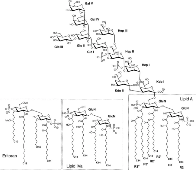
TLR4 and the coreceptor MD-2 comprise the physiological receptor for LPS. LPS is a complex glycolipid and the major component of the outer leaflet in the OM of most Gram-negative bacteria (Fig. 16). The structure
Fig. 16 Generic structure of LPS and lipid IVa from E. coli, and the synthetic antagonist Eritoran. No outer O-antigen polysac-charide is shown. Abbreviations: GlcN, 2-amino-2-deoxy-d-glucose; Kdo, 3-deoxy-d-manno-2-octulosonic acid; Hep, l-glycero-d-manno-heptose; Glc, D-glucose; Gal, d-galactose.
of LPS is highly variable, not only between different Gram-negative bacteria and different strains of the same bacteria, but can also vary within one strain depending upon growth conditions [136]; for example, depleting metal ions in the media, or growth in the presence of cationic host defense peptides (products of the innate immune system) can lead to rapid modification of LPS [137]. However, a common “lipid A” moiety can be recognized in all LPS molecules, which contains a bis-glucosamine disaccharide to which are typically attached 5–6 ester- and amide-linked lipid chains, as well as two phosphate groups, and sometimes other polar groups, such as phosphatidyl-ethanolamine (PE) and 4-amino-4-deoxy-l-ara-binose (Ara4N) (Fig. 16). Lipid A is not found naturally, but is released from LPS by mild acid hydrolysis. The lipid A moiety is linked to a carbohydrate core domain, typically comprising ≈6–8 sugar residues, including Kdo (3-deoxy-d-manno-oct-2-ulosonic acid) and Hep (l-glycero-d-mannoheptose), which are not generally found on mammalian cells. The core domain is then linked either to a long oligo-saccharide chain or to a highly immunogenic O-antigen polysaccharide, composed of repeat units that can be highly variable between different serotypes. The O-antigen is the most exposed segment of LPS on the bacterial cell surface. Overall, the LPS layer in the OM constitutes a major permeability barrier, which severely restricts diffusion of substances (e.g., antibiotics) across the OM and into the bacterial periplasm and cytoplasm.
LPS can stimulate a powerful immune response by signaling through TLR4. If the response is overwhelm-ing and uncontrolled, this immune protection mechanism can lead to fatal septic shock. LPS is extracted from the bacterial membrane and transferred to TLR4 by two accessory proteins, the LPS-binding protein (LBP) and CD14 [119]. Membrane-bound CD14 shuttles LPS to TLR4/MD-2 complexes at the cell surface, whereas a soluble form of CD14 enables cells that lack endogenous CD14 to also respond to LPS [122, 123]. Early work showed that the lipid A region is responsible for most of the immunostimulatory activity of LPS. The number of lipid chains has a large influence on inflammatory activity. Lipid A with six lipid chains has optimal inflam-matory activity, whereas with five chains the lipid A is ≈100-fold less active. With only four lipid chains, as found in lipids IVa and Eritoran (vide supra), the glycolipid typically has antagonist activity [138]. The mouse TLR4 complex appears to be more promiscuous in recognizing different forms of lipid A than human TLR4, and seems even to interact with the anticancer drug taxol [139]. However, minor changes in LPS structure can have large effects on immunostimulatory activity and even abolish endotoxic effects [140, 141].
2.4.1 The human MD-2/lipid IVa complex
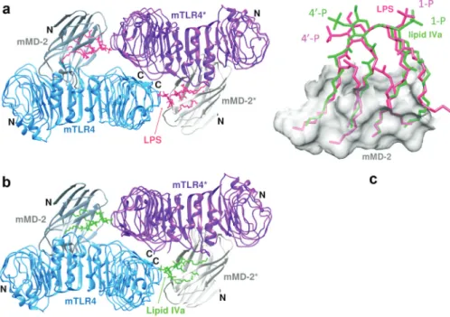
MD-2 is a ≈18 kDa secreted glycoprotein, which alone, as well as in the complex with TLR4, binds LPS with nanomolar affinity [85]. The number and length of the acyl chains have an important influence on signaling through the TLR4/MD-2 complex. For example, Escherichia coli lipid A is usually hexa-acylated and acts as a potent activator (agonist) of all mammalian cells. In contrast, tetra-acylated lipid A such as lipid IVa (Fig. 16), the biosynthetic precursor of E. coli LPS, acts as an agonist on mouse and as an antagonist (with antiendo-toxic) activity on human cells [142]. Lipid IVa does not induce formation of the human TLR4/MD-2 heterodi-mer complex, as required for signaling activity.
The first structural insight into how lipid A is recognized by MD-2 came from crystal structures of human MD-2 bound to lipid IVa [143]. MD-2 comprises a single immunoglobulin domain stabilized by three disulfide bonds. The protein consists of two β-sheets, one of three antiparallel β-strands, and the other of six antiparal-lel β-strands (Fig. 17a). Between the two sheets is a large, hydrophobic cavity with a volume of ca. 1710 Å3. The
overall structure is rather like a partly open clam, with the two shells represented by the two β-sheets. The four lipid chains of the ligand insert through the opening into the main body of the clam. In a crystal structure of MD-2 without bound lipid IVa, the MD-2 structure is not altered significantly; the lipid A binding pocket is still present, but is occupied by three myristic acid molecules that appear to have been purified and co-crystallized with MD-2. The lipid chains at positions 2 and 3 in lipid IVa are mostly fully extended and reach far into the hydrophobic binding pocket of MD-2 (Fig. 17b). The 2′ and 3′ lipid chains have a distinctly curved con-formation. The fatty acid chains in the cavity are packed next to each other and interact through van der Waals contacts. The size of the hydrophobic binding cavity is not large enough to accommodate the two additional lipid chains present in lipid A (and LPS). Rather, one of the additional lipid chains is found on the surface of MD-2 where it forms part of the contact interface with TLR4 in the TLR4/MD-2/LPS complex (vide supra).
Fig. 17 Structure of MD-2 with lipid IVa. (a) The four lipid chains of lipid IVa are buried between the two β-sheets in MD-2. (b) Surface representation of the complex. (c) Interactions between MD-2 and the head group [sugars (green/red) and phosphates (yellow/red)] of bound lipid IVa.
Fig. 18 Structure of the 1:1 TLR4/MD-2 complex with bound Eritoran (see text).
The 1- and 4′-phosphate groups and the two sugar residues in lipid IVa are at the solvent-exposed surface, in the opening of the clam-like structure. Here, hydrogen-bonding interactions occur between amide and ester carbonyls of the ligand and the edge of the β7 strand in the three-stranded β-sheet (Fig. 17c). Residues from Phe119-Gly123 are important for LPS recognition, and these residues with the exception of Lys122 are highly
conserved in MD-2. Overall, the entrance to the hydrophobic cavity has a positive electrostatic surface, which likely aids binding of the negatively charged ligand. However, neither of the phosphate groups is involved in close electrostatic contacts with atoms of MD-2.
2.4.2 The human TLR4/MD-2 with bound Eritoran
Eritoran (or E5564) is a synthetic molecule related to the lipid A of the nonpathogenic LPS of Rhodobacter
sphaeroides, with antagonist activity on human TLR4/MD-2. Eritoran contains a bis-glucosamine backbone
linked via amide or ether bonds to four lipid chains, one containing a cis-double bond, as well as two phos-phate groups (Fig. 16). However, neither Eritoran nor lipid IVa induces formation of the TLR4/MD-2 dimeric signaling complex (vide supra). The human MD-2 was crystallized with a hybrid hTLR4-hagfish-VLR fusion protein as a stable 1:1 complex, with Eritoran bound to MD-2 (Fig. 18) [102]. All four lipid chains in Eritoran are buried inside MD-2 and so are not available for interactions with TLR4. The glucosamine backbone in lipid IVa and Eritoran are superimposable when bound to MD-2. The shape of the lipid-binding pocket on MD-2 is essentially unchanged in the complexes with the two antagonists. Eritoran was in clinical development for use as a therapeutic against severe sepsis, however, the compound was recently withdrawn following incon-clusive phase III trials [111, 118].
2.4.3 The human TLR4/MD-2/LPS complex
LPS is extracted from the bacterial membrane and transferred to TLR4/MD-2 by LPS-binding protein and CD14. The crystal structure of the TLR4/MD-2/LPS complex has been very valuable in understanding ligand binding specificity and the mechanism of receptor activation. Interestingly, the signaling complex seen in the crystal structure is composed of two copies of TLR4/MD-2/LPS arranged in a symmetrical fashion (Fig. 19) [144].
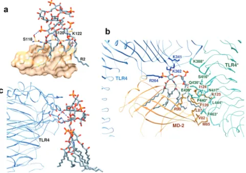
Fig. 19 Structure of the dimeric TLR4/MD-2/LPS signaling complex. TLR4 chains, blue; MD-2 chains, orange; lipid A, red; and outer sugar/phosphates, pink (see Section 2.4). (a) viewed from the side (membrane at the bottom), and (b) viewed from the top.
The two TLR4 chains each adopt a horseshoe-like shape, as seen with other TLRs, resembling in the complex the letter “m”, with both C-termini in the center and the N-termini located at each side of the “m” (Fig. 19). The two TLR4 chains contact each other in the center through LRR modules 15–17. MD-2 again has a clam-like shape composed of two antiparallel β-sheets, between which the hydrophobic lipid chains of LPS can bind. The two MD-2 chains are situated close to the center of each arm of the “m”. Each MD-2 makes contact with the side of one TLR4 chain and the exposed convex face of the other TLR4 chain. The number-ing system used for the lipid chains (R2, R3, R2′, R3′, R2″, and R3″) of the lipid A moiety is shown in Fig. 16.
Five of the six lipid chains of the lipid A are completely buried inside the hydrophobic pocket on MD-2, between the two β-sheets. However, the R2 chain is on the surface of MD-2 (Fig. 20a). This composite
Fig. 20 Structure of the dimeric TLR4/MD-2/LPS signaling complex. (a) Five lipid chains of LPS are buried and one (R2) is on the surface of MD-2. (b) The TLR4/MD-2 interface. (c) LPS sugar–TLR4 interactions.