Pure Appl. Chem. 2014; 86(10): 1435–1481
IUPAC Technical Report
Douglas M. Templeton* and Kerstin Moehle
Structural aspects of molecular recognition in
the immune system. Part I: Acquired immunity
Abstract: Humoral immunity allows the body to mount a defense against pathogens and foreign substances,
and to respond with memory to subsequent exposures. The molecular participants may also recognize self-structures, leading to attack on the body and autoimmune disease. The main players in humoral immunity are antibody-producing B lymphocytes, and several classes of T lymphocytes. This review deals with the molecular details of recognition of antigens by soluble antibodies, and of substances presented to recep-tors on the surfaces of T cells (TCRs). The prototype antibody consists of a dimer of dimers, two heavy (H) chains and two light (L) chains, with antigen recognition capacity lying in variable “head” regions of an H-L pair. Most crystallographic studies are done with this substructure, called a Fab fragment, bound in a soluble antigen complex. Homologous to this arrangement, the prototype TCR consists of two chains (α and β) that complex not soluble antigen, but usually a short peptide or other small molecule presented by proteins of the major histocompatibility complex. In each case a general background on the historical development of understanding the molecular recognition interface is given, followed by a number of examples of crystal structures from the recent literature that have allowed us to refine our understanding of the complex recog-nition process. Variations on the prototypical structures are also considered. The spectrum of recogrecog-nition strategies involves interplay of lock-and-key with flexibility, varying degrees of entropic and enthalpic contri-butions, surface shaping by entrapped water molecules, and combinations of stabilizing hydrogen bonding, electrostatic interactions, salt bridging, and van der Waals forces. Preeminent in the recent literature are details of antibody binding to influenza A and human immunodeficiency viral antigens. Both viral antigens and attempts to understand autoimmunity are prominent in the recent TCR literature.
Keywords: antigens, antigen–antibody recognition, immunity, IUPAC Chemistry and Human Health Division,
molecular recognition strategies, T-cell receptors, viral epitopes. DOI 10.1515/pac-2013-1020
Received October 18, 2013; accepted April 17, 2014
CONTENTS
1. General Introduction ...1436
2. Antigen–antibody (Ag-Ab) recognition ...1436
Information Boxes 1–3 ... 1436
2.1 Introduction ... 1437
2.2 Historical development ... 1439
2.3 Current perspectives ... 1441
Article note: Sponsoring body: IUPAC Chemistry and Human Health Division; see more details on p. 1479.
*Corresponding author: Douglas M. Templeton, Department of Laboratory Medicine and Pathobiology, University of Toronto, 1
King’s College Circle, Toronto M5S 1A8, Canada, e-mail: [email protected]
Kerstin Moehle: Chemistry Department, University of Zurich, Winterthurerstrasse 190, 8057 Zurich, Switzerland
2.4 Viral epitopes ...1451
2.4.1 Influenza A ...1451
2.4.2 HIV ... 1453
2.4.3 Other viruses ... 1456
2.5 Conclusions ...1460
3. T-Cell receptor (TCR) recognition ... 1460
Information Boxes 4,5 ...1460 3.1 Introduction ... 1461 3.2 Historical development ... 1461 3.3 Current perspectives ... 1463 3.3.1 General aspects ... 1463 3.3.2 Viral antigens ... 1465 3.3.3 Autoimmunity ...1468
3.3.4 Natural killer T (NKT) cell receptors ...1472
3.3.5 γδTCR structures ... 1476
3.3.6 Metal ion recognition ... 1476
3.4 Conclusions ... 1477
4. Abbreviations ...1478
5. Membership of sponsoring body ...1479
6. Acknowledgments ...1479
7. References ...1479
1 General introduction
The immune system provides humans, and a number of other species of higher organisms, with protection against a variety of microorganisms, biological molecules, and other chemical structures that it recognizes as being “foreign” to the self. The human immune system embodies an innate response that offers immedi-ate recognition and defense, as well as acquired immunity that provides defense upon recognition of repeat exposure. Acquired immunity involves both humoral immunity resulting from the production of antibodies by a class of white blood cells called B lymphocytes, as well as cell-mediated immunity achieved by activation of a group of thymocyte-derived cells – the T lymphocytes. Triggers and molecular mechanisms of activation of innate, humoral, and cell-mediated immunity are exceptionally complex, and are discussed in a number of good textbooks of immunology (see [1] for an excellent recent example). The aim of the present paper is not to provide background on mechanisms of immune activation, but rather to discuss structural aspects of molecular recognition within the immune system. Thus, we present principles and examples of recognition of antigens by B lymphocyte-derived antibodies, and of antigens presented in specific molecular complexes to receptors on T lymphocytes. A companion paper deals with interactions that mediate innate immunity.
2 Antigen–antibody (AG-AB) recognition
Information Box 1
Antigens, structures that are specifically recognized by and interact with receptors on the surfaces of cells of the immune system and with secreted antibodies, include protein epitopes, carbohydrates, lipids, nucleic acids, and small-molecule haptens. The recognizing antibody (immunoglobulin) consists of two main regions, called the variable (V) region and the constant (C) region. The variable region is the antigen-recognition site, and its variability from antibody to antibody allows the occurrence of antibodies that each can show speci-ficity against the nearly endless array of foreign and self-antigens that the immune system may encounter.
D.M. Templeton and K. Moehle: Molecular recognition in the immune system. Part I 1437 Information Box 1 (Continued)
The constant region is the effector part of the molecule that links the antigen–antibody complex to common immune system responses such as lymphocyte activation, phagocytosis, and the complement cascade.
Information Box 2
Human immunoglobulins (Ig) consist of two heavy (H) and two light (L) chains connected by disulfide bonds and are traditionally depicted as a planar “Y-shaped” structure (Fig. 1a). Based on the nature of the H chains, human antibodies are classified as IgG, IgA, IgE, IgM, and IgD, with H chains designated γ, α, ε, μ, and δ, respectively. The most widely studied, IgG, has subclasses IgG1–IgG4 based on the occurrence of H chains γ1–γ4. More detailed structural analysis reveals the L chains have an N-terminal V domain (VL) and a carboxy-terminal CL domain. As well as corresponding VH and CH1 domains, the H chains have additional CH2 and CH3 domains. An extended peptide region between CH2 and CH3 is referred to as the hinge region. Each domain is structured with two juxtaposed β sheets consisting of three and four anti-parallel β strands (five and four in the V domains), respectively, protecting interior hydrophobic residues. Domains VH-VL, CH1-CL, and CH3-CH3 are in close pairing contact; a variable carbohydrate chain on each CH2 domain disrupts CH2-CH2 contacts. Antigen recognition resides in the VL-VH domain pairing, with three regions of hypervariable amino acid sequence in each domain being the major determinants of recognition. Effector functions reside mainly in the CH2 domains. When cleaved from each other, the disulfide-bonded VL-VH-CL-CH1 fragment is referred to as Fab (frag-ment antigen binding) and the disulfide-bonded (CH2-CH3)2 fragment is denoted Fc (fragment crystallizable). While the traditional depiction is of a “Y-shaped” molecule, the structure has considerable flexibility in the hinge region (Fig. 1b). Bending (of the Fab at the elbow hinge), wagging (of both the Fab arm and the Fc region), and rotational motions (of the Fab arm) allow the Fab-Fab angle to more closely approximate a “T-shape”, the Fc component may rotate out of the Fab plane, and the CH2 domain may more closely approach one of the CH1 or CL domains.
Information Box 3
The variable regions in the Fab fragment binding structures are punctuated by even more variable (thus, hypervariable) regions, three each in the L chain and H chain. The high degree of amino acid sequence het-erogeneity in these so-called complementarity-determining regions (CDRs) provides much of the variability necessary to recognize the huge number of possible antigens with high specificity. Each of the CDRs, desig-nated L-chain CDR1-3 and H-chain CDR1-3, can be involved in antigen recognition. However, H-chain CDR3 is special: it is often dominant in binding, and it has an extended finger-like conformation that often allows it to penetrate into grooves and pockets of the antigen, providing multiple favorable contacts. It will be seen below that CDRs also occur in the T-cell receptor. As will be appreciated, the somatic gene rearrangements and sequence shuffling events that give rise to these hypervariable sequences are quite complex, and have been studied in detail, but they are beyond the scope of this discussion of molecular recognition.
2.1 Introduction
In general, recognition of a protein is the responsibility of the Fab end of the antibody and is strongly depend-ent on the hypervariable regions (complemdepend-entarity-determining regions or CDRs). A “goodness-of-fit” model applies, with thermodynamic affinity dependent on the number of favorable contacts. Concepts of protein– protein interactions apply, e.g., an increase in entropy when water is excluded from hydrophobic interfacial contacts decreasing the free energy and increasing affinity. Antigenicity refers to the “ability of an antigen
to bind to a specific antibody...often a measure of its ability to produce immunity” [3], the point being that strength of binding per se does not elicit the immune response, which is better referred to as immunogenicity.
The part of the antigenic molecule recognized by the antibody receptor is referred to as an epitope, and a protein antigen may have multiple epitopes that are recognized by distinct antibodies. Mapping the epitope surface of a protein with different antibodies is called epitope footprinting, and it may be observed that some epitope footprints overlap (for instance, when a particular amino acid is involved in two or more epitopes), while others do not. In antibody recognition of an antigenic protein, it is often noted that amino acid residues from well separated regions of the protein sequence are involved in an epitope, then said to be discontinu-ous, due to folding of the protein into a tertiary structure that brings these residues into spatial proximity. In special cases, several contiguous amino acids may constitute the epitope, and then the peptide that they define is said to represent a continuous or linear epitope.
A good discussion of the types of protein surface structures “seen” by antibodies is given in Delves et al. [1] (chapter 5). While complementarity of fit (shape) between antigen and antibody is an obvious considera-tion, flexibility is also important. Flexibility may allow the antibody to conform better to an epitope, or even represent a conformational change induced in the antibody by antigen. Often an antibody will recognize an extended surface, exploiting additivity of thermodynamically favorable contacts. Or, it may have an apparent cavity or groove, especially to accommodate a small molecule (hapten). The hypervariable region H-chain CDR3 is often a major contributor to recognition, and may take on an extended finger-like conformation that may fit into a pocket on a antigenic protein. A common antigenic motif is a loop protruding from the protein surface, which tends to allow more flexibility than the surrounding compact surface. Continuous epitopes are often well-structured, such as an α-helix preserved in the isolated peptide.
We now consider some historical developments that illustrate these points and the various approaches to antigen–antibody structural recognition.
H chain a b L chain Ag-binding site Papain digestion Intact lg Fc Fab S SS S SS SS SS SS
Fig. 1 Depictions of immunoglobulin structure. (a) Conventional schematic of a bivalent immunoglobulin showing
disulfide-bonded H chains (blue) and L chains (red), presenting two binding sites for antigen (green). Upon digestion with papain, cleav-age of the H chains releases two Fab and one Fc fragments. (b) A more realistic depiction based on the crystallographic data of [2]. The two L chains are shown in blue, the H chains in green.
D.M. Templeton and K. Moehle: Molecular recognition in the immune system. Part I 1439
2.2 Historical development
Houghten [4] presented a rapid method of solid phase peptide synthesis that allowed, as a secondary objec-tive, extending existing knowledge that antigenic determinants resided in protein sub-structures, by attempt-ing to identify sattempt-ingle amino acids involved in the interaction. The approach was to study monoclonal antibody binding by enzyme-linked immunoabsorbent assay (ELISA) to a series of peptide variants. The antibody was raised to the linear epitope representing amino acid residues 75–110 of influenza hemagglutinin, HA1, and 248 single amino acid variants of residues 98–110 were tested. The results showed that Asp101 was critical for antibody binding, while Asp104 and Ala106 also played significant roles, with the other amino acids having little influence on binding.
An early example of the use of synthetic peptides to probe the antigen–antibody interaction used four monoclonal antibodies recognizing linear epitopes at the 18-residue C-terminus of rhodopsin [5]. It was con-cluded that ionic interactions can dominate the interaction, but the antigenic determinant was not restricted to highly charged regions. Results were consistent with the view that the free energies of all contact point interactions are additive and a threshold energy must be exceeded to achieve binding. But a deeper under-standing of the binding interaction awaited crystallographic data.
At this time, creative use was being made of physical methods for the qualitative understanding of antigen binding. Surface plasmon resonance (SPR) continues to be useful in characterizing antibody-binding affini-ties, although a great deal of suspect data have probably been generated by over-interpretation of results (see D. G. Myszka’s entertaining Foreword in [6] for a thoughtful discussion). An early example of its use was gen-erated in-house at Pharmacia in Uppsala during the development of commercial instrumentation. Fägerstam et al. [7] immobilized an anti-mouse IgG1 on a dextran-derivatized sensor chip and then washed with either a monoclonal antibody to human immunodeficiency virus (HIV) core protein p24, or an unrelated antibody to block unoccupied IgG1 sites. Antigen (p24) was then passed over the surface, followed by a second distinct anti-p24 monoclonal antibody. This protocol allowed a type of epitope mapping by assessing the interactions of the first and second anti-p24 antibodies. Up to five antibodies could be bound sequentially, resulting in a hexamolecular complex of non-overlapping epitopes, and these could be mapped further using competing peptides from known sites on the antigen. Analysis of competition also allowed comment on partial epitope overlap. Although the authors point out the advantage of SPR in not requiring labeling of individual antibod-ies in these studantibod-ies, they also recognized the inherently qualitative nature of the results.
Atomic force microscopy was also being brought to bear. In early experiments [8], biotin linked by a spacer arm to albumin served as the antigen and was immobilized on the sensor tip of the atomic force microscope. Antibody (polyclonal anti-biotin IgG) was immobilized on a flat gold substrate with dithio-bis(succinimidylundecanoate) crosslinker to minimize antibody-surface contacts and potential attendant conformational changes. After binding, retraction forces in multiples of 60 ± 10 pN were observed, which were absent when the reaction was blocked with streptavidin, when albumin replaced biotinylated albumin, etc. In principle, with monoclonal antibodies and careful antibody orientation, it should be possible to cor-relate these force measurements with data from thermodynamic experiments.
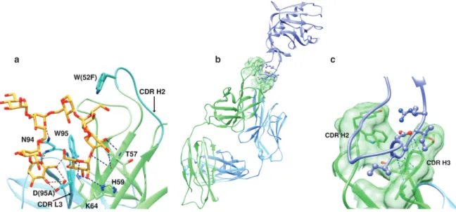
Notwithstanding the elegant use of these physical techniques, a more detailed appreciation of structural interaction of course awaited crystallographic studies. Not long after the early report of Houghten [4], X-ray structures of two crystal forms of monoclonal Fab HyHEL-5 bound to hen egg white lysozyme were published [9]. While structures of Fab fragments with haptens had been studied previously, and demonstrated the pres-ence of hapten binding pockets [10], the possibility that complexes with protein antigens might more resemble other protein–protein interfaces with exclusion of water and induced conformational changes in the antibody were considered. The results validated earlier epitope-mapping studies [11]. The lysozyme epitope consisted of three sequentially separated regions. A shallow groove accommodated positively charged Arg45 and Arg48 of lysozyme that were complemented by two negative Glu residues in the antibody heavy chain (Glu35 of H1 and Glu50 of H2) (Fig. 2). Shape and hydrogen bonding are also important, as change of Arg68 to lysine signifi-cantly decreases antibody affinity. Otherwise, the area of contact was relatively flat (about 1.9 × 2.6 nm), and contained no water in the interface region. The backbone structure of lysozyme was mostly unperturbed, and
Fig. 2 An epitope in hen egg white lysozyme (pink ribbon) lies in a shallow groove on the antibody surface (green). Two Arg
residues (N’s blue) from lysozyme contact two Glu residues (red) in the antibody H1 and H2 loops. The figure is based on the crystallographic data of [12].
Fig. 3 A more detailed look at the contact of a hen egg white lysozyme antibody with the lysozyme (brown) epitope reveals
multiple contacts with all 6 CDRs of the Fab (blue and green) that smooth the surface fit. See text for details. The figure is based on the crystallographic data of [13].
only 30 % of the residues in the CDRs were in contact with lysozyme. It was concluded [9] that binding of Fab to protein antigen shares with hapten binding charge neutralization at the interface, but results in much greater surface area of contact with exclusion of water. Variable elbow bends in the early structures were indicative of flexibility in this region of the antibody but did not correlate with binding affinity.
When Li et al. published a refined structure of Fab HyHEL-63 bound to lysozyme in 2000 [13] they noted that the three-dimensional structures of about 25 complexes between antibodies and various protein antigens by then had been determined. A significant difference between their experiments and those with the earlier lysozyme-antibody binding study was the ability to crystallize the Fab in the absence of antigen, and refine-ment of all structures to about 0.20 nm resolution. In addition, they introduced single Ala mutations into the lysozyme epitope to study energetic contributions of individual residues. Their results showed several notable differences from those discussed above. First, all six CDRs of Fab are in contact with the lysozyme epitope, and the interfacial interaction includes 24 hydrogen bonds, a salt bridge, and 170 van der Waals contacts (Fig. 3).
D.M. Templeton and K. Moehle: Molecular recognition in the immune system. Part I 1441
While the antibody side of the interface is predominantly hydrophobic, hydrophilic residues predominate on the lysozyme side, with charged residues tending to be at the periphery pointing away from the center of the interface. Alanine substitutions revealed that residues at the center of the interface contributed most to complex stability. Second, because the Fab antibody was crystallized with and without antigen, a compari-son of structures was able to reveal small conformational movements in the CDR loops and flexibility in side chains of amino acids in the interface region that enhanced complementarity of binding. Third, far from being devoid of water, 11 ordered water molecules could be ascertained in the interface region that were not observed at fixed sites in lower resolution. By filling gaps in the interface region, trapped water molecules increased surface complementarity. The authors suggested that in contrast to other protein– protein interactions that have maximized complementarity through coevolution, water molecules may serve to correct imperfections in antigen–antibody interfaces by improving fit and accommodating unpaired hydrogen-bonding residues.
2.3 Current perspectives
The above considerations have briefly set a context for a detailed examination of antigen–antibody interac-tions. Lock-and-key and induced-fit models are appropriate, and protein recognition by antibodies is firmly established as residing in the Fab domains, strongly involving the CDRs of the hypervariable regions. Ther-modynamic principles are useful in explaining binding affinities, with van der Waal’s contacts, hydrophobic and π interactions, hydrogen bonding, and electrostatic interactions all contributing in a roughly 2.5 × 2.5 nm2 surface; solvent (water) plays a significant role both entropically with respect to its exclusion, and also by its inclusion to shape the contact surface. Bending and rotating angles in the antibody are somewhat flexible, and such flexibility can contribute significantly to binding energy. Several hundred antigen–antibody crystal structures are available today, and newer structures are focusing not on complexes with model proteins such as lysozyme, albumin, and hemagglutinin, but rather on structures of therapeutic and biomedical signifi-cance, such as receptors and viruses. What more can be learned from these to guide future antibody design? Several recent examples are discussed here to illustrate some details of the binding interface, and they dem-onstrate how the field has quickly matured to studying structures of potential therapeutic significance. But, while each of these recent papers discussed below begins with a discussion of relevance to disease rather than addressing a question in physicochemical determinants of the antigen–antibody interaction, each nev-ertheless contributes to our growing understanding of the chemical nature of the antigen–antibody interface. A majority of therapeutic antibodies fall into the category of inhibitors of target protein function. Trans-membrane serine proteases are over-expressed on the plasma Trans-membrane of some cancer cells, where they may play a role in matrix remodeling and invasion. A phage display library created with a repertoire of Fabs from human B cells was used to identify Fab A11, which bound and inhibited with high specificity the trans-membrane type II serine protease, matriptase [14]. The crystal structures of matriptase complexed with A11 and with a previously known potent matriptase inhibitor, Fab S4, were determined at 0.21 and 0.15 nm, respec-tively, and compared. Both antibodies made multiple contacts with multiple surface loops on the protease and a total buried surface area 10–12 nm2.1 In both cases, H and L chains both contributed to surface coverage, with the H3 loop making the major contribution (63 % of the total for S4, 38 % for A11) (Fig. 4). Most of the A11 contacts are nonpolar, whereas S4 interacts with multiple polar interactions in a network of hydrogen bonds and π-stacking interactions. Binding had little effect on matriptase conformation remote from the binding site. In both cases, the H3 loop resides in the substrate-binding pocket, but the greater potency of the S4 inhibitor may be due to greater interaction of H3 with the active site. While H3 loops of both S4 and A11 form β-hairpin turns in the binding pocket, A11 makes fewer contacts and has less extensive coverage. These observations suggest a means of tailoring the H3 loop to increase inhibitor potency. In contrast, another
1 Note that above, a typical footprint of 2.5 × 2.5 nm = 6.25 nm2 was indicated, which would typically bury a surface area of about 12.5 nm2, taking both surfaces into account. The distinction between footprint and buried surface area should be kept in mind throughout this text.
transmembrane type II serine protease, hepsin, is prominent in prostatic cancer. A human monoclonal antibody, hH35, selectively inhibits hepsin, but does not appear to interfere directly with substrate binding, acting rather by allosteric inhibition [15]. Fab hH35 binds to an α-helix well away from the active center, but leads to major structural changes that inactivate the enzyme.
Given the importance of angiogenesis in cancer progression, a good deal of information has been pre-sented on the binding of the vascular endothelial growth factor (VEGF) family members to the VEGF receptors. The Fab portions of two antibodies that block VEGF binding to and/or signaling through VEGF receptor 2/KDR have been crystallized in complex with domain 3 of KDR and solved to 0.22 nm resolution [16]. The antibod-ies, 1121B and 6.64, had previously been shown to bind exclusively to domain 3, and to do so independently of one another. Fab 6.64 makes contact over an extended region of KDR domain 3 (coverage 9.82 nm2, total buried surface area 20.3 nm2), involving a highly discontinuous epitope, and contacts the C-terminus (representing the KDR domain 3/domain 4 interface) (Fig. 5). All six H- and L-chain CDRs make contact, and 45 % of contact is with the L chain. Fab 1121B binds mostly to a more localized region (nearly continuous β-hairpin epitope) at the N-terminus of domain 3, near the domain 3/domain 2 interface, accounting for the lack of competition of the two antibodies (Fig. 6). Binding is dominated by the H chain, with the L chain contributing one charge pair and a few hydrophobic interactions for a coverage of 10.0 nm2 (total buried surface 19.2 nm2). There is very slight change in Fab conformation on binding, and also not much change in KDR domain 3 structure, consistent with the localized binding. The results support two different mechanisms for inhibition. While Fab 1121B does not share binding residues with VEGF, it is immediately adjacent to the VEGF binding site and may sterically inhibit binding. On the other hand, Fab 6.64 is not near the VEGF binding site, but may induce conformational changes in the domain 3/domain 4 region that interfere with receptor dimerization.
Potent allergens in house dust mites include the cysteine proteases Der p 1 and Der f 1, which induce a strong response of IgE antibodies [17]. These antigens share 81 % sequence identity and elicit highly cross-reactive responses. A Fab fragment of a monoclonal antibody 4C1, which inhibits IgE binding, was used to identify a common epitope in IgE–Der f 1/p 1 interaction. The epitope was well away from the active center. Buried surface areas of 7.5–7.9 nm2 on the two antigens were found in the crystal structure at 0.205 nm resolu-tion. In coverage, 70 % was contributed by contacts with the H chain, mostly arising from the H-chain CDR3
Fig. 4 Complex of the serine protease inhibitor, matriptase, with inhibitory Fab S4. All three CDR loops of the H chain (H1–H3) and the L chain (L1–L3) make contact in the interfacial region, with the H3 loop dominating the interaction. The figure is based on the crystallographic data of [14].
D.M. Templeton and K. Moehle: Molecular recognition in the immune system. Part I 1443
Fig. 5 Structure of the anti-VEGF receptor KDR3 domain bound to Fab 6.64. Note the highly discontinuous epitope, which makes contact with all six H-chain (green) and L-chain (blue) CDRs. See text for more detail. From the crystallographic data of [16].
region being accommodated in a concave region of the antigen surface (Fig. 7). The antigen presented a central rigid surface of discontinuous residues. Overall conformation of the antibody and the conformations of its CDRs were similar in complexed and uncomplexed states, indicating little rearrangement upon binding. Conserved water molecules in the two antigens improved the fit at the interacting surfaces. Elbow angles (CL-CH1 and VL-VH) showed some flexibility (e.g., 146° and 139° in two different crystal forms of 4C1 Fab com-plexes), consistent with differences of 143° and 167° in two orthorhombic crystal forms of the uncomplexed Fab (Fig. 8). The authors [17] proceeded to analyse 16 available allergen–antibody complexes in the structure database and found a common theme that Tyr and Ser residues in the antibody played a dominant role in interaction with the allergen, followed by Arg, Asn, and Asp. In 4C1 interactions Tyr and Arg were particularly prominent, with potential contributions from hydrogen bonding, π bonding, and hydrophobic interactions. Interestingly, hydrogen-bonding networks in the binding interfaces of the 16 allergens were most frequently formed by Arg-Asp, Arg-Tyr, Asp-Arg, Asp-Tyr, Glu-Arg, Glu-Ser, and Gln-Ser pairs, distinct from a number of lysozyme–antibody complexes where Arg-Glu, Arg-Tyr, Asn-Gln, Asp-Ser, and Lys-Asp predominated [17].
A structure has been reported [18] at 0.28 nm resolution of the interaction of a single-chain variable domain (scFv) antibody, with interferon α (IFNα), of relevance to the study of the autoimmune disease, sys-temic lupus erythematosus. Single-chain Fv antibodies are fusion proteins of the H- and L-chain variable regions linked with a spacer sequence, isolated following a strategy of cloning variable-domain genes. The
Fig. 6 Structure of the anti-VEGF receptor KDR3 domain bound to Fab 1121B. Note the highly localized β-hairpin epitope in KDR3 that is predominantly bound to the H chain (contrast this with the binding mode described in Fig. 5). From the crystallographic data of [16].
structure of the complex revealed that three distinct regions in the interface contributed to high-affinity binding. In each case, a protruding side chain of one partner fits a cavity in the other – two cavities in IFNα and one in the antibody – allowing the structures to “zip together” (Fig. 9). Shape complementarity was thus a major determinant of binding strength, with an important salt bridge in one contact, numerous van der Waals and π-bonding interactions in another, and structural entrapment of an antibody Phe residue in an IFNα cavity in the third. Sixteen hydrogen bonds are included in the interactions.
The potentially therapeutic antibody ponezumab is directed against the β-amyloid peptide Aβ40, aggre-gation of which is implicated in the pathogenesis of Alzheimer’s disease. La Porte et al. [19] have reported the crystal structure of ponezumab Fab complexed with Aβ40 at 0.25 nm resolution. The antigen-binding site is in a cleft between the H- and L-chain variable domains, includes all six CDRs, and buries 7.59 nm2 of the peptide surface. Interaction is dominated by nonpolar amino acid residues, with 10 amino acids from Aβ40 and 24 from ponezumab, 12 of the latter contributed by each chain. The C-terminal carboxyl group plays a key role in binding as demonstrated by truncation studies and SPR, and in the crystal structure it is buried by contact with Lys, Arg, and two Tyr residues of the H-chain variable domains (Fig. 10).
Of relevance to another neurodegenerative disease, the monoclonal antibody 3B5H10 binds to huntingtin (htt), the protein implicated in Huntington’s disease. In common with several proteins implicated in neuro-degenerative diseases, htt contains stretches of polyglutamine that are expanded with increasingly severe manifestations of the disease. It is thought that expansion of the polyglutamine region leads to pathological protein self-aggregation with a cross-β-strand structure similar to β-amyloid. A crystal structure of 3B5H10 Fab at 0.19 nm resolution revealed enrichment of solvent-accessible aromatic residues in the H-chain CDRs, facilitating glutamine binding [20]. In fact, 30 % of the H-chain CDRs were aromatic, compared to 9 % in 100 closest homologues. In a second polyglutamine-recognizing antibody, MW1, this number is 39 % [20]. Upon recognition of the polygluatmine epitope, a 3° rotation between the H- and L chains results in a linear groove that accommodates the antigen. An unusual rigid loop structure in L-chain CDR3 forms part of the wall of
D.M. Templeton and K. Moehle: Molecular recognition in the immune system. Part I 1445
Fig. 7 A Fab fragment of a monoclonal antibody 4C1 was used to identify a common epitope in interaction of IgE with house dust mite antigens Der f 1/p 1 [17]. 70 % of surface contact was contributed by interactions with the H chain, mostly arising from the H-chain CDR3. Antibody contact surfaces within 0.5 nm distances to the antigen are colored dark green (H chain) and dark blue (L chain). Antigen contacts, orange.
the binding groove in both 3B5H10 and MW1. While only a single strand of polyglutamine is accommodated in the MW1 groove (Fig. 11a), in 3B5H10 a widened groove with an extended β-hairpin loop of H-chain CDR2 interacting with H-chain CDR1 and L-chain CDR3 (Fig. 11b) fits a compacted two-stranded hairpin in the polyglutamine peptide. Because recognition by 3B5H10 of htt with expanded polyglutamine sequences is a better predictor of neurodegeneration than recognition by MW1, the compacted polyglutamine loop may be of pathological significance.
The A2A adenosine receptor, A2AAR, is a G protein-coupled receptor involved in regulating cardiac blood flow and regulation of release of dopamine and glutamate neurotransmitters in the brain. It is antagonized by caffeine. A monoclonal antibody Fab fragment, Fab2838, blocks binding of receptor ago-nists, but not the antagonist ZM241385. A2AAR with bound ZM241385 has been crystallized in the pres-ence of Fab2838 and solved at 0.27 nm resolution [21]. ZM241385 occupies an extracellular ligand-binding pocket with hydrophobic (Phe and Ile) and hydrogen-bonding interactions (Asn). Fab2838 binds on the intracellular side of the receptor with an unusually long H-chain CDR3 fitting a pocket formed by four receptor helices (Fig. 12) and stabilizes an inactive form of the receptor. Interactions are mainly hydro-gen-bonding and van der Waals, with interaction between CDR3 and two of the helices stabilized by a hydrogen-bonding network containing two water molecules. Other CDRs form an additional 14 hydrogen bonds leading to a high-affinity interaction with Kd = 4.4 nM. For comparison, another antibody, Nb80, binds to the β2AR with a similar H-chain CDR3 β-hairpin and recognizes an active form of the receptor, whereas the β-hairpin Fab2838 CDR3 induces a different shape of binding pocket and locks the receptor in an inactive conformation [21].
Fig. 8 Flexibility of elbow angles occurs in two different crystal forms of Fab 4C1 directed against dust mite protease antigen. Superimposed structures are based on the crystallographic data of [17]. Angles are 143° and 167° for P212121 (blue) and C2221 (orange) forms, respectively.
Venoms that act as blockers of membrane channels have been another target for exploration of potential therapeutic antibodies. The scorpion Androctonus australis hector (Aah) produces lethal peptide toxins AahI, II, III, and IV that block mammalian voltage-gated Na+ channels. The peptides share a so-called cystine-stabilized α-helix structure consisting of an α-helix and an antiparallel three-strand β-sheet, cystine-stabilized by disulfide bridges. However, variation in the amino acid side chains among the peptides precludes cross-reac-tivity and prevents generation of a single antiserum against all the toxins. A high-affinity Fab was generated from a monoclonal antibody 4C1 to AahII, and the crystal structure of the Fab4C1–AahII complex was solved at 0.23 nm resolution [22]. Another antibody fragment, Fab9C2 directed against AahI, excluded its antigen from the crystal structure. The Fab4C1–AahII complex was described by the authors as resembling “an egg inserted small-end first in the egg cup”. The Fab4C1 “egg cup” consists of a binding pocket 1.3 nm deep and 1.2 nm wide formed by the six CDRs, with an elbow angle between the variable and constant domains of the H and L chains of 140.5° (Fig. 13). An extended L-chain CDR1 and an anionic H-chain CDR2, together with the long H-chain CDR3, form a boundary that “clamps” the cationic toxin. L-chain CDR2 and CDR3, and a hydropho-bic H-chain CDR1 serve as additional anchoring points. Strong complementarity buries about 25 % (10 nm2) of the venom surface at the epitope–paratope interface. A more planar, less anionic, hydrophobic binding surface in Fab9C2 may account for exclusion of antigen in attempts to crystallize its AahI complex. Also, an elbow angle of 171° in Fab9C2 suggests greater flexibility [22].
D.M. Templeton and K. Moehle: Molecular recognition in the immune system. Part I 1447
Fig. 9 Interaction of a single-chain variable domain (scFv) antibody with interferon α (IFNα), showing how the structures “zip” together. The antibody is depicted in a ribbon structure contacting IFNα (solid surface). A Met residue (magenta) from IFNα fits into a pocket formed by a Trp, an Arg, and two Asn residues of the antibody, while antibody H-chain (green) Phe and L-chain (blue) Tyr residues are accommodated in distinct cavities of IFNα. After [18].
Fig. 10 Structure of the β-amyloid peptide Aβ40 complexed with Fab ponezumab. The antigen binding site is in a cleft between the H- and L-chain variable domains (LV left, HV right). Dominating the interactions is that between the peptide carboxyl terminus (orange, O red, N blue) in contact with Lys, Arg, and two Tyr residues of the H-chain variable domains. From [19].
Complement factor D is an enzyme of the complement pathway of innate immunity. Due to a high turn-over rate, factor D is present at low concentrations in blood and becomes rate-limiting in the alternative complement pathway, making it a therapeutic target for inappropriate complement activation. Monoclonal antibody 166-32 binds to factor D with high affinity, and a humanized Fab fragment that blocks complement activation has been prepared by grafting the CDRs of 166-32 on human germ line immunoglobulin regions; this construct has been designated AFD. The crystal structures of AFD bound to human and cynomolgus monkey factor D have been solved at 0.24 and 0.23 nm, respectively [23]. The two structures superimposed closely, and no significant changes in factor D structure were induced upon binding. The buried surface of factor D is 9.01 nm2 (total of epitope plus paratope surface 16.5 nm2) with 4.85 and 2.63 nm2 covered on the AFD H chain and L chain, respectively. Eighty percent of the buried surface of factor D involves a protein loop sandwiched between the H chain and L chain and an Arg residue deeply buried in potential hydrogen
a b
Fig. 11 Huntingtin protein (htt) binding to Fab structures. (a) A polyglutamine sequence of htt lies along a groove formed across the VH–VL interface of antibody MW1 [20]. The strong interaction with L-chain CDR3 is somewhat unusual. (b) Note that in another htt-binding Ab, 3B5H10, the polyglutamine groove is widened by an extended H-chain CDR2 that interacts with H-chain CDR1 and L-chain CDR3, potentially accommodating a compact hairpin in the peptide (coordinates with peptide not available).
Fig. 12 Monoclonal Fab2838 blocks binding of agonists, but not antagonists, to the adenosine A2A receptor. An unusually long H-chain CDR3 loop (dark green) fits a pocket formed by four adenosine receptor helices (brown). The figure is based on the crystallographic data of [21].
bonding and electrostatic contact with two Glu residues on the H chain and Tyr and Asn residues on the L chain (Fig. 14). An additional three Asp residues on the L chain form charged interactions with a factor D Lys residue, and contribute to very high affinity binding (Kd < 10 pM). Interactions differ slightly with the
D.M. Templeton and K. Moehle: Molecular recognition in the immune system. Part I 1449
Fig. 13 The fitting of a scorpion toxin (red) into Fab4C1 makes contacts with all six CDR loops of the H chain (green) and the L chain (blue). The authors [22] have described this as “an egg inserted small-end first in the egg cup” (see text for details). The basket around the toxin represents the molecular surface of the protein in mesh style, and the surface around Fab antibody is rendered as a transparent solid.
Fig. 14 Crystal structure of complement factor D bound to the engineered antibody AFD (see text for details). A protein loop
of factor D (solid surface) is sandwiched between the H chain (green) and L chain (blue) of the antibody, and an Arg residue is in potential contact with two Glu residues on the H chain and Tyr and Asn residues on the L chain. The figure is based on the crystallographic data of [23].
cynomolgus protein, and the affinity is lower. The binding interface is well removed from the catalytic site, and inhibition of complement activation appears to be due to steric hindrance between factor D and its sub-strate, factor C3bB [23].
“Two-in-one” antibodies are known that can bind with the same site to two different epitopes. An inter-esting example is D11 [24]. An antibody was generated against the epidermal growth factor receptor (EGFR)/ human epidermal growth factor receptor-1 (HER1) from a library with diversity restricted to H-chain CDRs, so
a b
Fig. 15 Comparison of epitope-binding sites of the homologous receptors (a) HER3 and (b) EGFR. The surfaces of the two
recep-tors bound to Fab D11 (antibody not shown) are represented with contact atoms within 0.5 nm of the antibody colored blue. The figure is based on the crystallographic data of [24].
H7 H15 Group 2 H10 H3 H4 H14 H9 H8 H12 H11 H13 Group 1 H16 H6 H1 H2 H5
Fig. 16 Phylogenetic relationship among the influenza hemagglutinin subtypes. Adapted from [25].
it was assumed EGFR binding resided in the H chain. Mutations were then introduced in L-chain CDRs until a Fab was identified which bound the homologous HER3 receptor, without loss of EGFR binding. It is believed that binding to two distinct members of the HER receptor family will increase cancer activity of the anti-body. The epitopes on the two receptors do not exactly correspond (Fig. 15), but binding features are similar.
D.M. Templeton and K. Moehle: Molecular recognition in the immune system. Part I 1451
Surface coverage is somewhat higher in HER3 (8.9 vs. 8.0 nm2 in EGFR), accounted for by additional involve-ment of L-chain contacts, as expected.
2.4 Viral epitopes
2.4.1 Influenza A
Among the most widely studied viral antigens are the hemagglutinins of the influenza A virus, an RNA-based orthomyxovirus that causes most human influenza. The two major envelope proteins of influenza A are the glycoproteins hemagglutinin and neuraminidase, and serotypes of these proteins define the various infec-tious strains, H1N1, H2N2, H5N1, etc., denoting combinations of one of the 16 serotypes of hemagglutinin with one of at least seven neuraminidase variants. Hemagglutinins cluster phylogenetically into two distinct groups on the basis of their primary sequence; group 1 includes subtypes hemagglutinins H1, H2, H5, H6, H8, H9, H11, H12, H13, and H16; group 2 includes the remaining 6 subtypes (Fig. 16). Hemagglutinin is a lectin involved in binding to target cells through sialic acid residues, and so antibodies to hemagglutinin are an attractive therapeutic strategy for immunization against flu. A challenge is to develop a vaccine that neutral-izes multiple viral serotypes.
One challenge in developing an effective flu vaccine, then, has been to develop an antibody that reacts against both group 1 and group 2 hemagglutinins. Ekiert et al. [26] developed an antibody, CR6261, that rec-ognizes a highly conserved helical epitope in the hemagglutinin proximal membrane region. However, while CR6261 neutralizes most group 1 viruses, it fails to neutralizes those of group 2. The same laboratory subse-quently reported another antibody, CR8020, that neutralizes group 2 [25]. CR6261 and CR8020 recognize dis-tinct epitopes in the hemagglutinin stalk, with two overlapping amino acid residues, and binding is dominated by the Fab H-chain variable VH1-69 region (typically with extended CDR3) in each case. However, the neutraliza-tion patterns are distinct. These studies also suggest the importance of protein glycosylaneutraliza-tion in antigen escape. Further insights into viral resistance were achieved with the production of a monoclonal human anti-body, CH65, prepared by isolating rearranged H- and L-chains from single plasma cells of an individual who had been immunized with a trivalent influenza vaccine [27]. The H-chain CDR3 region of CH65 inserts into the sialic acid receptor binding pocket of hemagglutinin serotype 1 (H1). CH65 Fab was co-crystallized with a hemagglutinin ectodomain from H1N1 and the structure was solved to 0.32 nm resolution. N-linked glyco-sylation, an important mechanism of immune evasion, was visualized at all eight potential sites on hemag-glutinin. In contrast to CR6261 and CR8020, which bind to the hemagglutinin stem, three CH65 Fabs bind to the globular head region of the hemagglutinin 1 trimer, which encompasses the sialic acid receptor binding pocket (Fig. 17a). All three H-chain CDRs and L-chain CDR1 and CDR3 are involved in coverage (8.58 nm2 buried on the antibody surface, 7.48 nm2 on hemagglutinin 1), but as usual H-chain CDR3 dominates: it inserts into the receptor pocket where 7 of its 19 residues contribute 47 % of the complete interface (Fig. 17b). Interest-ingly, several contacts mimic those of a sialic acid-presenting analog. Affinity maturation of CH65 involved the appearance of Asp26 and Arg29 in L-chain CDR1 as well as subtle changes in H-chain CDR3. For instance, the backbone amide of antibody Val106 H-bonds to the carboxyl oxygen of a hemagglutinin Val residue, and the nonpolar side chain of this Val is in van der Waals contact with hemagglutinin Trp153 and Leu194; the amide and methyl moieties of the sialic acid acetamido group interact with the receptor in the same way. And, the sialic acid carboxylate has the same contact as does the side chain of H-chain CDR3 Asp107. CH65 neutralized 30 out of 36 H1N1 strains tested. Resistance appeared to be due to a single-residue insertion of either a basic Lys or Arg residue at a single site near the rim of the sialic-acid pocket [27].
By screening plasma cells isolated from infected or immunized donors, Corti et al. [28] have made a major advance by isolating a monoclonal neutralizing antibody (FI6) that cross-reacts with all 16 serotypes of hemagglutinin. A Fab fragment from this antibody was co-crystallized with both hemagglutinin H1 and H3 proteins, and the structures were solved at 0.34 nm resolution, revealing a common binding groove for the Fab H-chain CDR3 on both hemagglutinins. The CDR3 loop of FI6 crosses a helix (designated helix A) in the side of the groove of the hemagglutinin H2 peptide and makes hydrophobic contacts with Leu, Tyr, Phe, and
a
b
Fig. 17 Fab binding to influenza virus hemagglutinin 1. (a) Three molecules of the CH65 Fab (one copy depicted with L chain in blue and H chain in green) bind to the globular head region of the hemagglutinin 1 trimer. (b) Detail of the binding interaction shows dominance of the H-chain CDR3 (dark green) insertion into the receptor pocket. The spheres indicate the contact atoms with the Fab antibody in both panels; the majority are in close contact ( < 0.5 nm) with the H-chain CDR3 loop and these are colored violet in (b). Based on the crystallographic data of [27].
Fig. 18 The H-chain CDR3 loop (green) of the hemagglutinin-neutralizing Fab, FI6, crosses a helix (pink) in the hemagglutinin H2 peptide and makes hydrophobic contacts through its Leu, Tyr, Phe, and Trp residues. The figure is based on the crystallographic data of [28]. Residues labeled 100A, B, C... represent insertions between residues 100 and 101 (see [28]).
Trp residues in the Fab (Fig. 18). Binding is further stabilized with H-bonding contacts with a Thr and main chain carbonyl of hemagglutinin H1, and with polar interactions involving FI6 CDR3 carbonyls and Asn and Thr residues on the hemagglutinin helix. CDR3 buries about 0.75 nm2 of the surface of both hemagglutinin H1 and hemagglutinin H3 peptides. Hydrophobic and H-bonding contacts of L-chain CDR1 on the opposite surface of the helix are also described, burying an additional 0.19 nm2 on both peptides. Notably, Asn38 is gly-cosylated in hemagglutinin H3 but not H1; flexibility of this carbohydrate chain allows a rotation that intro-duces additional contacts with Asp and Asn residues in the H-chain CDR2 region. A difference in orientation
D.M. Templeton and K. Moehle: Molecular recognition in the immune system. Part I 1453
of about 90° of Trp21 between hemagglutinin H1 and H3 peptides is accommodated by local rearrangement in FI6 H-chain CDR3 that allows contact with a CDR3 Phe residue to be maintained by burying it about 0.2 nm deeper into the hemagglutinin H1 peptide. Binding to helix A and accommodation of structural differences in the region of Trp21 are suggested to be important determinants of a pan-influenza vaccine [28].
2.4.2 HIV
Perhaps currently the most avidly studied virus from the viewpoint of structural aspects of antibody design and vaccine development is the human immunodeficiency virus, HIV. This lentivirus member of the
Ret-roviridae family is the cause of acquired immunodeficiency syndrome, AIDS, and infects and kills cells of
the immune system, notably CD4+ T cells, dendritic cells, and macrophages. The viral envelope consists of phospholipid derived from the plasma membrane of the host cell. It presents two viral surface proteins, gp41, a 41 kDa glycoprotein stem that anchors to the envelope, and gp120, a cap glycoprotein of 120 kDa (Fig. 19). These glycoproteins are involved in attachment and fusion with target cells; in particular, gp120 contacts the CD4 receptor. Variable loops occur in the gp120 structure, and the third of these, V3, is especially involved in the processes of attachment and fusion. Because of their exposure on the surface of the viral particle, and their role in attachment, gp120, and to a lesser extent gp41, have been attractive targets for anti-HIV antibody and vaccine development. However, these proteins have efficient mechanisms of immune evasion, especially by expressing an extensive glycan coat. Of two subtypes of HIV, HIV-1 is most virulent, most widespread, and by far the most intensively studied from an immunological perspective.
A number of neutralizing antibodies to HIV-1 gp120 have been described (reviewed in [30]) including the glycan-reactive antibodies 2G12 and PGT121-137; those preferring quaternary structure – showing a higher affinity for the entire stem-plus-cap spike structure than for the gp120 monomer – including PG9, PG16, and CH01-04; and antibodies b12, HJ16, and VRC01-03, which are directed against the region of gp120 involved in initial contact with the CD4 receptor. Wu et al. [30] note an unusually high level of somatic mutation in the variable regions of the H chain during affinity maturation of these antibodies, especially in the CD4-contact region antibodies (40–46 %). Notably, VRC01, which neutralizes about 90 % of HIV-1 isolates, shows about 70 amino acid sequence changes during the maturation process. Wu et al. then isolated thousands of VRC01-like antibodies from dozens of donors to understand how extensive maturation leads to convergent recognition of an invariant gp120 domain. The crystal structure of a new donor antibody Fab, VCR-PG04, with gp120 was
Fig. 19 Density map for the HIV Env trimer. The location of the trimeric gp120 in the density map is shown in the red colors,
gp41 in green colors, and the viral membrane in gray. The CD4-bound gp120 core structure was fitted into the Env-trimer density and is shown as a dark red ribbon. The gp120 core is missing the V1, V2, and V3 variable regions. Based on data from [29].
Fig. 20 The anti-HIV antibody fragment NIH45-46 Fab, derived from donor serum, binds to the gp120 viral domain (surface depiction). H-chain (green) CDR3 insertion is prominent. CDR3 Tyr, Asp, and Arg residues are shown at atomic detail. They contact Ala carbonyl, Lys, and Asn residues in gp120, respectively. The figure is based on the crystallographic data of [31]. solved at 0.20 nm resolution and compared with that of the original VCR01. A further comparison was made with another donor-derived VCR03 Fab-gp120 complex at 0.19 nm. Despite a 50 % change in amino acids in the H-chain variable region between VCR-PG04 and VCR01, and a 49 % difference between VCR-PG04 and VCR03, a remarkably similar interface was observed for all three antibodies with similar pair-wise interac-tions and, for example, H-chain CDR2 and L-chain CDR3 Cα carbons showing root mean square deviainterac-tions of only 0.5–0.14 nm after superposition of VCR-PG04 and VCR01 on gp120. Binding energy during antibody convergence correlated with the average energy of hydrophobic interactions. It can be inferred that the multi-ple mechanisms of immune evasion protecting the CD4 binding site necessitate an unusual degree of affinity maturation to maintain critical hydrophobic contacts in a conserved site of gp120 vulnerability.
A more potent variant of VRC01 was isolated from the same donor and designated NIH45-46. It contains a four-amino-acid insertion in the H-chain CDR3. The crystal structure of NIH45-46 Fab alone at 0.26 nm and complexed with gp120 [31] revealed a similar structure to VRC01 and little change in antibody conforma-tion upon binding (with minor changes noted in L-chain CDR1 and H-chain CDR3). The primary target is the CD4 receptor binding site and outer domain loops, with H-chain CDR3 reaching into an inner domain, and the main interactions of VRC01 preserved. A main difference is in the H-chain CDR3 insertion, with three of the four residues making contact with gp120 (Fig. 20). First, a Tyr residue in the insertion hydrogen bonds with a main chain carbonyl, decreasing chances for evasion by gp120 mutation. Electrostatic Asp–Lys and hydrogen-bonding Arg–Asn interactions are also introduced. The insertion is additionally stabilized by two intramolecular hydrogen bonds, one with H-chain CDR2. Together, these interactions lead to a larger buried surface area on gp120 that more closely resembles the CD4 receptor footprint [31], and presumably account for the increased potency of NIH45-46 compared to VRC01. Importantly, Diskin et al. [31] then noted that a Phe residue on CD4 provides critical hydrophobic interaction in “welding” gp120 to CD4. By superimposing the structures of CD4-gp120 and NIH45-46-gp120, they identified an NIH45-46 Gly residue in proximity to the critical CD4 Phe residue. By substituting the NIH45-46 Gly with Trp, they achieved an increase in neutralizing potency of 10-fold, with neutralization of a greater number of strains.
D.M. Templeton and K. Moehle: Molecular recognition in the immune system. Part I 1455
While involvement of the gp120 V3 region in the CD4 binding domain has made it a popular target, its inherent variability would seem to implicate it in viral evasion and mitigate against development of broadly specific antibodies directed to this site. However, an interesting observation was made that a single immu-noglobulin gene locus (VH5-51) was responsible for encoding 18 of 51 monoclonal anti-V3 antibodies occur-ring naturally in patients’ serum [32]. This would suggest that preferential gene usage occurs in antibody production, but the corollary – important for development of anti-V3 gp120 antibodies – is that specific, conserved epitopes must exist within this variable region that guide gene usage. Gorny et al. [32] constructed an epitopic peptide mimetic (mimotope) that was recognized by several anti-V3 antibodies coded by VH5-51, but not by anti-V3 antibodies coded by other genes. The crystal structure of this mimotope complexed with the Fab of a monoclonal antibody encoded by VH5-51 showed similar interactions occurring with four differ-ent V3 peptides. The crystal structures of five differdiffer-ent VH5-51-derived Fabs with V3 peptides likewise showed good superimposition of H-chain CDR1 and CDR2, and L-chain CDR1 and CDR2. However, the often-important H-chain CDR3 did not superimpose, indicating its lack of relevance here for binding recognition, although it may contribute to the degree of neutralizing activity. Identical lengths of the H-chain CDR1, H-chain CDR2, and L-chain CDR2 point to a strong structural fit of antibody to antigen along the stable backbone conforma-tion of VH5-51 – derived immunoglobulins. Indeed, when the various VH5-51 – derived monoclonal antibod-ies were complexed with different V3 peptides, binding did not produce a significant shift in stable backbone conformation.
Another strategy to overcome immune evasion is to generate antibodies to the glycan coat of gp120 itself. These exist and provide additional insight into structural interactions available at the epitope interface. A class of antibodies designated PGT do just this. PGT128 Fab was crystallized first with a synthetic high-mannose glycan (Man9), and then with a fully glycosylated gp120 engineered with a truncated V3 loop [33]. Terminal mannose residues on the arms of the synthetic construct were extensively contacted with hydrogen-bonding interactions (11 of 16 total antibody hydrogen bond contacts). One of the arms of Man9 was bound by consecutive Asn, Trp, and Asp residues of the L-chain CDR3, while a six-amino-acid insertion in H-chain CDR2 contacted another arm (Fig. 21a), for a total of 0.39 nm2 buried surface on the carbohydrate. Several ordered water molecules were present in the interface. In addition to binding to two mannose glycans in the engineered gp120, the antibody penetrates the glycan shield to contact a short β-strand on gp120 V3.
a b c
Fig. 21 A class of antibodies designated PGT have been designed to bind to the glycan coat of HIV gp120. (a) Contact of L-chain
(blue) CDR3 and H-chain (green) CDR2 residues with a mannose arm (yellow, O red). (b) The antibody also penetrates the glycan shield to contact a short β-strand on gp120 region V3. C-terminal residues of V3 lie in a groove between H-chain CDR2 and CDR3, and the H-chain CDR3 contacts the apex of the V3 loop. (c) Close-up of the binding region in (b). The figure is based on the crystallographic data of [33].
C-terminal residues of V3 lie in a groove between H-chain CDR2 and CDR3, and the H-chain CDR3 contacts the apex of the V3 loop (Fig. 21b, c).
Variable regions V1 and V2 in gp120 show extensive sequence diversity that contributes to different pat-terns of N-linked glycosylation. Although not essential for viral entry, this 50–90 amino acid region, the most highly variable in gp120, is glycosylated on about 10 % of its residues, and is critically linked to viral evasion. Nevertheless, antibodies of the PG series (mentioned above) targeting the V1/V2 region quaternary struc-ture have been shown to be effective, and PG9 has been found to have broad capacity, neutralizing 80 % of HIV-1 isolates. Crystal structures of PG9 Fab were solved in complex with the gp120 V1/V2 domains of two HIV-1 strains, at better than 0.22 nm resolution [34]. To maintain the V1/V2 fold in native conformation, it was linked to small scaffold proteins, and chimeras that retained PG9 binding were selected for study. The V1/V2 structure exhibits a 4-β-strand structure with relatively conserved sequences in the strands, which are held together by intramolecular disulfide and hydrogen bonds. This allows a common structural fold despite overall sequence variation and variability of glycosylation. PG9 wraps a glycan moiety; its H-chain CDR3 hydrogen bonds with terminal mannose residues and extends into the protein. The L chain contributes further to a total of 11 hydrogen bonds and total surface coverage of 1.15 nm2 in the PG9-glycan interface; affinity maturation appears important for glycan recognition. However, the H-chain CDR3 also extends past the glycan barrier to contact the protein β-strand structure, where specific electrostatic interactions occur between cationic residues on V1/V2 and acidic residues, notably several sulfated Tyr’s on H-chain CDR3 that provide a closer length match to the side chains of V1/V2 Lys and Arg residues [34] (Fig. 22). H-chain CDR3 sequences from V1/V2-directed neutralizing antibodies isolated from several individuals showed common features of hairpin structures capable of penetrating the glycan shield and then presenting anionic moieties, including sulfated Tyr residues, for electrostatic interaction with the cationic protein surface.
2.4.3 Other viruses
Although influenza and HIV have obvious public health issues driving their study, a number of other viruses also present pressing concerns, and investigations into their structures also contribute insight into the nature of antigen–antibody interactions. The dengue virus (DENV) of the Flaviviridae genus is coated in a lipid bilayer that presents 180 copies of an envelope glycoprotein, E, required for receptor-mediated endocytosis of the
Fig. 22 PG9 Fab H-chain CDR3 (green) uses sulfated Tyr residues (Tys; sulfur yellow) to length-match residues in the gp120 V1/ V2 region. Contacts with Lys and Arg residues in the gp120 protein (brown) are shown. Hydrogen bonds, dashed lines. The figure is based on the crystallographic data of [34].
D.M. Templeton and K. Moehle: Molecular recognition in the immune system. Part I 1457
virus and fusion with the endosomal membrane. Immunity against one of the four serotypes of DENV appears to increase the severity of infection with other serotypes, and a safe vaccine should therefore protect against all four serotypes. Domain I (DI) of the DENV E protein contains a 9-stranded β-barrel (Fig. 23a). A monoclonal antibody, 5H2, against DI of serotype DENV-4 has been isolated from the chimpanzee, and a 5H2 Fab has been crystallized in complex with DENV-4 soluble E protein and analyzed at 0.32 nm resolution [35]. Conformational changes in E that are necessary for fusion are prevented by 5H2. As the epitope was found on DI, an engineered recombinant DI was then crystallized with 5H2 Fab at 0.27 nm resolution. DI Asp, Glu, Lys, and Arg residues form hydrogen bonds with the Fab, and a Lys174 residue forms a salt bridge with an Asp residue in the H-chain CDR3 (Fig. 23b). These residues are variable across DENV serotypes, except for conserved Glu172. In particular, Lys174 is replaced by Gln, Glu, or Ile in the other three DENV serotypes, and mutation to Glu in DENV-4 renders it non-binding to 5H2 [35]. However, subsequent study of a murine monoclonal antibody, 4E11, capable of neutralizing all four serotypes, gave additional insight. A single-chain variable fragment, scFv 4E11, was crys-tallized with the DIII domain of all four DENV E proteins at (0.16–0.21) nm [36]. A common hydrophobic core in all E proteins was recognized by the antibody, with critical Leu and Trp residues conserved in all four E proteins, although serotype-specific contacts were of course also present in all structures. It is concluded that the murine germ line contains gene segments coding for inherent high-affinity binding to DIII.
Another recent example shows how extreme conformational flexibility in a viral structure can work in favor of antibody recognition. Human noroviruses are common causes of outbreaks of gastroenteritis, and are genetically classified into two main groups, GI and GII, together representing about 25 known genotypes [37]. For example, the well-known prototype Norwalk virus is norovirus GI.1. The single capsid protein self-assembles into a virus-like particle thought to be structurally and antigenically similar to the intact virus, and consists of shell (S) and protruding (P) domains. The P domain is responsible for attachment and includes the
a
b
Fig. 23 The dengue virus E protein has three domains, and antigenicity appears to reside in domain I. (a) Domain structure of
the DENV E protein crystallized as a dimer. Note the β-barrel in domain I. (b) Interaction surface of the 5H2 antibody in complex with an engineered DENV E domain I (purple), showing potential hydrogen-bonding interactions of H-chain (green) CDR3 with protein Glu, Arg, and Asp residues, and a salt bridge involving peptide Lys174. See text for details. The figure is based on the crystallographic data of [35].
a b
Fig. 24 Norovirus GII structure and antibody binding. (a) Cryo-EM structure of GII-10, showing the S domain structure (yellow,
green) surrounded by 90 P1/P2 domain dimers (blue/pink). (b) Binding to antibody 5B18 Fab is dominated by interaction of the antigen P1 subdomain (magenta) with the κ L chain (blue). See text for details. Structures are derived from [37].
determinants of strain diversity. Several structural studies have shown that it may lie against the S domain, or be raised up to about 1.5 nm off the surface, and may show rotation when raised by up to 40°. It contains two subdomains, P1 consisting of an α-helix, and P2 with six anti-parallel β-strands. Hansman et al. [37] solved the crystal structure of strain GII.10 P domain complexed with a Fab of monoclonal antibody 5B18 (Fig. 24a). They note that as the resolution was 0.33 nm, water molecules were not added to the structure. The interface buried 15.0 nm2 of protein surface (7.7 nm2 on the P domain and 7.3 nm2 on the F
ab). Interestingly, the inter-action was dominated by interinter-action of the P1 subdomain with the κ L-chain (Fig. 24b), which contributed eight of nine hydrogen bonds, and binding was directed to two regions of negative charge on the VL surface. 5B18 binds numerous GII genotypes, but not GI, whereas several other cross-reactive antibodies bind only GI genotypes. However, these structural studies of 5B18 Fab–GII.10 P1 demonstrate an epitope in close proximity to those deduced from epitope mapping of other cross-reactive antibodies in the P1 subdomain of either GI or GII viruses [37]. While the epitope lies in an occluded region against the S domain surface, the conforma-tional flexibility of the P1 domain with respect to orientation to the S surface may render a highly conserved epitope accessible to broad antigenic recognition.
Another example of viral conformational flexibility that fails in immune evasion is provided by one of the pneumoviruses. The human metapneumovirus (HMPV) is a member of the pneumovirus subfamily of
Paramyxoviridae that causes lower respiratory tract infections. The paramyxovirus fusion protein, F, exists in
a pre-fusion conformation and undergoes extensive refolding during fusion; the pre-fusion conformation is a trimeric head-and-stalk structure (Fig. 25a) that becomes an asymmetric ‘T’ shape upon fusion with the host cell (Fig. 25b). The structure of anti-HMPV F protein monoclonal antibody DS7 Fab complexed with HMPV F was solved at 0.339 nm resolution [38]. The interface buries 16 nm2 of surface and involves 22 DS7 contacts with 27 residues in HMPV F. The DS7 H-chain CDR3 inserts into a hydrophobic pocket on the F-protein, while L-chain CDR1 spans a surface region. Importantly, the epitope lies in a structural feature that is conserved in pre- and post-fusion conformations of the F-protein.
Finally, another recent study shows the importance that one critical amino acid residue can take on in viral neutralization. Ebola, an RNA virus of the Filoviridae family, causes a highly lethal hemorraghic fever.
D.M. Templeton and K. Moehle: Molecular recognition in the immune system. Part I 1459
Its genome codes a trimeric glycoprotein called GP that is anchored in the viral membrane and is responsible for viral attachment and entry. However, due to transcriptional editing, only about 20 % of GP gene product ends up as GP, the remaining 80 % representing a secreted form, sGP. A consequence is that antibodies occurring naturally during infection preferentially react with sGP, and even those that cross-react with GP are absorbed by sGP [39]. Thus, a therapeutic challenge is to find antibodies specific for viral surface GP, and a main strategy has been to study antibodies that target unique 150-amino acid mucin-like sequences in GP that extend from its top and sides and are heavily covered in both N-linked and O-linked glycans. Olal et al. [39] have solved at 0.28 nm resolution the structure of a neutralizing monoclonal antibody 14G7 Fab bound to a linear epitope (residues 477–493) in this region of GP. The GP peptide adopts a tandem β-hairpin structure that extends to a depth of 1.4 nm into a pocket in the Fab Vh–VL interface (Fig. 26), and covers a total of 17 nm2 of protein surface (approximately equal areas from peptide and Fab). Seven hydrogen-bonding and 12 van der
a b
Fig. 25 Structure of the human metapneumovirus protein. The viral fusion protein F exists in a pre-fusion conformation
con-sisting of a trimeric head-and-stalk structure (a) that undergoes extensive refolding (b) during fusion with the host cell. A Fab epitope on F protein is conserved between the two conformations. Structures are based on data from [38].
Fig. 26 Glycoprotein GP from Filoviridae binds a neutralizing monoclonal antibody 14G7 Fab through a linear epitope (yellow chain). A tandem β-hairpin structure extends to a depth of 1.4 nm into a pocket in the Fab Vh–VL interface. Multiple contacts involve H-chain CDR1 (blue), CDR2 (green), and CDR3 (violet), as well as L-chain CDR1 (brown) and CDR3 (pink). The figure is based on the crystallographic data of [39].


![Fig. 11 Huntingtin protein (htt) binding to F ab structures. (a) A polyglutamine sequence of htt lies along a groove formed across the V H –V L interface of antibody MW1 [20]](https://thumb-eu.123doks.com/thumbv2/123doknet/14886819.647315/14.892.158.677.102.429/huntingtin-protein-binding-structures-polyglutamine-sequence-interface-antibody.webp)