Modulation of β‐adrenergic receptor subtype activities in perioperative medicine: mechanisms and sites of action
23
0
0
Texte intégral
(2) Zaugg et al. Table 1 Perioperative administration of atenolol and bisoprolol. *If p.o. administration is not feasible in the perioperative period, esmolol, metoprolol or atenolol should be administered i.v. to maintain a heart rate of 50±80 beats min±1. Patients. Perioperative atenolol142 Major non-cardiac surgery under general anaesthesia with tracheal intubation. Perioperative bisoprolol173 Vascular surgery under general or regional anaesthesia. With coronary artery disease (CAD). With mild to moderate ventricular wall-motion abnormalities as assessed by dobutamine stress echocardiography Concomitant cardiac risk factors: >70 yr diabetes angina prior myocardial infarction history of heart failure ventricular arrhythmias limited exercise capacity 5±10 mg bisoprolol p.o.* once a day 1 week before surgery continued for 30 days postoperatively (p.o.* or nasogastric tube, or metoprolol i.v.). or at least two risk factors for CAD: age >65 yr diabetes current smoking hypertension hypercholesterolaemia Dosing. Safety. 5±10 mg atenolol i.v. every 12 h 30 min before induction immediately after surgery until discharged (i.v. or 50±100 mg p.o.* every 12 h) heart rate >50 beats min±1 systolic blood pressure >100 mm Hg cave contraindications: active asthma, high-degree heart block, manifest congestive heart failure, allergies. heart rate >50 beats min±1 systolic blood pressure >100 mm Hg cave contraindications: active asthma, high-degree heart block, manifest congestive heart failure, allergies. cardiac death by 30%,176 and long-term treatment with bAAs after myocardial infarction reduces total mortality by more than 30%.74 Decreased elevation of the MB heterodimer of creatine kinase after coronary interventions associated with improved intermediate-term survival was reported in patients with prior b-AA therapy compared with those not on b-AAs.198 In addition, it has been estimated from crude annualized mortality rates, derived from trials with inhibitors of angiotensin-converting enzyme (ACE) and with b-AA conducted in heart-failure patients, that bAAs are more than twice as effective as ACE inhibitors in terms of average reduction in mortality.34 65 Thus, b-AAs are highly potent cardiovascular drugs. In spite of this overwhelming evidence, b-AAs are underused in current clinical practice, and physicians prescribe b-AAs only to approximately 50% of patients qualifying for this therapy.21 122 130 242 Notably, medical contraindications do not appear to explain the low use of bAAs, and it has been speculated that pharmaceutical industry competitiveness may have contributed to it by leading to exaggeration of the side-effects of b-AAs (harmful lipid pro®le, decreased sexual function, potential for precipitating congestive heart failure, decreased exercise performance).112 210 The proof of the concept of badrenergic antagonism in cardioprotection, however, is now also ®rmly established in patients with coexisting disease states that were traditionally considered as contraindications. Particularly, patients older than 80 yr with heart failure (ejection fraction less than 20%), non-Q-wave infarction, diabetes or chronic obstructive pulmonary dis-. ease have a disproportionate high bene®t from postinfarction b-adrenergic antagonism.74 205 Therefore, anaesthetists should assume the role of primary caregivers and initiate treatment with b-AAs in surgical patients with well-de®ned indications for b-AAs who are admitted to the hospital without proper treatment.78 232 Because gaining control over the autonomous nervous system constitutes a signi®cant part of perioperative medicine,59 184 b-adrenergic antagonism has been used traditionally to maintain blood pressure and heart rate within baseline values in various perioperative settings. In particular, b-AAs were successfully used to blunt haemodynamic responses to intubation,44 154 at the time of emergence caused by decreasing anaesthetic depth,67 and during electroconvulsive therapy.46 262 High-dose b-AA treatment is used to maintain deliberate hypotensive anaesthesia,105 219 and has been used most recently to enable multiple-vessel coronary artery bypass grafting (CABG) on the beating heart.170 152 Esmolol-enriched normothermic blood also resulted in better myocardial protection compared with crystalloid cardioplegia in patients undergoing CABG surgery.19 In addition, perioperative b-AAs reduce the incidence of atrial ®brillation after cardiac surgery,94 191 as well as after thoracotomy for lung resection.100 Nonetheless, it was not until the late 1970s that it became generally accepted that patients taking b-AAs preoperatively should be continued on b-AA treatment perioperatively. In one study, Slogoff and colleagues202 reported pre-bypass ischaemia in patients undergoing CABG surgery in 26% of patients with. 102.
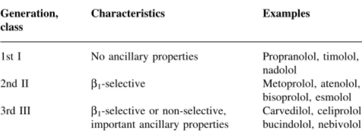
(3) b-Adrenergic antagonism in perioperative medicine Table 2 Classi®cation of b-adrenergic antagonists Generation, class. Characteristics. Examples. 1st I. No ancillary properties. 2nd II. b1-selective. 3rd III. b1-selective or non-selective, important ancillary properties. Propranolol, timolol, nadolol Metoprolol, atenolol, bisoprolol, esmolol Carvedilol, celiprolol, bucindolol, nebivolol. propranolol treatment continued in full dosage until operation, in 50% of patients with propranolol withdrawal and in 70% of patients with no b-AA treatment. Administration of perioperative b-AAs controls haemodynamic variables and successfully decreases the incidence of ischaemic events in patients with or at risk of coronary artery disease. This is particularly relevant as patients with perioperative ischaemia have a nine-fold increase in the risk of developing a serious adverse cardiac outcome during hospitalization and more than a two-fold increase in the risk of dying prematurely over the ®rst 6 months after surgery.140 Patients with a postoperative in-hospital myocardial infarction have a 28-fold increase in the rate of subsequent cardiac complications within 6 months, a 15fold increase within 1 yr and a 14-fold increase within 2 yr. Stone and colleagues212 administered a single oral preoperative dose of one of three different b-AAs (labetalol, oxprenolol, atenolol) to patients with mild uncontrolled hypertension undergoing non-cardiac surgery. The incidence of myocardial ischaemia was 28% in the untreated controls compared with 2% in the b-AA-treated patients, based on ECG criteria (P<0.001). Wallace and colleagues231 administered i.v. atenolol preoperatively and i.v. and oral atenolol for up to 7 days postoperatively in patients with or at risk of coronary artery disease. Myocardial ischaemia was assessed by continuous threelead Holter monitoring. Intraoperative myocardial ischaemia was reported to be 12% in each of the control and atenolol-treated groups. Conversely, the incidence of myocardial ischaemia was reported to be 34% in the control vs 17% in the treated patients in the ®rst 48 h after surgery (P<0.008) and 39 vs 24% over days 0±7 (P<0.029). More recent studies by Raby and colleagues179 and Urban and colleagues225 con®rm the powerful anti-ischaemic effect of perioperative b-adrenergic antagonism and re-emphasize the importance of stress-induced increases in heart rate in the pathogenesis of perioperative myocardial ischaemia. The association between the occurrence of perioperative myocardial ischaemic events, perioperative tachycardia and an adverse long-term cardiac outcome led to therapeutic trials with b-AAs. However, it should be noted at this point that the association between postoperative myocardial ischaemia and adverse cardiac events does not necessarily imply a causal relationship and that postoperative myocardial ischaemia may represent only a manifestation of the. underlying cardiac disease. Factors other than reduced ischaemia may contribute signi®cantly to the improvements in outcome observed after administration of b-AAs. These will be discussed extensively in this article. Using preoperative (1 h before surgery) and postoperative (until 7 days after surgery) atenolol (Table 1), Mangano and colleagues142 demonstrated in a well-designed study a signi®cant reduction in postoperative myocardial ischaemia in patients with or at risk of coronary artery disease. This reduction in ischaemic events was associated with a 55% decrease in overall mortality and a 65% decrease in cardiac mortality at 2 yr for the atenolol-treated patients. The protective effects of atenolol were evident in these patients 6 months after surgery (overall mortality 0 vs 8%, P<0.001) and were preserved over the 2-yr follow-up period (overall mortality 10 vs 21%, P<0.019). Recently, Poldermans and colleagues173 randomized patients undergoing vascular surgery with mild to moderate positive stress echocardiography to preoperative (from 1 week before surgery) and postoperative (until 30 days after surgery) bisoprolol treatment or placebo (Table 1). After the inclusion of 112 patients, this study was halted for ethical reasons associated with large differences in morbidity and mortality rates between the placebo and bisoprolol arms of the study. Notably, the study reports a 10-fold decrease in the 30-day perioperative incidence of death from cardiac causes and non-fatal myocardial infarction in bisoprolol-treated patients (3.4 vs 34%, P<0.001). A critical evaluation of these studies has been published recently in the British Journal of Anaesthesia and can be recommended as additional reading.96 Taken together, these data indicate that b-adrenergic antagonism remains the sole proven pharmacological means of reducing perioperative cardiovascular short- and longterm cardiac morbidity and mortality in patients with or at risk of coronary artery disease. In the light of the bene®ts of perioperative b-AAs and the exceptionally low complication rate associated with the perioperative use of b-AAs, future trials have to clarify whether the cumulative morbidity and mortality associated with sophisticated and expensive preoperative testing can be justi®ed in high-risk cardiac patients undergoing surgery.128 144 190 New aspects of perioperative b-adrenergic antagonism have emerged recently. Johansen and colleagues102 103 demonstrated that esmolol could potentiate the reduction in minimum alveolar concentration for iso¯urane by alfentanil (±26%) and decrease anaesthetic requirements for skin incision during propofol/nitrous oxide/morphine anaesthesia (±27%) in patients. The clinical utility of this effect was subsequently demonstrated by Zaugg and colleagues254 in a study with elderly surgical patients that evaluated three anaesthetic regimens, two of them with atenolol. High-dose intraoperative administration of atenolol decreased iso¯urane requirements by 37% and still allowed an adequate depth of anaesthesia, as assessed by bispectral analysis (mean bispectral index »50±60). Pre- and postoperative atenolol as well as high-dose intraoperative atenolol also. 103.
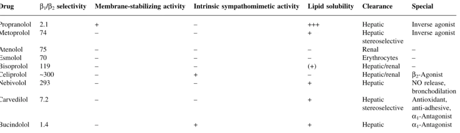
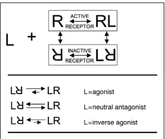
(4) Zaugg et al. Table 3 Ancillary properties of clinically used b-adrenergic antagonists. +=effect present; ±=effect absent Drug. b1/b2 selectivity. Membrane-stabilizing activity. Intrinsic sympathomimetic activity. Lipid solubility. Clearance. Special. Propranolol Metoprolol. 2.1 74. + ±. ± ±. +++ +. Inverse agonist Inverse agonist. Atenolol Esmolol Bisoprolol Celiprolol Nebivolol. 75 70 119 ~300 293. ± ± ± ± ±. ± ± ± + ±. ± ± (+) ± +. Hepatic Hepatic stereoselective Renal Erythrocytes Hepatic/renal Hepatic/renal Hepatic. Carvedilol. 7.2. ±. ±. +. Hepatic stereoselective. Bucindolol. 1.4. ±. +. +. Hepatic. decreased requirements for intraoperative fentanyl (±27%) and postoperative morphine (±40%). As a consequence, extubation time and recovery in the postanaesthesia care unit were signi®cantly faster in patients treated with atenolol.. The present armamentarium of b-adrenergic antagonists Although all b-AAs are able to antagonize the transduction of the b-adrenergic receptor signal (b-AR), this class of drugs is far from being homogeneous. Recently, antisense oligonucleotides against b1-adrenergic receptor (b1-AR) mRNA, which suppress protein translation at the ribosomes, have been constructed and used successfully to treat hypertensive rats.258 Currently used b-AAs, however, competitively antagonize b-ARs and can be roughly classi®ed into three generations depending on their ancillary properties (Table 2).28 29 The main ancillary properties of individual agents include partial agonist activity (intrinsic sympathomimetic activity), b-receptor subtype speci®city (b1 vs b2), lipophilicity and membrane-stabilizing activity (Table 3). Other ancillary properties include vasodilator effects [b2 (celiprolol)-, anti-a1 (carvedilol)- or nitric oxide (NO)-mediated effects (nipradilol, nebivolol)], class III anti-arrhythmic activity (sotalol), antioxidant effects (carvedilol) and stereoselective hepatic metabolism (carvedilol, metoprolol). Accordingly, carvedilol by oral administration exerts equal effects on a- and b-ARs, whereas carvedilol by i.v. administration exerts more b-AR effects than a-AR effects because of decreased stereoselective hepatic metabolism of the b-AR-speci®c S-isomer of carvedilol.164 Interestingly, stereoselective metabolism of metoprolol may result in insuf®cient b-adrenergic antagonism in `poor metabolizers' (S/R isomer ratio <1).201 203 Recent research in transgenic animal models also emphasizes the importance of the two-state model of bAR activation in characterizing b-AAs (Fig. 1). This model proposes an equilibrium between an inactive and an active conformation of the receptor, which is differentially modulated by various ligands (concept of inverse agonism:. ± ± ± b2-Agonist NO release, bronchodilation Antioxidant, anti-adhesive, a1-Antagonist a1-Antagonist. neutral antagonist vs inverse agonist).18 Notably, it predicts spontaneous activation of b-ARs, which was veri®ed recently for the b2- but not the b1-AR.261 This model also explains the inability of some b-AAs with pronounced neutral antagonism to block the effects of receptor overexpression fully, as neutral antagonists counteract activation by endogenous catecholamines but not activation by spontaneous transition into the active receptor conformation.134 The physiological consequences are not yet determined fully but may be of clinical relevance with respect to the tolerability of various b-AAs and the treatment of the withdrawal syndrome. Finally, there is growing evidence that b-ARs differentially couple to various G-proteins depending on the speci®c properties of the ligand, thereby stimulating differential cellular responses.233 Interestingly, a meta-analysis of randomized controlled trials revealed differential effects on cardiovascular events, such as reinfarction and sudden cardiac death, metoprolol being more effective than atenolol or propranolol. This led the authors to conclude that the so-called class effect of bAAs may be less important than ancillary properties.204 However, the mechanistic concept of the class effect is greatly supported by the observation that selective as well as non-selective b-AAs decrease mortality signi®cantly in chronic heart failure.121 Nonetheless, ancillary properties are important with respect to the side-effects and tolerability of the speci®c agents, which will be discussed separately.. Mechanisms and sites of action The following sections will focus on the mechanisms and sites of action elicited by b-AAs.. Cardiac considerations Bradycardia, the link to many cardioprotective effects of b-adrenergic antagonists. Elevated heart rate is a well-established independent predictor of coronary artery disease and cardiovascular morbidity and mortality.168 Also, delayed decrease in heart. 104.
(5) b-Adrenergic antagonism in perioperative medicine. Fig 2 Myocardial oxygen balance. In patients with coronary artery disease, tachycardia decreases myocardial oxygen supply and concomitantly increases oxygen demand.. Fig 1 Two-state model of b-adrenergic receptor activation by competitive ligands. Signalling at the receptor includes binding of the ligand to the extracellular binding domain, transduction of the signal through conformational changes of the receptor and activation of the effector (G-protein complex). In the absence of a ligand, the receptor can undergo spontaneous transition from the inactivated to the activated state. Ligands can be classi®ed into agonists, neutral antagonists and inverse agonists according to their tendency to shift this equilibrium. The agonist shifts the equilibrium towards the active state and the inverse agonist shifts it to the inactive state. Although most b-adrenergic antagonists (b-AAs) act as inverse agonists, some b-AAs with weak inverse agonism may be classi®ed as neutral antagonists.18 The relative degree of inverse agonism increases in the following order: bucindolol<carvedilol <propranolol<metoprolol.249 This model explains the observed lower tolerability of patients treated with inverse b-AAs: they shift the receptor population almost completely to the inactive state, particularly when the sympathetic basal tone is low. On the other hand, b-AAs with weak inverse agonism leave a sizeable fraction of the receptor in the active state, thus explaining their better tolerability in clinical use.. rate after graded exercise predicts cardiovascular mortality.38 Bradycardia is suggested to be one important mechanism of cardioprotection elicited by b-AAs. Importantly, a negative chronotropic response to b-AAs is preserved among diabetic patients with progressive autonomic dysfunction.111 In the perioperative period, increased heart rate is strongly associated with myocardial ischaemia.141 143 Accordingly, myocardial oxygen balance is closely related to heart rate and must be examined on a beat-to-beat basis (Fig. 2). Increased heart rate results in elevated myocardial oxygen demand via the Bowditch effect, which is, however, nearly offset by decreased oxygen demand caused by the lower ventricular wall tension at higher heart rates. Because increased heart rate is usually accompanied by increased inotropy and the length of diastole is signi®cantly decreased in tachycardia, myocardial oxygen balance can deteriorate seriously at higher heart rates in patients with coronary. artery disease. Increased left ventricular stiffness further exacerbates impairment of ventricular ®lling. Importantly, the heart rate at which patients are considered at risk of developing ischaemia is not absolute and must be individualized. Patients with severe angiographic narrowing of the coronary arteries show a gradual decrease in crosssectional area by 32% when heart rate reaches 90 beats min±1.160 Therefore, it is not surprising that patients with coronary artery disease and a heart rate greater than 100 beats min±1 almost inevitably develop myocardial ischaemia.69 The deleterious effects of an increased heart rate on infarction size have been reported. Augmentation of heart rate after experimental coronary occlusion in dogs, by ventricular pacing, isoproterenol or atropine, leads to increases of 40, 70, and 40% respectively in myocardial necrosis when compared with control.199 Consistent with this notion, thiopental given during coronary occlusion doubles infarction size by increasing heart rate. Notably, tachycardia also accentuates endomyocardial to epimyocardial maldistribution of ventricular blood ¯ow in ischaemia.12 b-AAs effectively reverse all these untoward effects by lowering heart rate.227 b-AAs also exert a bene®cial effect in coronary artery disease by decreasing the stiffness of atherosclerotic plaques, which results in increased tensile strength.127 The stiffness of ®brous caps of human atherosclerotic plaques is directly related to heart rate, and increased heart rate promotes the ®ssuring of atherosclerotic plaques. b-AAs prevent the rupture of vulnerable atherosclerotic plaques, which leads to less in¯ammation in the plaque and decreases the gradual narrowing of the vessel lumen.63 66 178 Furthermore, tachycardia causes activation of platelets.54 When coronary blood ¯ow increases, platelets can be traumatized and activated across the coronary bed, particularly at sites with signi®cant narrowing. Histopathological analyses of perioperative myocardial infarction stress the importance of plaque disruption and thrombosis as pivotal steps in the pathogenesis of perioperative myocardial infarction.48 Alternatively, long-duration subendocardial ischaemia and. 105.
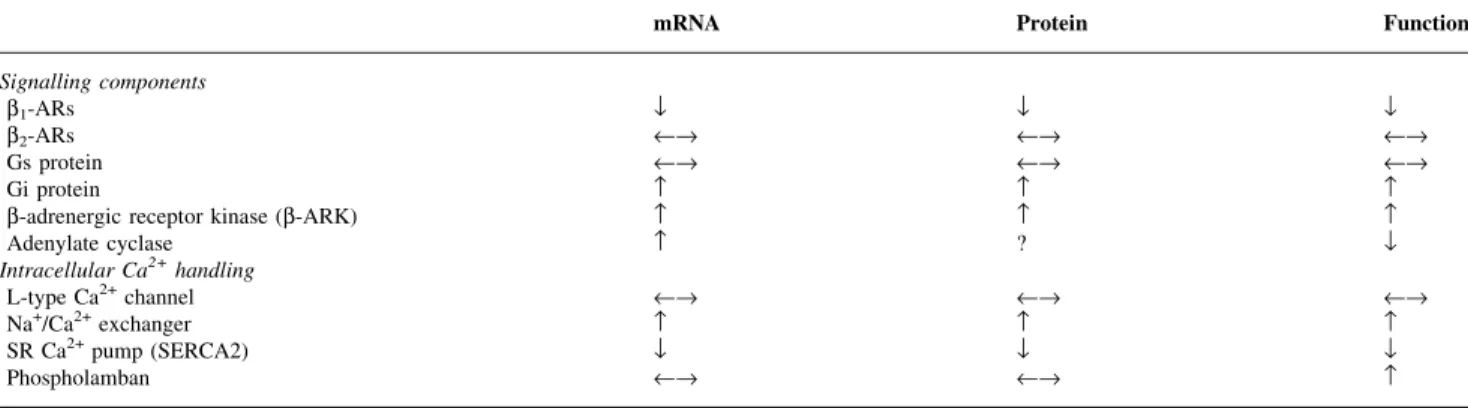
(6) Zaugg et al.. subsequent non-Q-wave infarction resulting from prolonged tachycardia were also proposed as the underlying mechanism of perioperative myocardial infarction.124 The view that postoperative myocardial ischaemia is a mere manifestation of the underlying cardiac disease is speci®cally supported by the ®ndings of the post-mortem study by Dawood and colleagues.48 In this study, fatal postoperative myocardial infarctions were associated with evidence of unstable plaques in 55% of the patients. In contrast, imbalance of myocardial oxygen supply/demand may play a causal role in the pathogenesis of postoperative cardiac events. This view is supported by the fact that most postoperative myocardial infarctions are non-Q-wave infarctions that are preceded by long-duration (>2 h) postoperative ischaemia.125 Because cardiac complications are preceded by long-duration STsegment depression rather than elevation, it seems plausible that the cascade of events leading to postoperative cardiac complications does not begin with acute coronary occlusion but with long-duration subendocardial ischaemia. This is further supported by a recent study by Badner and colleagues,8 which determined the incidence of postoperative myocardial infarction after non-cardiac surgery in a large group of patients at high risk. This study also reports the preponderance of non-Q-wave infarction, which differs from that seen in non-surgical patients presenting to the emergency room. From a mechanistic point of view, non-Q-wave infarctions result from prolonged ischaemia rather than from total occlusion of the coronary arteries. Certainly, further studies are needed to elucidate the role of postoperative myocardial ischaemia in the cascade of events leading to perioperative cardiac morbidity and mortality. The institution of bradycardia was recently found to cause restoration of contractile function in a canine model of mitral regurgitation-induced left ventricular dysfunction.161 In this model, optimized myocardial Ca2+ handling and bioenergetics are direct consequences of bradycardia and are suggested to be responsible for the observed improvement in contractility.257 At the cellular level, rapid electrical stimulation of contraction reduces the density of b-ARs and their responsiveness,116 which appears to be associated with disassembly of microtubules secondary to undue micromechanical stress.248 Cardioprotective effects of b-adrenergic antagonists not apparently associated with bradycardia. Stangeland and colleagues207 addressed the important question of whether decreased heart rate is the only mechanism responsible for cardioprotection elicited by bAAs. They treated anaesthetized cats with alinidine (a clonidine analogue that decreases heart rate independently of b-ARs) or timolol. Heart rate was similarly reduced by 40 beats min±1 in the treatment groups compared with the control group, and regional ischaemia was induced by occluding the left descending coronary artery. After 6 h of ischaemia, the necrotic tissue was measured and expressed. as a percentage of necrotic tissue in the area at risk. Notably, alinidine signi®cantly decreased necrosis from 87% present in the control group to 77% (P<0.01), whereas timolol decreased necrosis to 65% (P<0.001). This observation clearly indicates that mechanisms other than decreased heart rate contribute substantially to cardioprotection by b-AAs. b-Adrenergic signal transduction in cardiomyocytes. Biological responses mediated by b-ARs involve positive chronotropy, dromotropy, inotropy and cardiomyocyte growth and death (Fig. 3). b-ARs are members of the Gprotein-coupled superfamily, which share the characteristic feature of the seven-transmembrane-spanning domains. In healthy mammalian cardiomyocytes, b1-ARs constitute around 70±80% of the b-ARs in human and rat hearts.52 236 In many disease states with heightened sympathetic drive, b1-ARs are down-regulated by phosphorylation (desensitization), translocation (sequestration) and ®nally by degradation of the receptor.88 Conversely, b2ARs do not decrease in number; however, they show some loss of contractile response to agonist stimulation as a result of the up-regulation of b-AR kinases (b-ARK) and Gi proteins (Table 4).26 In the failing human heart, b2-ARs represent 40% of b-ARs and are of great importance in mediating inotropic and chronotropic responses.25 Key steps in signal transduction of b1- and b2-ARs involve coupling to G-proteins and activation of the cAMP/protein kinase A (PKA) pathway, which leads to phosphorylation of target proteins such as phospholamban, the ryanodine receptor, troponin I and L-type Ca2+ channels.209 244 However, apart from changes in myocardial contractile function, b-ARs exert important effects on cellular metabolism, growth and death (gene expression) through the activation of PKA and protein kinase C (PKC).90 Because the b1-AR and the b2-AR share only 54% of amino acid sequences overall, it is possible that b-AR subtypes couple to distinct signal transduction pathways.243 245 Although both b1- and b2-ARs increase the contractile response and hasten relaxation in ventricular myocytes, several striking differences with respect to G-protein binding characteristics and signal transduction downstream from the receptor have been revealed. In contrast to b1-ARs, b2-ARs exhibit dual coupling to Gs and Gi that can completely negate Gsmediated responses. Also characteristic of b2-mediated signalling is the exceptionally modest increase in cAMP and the compartmentalized increase in PKA activity, which is restricted to the vicinity of L-type Ca2+ channels.124 260 Finally, b2-ARs may also bind to Gq-activating phospholipase C (Fig. 3). These data indicate that b-AR subtypes differentially modulate cardiac function and cardiomyocyte phenotype. Therefore, the subtype speci®city of various bAAs affects biological responses signi®cantly. Little is known about the role of b3-ARs in cardiomyocytes. Whereas b3-ARs are known to exert important physiological effects in brown adipose tissue, gut relaxation and vasodilation, b3-ARs mediate negative inotropy by a NO-dependent pathway in cardiomyocytes.70 226 Studies. 106.
(7) b-Adrenergic antagonism in perioperative medicine. Fig 3 b-Adrenergic signalling cascades in cardiomyocytes. (A) Binding of an agonist (L) to either the b1- or the b2-adrenergic receptor (b1-AR, b2AR) stimulates Gs protein, which dissociates from the receptor and binds to adenylate cyclase (AC), causing production of cAMP from ATP and activation of protein kinase A (PKA). G-protein-coupled receptors interact by their intracellular loop 3 with the heterotrimeric G complex and promote GDP release. PKA phosphorylates the voltage-dependent L-type Ca2+ channels, the Na+/H+ exchange channels and the Na+/K+ pump at the sarcolemma, phospholamban (PLB) and the ryanodine receptor (RYR) at the sarcoplasmic reticulum (SR) and cardiac troponin-I (cTnI) in the sarcomeres, leading to increased inotropic and lusitropic (relaxation) responses. In contrast, the b2-AR is also able to couple to Gi or Gq proteins. Gi inhibits adenylate cyclase (AC) and opposes the effects of Gs. Gq activates phospholipase C (PLC). By splitting phosphatidylinositol bisphosphate, PLC liberates the two intracellular second messengers diacylglycerol (DAG) and inositol trisphosphate (IP3). IP3 binds to the IP3 receptor (IP3R), which releases Ca2+ from the SR. Ca2+ combines with calmodulin (CaM) and directly activates the sarcolemmal Ca2+ pump as well as several CaMdependent protein kinases (CaMKs). This signalling pathway leads to phosphorylation of PLB, ventricular myosin light chain 2 (MLCV2) and the Na+/Ca2+ exchanger. DAG and CaM together activate PKC, which in turn phosphorylates the mitochondrial ATP-dependent K+-channel and MLCV2. In its unphosphorylated state, the regulatory protein PLB is bound to the SR Ca2+-pump (SERCA2), inhibiting its activity. When phosphorylated by PKA and/or CaMK, it dissociates from SERCA2, relieving the inhibitory effect. On the other hand, direct phosphorylation of RYR at Ser-2809 dissociates the regulatory component FKBP (FK506 binding protein), leading to increased activity of the RYR channel. PKA and PKC both affect gene expression in the cell nucleus via the common MAPK (mitogen-activated protein kinases) signalling pathway: Ras (monomeric GTPase), Raf (a MAPKKK), MEK (mitogen-activated ERK activating kinase) and ERK (extracellular signal regulated kinase). To protect the cardiomyocyte from b-adrenergic overstimulation, a negative feed-back loop (not shown in the diagram) is built in, which degrades cAMP to AMP by means of CaMactivated phosphodiesterase III. (B) Diagram showing in more detail the dissociation of the heterotrimeric G complex from the b-AR upon binding of the agonist (L). After this dissociation, the adenylate cyclase (AC) becomes activated by binding to the a-subunit. Desensitization of the b1- and b2-ARs is mediated by phosphorylation of the intracellular C-terminal part of the receptor by either PKA or the b-adrenergic receptor kinase (b-ARK, GRK2 and 3). Binding of arrestin and clathrin to the phosphorylated b-AR mediates internalization to the endosome. After dephosphorylation, b-ARs may be either degraded or resensitized. Note that an additional negative feed-back loop leads to inhibition of the b-ARK via increased Ca2+calmodulin (CaM).17 58 90 147 148 171 189. 107.
(8) Zaugg et al. Table 4 Major changes in components of b-adrenergic signalling in the failing human heart28. Signalling components b1-ARs b2-ARs Gs protein Gi protein b-adrenergic receptor kinase (b-ARK) Adenylate cyclase Intracellular Ca2+ handling L-type Ca2+ channel Na+/Ca2+ exchanger SR Ca2+ pump (SERCA2) Phospholamban. 147 171. mRNA. Protein. Function. ¯ ¬® ¬® . ¯ ¬® ¬® ?. ¯ ¬® ¬® ¯. ¬® ¯ ¬®. ¬® ¯ ¬®. ¬® ¯ . Fig 4 Cardiotoxicity of catecholamines. Adult rat ventricular myocytes (ARVMs) grown on coverslips were exposed to norepinephrine (NE) (10 mmol litre±1) alone or in the presence of atenolol (AT) (10 mmol litre±1) for 12 h and subjected to TUNEL (terminal dUTP nick end labelling) staining, which is speci®c for apoptotic cell death. (A) Mean percentage of TUNEL-positive ARVMs on coverslips. Data are mean (SEM). *P<0.0001 vs control; P<0.0001 vs NE. (B) Control ARVMs with rod-shaped morphology. (C) NE-exposed ARVMs with rounded morphology and black apoptotic nuclei. (D) NE+AT-treated ARVMs. Note preservation of rod-shaped morphology with NE treatment (reproduced with permission from Circulation255).. evaluating the distribution and quanti®cation of b1/b2/b3AR subtypes in heart tissue have revealed subtype proportions for the left porcine ventricle as follows: b1:b2:b3=72%:28%:0.25%.151 This implies that, in the normal myocardium, b3-AR may be of less importance. However, recent observations in heart failure patients demonstrate that opposite changes in the abundance of b1AR (down-regulation) and b3-AR (up-regulation) occur and may play a role in the progressive functional degradation in the failing human heart.155 Cell death signalling: apoptosis and necrosis. Catecholamines, although bene®cial in the short-term cardiovascular response, exert signi®cant cardiac toxicity. The toxic effects of catecholamines on cardiomyocytes have been known since the beginning of the 20th century.82 However, necrotic and apoptotic cell death has been closely related to enhanced b-adrenergic signalling only recently.145 Apoptotic cardiomyocyte death by activation of the b-adrenergic signalling pathway was reported in norepinephrine-stimulated adult rat ventricular myocytes.39. Zaugg and colleagues255 further demonstrated that apoptotic cardiomyocyte cell death is dissociated from b2-ARs and selectively mediated by b1-ARs in adult ventricular myocytes (Fig. 4). This is in line with clinical observations that cardiac lesions associated with massive catecholamine bursts were prevented with atenolol in patients with subarachnoid haemorrhage.43 Communal and colleagues40 and Chesley and colleagues36 further showed that b2-AR stimulation may protect cardiomyocytes from apoptosisinducing stimuli. The abilities of b-AR stimulation and tachycardia to induce cardiomyocyte apoptosis were addressed by Shizukuda and colleagues200 in an in vivo rat model. Rats were treated with placebo or isoproterenol to establish whether catecholamines per se in the absence of signi®cant increases in systolic load and tachycardia induce myocardial damage via apoptosis. After only 24 h of isoproterenol treatment, a signi®cant increase in apoptotic events was detected. Animals exposed to ventricular pacing to induce tachycardia equivalent to that produced by isoproterenol treatment did not show an increase in. 108.
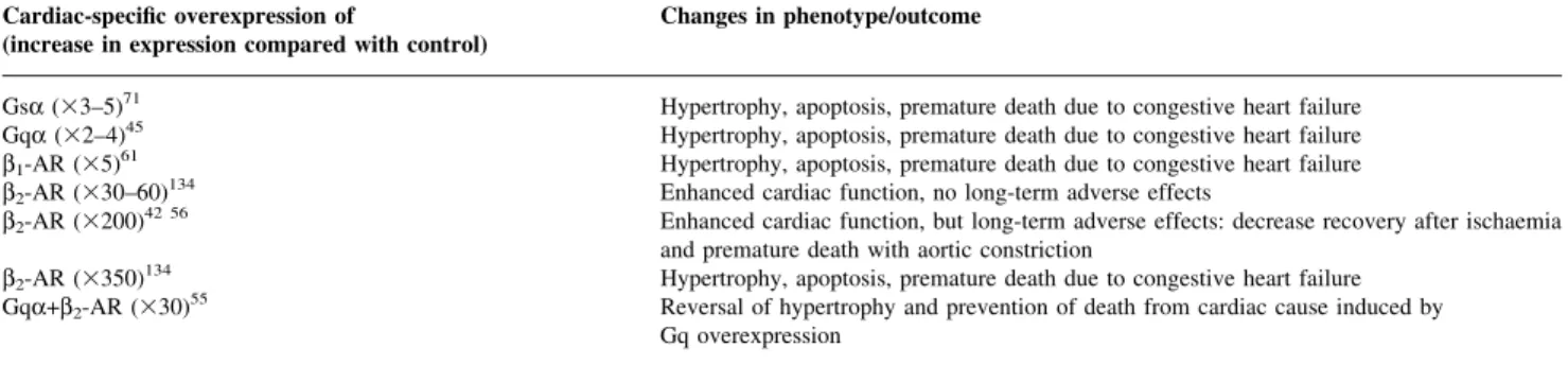
(9) b-Adrenergic antagonism in perioperative medicine Table 5 Gene-targeted mice mimicking enhanced b-adrenergic signalling Cardiac-speci®c overexpression of (increase in expression compared with control). Changes in phenotype/outcome. Gsa (33±5)71 Gqa (32±4)45 b1-AR (35)61 b2-AR (330±60)134 b2-AR (3200)42 56. Hypertrophy, apoptosis, premature death due to congestive heart failure Hypertrophy, apoptosis, premature death due to congestive heart failure Hypertrophy, apoptosis, premature death due to congestive heart failure Enhanced cardiac function, no long-term adverse effects Enhanced cardiac function, but long-term adverse effects: decrease recovery after ischaemia and premature death with aortic constriction Hypertrophy, apoptosis, premature death due to congestive heart failure Reversal of hypertrophy and prevention of death from cardiac cause induced by Gq overexpression. b2-AR (3350)134 Gqa+b2-AR (330)55. apoptosis. The authors concluded that apoptotic cardiomyocyte death resulting from isoproterenol treatment may not be explained by increased heart rate alone. Conversely, in a canine model, rapid ventricular pacing per se increased apoptotic cell death and led to cardiac myopathy.136 Importantly, decreased apoptosis has been reported in carvedilol- and propranolol-pretreated rabbit hearts subjected to ischaemia±reperfusion injury.252 Because apoptotic cell death occurs in only a few hours, apoptosis may be an important mechanism for loss of viable cardiomyocytes and myocardial dysfunction in the immediate perioperative period.253 New insight from gene-targeted animals. While experimental results indicate incontrovertibly that enhanced b1AR signalling is exceptionally cardiotoxic, data on the effects of the b2-AR with respect to bene®cial and detrimental effects are contradictory. Recent research in the ®eld of heart failure tried to construct genetically altered mouse models mimicking increased sympathetic nervous system activity (Table 5). In particular, transgenic mouse models with cardiac-speci®c overexpression of various Gproteins and b-AR subtypes were constructed.3 45 61 71 134 153 Mice with Gsa overexpression typically develop a characteristic hypertrophic cardiomyopathy at 15 months of age. Sections of these hearts reveal hypertrophic myocytes with increased cross-sectional areas and an increased number of apoptotic myocytes. Importantly, propranolol, a non-selective b-AA, abolished the hypertrophic response and the development of dilated chambers, thereby improving survival. Similar results were reported for mice overexpressing Gqa- and b1-AR. Notably, mice with only ®ve-fold overexpression of the b1-AR develop fatal cardiomyopathy,61 whereas mice with 30- to 60-fold overexpression of the b2-AR exhibit enhanced cardiac function and do not develop overt cardiomyopathy.134 Although no long-term toxic effects were reported by some authors in mice with 200-fold overexpression of the b2-AR,209 244 increased susceptibility to ischaemic injury42 and augmented afterload56 were clearly observed. Nonetheless, it was suggested that manoeuvres that serve to augment b2-adrenergic signalling, which improves systolic and diastolic function, may offer a potential therapeutic approach in patients suffering from impaired cardiac function. For this purpose,. pharmacological means and ultimately in vivo gene transfer strategies were proposed and investigated.110 150 196 Accordingly, the contractility of single myocytes isolated from the ventricles of rabbits chronically paced to produce heart failure can be functionally restored by adenovirusmediated transfer of b2-ARs.4 Consistent with this notion, a dual-expressing `designer' mouse with cardiac-speci®c Gqa expression and concomitant b2-AR expression at low (303), medium (1503) and high levels (10003) was constructed recently.55 Gqa mice with low concomitant expression of b2-ARs, i.e. a ~30-fold increase in b2-AR compared with wild type, displayed rescue of hypertrophy and ventricular function. Importantly, these effects occurred in the absence of any improvement in basal or agoniststimulated adenylate cyclase (AC) activity, indicating the restoration of a compartmentalized b2-AR±AC signalling pathway.124 260 The summarized experimental results, which clearly demonstrate bene®cial effects of modest b2adrenergic signalling in an animal model of heart failure, have found their clinical counterpart very recently.30 Studies of b2-AR gene variations in twins revealed that speci®c b2-AR polymorphisms, which resulted in enhanced down-regulation of the b2-AR, increased cardiac dimensions (septum thickness, posterior wall thickness, left ventricular mass). In summary, the concept that b-adrenergic signalling may not mediate deleterious effects exclusively but may also have bene®cial effects in the compromised heart is based on several experimental observations. In this regard it is interesting to note that, in patients with symptomatic heart failure, pan-adrenergic antagonism using central sympatholysis (moxonidine, an a2-agonist) was terminated because of excess mortality.121 b2-AR agonism as an adjunct to b1AR antagonism may therefore have the potential to improve the therapeutic tolerance, particularly during the initiation of b-AA therapy, and to improve survival in the treatment of the failing heart. Interestingly, b1-AR antagonism enhances the b2-AR-mediated inotropic response to catecholamines, which may, in part, also explain the better tolerability of selective b1-AAs.84 Mechanical unloading and modulation of gene expression. b-AAs allow the heart to `rest' by emulating a state of ventricular unloading. Accordingly, the expression of. 109.
(10) Zaugg et al.. tumour necrosis factor a (TNF-a), an indicator of increased mechanical load, is similarly reduced by b-AAs and by mechanical circulatory support.174 221 High-dose atenolol also prevents angiotensin II- and tachycardia-induced activation of metalloproteinases and diastolic stiffening.195 Changes in gene expression caused by b-AAs also involve a decrease in endothelin-168 and sarcoplasmic reticulum proteins217 and an increase in atrial natriuretic factor.250 Furthermore, carvedilol decreases Fas receptor expression (cell death signalling receptor) after ischaemia±reperfusion injury, which leads to decreased apoptotic cell death.252 Taken together, these observations suggest altered gene expression as a potential site of cardioprotection by b-AAs. Platelet aggregability and coagulation. The adrenergic system in¯uences coagulation and ®brinolysis, particularly during episodes of heightened adrenergic drive, but contributes much less to baseline levels of coagulatory and ®brinolytic function. Epidemiological studies revealed a signi®cant morning increase in the incidences of infarction, sudden cardiac death and transient myocardial ischaemia, which appears to be related to increased morning catecholamine levels and coagulation.158 238 Perioperatively, increased catecholamine levels and tissue damage greatly increase the propensity to coagulation. Propranolol decreases thromboxane synthesis and platelet aggregation in patients receiving long-term propranolol treatment.32 One study evaluating the effect of metoprolol on platelet function did not ®nd any inhibitory effect,239 whereas another study reports decreased platelet aggregability in patients with stable angina being tested for exercise stress.241 Metoprolol also prevents stress-induced endothelial injury by increasing prostacyclin biosynthesis75 and decreasing epinephrine-induced increases in von Willebrand factor antigen.126 Esmolol has in vivo inhibitory action on neutrophil superoxide generation and platelet aggregation in a canine model of myocardial ischaemia±reperfusion.186 b-AAs also decrease the af®nity of lowdensity lipoprotein to arterial proteoglycans and endothelial wall damage by reducing plasminogen activator inhibitor1.216 Conversely, increased platelet aggregability and decreased platelet cAMP production were reported after timolol treatment.240 b-Adrenergic receptor down-regulation and target protein hyperphosphorylation. Prolonged and intensive perioperative agonist stimulation leads to desensitization and down-regulation of b-ARs, which may seriously impair cardiac function.28 29 Conversely, down-regulation of the sensitivity and number of receptors may be bene®cial with respect to arrhythmogenicity.251 Tachycardia,116 free oxygen radicals133 and increased serum levels of TNF-a,79 which are, notably, all factors signi®cantly affected by bAAs, were further implicated in the down-regulation of bARs and the subsequent attenuated cardiovascular response. Importantly, down-regulation occurs after only a few hours of agonist stimulation.88 At the molecular level, the process involves uncoupling of the b-AR from Gs-proteins by PKA. and b-adrenergic receptor kinase 1 (b-ARK) and binding of inhibiting arrestin to the receptor, which is followed by internalization and degradation or resensitization of the receptor (Fig. 3B). Early uncoupling and late down-regulation of myocardial b-ARs were reported after cardiopulmonary bypass.72 193 Similarly, persistent down-regulation and desensitization of b-ARs were reported after thoracotomy or laparotomy throughout the ®rst week after surgery.5 Hyperphosphorylation of channels and regulatory proteins such as the sarcoplasmic ryanodine receptor (RYR) may occur, resulting in hypersensitivity to cytosolic Ca2+.147 148 b-AAs potentially prevent hyperphosphorylation in the perioperative period, which is similar to their effects in congestive heart failure.91 Anti-arrhythmic effects. Sustained arrhythmias may be haemodynamically relevant and may affect the outcome adversely.13 Almost half of all high-risk cardiac patients undergoing non-cardiac surgery have ventricular ectopic beats or some sort of ventricular tachycardia.166 Patients undergoing cardiac surgery have a high risk of developing new-onset atrial ®brillation.6 From a mechanistic point of view, sympathetic tone plays an important role in most ventricular as well as atrial arrhythmias.192 b-AAs shift the autonomic balance towards a higher vagal and lower sympathetic tone.224 Studies on infarctions in pigs clearly showed that b-adrenergic mechanisms play a major role in ventricular ®brillation threshold during experimental coronary occlusion.119 This is consistent with the notion that bAAs prevent sudden electrical cardiac death.62 112 Regarding atrial arrhythmias, b-AAs may be superior to newer class-III anti-arrhythmic drugs in the treatment of perioperative atrial ®brillation, as these drugs carry the risk of drug-induced polymorphic ventricular tachycardia. Notably, b-AAs also counteract epinephrine-induced hypokalaemia, which signi®cantly predisposes to arrhythmias. Bioenergetics. b-AAs are known to reduce NADH oxidase activity in mitochondria, which may lead to an energysparing effect.177 Also, b-AAs shift cellular metabolism from fatty acid oxidation to glucose utilization, which effectively reduces the myocardial oxygen requirement.22 Recently, oxidative metabolism was evaluated in patients with ventricular dysfunction using C-11-acetate positron emission tomography. The results of this study showed a signi®cant reduction in cellular oxidative metabolism under metoprolol treatment.10 Accordingly, in patients undergoing cardiopulmonary bypass, chronic propranolol treatment reduces oxygen consumption.108 Interestingly, patients receiving chronic b-AA treatment compensate for reduced arterial oxygen content by increases in cardiac output and oxygen extraction, whereas patients not receiving b-AA treatment demonstrate only an increase in oxygen extraction.206 Reduced production of lactate during exercise and increased oxygen extraction as a result of decreased cardiac output were reported previously under b-adrenergic antagonism.175 b-AAs also prevent the decrease in mitochondrial CK activity after myocardial infarction.97. 110.
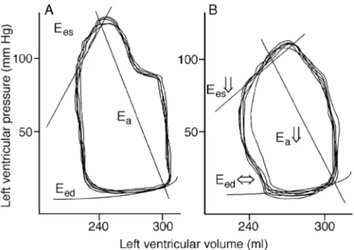
(11) b-Adrenergic antagonism in perioperative medicine. Fig 5 Schematic depiction of left ventricular volume±pressure loops in a patient with heart failure. (A) Volume±pressure loop without b1adrenergic receptor (AR) antagonism. (B) Volume±pressure loop with acute exposure to b1-A antagonism. End-systolic elastance (Ees) is decreased under b1-AR antagonism, which is accompanied by decreased cardiac output, decreased stroke volume and decreased +dP/ dtmax. In contrast, whereas the duration of active isovolumetric relaxation may increase only slightly (decrease in ±dP/dtmax), passive diastolic function, as indicated by end-diastolic elastance (Eed, chamber stiffness), remains largely unaffected by b1-AR antagonism. Importantly, afterload, as indicated by arterial elastance (Ea), is decreased by b1-AR antagonism. Also, the ratio Ees/Ea, which represents ventriculoarterial coupling (the relationship between systolic function and afterload), is well preserved. Note that the area enclosed by the volume±pressure loops is closely related to myocardial oxygen consumption and is markedly reduced by b-AAs.. Neuroendocrine stress response. There is a reduction in renin activity by selective as well as non-selective b-AAs mediated by antagonizing b-ARs, and some studies have even reported decreased catecholamine release after initiation of b-adrenergic antagonism.157 Most studies, however, did not observe a decrease in catecholamine serum levels254 but rather an increase.93 One study in pigs reported decreased neuropeptide Y serum levels associated with increased heart rate variability after treatment with metoprolol.1 Preconditioning. Theoretically, b-AAs may prevent preconditioning of the heart, which renders it more resistant to subsequent sustained ischaemia. However, metoprolol does not neutralize the favourable effects of preconditioning.237 On the contrary, nipradilol, a nitric oxide-generating b-AA, clearly induces preconditioning by itself.95. Non-cardiac considerations Effects of b-adrenergic antagonism on core components of an anaesthetic regimen. Anaesthetic and analgesic requirements. Previous studies focused on the anti-ischaemic properties of peri- and intraoperative b-adrenergic antagonism. Recently, it was shown that esmolol can potentiate the reduction in the minimum alveolar concentration (MAC) for iso¯urane. (±26% at esmolol 250 mg kg±1 min±1) and decrease anaesthetic requirements for skin incision during propofol/ nitrous oxide/morphine anaesthesia (±27% at esmolol 250 mg kg±1 min±1).102 103 Esmolol also decreases nociception in a variety of experimental settings, suggesting the potential to decrease the intraoperative anaesthetic requirements.47 Altered distribution and decreased metabolism of opioids by b-AAs may underlie this anaesthetic-sparing effect.182 Furthermore, although b-AAs per se do not provide analgesia or hypnosis, they are known to have central nervous system modulating activities and anxiolytic effects.76 146 158 b-AAs potentially affect central nervous system pathways, which include neurones in the hypothalamus, hippocampus and cerebral cortex.120 234 Accordingly, the favourable changes in heart rate variability after b-AA treatment are ascribed to lower activity of the central sympathetic nervous system. In mice and rats, the locus coeruleus-associated noradrenergic system participates in arousal, and b-adrenergic antagonism within this region reduces forebrain electroencephalographic activity.16 Similarly, amphetamine-induced activation of the rat forebrain is clearly inhibited by timolol, and in humans norepinephrine is known to enhance the responsiveness of the cerebral cortex to excitatory neuronal transmission.15 181 Notably, pure b-adrenergic antagonism is crucial for the observed anaesthetic-sparing effect because labetalol increases the anaesthetic requirement.47 Even though esmolol and atenolol are hydrophilic b-AAs, they produce the same plasma/cerebrospinal ¯uid ratio as lipophilic b-AAs, thereby affecting the centrally located surface b-ARs.120 Another mechanism that may signi®cantly contribute to the anaesthetic-sparing effect elicited by b-AAs involves decreased excitatory stimulation of central nervous effector sites of hypnosis and somatic response. In this case, peripheral interruption of centripetal b-adrenergic autonomic pathways, like spinal and epidural anaesthesia, decreases afferent input and anaesthetic requirement.163 Memory storage. b-AAs also possess attenuating effects on memory storage. Effects of opioids on memory are known to be mediated through noradrenergic in¯uences.98 Importantly, propranolol was reported to impair memory storage of particularly emotional events in humans.31 Also, b-AAs impair arousal-induced enhancement of working memory in elderly patients.165 As intraoperative recall and subconscious processing of information is particularly increased for emotionally charged information,132 it is tempting to speculate that b-AAs, as anaesthetic adjuvants, might actually decrease the risk of intraoperative awareness and recall. However, this may be different for haemodynamically compromised patients with concomitant cardiovascular medication. Nonetheless, adequate depth of anaesthesia, as indicated by the bispectral index, was achieved in a group of elderly patients using high-dose intraoperative atenolol and a restricted amount of anaesthetic.254. 111.
(12) Zaugg et al. Table 6 Haemodynamic effects of b-adrenergic antagonists (b-AA), phosphodiesterase 3 inhibitors (PDE3I) and their combination in heart failure patients Variable. b-AA. PDE3I. b-AA+ PDE3I. Heart rate Systolic function Diastolic function End-diastolic pressure Myocardial oxygen consumption Propensity to arrhythmia. ¯ ¯ then ¬® or ¬® then ¯ ¯ ¯. ¯ ¬® or . ¯ ¯ ¯ ¬®. require additional monitoring. Even when started acutely with high doses and in combination with potentially negative inotropic agents, b-AAs were well tolerated in compromised patients. Nonetheless, certain contraindications to b-AAs must be considered.. Haemodynamics. Recovery. Faster recovery from anaesthesia was reported in patients receiving propranolol or metoprolol101 208 and in patients receiving intra- or perioperative atenolol.254 Titration of anaesthetics to heart rate and blood pressure without administration of b-AAs may lead to prolonged recovery from anaesthesia as a result of `relative overdosing' with administered anaesthetics (MACBAR> MACAWAKE).183 Furthermore, speci®c properties of bAAs may alleviate recovery from anaesthesia. In cats receiving atenolol, waking times were signi®cantly prolonged,92 and human sleep disturbance is a well-known side-effect of b-AAs.159 Immune response and b-adrenergic antagonism. The perioperative stress response impairs immune competence, particularly natural killer cell cytotoxic activity.220 In the experimental setting, reduction of natural killer cell cytotoxicity was achieved by electrical stimulation of the splanchnic nerve in rats, and was completely antagonized by nadolol.109 Recently, Ben-Eliyahu and colleagues14 reported that hypothermia in barbiturate-anaesthetized rats suppresses natural killer cell cytotoxic activity and thereby accelerates the spread of tumour cells. Interestingly, nadolol attenuated the effect of hypothermia on natural killer cells and increased resistance to tumour metastasis.. Tolerability of perioperative administration of b-adrenergic antagonists Contraindications to the use of b-AAs result directly from their anti-adrenergic action. Drug intolerance greater than 20% as a result of decreased contractile function and increased afterload were previously reported in ®rst-generation compounds.215 However, drug tolerability for secondgeneration compounds is 80±100% and for third-generation compounds 90±100%.27 Recent research has revealed an exceptionally low complication rate associated with the use of b-AAs in heart failure patients as well as perioperatively in high-risk cardiac patients.142 173 179 225 231 254 Speci®cally, these studies do not report an increased number of episodes with severe hypotension, bradycardia or bronchospasm. Therefore, administration of b-AAs according to the reported dosing by Mangano and colleagues142 and Poldermans and colleagues173 does not. Excessive sympatholysis is undesirable in patients who depend heavily on central sympathetic tone for adequate circulatory function. While chronic administration of bAAs improves systolic and diastolic function in heart failure patients,73 115 228 acute exposure to b-AAs may lead to intolerable bradycardia and arterial hypotension and potentially result in an adverse outcome.215 Accordingly, eyedrops with b-AAs were implicated in the progression of ischaemic optic nerve disorders and the progression of visual loss attributable to recurrent nocturnal hypotensive episodes.89 Therefore, patients with advanced conduction defects or symptomatic bradycardia should not receive bAAs without concomitant pacemaker therapy. However, patients with a resting heart rate below 60 beats min±1 may receive therapy with caution. One study evaluating the tolerability of b-AA titration in patients with idiopathic dilated cardiomyopathy found that generally accepted measures of the severity of heart failure were not predictive of problematic up-titration of b-AAs.7 A low systolic blood pressure (<120 mmHg) was the strongest predictor of complications. The mechanisms underlying the good tolerability of initiation of b-AAs in patients with heart failure was previously investigated by Halpern and colleagues81 (Fig. 5). Acute effects of metoprolol on systolic and diastolic function as well as on ventriculoarterial coupling were evaluated using volume±pressure loops. The results of these studies indicate that decreased afterload, as assessed by arterial elastance and the preservation of ventriculoarterial coupling and passive ventricular properties, explain the excellent tolerance of b1-AR antagonists in heart failure patients (Fig. 5). Notably, b1-AR antagonists also leave inotropic responses to b2-AR receptors intact and thereby produce less cardiac depression and vasoconstriction. Conversely, studies using a pulmonary artery catheter technique showed an increase in systemic vascular resistance (SVR) after metoprolol administration in chronically treated, mostly NYHA III heart-failure patients.123 Similarly, acute graded administration of esmolol in patients with severe ventricular dysfunction increased SVR to compensate for the decrease in cardiac output.99 Intraoperatively, SVR decreases signi®cantly during undisturbed anaesthesia, but increases markedly under surgical stimulation in metoprolol as well as placebo-treated patients.139 However, SVR depends on loading conditions and contractility, which were not measured in all these studies. Therefore, SVR may not properly re¯ect the effects of b-AAs on distension in the arterial system, i.e. afterload. Accordingly, in mitral regurgitation-induced left ventricular. 112.
(13) b-Adrenergic antagonism in perioperative medicine. dysfunction b-AAs decrease left atrial hypertension independently of changes in heart rate, which is thought to result from decreased afterload.33 228 Experience with recent large trials indicates that fewer than 5% of patients need to be withdrawn from b-AAs because of worsening heart failure when they are carefully monitored. The favourable haemodynamic effects and safety of perioperative b-AA administration is now also documented in several studies with brittle elderly surgical patients.142 173 179 225 231 254 As the potentially adverse clinical effects of esmolol completely disappear within minutes of its discontinuation, current experience suggests the initiation and up-titration of this short-acting b1-AR antagonist even in compromised patients perioperatively.9. Coronary vascular resistance Sympathetic nerve stimulation in the presence of propranolol was reported to increase diastolic coronary resistance by 30%.64 This coronary vasoconstriction is less likely to occur after administration of b1-AR antagonists.2 More importantly, in a study that measured simultaneously total coronary ¯ow (sinus out¯ow) and local tissue ¯ow (heated thermocouples), sympathetic stimulation after b-AA administration resulted in a decrease in sinus out¯ow but an increase in the nutritional microcirculatory ¯ow.107 This implies that a reduction in total coronary out¯ow does not necessarily parallel a decrease in tissue ¯ow at the microcirculatory level. Also, the mechanism of the reduction in blood ¯ow after administration of b-AAs is likely to be a result of decreased myocardial oxygen demand. This is strongly supported by the notion that atenolol decreases myocardial blood ¯ow by 16% and increases coronary vascular resistance by 23%.211 At the same time, however, the myocardial arteriovenous oxygen difference remains unaltered. Furthermore, when paced at the pre-atenolol heart rate, there is no decrease in coronary blood ¯ow or increase in vascular resistance under atenolol treatment, which again clearly indicates that the observed decreases in coronary ¯ow can be ascribed solely to decreased left ventricular work and myocardial oxygen demand. Adverse effects of b-AAs on feed-forward coronary vasodilation (mediated by b2-ARs) clearly do not occur. Importantly, even in patients with vasospastic angina, administration of propranolol does not precipitate coronary spasms.51. Strategies to treat bradycardia and hypotension caused by b-adrenergic antagonists In general, untoward circulatory effects can be treated easily with vagolytic drugs (atropine) or can be overcome pharmacologically with inotropic agents. If atropine is not effective in treating bradycardia, i.v. glucagon 2.5 mg kg±1 may be administered.137 187 The haemodynamic improvements after glucagon treatment result mainly from its pronounced chronotropic effect. Importantly, b-adrenergic. agonists are not the inotropic agents of choice in treating cardiac decompensation from b-adrenergic antagonism.138 In the presence of fully established b-adrenergic antagonism, high doses of catecholamines, which signi®cantly increase afterload and pulmonary artery pressure, have to be administered to overcome the receptor antagonism.137 138 222 In metoprolol-treated patients, the response to phosphodiesterase III inhibitor (PDE3I) is superior to the response to dobutamine. Milrinone 25 mg kg±1 given over 10 min is well tolerated in patients with heart failure under carvedilol treatment and signi®cantly improves the cardiac index with virtually no changes in heart rate and mean arterial pressure. PDE3Is retain their full haemodynamic effects in the face of b-adrenergic antagonism, because their site of action is beyond b-ARs. PDE3Is act speci®cally on the phosphodiesterase III isoenzyme, which is anchored to the sarcoplasmic reticulum.246 Inhibitory effects on degradation of cAMP remain compartmentalized and thereby lead selectively to increased activity of sarcoplasmic reticulum PKA, which preferentially improves systolic and diastolic function.129 Notably, the combination of glucagon and milrinone effectively restores cardiac output, but may increase heart rate excessively.187 Interestingly, the combination of PDE3I and b-AA, administered on a short- or long-term basis, confers additive bene®cial but subtractive adverse effects (Table 6).197 Accordingly, concomitant administration of PDE3Is was used to improve the tolerability of starting b-adrenergic antagonism in patients with severe heart failure by counteracting the myocardial depressant effect of adrenergic withdrawal. The results of these studies suggest that the combined treatment may have bene®cial effects beyond those produced by b-AAs. Similarly, the use of prophylactic concomitant PDE3I administration may have the potential to facilitate the introduction of acute preoperative b-adrenergic antagonism. Along with their favourable effects on cardiac function, PDE3Is also partially activate protein kinase G in bronchial smooth muscle, counteracting the bronchoconstriction of bAAs. In the future, adverse negative inotropic effects of bAAs may be reversed by the transient use of Ca2+ sensitizers, which are currently under investigation.194. Management of acute poisoning with b-adrenergic antagonists Most poisoning is uneventful, but serious effects of agents with membrane-depressant effects, such as propranolol and oxprenolol, have been reported.41 In very high doses, respiratory and cardiovascular depression occurs and arti®cial ventilation may be necessary.218 Insulin improves survival in a canine model of acute b-AA toxicity and seems to be a better antidote than glucagon or epinephrine.114 In an animal model of spontaneously breathing rats and propranolol intoxication, administration of catecholamines (dopamine and isoprenaline) signi®cantly reduced survival time.218 Importantly, an effect of extracellular ions on b-. 113.
(14) Zaugg et al.. AA cardiotoxicity had been described.113 Low extracellular K+ and high extracellular Na+ may reverse refractoriness to pacing associated with atenolol and propranolol, which is consistent with the hypothesis that b-AA toxicity is mediated by membrane hyperpolarization.. Drug interactions and the withdrawal phenomenon Concurrent administration of b-AAs with drugs that alter gastric, hepatic or renal function may affect the blood levels, duration of action and ef®cacy of b-AAs. In general, doses of lipophilic b-AAs must be reduced in the presence of liver dysfunction, whereas doses of hydrophilic b-AAs need to be adjusted in renal dysfunction. Although lipophilic substances generally have larger volumes of distribution, they usually have shorter plasma elimination half-lives because of the greater capacity for drug clearance by the liver than the kidneys. Polymorphic metabolism of b-AAs is of clinical relevance. In particular, lipophilic b-AAs are metabolized by oxidative pathways and glucuronidation, and oxidative clearance is in¯uenced by the debrisoquine hydroxylation gene.104 201 203 Poor metabolizers with polymorphic variants of cytochrome P450 may therefore have increased plasma levels. Also, changes in metabolism related to the genetic background appear to be responsible for decreased ef®ciency of b-AAs in black patients. Notably, bisoprolol is independent of any genetic polymorphism of oxidation.131 Concomitant administration with Ca2+ channel blockers results in additive myocardial depression, and QT prolongation may occur with hypokalaemia, particularly if treatment with b-AAs is associated with the use of diuretics.118 Sinus arrest and atrioventricular block were described under combined administration of diltiazem and b-AAs, but this seems to occur only rarely.156 A combined infusion of nifedipine and metoprolol has been used successfully in patients undergoing CABG surgery.172 Importantly, sinus node dysfunction may be further enhanced by digoxin, guanidine, procainamide, disopyramide, methyldopa, reserpine, clonidine, cimetidine, lithium and lidocaine. The practice of concomitant amiodarone and b-AA treatment is not hazardous.20 However, the potential for conduction abnormalities and arterial hypotension must be considered carefully. In patients receiving b-AAs, the duration of regional anaesthesia is prolonged by 30±60% after administration of local anaesthetic containing epinephrine.256 Severe complications were reported in cancer patients receiving b-AA treatment after administration of aminoglutethimide as a hormonal cancer therapy.85 Although the novel ultra-shortacting opioid remifentanil is metabolized by the same nonspeci®c esterases in the blood as esmolol, there is no apparent pharmacokinetic interaction between remifentanil and esmolol in a rat model.83 Importantly, the use of nonsteroidal anti-in¯ammatory drugs offsets the antihypertensive effects of b-AAs.57 Carvedilol may increase plasma. levels of digoxin by 15%. Conversely, the bioavailability of digoxin may be decreased after oral administration of talinolol, a b1-AR antagonist, as a result of competition for intestinal P-glycoprotein between digoxin and talinolol.235 Interference of propofol and volatile anaesthetics with badrenergic signal transduction has been reported and may modulate the response to b-AAs.86 253 259 When combining a2-agonists and b-AAs, the following issues need to be addressed. Concomitant administration of sotalol and clonidine produces an increase in blood pressure. Conversely, propranolol potentiates clonidineinduced decreases in blood pressure, which is even more pronounced with atenolol.135 Special caution must be used in treating withdrawal syndromes from a2-agonists, as a2agonists and b-AAs cannot be used interchangeably. Whereas clonidine successfully blunts b-AA withdrawal, b-AA substitution in clonidine withdrawal provokes hazardous hypertension.106 Abrupt autonomic changes occur with b-AA withdrawal, and sudden cardiac death may occur.223 230 This withdrawal phenomenon is virtually absent in b-AAs with intrinsic sympathetic activity. Importantly, inadvertent withdrawal, particularly from hydrophilic b-AAs, must be considered after massive intraoperative transfusion. Although ACE inhibitors may induce catecholamine-resistant intraoperative hypotension, preoperative withdrawal of ACE inhibitors may lead to decreased b-AR responsiveness and down-regulation.247. Renal function In general, b-AAs decrease renal blood ¯ow and glomerular ®ltration rate as a result of decreased cardiac output.11 However, this is not of clinical signi®cance, and most bAAs, particularly the b1-AR antagonists, alter renal haemodynamics only slightly. b-AAs reduce tubular reabsorption of ¯uid and electrolytes leading to reduced sodium and water retention, while renal function is well maintained.169 213 This effect is bene®cial, particularly in the perioperative period. Very few cases of clinically evident deterioration in patients with already impaired renal function have been reported.. Pulmonary function Chronic obstructive pulmonary disease is not a contraindication for perioperative b-adrenergic antagonism, and even patients with inactive stable asthmatic disease might be given a trial of a low dose of a highly selective b-AA with appropriate ancillary properties [nebivolol (NO release), celiprolol (b2 stimulation), bisoprolol].35 However, patients with active asthma and a demonstrable bronchodilator response should not receive b-AAs. Celiprolol (200 mg day±1) and atenolol (25 mg day±1) can be used relatively safely in stable asthmatic patients, while metoprolol signi®cantly increases airway resistance.214 Albuterol administered as four puffs before tracheal intubation blunts. 114.
(15) b-Adrenergic antagonism in perioperative medicine. airway response signi®cantly in patients with reactive airway disease and should be used prophylactically in these patients.149 Interestingly, b-AAs increase hypoxic pulmonary vasoconstriction, which may be favourable in patients undergoing one-lung ventilation.24. Metabolic changes associated with b-adrenergic antagonists Several large studies indicate that there is some hyperglycaemic effect in patients receiving b-AAs.77 However, as pointed out earlier, this is not a reason to withhold b-AA treatment. Early treatment of myocardial infarction with bAAs results in a 13% reduction in mortality in all patients compared with a 37% reduction in diabetic patients.117 Similarly, reinfarction is reduced by 21% in patients without diabetes compared with 55% in diabetic patients.80 Nonetheless, b-AAs impaired glucose tolerance and appeared to increase the risk of diabetes on a long-term basis by 28% in a recent study.77 Other studies (atenolol, acebutolol) did not show an increased risk of hyperglycaemia or diabetes in subjects taking long-term b-AA treatment.162 188 Importantly, prolonged hypoglycaemia with delayed recovery may complicate b-AA treatment in diabetic patients, particularly those with non-insulindependent diabetes mellitus.50 Sweating, but not tachycardia or palpitations, may be present in hypoglycaemic episodes.229 Also, diastolic blood pressure may be increased signi®cantly as a result of unopposed epinephrine-induced a-AR stimulation. Hypoglycaemia as a complication of bAA therapy is virtually absent with b-AAs that have intrinsic sympathetic activity. Although not of immediate concern in the perioperative period, unfavourable changes in lipid metabolism have been reported with b-AA treatment. b1-AR antagonists and labetalol do not increase triglycerides but may elevate very low density lipoproteins.49 Atenolol does not affect high-density lipoproteins, whereas metoprolol decreases them.185 However, total cholesterol, is largely unaffected by b-AA treatment and a myriad of clinical and experimental studies document the anti-atherosclerotic effect of b-AAs.. Cognitive dysfunction The association between b-adrenergic antagonism and depression remains controversial. More recent studies did not ®nd any relationship between b-AA use and depression or cognitive impairment.23 53 However, sleep disturbances, dif®culty in falling asleep and vivid dreams with nightmares are clearly associated speci®cally with the use of lipophilic b-AAs.. Vascular complications Large studies on patients with peripheral occlusive artery disease do not show any adverse effects of b-AAs on. walking capacity or symptoms of intermittent claudication, even in patients with severe disease.180 Also, b-AAs do not increase vascular complications in these patients.87 Because of epinephrine-induced effects mediated by the b2-AR, the administration of b1-AR antagonists may even result in decreased arterial resistance, which may increase nutritive blood ¯ow.. Conclusions The collective interpretation of the experimental and clinical data summarized here is that the consistently demonstrable bene®cial effects of b-adrenergic antagonism on the cardiovascular system, as well as on stress acting through the nervous system, translates into favourable changes in outcome. b-Adrenergic antagonism should therefore be employed more generously in the stressful perioperative period. Many favourable effects on the biology of cardiomyocytes are closely related to bradycardia. By cautious dose titration and selection of a highly speci®c b1-AA, the majority of patients, even those with impaired ventricular function, can be started safely on bAAs and up-titrated successfully to cardioprotective doses. However, b-AAs with strong inverse agonism should be avoided in these patients. Diabetes and chronic obstructive pulmonary disease are not contraindications to perioperative b-AA therapy. Patients with obstructive pulmonary disease should be treated with a highly selective b1-AA with bronchodilating ancillary properties (b2-adrenergic agonism, NO release). Many important questions regarding the optimal drug pro®le of b-AAs for speci®c patient subpopulations and the optimal dosing and duration of perioperative b-AA treatment remain to be addressed in future studies. For improved safety, the potential bene®t of combined treatment with b-AAs and PDE3Is should be evaluated in severely compromised patients. If we accept that b-AAs act on fundamentally detrimental biological processes, we will use them more comfortably perioperatively.. Addendum During the review process of this article, two important studies concerning perioperative b-AAs were published and need consideration.263 264 Poldermans and colleagues reported that postoperative continuation and up-titration of perioperative bisoprolol treatment enhanced the protective effects in the study population described previously.173 Most of the effect was observed in the ®rst 6 months after surgery and was preserved over the following 2 yr. Therefore, combined perioperative and long-term bisoprolol treatment leads, in this highly selected patient population, to a reduction of more than 50% in cardiovascular morbidity and mortality. Using a retrospective and non-randomized approach, Boersma and colleagues264 investigated 1351 consecutive. 115.
Figure

+6


Documents relatifs
(CPT1), non-esterified fatty acid (NEFA), lipase, fatty acid synthase (FAS), and fat accumulation; in carbohydrate metabolism including
A wide variety of mutations has been detected in the various target genes of a wide range of gram-positive and gram-negative bacteria of human and veterinary impor- tance [16, 23,
The documents may come from teaching and research institutions in France or abroad, or from public or private research centers.. L’archive ouverte pluridisciplinaire HAL, est
Increased expression of the auxiliary ß 2 -subunit of ventricular L-type Ca 2+ channels leads to single-channel activity characteristic of heart failure.. Hoch B, Meyer R, Hetzer
Ist die Indikation zum Ausbau der OSG-TP und Konversion in eine Arth- rodese gestellt, präsentieren sich dem or- thopädischen Chirurgen diverse Proble- me: Einerseits
- la part de demandeurs d’emploi indemnisés au titre du chômage, qui rapporte le nombre de demandeurs d’emploi indemnisés (soit par le régime d’assurance chômage, soit par
Midi
Si 3 lanternes identiques coˆ utent 259,80 $, combien coˆute chaque
