THE JOURNAL OF INFECTIOUS DISEASES • VOL. 155, NO.5. MAY 1987
© 1987 by The University of Chicago. All rights reserved. 0022-1899/87/5505-0021$01.00
Nonsteroidal Anti-Inflammatory Agents in the Therapy for Experimental
Pneumococcal Meningitis
Elaine Tuomanen, Bruno Hengstler, Roland Rich, Michael A. Bray, Oto Zak,
and Alexander Tomasz
From the Laboratory of Microbiology, the Rockefeller University, New York, New York; and the Research Departments, Pharmaceuticals Division, Ciba-Geigy Limited, Basel, Switzerland An increased inflammatory mass in the subarachnoid space during bacterial meningitis
may correlate with a poor outcome of disease. Using a rabbit model of pneumococcal meningitis, we sought to reduce this inflammatory process. The ability of the pneumo-coccal cell wall to cause death and to generate leukocytosis and abnormal chemistry in cerebrospinal fluid was prevented when animals were treated with inhibitors of the
cy-clooxygenase pathway of arachidonate metabolism. Bacterial lysis by ampicillin led to release of cell wall that caused a significant, transient increase in meningeal inflamma-tion. This inflammatory burst was also prevented by administering cyclooxygenase inhib--itors concurrently with the antibiotic.
The morbidity and mortality associated with pneu-mococcal meningitis is directly proportional to the amount of inflammation in the subarachnoid space [1]. Much of the inflammatory response to the pneu-mococcus arises from the triggering of local host defenses by the pneumococcal cell surface, particu-larly the cell wall, which underlies the capsule [2, 3]. Both the complement and the arachidonic acid pathways generate chemotactic factors in response to the bacterial cell wall [4]. Because the target of the~-lactamfamily of antibiotics is the bacterial cell wall, current modes of chemotherapy enhance the release of cell wall products during bacterial lysisand killing. Many of these products are highly inflam-matory when instilled into the CSF space [3]. Al-though effective bacterial killing can be achieved by current antibiotic therapy, inflammation may actu-ally be transiently enhanced as the bacteria lyse. We explored this possibility by using a rabbit model of pneumococcal meningitis. Because cell wall-induced inflammation involves the generation of chemotac-tic factors from the arachidonic acid pathway, these studies were designed to determine if modulation of antibiotic-induced enhancement of inflammation
Received for publication 4 August 1986, and in revised form 10 October 1986.
This work was supported in part by the Irma T. Hirschi Re-search Award to E. T., by a matching grant from the Infectious Diseases Society of America, and by grants 16794 and AI-23459 from the National Institute of Allergy and Infectious Dis-eases.
Please address requests for reprints to Dr. Elaine Tuomanen, The Rockefeller University,Box 152,1230YorkAvenue, NewYork, New York 10021.
could be achieved by administering nonsteroidal, anti-inflammatory agents that block the generation of some arachidonate metabolites. This treatment regimen may improve the prognosis for patients with meningitis; morbidity has remained stable at 30070 despite achievement of excellent bactericidal activ-ity in CSF.
Materials and Methods
Streptococcus pneumoniaestrain SIll is a type III clinical strain from Dr. M. Sande (San Francisco, Calif). Bacteria were grown in casein hydrolysate medium at 37 C [2], and midlogarithmic-phase or-ganisms were harvested, washed in saline, and inocu-lated into the cisterna magna of rabbits at a concen-tration of 103 cfu in 0.2 ml of pyrogen-free saline
(Ciba-Geigy, Basel, Switzerland). Bacterial concen-trations were titrated for cfu on trypticase-soy agar containing 5% sheep's blood.
Cell walls from unencapsulated S.pneumoniae strain R6 were prepared by extraction of disrupted cells with boiling in SDS, digestion with nucleases and proteases, repeat SDS extraction, and extensive washing [4]. Lyophilized cell walls were suspended in pyrogen-free saline (Ciba-Geigy) at a concentra-tion of 30 J.1g in 0.2 ml, were sonicated briefly, and were instilled into the cisterna magna of rabbits. This concentration corresponds to "-'108whole cell
equiva-lents and is known to cause a brisk inflammatory response in CSF within 5 hr of challenge [2].
The rabbit model of meningitis was similar to Dacey and Sande's [5], as described [2, 3]. Male chin-chilla rabbits weighing 2 kg (Thome Farm, Biebarach 985
der Riss, FRG) were tested in groups of four animals each. Ampicillin (50 mg/kg) was administered iv at 18 hr postinfection by bolus infusion in saline into an ear vein for 10 min. Table 1 shows the dosing schedule for animals receiving either steroids or non-steroidal anti-inflammatory agents. All medications were obtained from Ciba-Geigy.
The density of leukocytes in CSF was determined in serial samples by using a Coulter counter, and the concentration of protein in CSF was determined by using the Lowry microdilution method [2]. To de-termine the concentration of arachidonic acid-path-way metabolites, we centrifuged samples of CSF (200 J..I.I) at 10,000 g for 5 min to remove leukocytes, and the supernatant fluids were frozen at - 70 C for up to three months before analysis. The samples were thawed, precipitated in 5 volumes of methanol, cen-trifuged at 5,000 g for 30 min at 4 C, and dried un-der argon evaporation. Samples were suspended in PBS and assayed in duplicate. The presence of leu-kotriene B4(LTB4 ) was determined both directly by
bioassay of methanol-extracted supernatants and in-directly after high-pressure liquid chromatography (HPLC) and bioassay of the fraction containing LTB4 ,as determined from a synthetic standard; the
bioassay used aggregation of rat peritoneal neu-trophils (sensitive to the picogram level) [6]. The con-centration of prostaglandin E2(PGE2 )in
methanol-extracted samples was determined by standard RIA (sensitive to the picogram level) [7].
Statistical analysis of a single parameter between groups was performed by the two-tailed Student's t test.
Results
Effect ofinhibitors ofthe arachidonic acid meta-bolicpathway on inflammation. Intracisternal chal-lenge with 108cell equivalents of pneumococcal cell
wall (30 J.1g) rapidly induced the influx of PMNLs and protein into the CSF. With a single dose of cell walls, the CSF abnormalities were transient, with the peak occurring at 7 hr postchallenge. Table 1 sum-marizes the effect of inhibition of the arachidonic acid pathway on the degree of inflammation after an intracisternal challenge with lOS whole cellequiva-lents of pneumococcal cell walls. In all cases, pro-tein concentrations closely paralleled the number of leukocytes. The lipoxygenase inhibitor nordi-hydroguaiaretic acid did not significantly decrease the CSF inflammatory response to cell walls. In con-trast, the most-effective inhibitors of this response were methylprednisolone and the cyclooxygenase in-hibitor oxindanac (CGP 6258), which decreased the number of leukocytes by >900/0. The cyclooxygenase inhibitors diclofenac sodium and indomethacin also reduced inflammation, but not as effectively as methylprednisolone and oxindanac. The effects of oxindanac and indomethacin were dose-dependent, as shown in figure 1. As the doses of drug were de-creased, the mean number of leukocytes present in CSF increased toward control values.
Four rabbits were challenged with repeated in-tracisternal doses of cell walls (200 J.1g) at 0, 5, 8, and 24 hr. These repeated doses led to a greater and more-sustained CSF leukocytosis than did single doses (mean ± SD): 5,984± 1,566, 19,155± 3,875,
Table 1. Effect of cyclooxygenase and lipooxygenase inhibitors on CSF leukocytosis in rabbits after intracisternal injection of 30 J.1g of pneumococcal cell walls.
CSF leukocytosis aftert Compound Dose (rug/kg), route Time of administration* 5hr 7 hr 24hr
Control 677 ± 34 1,545 ± 90 870 ± 92
Methylprednisolone 30im -1 50 ± 20 198 ± 30* 54 ± 20
Diclofenac sodium 5iv -I, +2 110 ± 40 433 ± 210* 1,183 ± 150
Indomethacin 5po§ -I, +2, +5 155 ± 60 300 ± 50* 320 ± 50
Nordihydroguaiaretic acid 5po -I, +2, +5 1,093 ± 500 870 ± 240
Oxindanac 5po -I, +2, +5 118 ± 57 59 ± 14* 500 ± 325
NOTE. po = perorally.
* In reference to time, in hours, of cell wall challenge dose.
tMean of leukocytes/ul of CSF ± SD; value at 0 hr, 28 ± 14cells/ul, *Significant differences, P<0.01 compared with control at 7 hr. § Administered by bolus injection through feeding tube.
Anti-Inflammatory Agents and Pneumococcal Meningitis 987
Hours
Figure 1. Inhibition of cell wall-induced leukocytosis in CSF by inhibitors of arachidonate metabolism. Groups of four rabbits were challenged intracisternally with 30 ug of pneumococcal cell wall at time O. The leukocyte den-sity appearing in CSF over 24 hr was determined in groups receiving the following treatments at -1,2, and 5 hr: no drug (A), oxindanac po at 1.5mg/kg(0)or 5 mg/kg(e), and indomethacin po at 1.5 mg/kg (D) or 5 mg/kg (.). Values for oxindanac and indomethacin at 5 mg/kg at 3, 5, and 7 hr were statistically different from controls (P
<
.01). 10 o o-
)( 1 3 5 7 24and the number of leukocytes appearing in CSF af-ter challenge with cell walls or live pneumococci. Control preparations of whole pneumococci, cell walls, or diclofenac sodium did not interfere with the assay when tested directly (i.e., PGE2values, <1
ng). Five hours after intracisternal challenge with cell walls or living organisms, large increases in leuko-cyte density (40-100-fold) and PGE2concentration
(20-60-fold) occurred in CSF. In contrast, both the leukocyte number and the PGE2concentration
re-mained low when the cyclooxygenase inhibitor di-clofenac sodium was administered in conjunction with cell walls. No CSF samples tested showed any detectable LTB4 bioactivity either in
methanol-extracted supernatants or after HPLC purification. Thus, the activity of inhibitors of the cyclooxygenase pathway in decreasing the magnitude of inflamma-tion due to pneumococcal cell walls appeared to correlate with low concentrations of PGE2in CSF. Effect of bacterial lysis on CSF inflammation. Figure 2 demonstrates the effect of bacterial lysis caused by ampicillin on the density of leukocytes in CSF. At 30 min postinfusion, the peak ampicillin concentration was reached (0.5 ug/ml) and exceeded the MIC for strain SIll (0.01 ug/ml) by 50-fold. Bac-terial killing ensued (decrease in cfu from 105
.8
cfu/ml before ampicillin to 102 cfu/ml 5 hr after treatment with ampicillin). Bacterial lysis was documented by the appearance of debris in CSF sam-ples examined by light microscopy. As lysis pro-gressed, an abrupt influx of PMNLs occurred, which
Table 2. Correlation between CSF leukocytosis and concentration ofPGE2in CSF of rabbits challenged with
pneumococci or cell walls.
* Arithmetic mean of two challenges; SD ± <200 for leuko-cyte number and ± <0.5 for PGEzconcentration.
t ic = intracisternally.
tSignificantly different from group treated with cell wall alone,P<.01.
Mean values in CSF at 7 hr postchallenge* and 12,540 ± 1,819 cells/ul in CSF samples taken
just before the wall challenges at 5, 8, and 24 hr, respectively. Rabbits receiving repeated doses of cell walls exhibited opisthotonus and paralysis, and two of four animals died after the fourth dose.
When repeated intracisternal doses of cell walls were adminstered to rabbits that were pretreated orally with oxindanac 1 hr before cell wall challenge, the outcome was quite different. Leukocytosis was less marked at 5, 8, and 24 hours: 5,330 ± 1,771, 9,755 ± 1,734, 5,515 ± 3,523 cells/ul, respectively
(P< .05 at 8 and 24 hr). Meningeal signs were again seen in the animals, but none of the animals experi-enced paralysis or died. Thus, oxindanac decreased both the leukocyte number in CSF and the incidence of severemeningeal signs such as paralysis and death. Table 2 compares the CSF concentration of PGE2
Treatment conditions Control (saline alone) 107
.5 Live pneumococci
Purified cell wall (30 ug) Diclofenac sodium (iv) plus
30 ug of cell wall (ic)t
Leukocytosis (cells/ul of CSF) 40 4,570 1,545 PGE2 (ng/ml of CSF) 0.3 18.6 6.1
20 o o o 15 x 10 Hours
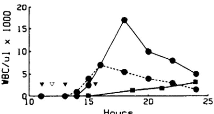
Figure 2. Effect of cyclooxygenase inhibitor on CSF leu-kocytosis after challenge with pneumococcal strain Sm. Groups of five rabbits were challenged at time 0 with 103
pneumococci. The density of leukocytes in CSF was stud-ied over 24 hr in groups treated as follows: no treatment (_); ampicillin (. .), 50 mg/kg iv at the time in-dicated by the open triangle; or ampicillin plus oxindanac (. - - - .), 5 mg/kg po at the time indicated by the closed triangles.
was absent in control animals not receiving ampicil-lin (in which bacterial multiplication continued). Thus, lysis and killing of pneumococci in the CSF space was accompanied by a transient burst of in-creased inflammation.
To determine if the anti-inflammatory effects of cyclooxygenase inhibitors would modulate the in-flammation associated with bacterial lysis in vivo, we used oxindanac and ampicillin to treat animals with established pneumococcal meningitis. As shown in figure 2, the number of leukocytes appearing in CSF after a dose of ampicillin was significantly lower in the animals that also received oxindanac. Oxin-danae did not alter the concentration of ampicillin in the CSF (data not shown). Importantly, effective bacterial killing occurred in oxindanac-treated animals despite the lower density of CSF leukocytes; a~3log killing occurred by 2 hr postampicillin treat-ment in both the control and the oxindanac-treated animals.
Discussion
The mortality from pneumococcal meningitis is directly correlated with the amount of meningeal in-flammation [1]. Two parameters used to quantitate the inflammatory response are the density of CSF leukocytes and the protein concentration, both of which are increased during natural infection. Our previous studies indicated that these abnormalities
in CSF cytochemistry in experimental meningitis can be generated by the interaction of host defenses with the pneumococcal cell wall [2-4]. The current results indicate that mortality is also associated with the per-sistent presence of bacterial cell wall in the subarach-noid space. Bacterial lysis in vivo and in vitro involves the explosive release of cell wall degradation prod-ucts into the surrounding environment. The results reported here show that ampicillin-induced killing of pneumococci in vivo, which entails such lysis, is associated with a transient enhancement of the in-flammatory response in the CSF space. This is con-sistent with the fact that the major products of pneu-mococcal autolysis are components of the cell wall that are known to be highly inflammatory in this meningitis model [2, 3].
Because enhancement of inflammation associated with bacteriolysis may contribute to the poor out-come of meningitis despite effective bacterial sterili-zation, it was important to determine if the host response to this transient inflammatory stimulus as-sociated with chemotherapy could be modulated. The present studies indicate that it is possible to re-duce the inflammatory response in the CSF space by inhibiting noncomplement-mediated host de-fenses. Several inhibitors of the cylcooxygenase path-way of arachidonic acid metabolism proved to be highly effective in reducing the influx of leukocytes and the elevation of protein concentration associated with intracisternal inoculation with cell walls. Methylprednisolone and oxindanac were particularly effective, whereas indomethacin and diclofenac so-dium were moderately effective. In contrast, inhibi-tors of primarily the lipoxygenase pathway were ineffective in preventing cell wall-induced inflam-mation. When tested against the natural infection, administering cyclooxygenase inhibitors in conjunc-tion with 13-lactam antibiotics also markedly reduced inflammation associated with release of inflamma-tory bacterial products during bacterial lysis byam-picillin.
Surprisingly, the arachidonic acid pathway that ap-pears to playa significant role in generating inflam-mation in the subarachnoid space of the rabbit is not the lipooxygenase pathway, which generates the powerful chemotaxin leukotriene B4 ,but rather, the
cyclooxygenase pathway. This conclusion is based on several lines of evidence. First, direct measurement of arachidonic acid pathway metabolites suggested a positive correlation between the concentration of POE] and the number of leukocytes that appeared
Anti-Inflammatory Agents and Pneumococcal Meningitis
in CSF in response to either living pneumococci or purified pneumococcal cell walls. Secondly, inhibi-tors of the cyclooxygenase pathway reduced both PGEzconcentrations and CSF inflammation in re-sponse to pneumococcal challenge. Prostaglandins of the E series are known to induce fever in response to endogenous pyrogen and to increase in concen-tration in response to challenge of rabbits with Esch-erichia coli or Shigella endotoxin [8, 9]. PGE:z are also known to have direct effects on PMNLs, includ-ing inhibition of chemotaxis in response to immune complexes and inhibition of degranulation of leu-kocytes activated by the chemotactic peptide formyl-methionyl-Ieucyl-phenylalanine [10, 11]. These direct effects would not, however, explain the correlation of high concentrations of PGE:z with increased leu-kocytosisinCSE Thus, our results do not distinguish whether PGE:z causes a previously undescribed en-hancement of leukocyte chemotaxis in this particu-lar site of infection or whether other cyclooxygenase metabolites are the direct chemotaxins, and the PGE:z response is correlated but secondary.
The approach we have taken to identify modes of therapy that would decrease the currently stable mor-bidity and mortality of meningitis has rested on the thesis that decreased inflammation is a new goal to be achieved in an era of chemotherapy in which bac-terial killing is easily attained. Leukocytes in inflam-matory exudates can most certainly contribute directly to local tissue injury, particularly if host cell membranes are coated with inflammatory bacterial components (i.e., cell walls and lipid-linked cell wall congeners). Cell walls can generate leukocytosis by activating several host defenses [4, 12], and from the present study, these components can even induce death. The most-significant finding of this study is that inhibiting the activity of one arm of the CSF defense system, the cyclooxygenase pathway, appears to reduce both the total CSF inflammatory mass and to improve survival. Preliminary studies have con-firmed and extended this association between cyclo-oxygenase inhibitors and enhanced survival in other instances of experimental meningeal inflammation (0. Z., unpublished observations).
To further improve the outcome of bacterial meningitis, we must identify the pathological lesions associated with morbidity and mortality. Leukocyte-induced damage is almost certainly not the only source of destructive lesions. For instance, from the studies of Tauber et al. [13, 14], CSF pressure is known to correlate with bacterial density and with
989
the concentration of cell walls in the CSF space. Brain edema does not appear to show this correla-tion. Yet, neither of these pathological lesions nor even simple abnormalities in CSF chemistry are directly dependent on leukocytes because all occur in neutropenic animals[13-15].Furthermore, our re-cent studies indicate that death can be caused by in-tracisternal inoculation of cell walls in both normal and neutropenic animals (E. T., unpublished obser-vations). Thus, the processes that generate leukocy-tosis may also generate, in parallel, tissue damage independent of leukocytes. In this regard, the abil-ity of the arachidonic acid cascade to generate medi-ators capable of altering vascular permeability, plate-let function, etc. in addition to chemotaxins, may be important. Thus, it may be the multiplicity of ef-fects of inhibitors of the arachidonic acid pathway that is critical to linking decreased leukocytosis, de-creased tissue damage, and enhanced prognosis. The combination of effects of individual inhibitors varies, and the relative proportion of these effects with the greatest benefit for outcome of disease re-mains to be determined. Cyclooxygenase inhibitors such as diclofenac sodium and oxindanac, however, appear to be excellent candidates for improving out-come of therapy when used in conjunction with ~
lactam antibiotics to achieve bacterial killing dur-ing pneumococcal mendur-ingitis.
References
1. McAllister CK, O'Donoghue JM, Beaty HN. Experimental pneumococcal meningitis. II. Characterization and quan-titation of the inflammatory process.JInfect Dis 1975; 132: 355-60
2. Tuomanen E, Tomasz A, Hengstler B, Zak O. The relative role of bacterial cell wall and capsule in the induction of inflammation in pneumococcal meningitis. J Infect Dis 1985;151:535-40
3. Tuomanen E, Liu H, Hengstler B, Zak 0, Tomasz A. The induction of meningeal inflammation by components of the pneumococcal cell wall. J Infect Dis 1985;151:859-68 4. Tuomanen E, Hengstler B, Zak 0, Tomasz A. The role of complement in inflammation during experimental pneu-mococcal meningitis. Microbial Pathogenesis 1986;1:15-32 5. Dacey RG, Sande MA. Effect of probenecid on cerebrospi-nal fluid concentrations of penicillin and cephalosporin derivatives. Antimicrob Agents Chemother 1974;6:437-41 6. Bray MA. Retinoids are potent inhibitors of the generation of rat leukocyte leukotriene Bs-like activity in vitro. Eur J Pharmacol 1984;98:61-7
7. White HL, Glassman AT. A simple radiochemical assay for prostaglandin synthetase. Prostaglandins 1974;7:123-9 8. Bernheim HA, Gilbert TM, Stitt JT. Prostaglandin E levels
fever and changes in body temperature. J Physiol 1980; 301:69-78
9. Philipp-Dormston WK, Siegert R. Prostaglandins of the E and F series in rabbit cerebrospinal fluid during fever in-duced by Newcastle Disease virus,E.Coli-endotoxin, or endogenous pyrogen. Med Microbiol Immunol 1974; 159:279-84
10. Fantone JC, Marasco WA, Elgas LJ, Ward PA. Anti-inflam-matory effects of prostaglandin E1 :in vivo modulation of
the formyl peptide chemotactic receptor on the rat neu-trophil. J Immunol 1983;130:1495-7
11. Kaplan HB, Edelson HS, Korchak HM, Given WP, Abram-son S, Weissmann G. Effects of non-steroidal anti-inflammatory agents on human neutrophil functions in vitro and in vivo. Biochem Pharmacol 1984;33:371-8 12. Ernst JD, Hartiala KT, Goldstein 1M, Sande MA.
Comple-ment (C5)-derived chemotactic activity accounts for
ac-cumulation of polymorphonuclear leukocytes in cerebro-spinal fluid of rabbits with pneumococal meningitis. Infect Immun 1984;46:81-6
13. Tauber MG, KhayBashi H, Sande MA. Effects of am-picillin and corticosteroids on brain water content, cere-brospinal fluid pressure, and cerecere-brospinal fluid lactate lev-els in experimental pneumococal menigitis. J Infect Dis 1985;151:528-34
14. Tauber MG, Tuomanen E, Zak0, Sande MA. Increased in-tracranial pressure induced by pneumococcal cellwalls [ab-stract no. 683]. In: Program and ab[ab-stracts of the 25th In-terscience Conference on Antimicrobial Agents and Chemotherapy. Washington, DC: American Society for Microbiology, 1985
15. Ernst JD, Decazes JM, Sande MA. Experimental pneumo-coccal meningitis: role of leukocytes in pathogenesis. In-fect Immun 1983;41:275-9