HAL Id: hal-02301250
https://hal.archives-ouvertes.fr/hal-02301250
Submitted on 27 Nov 2020
HAL is a multi-disciplinary open access
archive for the deposit and dissemination of sci-entific research documents, whether they are pub-lished or not. The documents may come from teaching and research institutions in France or abroad, or from public or private research centers.
L’archive ouverte pluridisciplinaire HAL, est destinée au dépôt et à la diffusion de documents scientifiques de niveau recherche, publiés ou non, émanant des établissements d’enseignement et de recherche français ou étrangers, des laboratoires publics ou privés.
Giant Core–Shell Nanospherical Clusters Composed of
32 Co or 32 Ni Atoms Held by 6 p - tert
-Butylthiacalix[4]arene Units
Alexandre Gehin, Sylvie Ferlay, Jack Harrowfield, D. Fenske, Nathalie
Kyritsakas, Mir Wais Hosseini
To cite this version:
Alexandre Gehin, Sylvie Ferlay, Jack Harrowfield, D. Fenske, Nathalie Kyritsakas, et al..
Giant Core–Shell Nanospherical Clusters Composed of 32 Co or 32 Ni Atoms Held by 6 p tert -Butylthiacalix[4]arene Units. Inorganic Chemistry, American Chemical Society, 2012, 51 (9), pp.5481-5486. �10.1021/ic300550v�. �hal-02301250�
Giant core-shell nanospherical clusters composed of 32 Co or
32 Ni atoms held by 6 p-tert-butylthiacalix[4]arene units
Alexandre Gehin,a Sylvie Ferlay,a* Jack M. Harrowfield,b D. Fenske,c Nathalie
Kyritsakasa and Mir Wais Hosseinia*
a Laboratoire de Chimie de Coordination Organique, UMR CNRS 7140, Université de Strasbourg, F-67000 Strasbourg, France
b Laboratoire de Chimie Supramoléculaire, Institut de Science et d'Ingénierie
Supramoléculaires, Université de Strasbourg, 8, allée Gaspard Monge, 67083 Strasbourg, France
c Institut für Anorganische Chemie, Unversität Karlsruhe, D 76128 Karlsruhe, Germany
RECEIVED DATE (will be automatically inserted after manuscript is accepted)
Using combinations of p-tert-butylthiacalix[4]arene (TCA) and [M(DMSO)6(BF4)2] salts (M = Co(II) or
Ni(II)), two almost isostructural core-shell type thermally stable giant nanoclusters, composed of 32 metal centres, 6 deprotonated calix units binding the metal centres by both their O and S atoms, 24 m-oxo or m-hydroxo bridging groups and 6 MeOH molecules, have been prepared under mild and reproducible conditions. For both giant clusters, the oxidation state II (MII32O16(OH)8(CH3OH)6TCA6 (M =
Co, Ni)) for the metal centre was demonstrated by XPS and electronic absorption spectroscopies.
Introduction
Polynuclear transition-metal compounds, in particular m oxo and/or hydroxo bridged polyoxometallate type compounds, are of interest for their chemical and physical properties.1 Several
elegant strategies have been reported for the preparation of giant polynuclear species such as of, for example, iron2, molybdenum3, manganese4 and
cadmium5 .
The calix[4]arene backbone 1 (Fig. 1 left)6 is an
interesting unit for the design of polynucleating ligands. In particular, calix[4]arene derivatives in the cone conformation (Fig. 1 right) offer four OH groups oriented in a convergent manner. This feature has been explored for the formation of koilands, which are face to face calix[4]arenes doubly fused by two Si(IV)7, two Ti(IV)8, one Si(IV) and one Ti(IV)9, two
Al(III)10, two Zn(II)11, two Eu(III)12 or four Nb(V)13 ions.
The thiacalix[4]arene backbone 214 (TCA) for which
the four CH2 groups connecting the aromatic moieties
are replaced by sulphur atoms, offers four additional coordinating sites, i.e. the four S atoms15 and is a
candidate of choice for the generation of high nuclearity complexes. From 2, which also adopts the cone conformation,16 tetranuclear Cu(II)17 and tetra-
and hexa-nuclear Hg(II)18 species have been
generated. Using dimercapto-19 and
tetramercapto-calix[4]arene derivatives20 3 and 4 respectively,
mononuclear and binuclear Hg(II) complexes have been reported. The synthesis and structural analysis of the tetramercapto-tetrathiacalix[4] arene 521 and
some Hg(II)18 and low valence Mo and W22 or Ir and
Rh23 organometallic complexes have been also
published.
The coordination ability24 of 2 has been used for the
preparation of other polynuclear species such as Cu4,17 Mn4,25 Ln4,26 Ni627, V6,28 Fe8,29 Mn4Ln4,30 Cu931,
Cu1032 and Co2433. Recently, a new heterometallic
cationic cluster [CoII24(MVIO4)8TCA6Cl6]2+ combining
24 Co(II) and 8 M(VI) (M = Mo or W) atoms was published.34 The highest nuclearity homometallic
clusters reported are those containing 32 cobalt35
(CoII24CoIII8O24(OH2)24TCA6) or nickel atoms
(NiII32(OH)40TCA6) 27.
Figure 1: Chemical formulae of calix[4]arene, thiacalix[4]arene and mercaptathiacalix[4]arene derivatives (left) and a schematic representation of their cone conformation (right).
Here, we report on the synthesis, structural studies, thermal and XPS characterization of two new core-shell nanospherical clusters composed of 32Co(II) or 32Ni(II) held by 6 p-tert-butylthiacalix[4]arenes.
Experimental section
Preparation of Co32: In a crystallisation tube (1 cm
diameter), exposed to the atmosphere and at room temperature, diffusion of a DMF solution (5 mL) of Co(DMSO)6(BF4)2 (230 mg, 0.28 mmol) into a DMF
solution (1 mL) containing the thiacalixarene 2 (20 mg, 2.78x10⁻² mmol) and Et3N (0.1 mL), provided a
green solution after ca one hour. This was filtered before MeOH (1 mL) was carefully added. After a week, a fine pale green precipitate had formed at the bottom of the tube. Upon standing further for ca 3 weeks, large pink crystals, suitable for single crystal X-ray diffraction, were formed on the wall of the tube. 97 mg, (48 % yield). MS (ESI (DMSO), m/z): 2285.7
(calculated for
Co32(TCA)6(CH3OH)6O16(OH)8(KNa2)3+, m/z =
2285.6); IR (n, cm-1): 752.3; 1092.7; 1255.7; 1448.6;
1672.3; 2956.0.
Synthesis of Ni32: In a crystallisation tube (1 cm
diameter), exposed to the atmosphere and at room temperature, diffusion of a DMF solution (5 mL) of Ni(DMSO)6(BF4)2 (230 mg, 0.28 mmol) into a DMF
solution (1 mL) containing the thiacalixarene 2 (20 mg, 2.78x10⁻² mmol) and Et3N (0.1 mL), provided a
green solution after ca one hour. This was filtered before MeOH (1 mL) was carefully added. After a week, a fine green precipitate had formed at the bottom of the tube. Upon standing further for ca 3 weeks, large green crystals, suitable for single crystal X-ray diffraction, were formed on the wall of the tube. 95 mg, (ca 45 % yield). MS (ESI (DMSO), m/z): 3385.7 (calculated Ni32(TCA)6(CH3OH)6O16(OH)8(LiH)2+, m/z = 3385.6) ;
6788.2 (calculated for Ni32(TCA)6(CH3OH)6O16(OH)8Na+, m/z = 6787.8). IR
(n, cm-1): 750.3; 1058.9; 1257.6; 1438.9; 1666.5;
2958.9.
Physical measurements Single-Crystal Studies.
Data were collected at 173(2) K on a Bruker APEX8 CCD Diffractometer equipped with an Oxford Cryosystem liquid N2 device, using
graphite-monochromated Mo-Kα (l = 0.71073 Å) radiation. For both structures, diffraction data were corrected for absorption. Structures were solved using SHELXS-97 and refined by full matrix least-squares on F2 using
SHELXL-97. The hydrogen atoms were introduced at calculated positions and not refined (riding model).36
In the case of Ni32, due the disorder of solvent molecules, the SQUEEZE command was used.37
CCDC XXX and CCDC XXX contain supplementary crystallographic data for the Co32 and Ni32
nanospheres, respectively. They can be obtained free of charge from the Cambridge Crystallographic Data Centre via www.ccdc.cam.ac.uk/datarequest/cif.
Powder diffraction studies (PXRD)
For both Co32 and Ni32 nanoclusters, PXRD
diagrams for the crystalline and powdered materials were collected on a Bruker D8 diffractometer using monochromatic Cu-Ka radiation with a scanning range between 3.8 and 30° and a scan step of 2°/mn. The desolvated forms of Co32 and Ni32
nanospheres (Ni32(des) and Co32(des)) were
generated upon heating the sample at 170 °C and the crystallinity was studied by PXRD. The thermal stability of Co32 and Ni32 nanoclusters was studied by
first heating the powder to 300 °C and then measuring PXRD patterns at room temperature.
Thermogravimetric (TGA) Studies.
TGA measurements have been performed on both powdered single crystal samples of Co32 and Ni32
nanoclusters using a Pyris 6 TGA Lab System (Perkin-Elmer), using a N2 flow of 20 mL/min and a
heat rate of 10 °C/min.
XPS measurements.
XPS (X-ray photoelectron Spectroscopy) measurements were performed on powdered single crystal samples of both Co32 and Ni32 nanoclusters
using a ThermoVGScientific photoelectron spectrometer equipped with a twin anode providing both unmonochromated Al Kα and Mg Kα radiation (1486.6 and 1453.6 eV, respectively). The spectrometer, equipped with a multichannel detector, was used in the constant resolution mode with a pass energy of 20 eV. The total resolution of the system was estimated at 0.55 eV. Spectra were referenced to the aliphatic hydrocarbon C1s signal at 285 eV.
Solid state UV measurements
UV/Visible spectra were obtained on powdered samples of single crystals of Co32 and Ni32
nanoclusters using a Shimadzu UV3600 spectrometer (data were recorded in the reflection mode using a 150 mm integrating sphere, with a resolution of 4 nm and a sampling rate of 300 nm min-1)
Infra-red Studies
IR spectra were recorded on powdered samples of single crystals of Co32 and of Ni32 nanoclusters using
a Perkin Elmer Spectrometer RX1.
Results and discussion
Using the p-tert-butylthiacalix[4]arene 2 in its cone conformation (Fig. 1), a reproducible synthetic procedure using identical conditions was established for the formation of giant clusters composed of 32 Co or 32 Ni atoms. The formation of these polynuclear assemblies displaying almost identical structures was
achieved under mild conditions. Upon slow diffusion at room temperature of a DMF solution containing the tetrafluoroborate salt of Co(II) or Ni(II) into a DMF solution of the TCA 2 and Et3N as base, a powder
was obtained which upon standing in MeOH afforded pink and green crystalline materials in the case of Co and Ni, respectively (see experimental section). It is worth noting that these species could only be produced when using [MII(DMSO)6(BF4)2] (M = Co or
Ni) salts. Interestingly, the same type of crystalline material was obtained using EtOH or i-PrOH, however with much lower crystalline quality. Both clusters have been characterized by X-ray diffraction on single crystals.‡
In both cases, the diffraction data obtained were treated supposing the presence of discrete molecular entities with the following formula ((32M)(16O2-)(8OH
-)(624-)(6CH3OH). The giant clusters, with the longest
distance between opposite CH3 groups of ca 24.7 and
of 23.7 Å for Co32 and Ni32 species, respectively,
display almost identical connectivity patterns (Fig. 2 left). Whereas for Co32 ((32Co)(16O2-)(8OH-)(6 TCA
4-)(6CH3OH).3DMSO.3DMF) the solvent molecules (3
DMF and 3 DMSO) could be localised, for Ni32, due
to disorder of solvents, the structure was refined using the SQUEEZE procedure.38
Thus, although both species crystallise in the same space group (P21/n), since the solvent molecules
could not be identified in the case of Ni32, the two
crystals may or may not be isomorphous. The overall assembly in both cases may be described as a cubic metallic core composed of 8 Co or Ni centres located within a shell composed of 24 metal cations (Co or Ni) held together by 6 deprotonated thiacalix 2
4-occupying the apices of an octahedron and forming an organic envelope (Fig. 2 right).
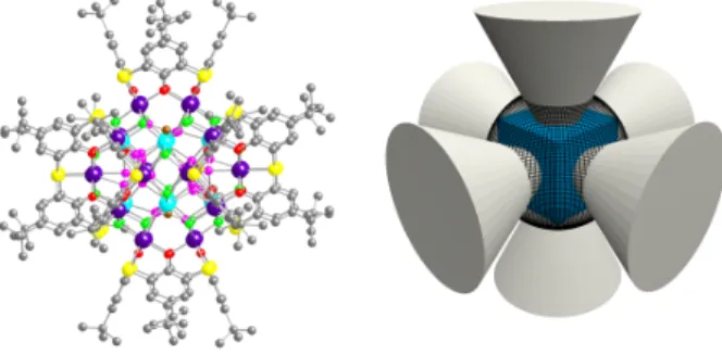
Figure 2: Crystal structure of the M32 (M = Co or Ni) nano cluster.
For the sake of clarity, the 8 core (light blue) and 24 shell (violet) metal centres are differentiated by colour. The 24 O atoms of the calix units (red), 24 m-oxo and m -hydroxo (green) and of MeOH units (pink) are also differentiated by colour. The C atom of the 6 MeOH molecules is coloured in brown. Solvent molecules are omitted (left) and schematic representation of the giant M32
nanoclusters (M = Co or Ni) composed of a cubic M8 core (blue)
inserted inside of a spherical M24 shell (black) surrounded by 6
thiacalix[4]arenes (grey) (right).
The aesthetically appealing structure of both clusters is rather complex. They will be described starting from their organic envelope and zooming towards their core.
As for the parent compound 2,14 all (here,
deprotonated) calix units (24-) adopt the cone
conformation (Fig. 1 right) with typical C-O distances in the 1.333(7)-1.368(8) and 1.281(13)-1.382(11) Å range for Co and Ni respectively and CS distances in the 1.773(6)-1.804(8) and 1.705(17)-1.844(13) Å range for Co and Ni, respectively. Among the 6 calix units, the t-butyl groups were found to be disordered over two positions (two calix with 4 and the remaining four with 2 disordered groups located on adjacent aromatic moieties). In the case of the Co32 cluster,
the three DMF solvent molecules are located within the cavities of the six calix units but with an occupancy of 50 %, i.e. one DMF molecule is disordered over two calix units. The three DMSO solvent molecules are located in the proximity of the metallic core with a Co…O distance of ca 3.4 Å. One of them is disordered over two positions with an occupancy of 50 %.
Each calix moiety behaves as a cluster keeper and binds 4 metal centres forming a square through both their phenoxide groups (O-M distance in the 2.022(4)-2.061(4) Å and 1.968(8)-2.060(9) Å range for Co and Ni respectively) and their sulfur atoms (S-M distance in the 2.4432(17)-2.4543(18) Å and 2.409(3)-2.439(3) Å range for Co and Ni, respectively). This motif is reminiscent of that found in the reported tetranuclear Cu(II)17 and Hg(II)18 species (Fig. 3).
Figure 3: A portion of the crystal structure of the M32 (M = Co or Ni)
nano cluster showing the 24 metal centres (violet) forming the shell of the giant cluster hold by the 6 thiacalix units. For sake of clarity, the 24 O atoms of the calix units (red), 24 m-oxo and m -hydroxo (green) are differentiated by colour. Solvent molecules are omitted.
The 6 tetranuclear M4 units are further
interconnected by 16 m-oxo and 8 m-hydroxo groups forming thus a metallic shell of the sodalite type with a diameter of ca 10.63 and 10.38 Å (longest M-M distance) for Co32 and Ni32 respectively. The
protonation state of the above-mentioned bridging O atoms cannot be specified. The M-O distances are in the 2.031(5)- 2.062(5) and 1.989(7)-2.061(6) Å range for Co and Ni respectively. The coordination sphere around the metal centres is composed of 5 oxygen and 1 sulphur atoms. All metal centres adopt a distorted octahedral coordination geometry (Fig. 4).
Figure 4: A portion of the crystal structure of the M32 (M = Co or Ni)
nano cluster showing the 24 metal centres (violet) forming the shell and their surroundings. The O atoms are differentiated by colour (red and green) for clarity. Solvent molecules are omitted.
The metallic shell defines a cavity occupied by a octanuclear M8 core. The 8 metallic centres
occupying the corners of a distorted cube (M-M distance in the ca 4.20-4.38 Å range) are connected to the shell through the 24 m-oxo or m-hydroxo bridging groups (three O atoms per metal centre) with a O-M distance in the 2.057(5)-2.323(14) Å and 1.989(7)-2.24(2) Å range for Co and Ni, respectively.
Figure 5: A simplified view of the M32 (M = Co or Ni) nanocluster
found in the crystal lattices, showing the 24 metal centres (violet) forming the sodalite type shell and the 8 metals (light blue) forming a cubic core located at the centre of the giant cluster. The O atoms are differentiated by colour (red and green) for clarity. Solvent molecules are omitted.
Finally, within the cubic core, the 8 metal centres are connected by 6 MeOH molecules, with M-O distances in the 2.169(14)- 2.323(14) and 2.072(7)-2.39(2) Å range for Co32 and Ni32 respectively.
In regard to the organic envelope and metallic shell, the two clusters are very similar but they diverge in their cubic cores (Fig. 6). For Co32 cluster, of the six
disordered MeOH molecules occupying the faces of the cubic arrangement, the oxygen atoms of two of them are disordered over four positions, whereas for the remaining four molecules, the oxygen atoms are disordered over three positions (Fig. 6 left). In the case of Ni32 cluster, the reverse is observed (Fig. 6
right).
Figure 6: Portions of the crystal structures of the nanoclusters showing the Co8 (left) and Ni8 (right) cubic metallic core
interconnected by 6 MeOH (disordered O atoms in pink and C in brown). H atoms are omitted. For differences see text.
In the Co species, the inner Co8 unit atoms have
distorted octahedral O6 coordination geometry, while
in the Ni analogue, six have this form and two have distorted square-pyramidal O5 coordination.
The packing of the two clusters is identical with no specific interactions between the polynuclear species (Fig. S1).
It is interesting to note that for the reported analogous but mixed oxidation state Co cluster,34 48
m-oxo or m-hydroxo bridging anions are involved in the coordination sphere of the metal centres, whereas for the Ni analogue,27 40 m-hydroxo groups were
found to be bound to the metal. For both Co32 and
Ni32 species reported here, in addition to six MeOH
molecules bound to the metallic core, only 24 m-oxo or m-hydroxo groups are connected to metal centres.
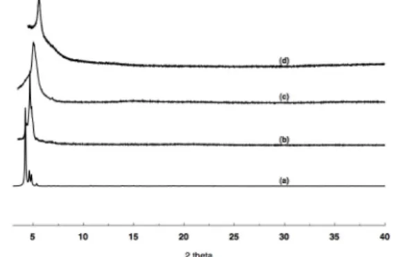
The crystallisation process used for the preparation of both Co- and Ni-based nanospheres led to single crystals attached to the walls and a powder deposited at the bottom of the tube. The crystals and powder were separated and dried in air. Both phases were analysed using PXRD (Fig. 8 and 9) and similar patterns were observed. For the Co32 nanosphere
dried in air, the observed pattern (Fig. 7b) fits well with the simulated one (Fig. 7a). Interestingly, the powder sample heated at 170 °C to afford the desolvated material also displayed a similar pattern (Fig. 8c bottom) indicating the conservation of the crystallinity of the sample. Retention of the same pattern even after heating at 300 °C (Fig. 7d) showed the thermal stability of the sample. The same behaviour was observed for the Ni32 nanosphere
Figure 7: Comparison of the simulated (a) and recorded PXRD patterns for Co32 at RT (b), after heating at 170 °C (c) and after
heating at 300 °C (d).
Figure 8: Comparison of the simulated (a) and recorded PXRD patterns for Ni32 at RT (b), after heating at 170 °C (c) and after
heating at 300 °C (d).
The thermal stability of both species was also investigated by thermogravimetric analysis. For both
Co32 and Ni32 species, in the 30 °C - 460 °C
temperature range, a weight loss of ca 25 % was observed prior to decomposition which appeared at 440 °C for Ni32 and at ca 460 °C for Co32 (see figure
S2). Although the weight loss prior to decomposition was assumed to be due to volatilisation of solvents, steps corresponding to the loss of different species were not discerned.
In order to clarify the oxidation state of the Co and Ni cations, X-Ray Photoelectron Spectroscopy (XPS) was used. Measurements were carried out on single crystals, using as reference salts Co(OH2)6•(NO3)2 for
Co32 and Ni(OH2)6•SO4 for Ni32. For both Co32 (Fig. 9
top) and Ni32 (Fig. 10 bottom) giant clusters, the
spectra displayed the M2p3/2, M2p1/2 and M2s
signatures. Significantly, the match, in terms of binding energy and shape, between the recorded spectra for the two clusters and the corresponding reference compounds indicates that, in both cases, the metal centres are in the oxidation state II with a deformed octahedral environment.
Figure 9: The X-Ray Photoelectron Spectra (XPS) of the polycrystalline samples Co32 (top, a) and Ni32 (bottom, a) exhibiting
the M2p3/2, M2p1/2 and M2s signatures (M = Co or Ni). The spectra
are compared to those of Co(OH2)6SO4 (top, b) for Co32 and
Ni(OH2)6(NO3)2 (bottom, b) for Ni32.
The UV-Visible reflectance spectra of M32 (M = Co
or Ni) clusters were recorded in the solid state using polycrystalline samples (Fig. 10). In agreement with the observations made by XPS spectroscopy mentioned above, in both cases, the recorded spectra are typical of M(II) species. For the Co32 giant cluster,
considering the oxidation state II and an octahedral geometry, three transitions in the 250-800 nm region are expected. Indeed, three major bands at 320nm, assigned to the 4T1g (F) ® 4T2g (F) transition and at
550 nm to the 4T1g (F) ® 4T1g (P) and 4T1g (F) ® 4A2g
(F) transitions, were observed. For Ni32, again
considering a Ni(II) cation in octahedral geometry, two transitions in the 250-800 nm region are expected and were indeed observed at 330 nm for the high energy
3A2g®3T1g(P) transition and at 670 nm for the low
energy band assigned to the 3A2g®3T1g(F) transition.
The presence of shoulders at ca 400 nm probably results from the presence of M centres in a significantly deformed octahedral geometry as discussed in the section dealing with structural analysis.
Figure 10: UV-Vis reflectance spectra of Ni32 (green) and of Co32
(pink) recorded in the solid state at RT.
Finally, in support of the above mentioned experimental facts, using the observed M-O or M-S distances, a BVS calculation38 on both clusters
revealed that for none of the metal centres was an oxidation state higher that 2.3 appropriate (see supporting information).
Conclusions
A mild, reproducible procedure for obtaining thermally stable, giant core-shell nanoclusters containing 32 Co or Ni atoms enclosed by six tetrathiacalix[4]arene units has been established. The structure of both species was elucidated using X-ray diffraction techniques both on single crystals and on powdered crystalline samples. Both clusters are based on a cubic metallic core composed of 8 metal centres located in the centre of a sodalite type shell composed of 24 metals surrounded by 6 calix units as an organic envelope (Fig. 2 right). In marked contrast with a reported analogous cluster,35 obtained under
hydrothermal conditions and which contains both Co(II) and Co(III) centres, for the Co32 and Ni32
clusters reported here, the metal centres are clearly in the oxidation state II.
Using the same methodology, the formation of analogous nanoclusters using other calixaerene derivatives and metal centres is currently under investigation.
ACKNOWLEDGMENT
This work was financially supported by the University of Strasbourg (UdS), the Centre National de la Recherche Scientifique (CNRS), Institut Universitaire de France (IUF) and the International Centre for Frontier Research in Chemistry (FRC), Strasbourg. Pierre Bernardt (LMSPC, Strasbourg) is warmly acknowledged for the XPS measurements
Supporting information
TGA traces for Co32 and Ni32, as well as BVS calculations. ‡ (a) Crystal data for Co
32:
(C40H44O4S4)6Co32(CH3OH)6 O16(OH)8, (C3H7NO)3
(C2H6OS)3, M = 7225.70, monoclinic, space group
P21/n, a = 24.7390(7) Å, b = 26.8037(7) Å, c =
32.6310(9) Å, β = 94.2370(10)°, V = 21578.4(10)Å3, T
= 173(2) K, Z = 2, Dc = 1.112 g·cm-3, μ = 1.371 mm-1,
149389 collected reflections, 48887 independent [R(int) = 0.0463], GOF = 1.052, R1 = 0.0909, wR2 =0.2687 for I > 2σ(I) and R1 = 0.1398, wR2 = 0.3015 for all data ;
(b) Crystal data for Ni32: (C40H44O4S4)6Ni32(CH3OH)6
O16(OH)8, M = 6732.73, monoclinic, space group
P21/n, a = 24.5603(5) Å, b = 26.6097(6) Å, c =
32.2446(7) Å, β = 93.4090(10)°, V = 21035.9(8)Å3, T
= 173(2) K, Z = 2, Dc = 1.040 g·cm-3, μ = 1.557 mm-1, 163687 collected reflections, 48655 independent [R(int) = 0.0852], GOF = 1.366, R1 = 0.1074, wR2 =0.2613 for I > 2σ(I) and R1 = 0.2169, wR2 = 0.2949 for all data.
References
[1] (a) Fenske, D.; Ohmer, J.; Hachgenei, J. Angew. Chem., Int. Ed. Engl. 1985, 24, 993-995; (b) Liu, T.; Zhang, Y.-J.; Wang, Z.-M.; Gao, S. J. Am. Chem. Soc. 2008, 130, 10500-10501; (c) Alborés, P.; Rentschler, E. Angew. Chem., Int. Ed. 2009, 48, 9366-9370.
[2] Taft, K. L.; Lippard, S. J. J. Am. Chem. Soc. 1990, 112, 9629-9630.
[3] (a) Müller, A.; Kögerler, P. Coord. Chem. Rev. 1999, 182, 3-17; (b) Müller, A.; Roy, S. Coord. Chem. Rev., 2003, 245 153-166; (c) Long, D. L.; Tsunashima, R.; Cronin, L. Angew. Chem. Int. Ed. 2010, 49, 1736-1758.
[4] (a) Goldber, D. P.; Caneschi, A.; Delfs, C. D.; Sessoli, R.; and Lippard, S. J. J. Am. Chem. Soc. 1995, 117, 5789-5800; (b) Bagai, R.; Christou, G. Chem. Soc. Rev. 2009, 38, 1011-1026. [5] Argent, S. P.; Greenaway, A.; Gimenez-Lopez, M.; Lewis, W.;
Nowell, H.; Khlobystov, A. N.; Blake, A. J.; Champness, N. R. and Schröder, M. J. Am. Chem. Soc., 2012, 134, 55-58. [6] (a) Gutsche, C. D. Calixarenes Revisited: Monographs in
Supramolecular Chemistry;Royal Society of Chemistry: Cambridge, U.K., 1998; (b) Calixarenes 2001; Asfari, Z.; Böhmer, V.; Harrowfield, J.; Vicens, J., Eds.; Kluwer: Dordrecht, The Netherlands, 2001.
[7] Delaigue, X.; Hosseini, M. W.; De Cian, A.; Fischer, J.; Leize, E.; Kieffer, S.; Van Dorsselaer, A. Tetrahedron Lett. 1993, 34, 3285-3288.
[8] Olmstead, M. M.; Sigel, G.; Hope, H.; Xu, X.; Power, P. P. J. Am. Chem. Soc. 1985, 107, 8087-8091.
[9] Delaigue, X.; Hosseini, M. W. Tetrahedron Lett. 1993, 34, 7561-7564.
[10] Atwood, J. L.; Bott, S. G.; Jones, C.; Raston, C. J. J. Chem. Soc., Chem. Commun. 1992, 1349-1351.
[11] Atwood, J. L.; Junk, P. C.; Lawrence, S. M.; Raston, C. L. Supramol. Chem. 1996, 7, 15-17.
[12] Bilyk, A.; Harrowfield, J. M.; Skelton, B. W.; White, A. H. J. Chem. Soc., Dalton Trans. 1997, 4251-4256.
[13] Corazza, F.; Floriani, C.; Chiesi-Villa, A.; Guastini, C. Chem. Commun. 1990, 1083-1084.
[14] Kumagai, H.; Hasegawa, M.; Miyanari, S.; Sugawa, Y.; Sato, Y.; Hori, T.; Ueda, T.; Kamiyama, H.; Miyano, S. Tetrahedron Lett. 1997, 38, 3971-3971.
[15] Hosseini, M. W. in Calixarenes 2001; Asfari, Z.; Böhmer, V.; Harrowfield, J.; Vicens, J., Eds.; Kluwer: Dordrecht, The Netherlands, 2001. 110-129.
[16] Akdas, H.; Bringle, L.; Graf, E.; Hosseini, M. W.; Mislin, G.; Pansanel, J.; De Cian, A.; Fischer, J. Tetrahedron Lett. 1998, 39, 2311-2314.
[17] Mislin, G.; Graf, E.; Hosseini, M. W.; Bilyk, A.; Hall, A. K.; Harrowfield, J. M.; Skelton, B. W.; White, A. H. Chem. Commun. 1999, 373-374.
[18] Akdas, H.; Graf, E.; Hosseini, M. W.; De Cian, A.; Bilyk, A.; Skelton, B. W.; Koutsantonis, G. A.; Murray, I.; Harrowfield, J. M.; White, A. H. Chem. Commun. 2002, 1042-1043.
[19] (a) Delaigue, X.; Hosseini, M. W.; Kyritsakas, N.; De Cian, A.; Fischer, J. Chem. Commun. 1995, 609-610; (b) Rao, P.; Enger, O.; Graf, E.; Hosseini, M. W.; De Cian, A.; Fischer, J. Eur. J. Inorg. Chem. 2000, 1503-1508.
[20] Delaigue, X.; Harrowfield, J. McB.; Hosseini, M. W.; De Cian, A.; Fischer, J.; Kyritsakas, N. Chem. Commun. 1994, 1579-1580. [21] Rao, P.; Hosseini, M. W.; De Cian, A.; Fischer, J. Chem.
Commun. 1999, 2169-2170.
[22] Buccella, D.; Parkin, G. Chem. Commun. 2009, 289-291. [23] Hirata, K.; Suzuki, T.; Noya, A.; Takei I.; Hidai, M. Chem.
Commun. 2005, 371-3720.
[24] Kajiwara, T.; Iki, N.; Yamashita, M. Coord. Chem. Rev. 2007, 251, 1734-1746.
[25] (a) Desroches, C.; Pilet, G.; Borshch, S. A.; Parola, S.; Luneau, D. Inorg. Chem. 2005, 44, 9112-9120; (b) Karotsis, G.; Teat, S. J.; Wernsdorfer, W.; Piligkos, S.; Dalgarno, S. J.; Brechin, E. K. Angew. Chem., Int. Ed. 2009, 48, 8285-8288.
[26] (a) Bi, Y. F.; Wang, X. T.; Liao, W. P.; Wang, X. W.; Deng, R. P.; Zhang, H. J.; Gao, S. Inorg. Chem. 2009, 48, 11743-11747 ; (b) Bilyk, A.; Dunlop, J. W.; Fuller, R. O.; Hall, A. K.; Harrowfield, J. M.; Wais Hosseini, M. W.; Koutsantonis, G. A.; Murray, I. W.; Skelton, B. W.; Sobolev, A. N.; Stamps, R. L.; White, A. H. Eur. J. Inorg. Chem. 2010, 2127-XXX.
[27] Bilyk, A.; Dunlop, J. W.; Fuller, R. O.; Hall, A. K.; Harrowfield, J. M.; Wais Hosseini, M. W.; Koutsantonis, G. A.; Murray, I. W.; Skelton, B. W.; Stamps, R. L.; White, A. H. Eur. J. Inorg. Chem. 2010, 2106-2126.
[28] Aronica, C.; Chastanet, G.; Zueva, E.; Borshch, S. A.; Clemente-Juan, J. M.; Luneau, D. J. Am. Chem. Soc. 2008, 130, 2365-2371.
[29] Desroches, C.; Pilet, G.; Szilágyi, P.; Molnár, G.; Borshch, S. A.; Bousseksou, A.; Parola, S.; Luneau, D. Eur. J. Inorg. Chem. 2006, 357-365.
[30] Karotsis, G.; Kennedy, S.; Teat, S. J.; Beavers, C. M.; Fowler, D. A.; Morales, J. J.; Evangelisti, M.; Dalgarno, S. J.; Brechin, E. K. J. Am. Chem. Soc. 2010, 132, 12983-12990.
[31] Karotsis, G.; Kennedy, S.; Dalgarno, S. J.; Brechin, E. K. Chem. Commun. 2010, 3884-3886.
[32] Kajiwara, T.; Kon, N.; Yokozawa, S.; Ito, T.; Iki, N.; Miyano, S. J. Am. Chem. Soc. 2002, 124, 11274-11275.
[33] Bi, Y. F.; Xu, G. C.; Liao, W. P.; Du, S. C.; Wang, X. W.; Deng, R. P.; Zhang, H. J.; Gao, S. Chem. Commun. 2010, 6362-6364. [34] Bi, Y. F.; Du, S. C.; Liao, W. P. Chem. Commun. 2011,
4724-4726
[35] Bi, Y. F.; Wang, X. T.; Liao, W. P.; Wang, X. F.; Wang, X. W.; Zhang, H. J.; Gao, S. J. Am. Chem. Soc. 2009, 131, 11650-11651.
[36] Sheldrick, G. M. : Program for Crystal Structure Solution; University of Göttingen: Göttingen, Germany, 1997.
[37] Spek A. J. Appl. Cryst. 2003, 36, 7-13
[38] Willis, A. S. Brown, I. VaList, version 3.0.15; CEA: Saclay, France, 1999.
Table of Contents :
Thermally stable, giant core-shell nanoclusters containing 32 Co or Ni atoms enclosed by six tetrathiacalix[4]arene units are generated under mild and reproducible conditions.
![Figure 1: Chemical formulae of calix[4]arene, thiacalix[4]arene and mercaptathiacalix[4]arene derivatives (left) and a schematic representation of their cone conformation (right)](https://thumb-eu.123doks.com/thumbv2/123doknet/14697456.563652/2.918.483.814.671.1024/chemical-formulae-thiacalix-mercaptathiacalix-derivatives-schematic-representation-conformation.webp)