Development of polymeric quantum dot ligands for biological imaging in the short-wave infrared
by
Daniel Mauricio Montana Fernandez
Submitted to the Department of Materials Science and Engineering on June 2 0th, 2018, in partial fulfillment of the requirements for the degree of
Doctor of Philosophy
ABSTRACT
The short-wave infrared region (SWIR; 1000-2000 nm) has excellent properties for in vivo imaging: low autofluorescence, reduced scattering, and little light absorption by blood and tissue. However, broad adoption of SWIR imaging in biomedical research is hampered by the availability of versatile and bright contrast materials. Quantum dots (QDs) are bright, compact SWIR emitters with narrow size distributions and emission spectra, qualities that make them ideal for labeling and multiplex SWIR imaging. Nevertheless, SWIR QDs have limited applications due to the shortcomings of established ligand systems. Established ligands result in QD probes with limited colloidal stability, large size and broad size distribution, or all three limitations. To address these limitations, we turned to polymeric ligands, beginning with the polymeric imidazole ligand (PIL) initially developed for visible-emitting CdSe/CdxZnl-xS QDs with L-type native ligands. We studied ligand exchange with PIL and InAs/CdSe/CdS SWIR QDs with native X-type ligands in a variety of conditions but only saw limited exchange. Our results combined with reports in the literature suggest that the mechanism of X-to-L ligand exchange is not amenable to polymeric ligands. These results led us to the concept of ligand-type matching: for straightforward exchange,
QD native ligands should be the same type as the binding groups on the polymer. Thus, we
synthesized InAs/CdSe/ZnS with L-type native ligands, which exchanged readily with PIL to produce probes with (<14 nm hydrodynamic diameter, Hd). We also synthesized a new ligand that
is compatible with oleate-capped QDs: the polymeric acid ligand (PAL), which features carboxylic acids as the binding group and PEG1 chains to solubilize the QD-ligand construct. We exchanged PAL with oleate-capped PbS and PbS/CdS QDs, resulting in compact probes (<11 nm Hd) with narrow size distribution. The small size and narrow size distribution of these constructs are preserved for several months when stored in isotonic saline solution in air, addressing the size and stability limitations of existing ligand systems for SWIR QDs. Our constructs are bright in vivo and to demonstrate their suitability for imaging, we performed whole-body imaging as well as lymphatic imaging, including visualization of lymphatic flow.
Thesis supervisor: Moungi G. Bawendi Title: Lester Wolfe Professor of Chemistry
Acknowledgments
I have been incredibly lucky to call MIT my home for many years as an undergraduate and as a
graduate student. In those many years, countless people played a role in my development as a student, as a researcher, and as a person. I am forever indebted to all of you for shaping who I am and for making this long journey a worthwhile one.
The Bawendi lab has been a great place to work and learn over the last few years. Under Moungi's tutelage, I have come a long way as a researcher and have learned to think about problems more deeply. My mentors Hee-Sun Han and Jose Cordero helped me get my bearings when I switched into the group. Jose in particular has been a great friend and sounding board for my ideas -and the only person with whom I could speak in my native Spanish. The biology subgroup as a whole were a great group to work with and I am a little sad to leave the lab as they go in exciting new directions. Finally, Lea Nienhaus and Whitney Hess are wonderful friends who made my time in the lab much more enjoyable.
Although my lab was in the Chemistry department, I had a lot of support from my home department, DMSE. In particular, Angelita Mireles, Prof. Geoffrey Beach, and Prof. Krystyn Van Vliet were wonderful resources in navigating graduate school.
Finally, I want to thank the people who made my life outside of the lab special. My ballroom dance coaches, dance partners, and other friends on the dance team played a huge role in my mental health and in dance I found a space to recharge from work. My family has always been a rock in my life: I have been able to count on their constant love and support. And last, my fiancee Julia, who was a wonderful partner for the last two years of this adventure -and for many years to come.
Le dedico esta tesis a mifamilia, que me lo ha dado todo, y a mipas, Colombia, que siempre llevar en mi coraz6n
Table of contents
Chapter 1: an introduction to short-wave infrared imaging, quantum dots, and their ligands...11
1.1 Short-wave infrared (SWIR) imaging ... 11
1.1.1 Fluorescence imaging and optical properties of tissue ... 11
1.1.2 Current SWIR fluorophores... 14
1.2 An introduction to quantum dots ... 18
1.3 Quantum dots and their ligands ... 22
1.3.1 L igand theory ... 22
1.3.2 Ligand kinetics and exchange ... 24
1.3.3 Ligands for biological probes... 27
1.4 Conclusion and motivation for the original work ... 35
1.5 B ibliography ... 37
Chapter 2: the polymeric imidazole ligand and its deployment on InAs-based SWIR QDs ... 44
2.1 Introduction and Motivation... 44
2.1.1 The Polymeric Imidazole Ligand (PIL) and its properties ... 44
2.1.2 Changes to PIL since its publication ... 44
2.1.3 The ligand exchange and characterization of QD ligands ... 48
2.2 Ligand exchange with oleate-capped InAs/CdSe/CdS SWIR QDs ... 53
2.3 Ligand exchange with NIR QDs capped with L-type ligands ... 56
2.4 Assisted ligand exchange with oleate-capped dots ... 61
2.5 Ligand-type matching: a new perspective for the design of QD-polymer constructs...65
2.6 Ligand exchange with SWIR QDs capped with L-type ligands ... 71
2.6 Conclusion and outlook... 77
2.7 Chapter acknowledgments... 78
2.8 Appendix I: Supporting Information... 79
2.8.1 'H NMR, 13C NMR, and ESI/FT-MS spectra of chemical compounds ... 83
2.8.2 Materials and Methods...92
2 .9 B ibliography ... 103
Chapter 3: The Polymeric Acid Ligand (PAL) ... 107
3.1 Introduction and motivation...107
3.2 R esults and D iscussion ... 111
3.2.3 Ligand exchange and characterization of QDs in aqueous solution ... 118
3.2.4 Comparison to other solubilization methods (micelle encapsulation, DHLA)...129
3.2.5 Anim al im aging...131
3.2.6 Further characterization and potential lim itations...135
3.3 Conclusion ... 137
3.4 Chapter acknowledgm ents...138
3.5 Appendix I: PAL for coupling ... 139
3.5.1 M otivation...139
3.5.2 Strategy, results, and discussion...139
3.6 Appendix II: Polym er length as a variable in QD ligand design ... 145
3.6.1 M otivation...145
3.6.2 Results and discussion ... 146
3.6.3 Conclusion ... 149
3.7 Appendix III: Supporting inform ation ... 151
3.7.1 1H NMR, 13C NMR, and ESI/FT-MS spectra of chemical compounds ... 157
3.7.2 M aterials and M ethods...173
Chapter 1: an introduction to short-wave infrared
imaging, quantum dots, and their ligands
1.1
Short-wave
infrared (SWIR) imaging
1.1.1 Fluorescence imaging and optical properties of tissue
Since its beginnings in 1665-1683 with the invention of light microscopy, optical imaging has
played a key role in biological research. The combination of optical imaging with fluorescence, a
phenomenon first described by Stokes in 1852, has driven important discoveries in cell and
molecular biology. In the 2 0th century, fluorescence microscopy harnessed decades of work by
chemists and materials scientists who developed visible-emitting fluorophores (both molecules
and nanoparticles): biologists have used these fluorophores to label molecules and structures of
interest in order to understand biological processes in cells and various model organisms. More
recently, the discovery and subsequent engineering of genetically-encoded fluorescent proteins
has made optical imaging an even more powerful technique. Now, researchers can carefully
control the conjugation and localization of the fluorophore in the cell (e.g., by encoding fusion
proteins), they can use fluorescent protein synthesis as a readout for gene expression, and can even
encode multiple fluorescent proteins to carry out multiplexed imaging.
However, despite all the advances in optical fluorescence imaging up to the present day, this
technique has substantial intrinsic limitations. In the visible band of the electromagnetic spectrum, tissue has significant optical absorption and scattering (which reduce imaging depth, speed, and
resolution), and biomolecules have high autofluorescence (which creates a high background and
procedures like implanting windows in model organisms) have helped mitigate the problems
associated with the optical properties of tissue, but ultimately, visible fluorescence imaging is
ill-suited to examine thick tissue samples or deep structures in living organisms.
In order to circumvent some of the inherent limitations of visible fluorescence imaging, researchers
developed contrast agents and imaging technology for the near infrared band (NIR, 650-1000 nm), where tissue scattering and absorbance are reduced compared to the visible band. Despite these
improvements, tissue autofluorescence is still present in the NIR, so the signal-to-noise ratio
remains low despite enhanced penetration depth and spatial resolution. However, by venturing
further out in the infrared (IR) region of the spectrum we find a band where the limitations for
fluorescence imaging in tissue are suppressed: the short-wave infrared (SWIR, 1000-2000 nm).
In 2003, Lim et al. used theoretical models to predict the optical behavior of different compositions
of tissue between 400 and 2000 nm.1 Their study concluded that much of the SWIR band would have higher photon transmission than the NIR and visible bands, as shown in Figure 1.1. In other
words, both the absorption and scattering of light by tissue are vastly diminished in much of the
SWIR. Within a few years, the first demonstrations of SWIR imaging in model organisms were
published, confirming that indeed, fluorescent imaging in this wavelength range could allow
visualization of anatomical features like blood vessels through the intact skin of mice.2 Additional
studies have experimentally examined the SWIR optical properties of tissue, confirming the high
optical transmission predicted by Lim et al. and quantifying tissue autofluorescence across the
SWIR.3
- Fluorescence SWIR imaging is a growing area of research both in the development of
demonstrated targeted labeling, imaging of the vasculature through the intact skin and skull, contactless cardiography in awake and sedated animals, and flow velocimetry in blood.9-12
1.0- 0.5- 0-400 800 1200 Wavelength (nm) 1 1600 2000
Figure 1.1 Light transmission through tissue from 400-2000 nm according to a theoretical
model formulated by Lim et al. Notice that the NIR band (650-1000 nm) has increased transmission compared to the visible (VIS) band. However, the SWIR band (1000-2000 nm) has wide regions with much higher transmission. This means that imaging in the SWIR can result in improved imaging depth and speed (from reduced absorption) and improved spatial resolution (from reduced scattering). Data extracted from Lim et al. and used to construct own plot.
Until recently, a major limitation for SWIR imaging was the availability of high-quality and
affordable InGaAs detectors sensitive in that wavelength range. Because of their military
applications, these detectors have been tightly controlled by U.S. defense policies like the
International Traffic in Arms Regulations. However, as of 2018, over 20 SWIR cameras are
C6) 0 .-0 0 E Cn C L.a I I
VIS
:
NIR
ISWIR
I I I I I I I I I I I I I I I I I I I I I I I I I I I I I I I I I I I I I I I I I I I I I I I I I I I I
improvements in sensor fabrication and a growing market and supply have decreased the cost of
these cameras.5 A major challenge that remains for SWIR imaging is the development of fluorophores that are well-suited for biological studies. In the last decade, much work has been
done in this area in several materials classes, but much remains to be done to improve
fluorophores' brightness and functionality. We will discuss the major candidate materials in the
next section.
1.1.2 Current SWIR fluorophores
Before launching into a discussion of the different SWIR fluorophores published as of this writing, we will briefly discuss some important parameters and figures of merit for fluorophores in biology.
The fundamental roles for a fluorophore are 1) to absorb excitation light efficiently and 2) to emit
light at the desired wavelengths efficiently. Performance in 1) can be quantified by the material's
extinction coefficient at the excitation wavelength and this is typically normalized by weight or
mass of the fluorophore. For contrast applications (e.g., fluorescence angiography), the appropriate
normalization is by weight; for labeling applications, where a discrete number of fluorophores bind
to a target molecule, the appropriate normalization is by mole, since we are concerned with the
number of emitters bound to their target, not the mass of emitters bound. Performance in 2) can be
quantified by the material's quantum yield. Metrics 1) and 2) can be packaged into a single figure
called the quantum efficiency (QE) of the material, allowing for easier comparison of different
candidate fluorophores.13"4 Unfortunately, while QY values for many SWIR-emissive materials are widely reported, only some of these reported values were measured in aqueous solution.
Furthermore, few materials have reported extinction coefficients at all, let alone at wavelengths
parameters for fluorophores are the tunability of their fluorescence and the width of their emission
curve. These factors are key to enable multicolor SWIR imaging (known as multiplexing).
Another critical parameter for biological contrast agents is their hydrodynamic diameter (Hd), as
the size of fluorophores is a major determinant of their behavior in vivo.' 5-9 The clearance
mechanisms and localization of contrast agents are, to an extent, dependent on their size. Likewise,
a contrast agent's suitability for labeling applications is determined by its size. For example, as we
discuss later in this chapter, large fluorophores can change the biological behavior of cells once
bound to their membrane. Design variables for SWIR fluorophores include a range of optical and physical properties, which makes the choice of contrast agent dependent on the specific imaging
application. SWIR fluorophores published as of this writing come from several material classes discussed below:
Gold nanoparticles
Gold nanoparticles (AU NPs) have been used as visible and NIR fluorophores for years20-2 2, but
our group recently demonstrated that they can be synthesized to emit in the SWIR, albeit with a low QY in aqueous solution (Em=1000 nm, 0.6% QY).23 Despite this modest QY, they are attractive candidates because of their low toxicity, and their small size allows for fast renal clearance. This fast clearance can allow researchers to achieve good target-to-background signal quickly, making gold NPs a good platform for labeling.
Rare-earth doped nanoparticles
Rare-earth doped NPs have been synthesized to emit between 1000 and 1600 nm with QY up to
90% in organic solvents.2
them good candidates for biological applications. However, light absorption in these NPs comes
from active dopant centers and is fairly weak, which leads to a low extinction coefficient and
reduced QE for SWIR imaging.
Organic dyes
Organic dyes are good candidate fluorophores because they are small in size compared to other
SWIR emitters, can be easily conjugated to targeting biomolecules, and one is already
FDA-approved for use in humans (ICG) while another is in clinical trials (IRDye 800).12 However, organic dyes have low QYs (on the order of 1% in aqueous solution) and limited photostability.
Finally, for many well-established dyes, peak emission is close to or bluer than 1000 nm, so the
signal collected in the SWIR band comes from the dye's emission tail. Researchers have
synthesized dyes whose emission peak lies in the SWIR but these molecules also very low QY
(0.3%) ."
Carbon nanotubes
CNTs were employed in the first demonstrations of in vivo SWIR imaging and since then have
been key in showcasing exciting capabilities of this imaging technique, like imaging through intact
skin and skull.2'10'- 3 2 Despite their historical importance, CNTs have some serious drawbacks. First, they have low QY (0.1%) and the exciton diffusion length in CNTs is -100 nm, which
implies that nanotubes of that length or shorter will have low quantum efficiency.3 3-3 Although
the diameter of nanotubes is on the order of 1-3 nm, the length required for good quantum
efficiency means that CNT constructs have a large hydrodynamic size. An additional problem is
order to keep the CNTs pristine, they can only be noncovalently coated and solubilized. This
process can be quite damaging to CNTs (for example, methods that use sonication to encapsulate
tubes lead to a 30-fold decrease in QY), although milder but more cumbersome alternatives exist.3 3
Quantum dots
Quantum dots (QDs) are good candidates for SWIR fluorophores because of their small size
(<10 nm inorganic diameter), narrow size distribution, high QY, and narrow, tunable emission
spectra. A few compositions have been reported in the literature. InAs-based QDs have good
tunability (600-1600 nm) and high QY in organic solvents (2.5-90%), although their QY declines
as the emission wavelength increases.36-39 PbS-based QDs also show broad tunability (900-1900 nm) with good QY (30-90%), although they are brighter in the NIR than in SWIR.4015 PbS-based
QDs are vulnerable to oxidation, so they must be synthesized as core-shell QDs for biological
applications. Ag2S QDs have facile synthesis compared to other compositions and low in vitro
toxicity, but they have low QY (<1.5%) and only narrow tunability (1000-1200 nm).4
6-48 Other
QD compositions have been studied (like HgTe and Cd3As2) but they have not been the focus of
the same intense research as those above.
The focus of this thesis is to assist the development of SWIR biological imaging by developing
polymer coatings for QDs. In the following sections, we will discuss the inorganic and surface
1.2 An introduction to quantum dots
Quantum dots are semiconductor nanoparticles whose diameter is comparable to or smaller than
the exciton Bohr radius in the bulk material. In this range of dimensions, the crystal surface
imposes boundary conditions on the electron and hole wavefunctions such that the electronic states
of the nanoparticle differ significantly from those of the bulk. In the bulk material, we solve for
these wavefunctions by making the assumption that the crystal is infinite and use periodic
boundary conditions; in contrast, the amplitude of the electron and hole wavefunctions have a hard
boundary at the QD surface, where their amplitude must be zero -similar to the particle in a box
model in introductory quantum physics. This confinement of the charge carriers gives rise to a
confinement energy and concomitant blueshift of the transition between the ground state and the
first excited state of the QD. The effective band edge of the quantum dot has three components:
E Eg,bulk + Econfinement + ECoulomb
h2
7 2 1 1 1.8q2 E =~Eg,bul k + -- + -
-2R2 \me mf ER
where R is the radius of the QD, me is the mass of the electron, and mh is the hole mass. From the
equations above, it is clear that the band edge of QDs depends on their radii, which leads us to an
important result: the fluorescence emission of QDs can be tuned by changing their physical size.
As the size of the QD grows, the band edge of the QD approaches the bulk band gap. This is has
two important implications: 1) the size range for quantum confinement is different for different
materials (because it depends on the exciton Bohr radius) and 2) the bulk band gap of the material
is an important parameter in determining in what wavelength range QDs can be used as emitters.
span across semiconductor families, including Ill-V (InAs, InP), II-VI (CdS, CdSe, CdTe), and
IV-VI (PbS, PbSe, PbTe) materials.
Exciton Bohr Band gap (eV) Lattice constant Crystal
radius (nm) at 300K (A) at 300K structure
InAs 34 0.36 6.0584 Zincblende CdSe 5.6 1.74 6.050 Zincblende CdS 2.8 2.42 5.8320 Zincblende a=4.160, c=6.756 Wurtzite ZnS 2.4 3.54 5.420 Zincblende 3.91 a=3.82, c=6.26 Wurtzite PbS 20 0.37 5.9362 Rock salt
Table 1.1 A compilation of crystal structures, lattice constants, Bohr radii, and band gaps for
semiconductor materials commonly used in QD synthesis
There are various methods of synthesizing QDs, but the most common method at laboratory-scale is solution synthesis. Colloidal solution synthesis involves mixing of precursor compounds in a high boiling point solvent at elevated temperatures (~100 C up to 350 'C) while in the presence of organic molecules that bind to the surface of the nanoparticles (these organic molecules will be henceforth referred to as ligands). Ligands are used to control the rate of growth of nanoparticles after particle nucleation, and they can also be used to control the shape of the confined structure. This shape control is possible because ligands have different binding strengths for different crystallographic planes of the semiconductor, and quantum-confined structures including QDs, rods, platelets, and tetrapods have been synthesized.49-5 1 Commonly-used ligands include fatty
acids (carboxylic and phosphonic acids), fatty amines, phosphines, and phosphine oxides. QD synthesis goes beyond core-only nanocrystals to nanoparticles with one or more semiconductor shells. Just as the small size of QDs allows us to tune their fluorescence, the small size of the crystal introduces synthetic challenges. Dangling bonds at the QD surface and other defects can give rise to trap states that result in non-radiative recombination of the charge carriers. Another
challenge is that some compositions of QDs are sensitive to oxidation in air. Thus, in order to
improve QDs' optical properties and stability (and for some compositions, chemical stability), a
semiconductor shell is often grown to coat the core. In selecting a shell material, there are two
important considerations: 1) a lattice structure match and close matching of the lattice constants to
prevent the formation of defects in the nanoparticle and 2) adequate alignment of the valence and
conduction bands between the core and shell materials.52 Band alignment is used to control the localization of the charge carriers either by confining the exciton to the core and preventing
interaction with surface defects (known as type I alignment) or by spatially separating the electron
and hole in order to achieve emission at even longer wavelengths (known as type II alignment).
At present, colloidal synthesis methods allow researchers to synthesize some compositions of QDs
with very low size dispersity (standard deviation of diameter ~4-5%), QY approaching unity, and
ensemble linewidths approaching the single-QD linewidth.51 High QYs have also been achieved
for SWIR-emissive QDs as discussed in the previous section.
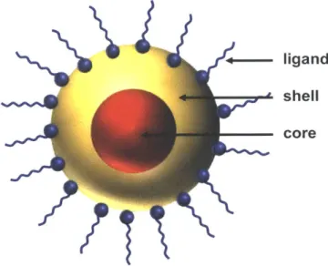
+-- ligand
shell core
Although much of QD synthesis is centered on the semiconductor materials (their precursors, composition, and crystal structure) in order to obtain specific optical properties, it is important to remember that the ligands are more than just tools to control the synthesis of nanoparticles. The final product of QD synthesis is a true mixed species where both the semiconductor and the organic ligands play pivotal roles in the behavior of the QDs. Before discussing the original work in this thesis, we will examine QD ligands and specifically ligands for biological applications in the following section.
1.3 Quantum dots and their
ligands
The organic molecules that are bound to the surface of QDs are an integral part of the nanoparticle.
As discussed in the previous section, ligands play a key role in controlling QD synthesis and in the
optical properties of their hosts. In addition, ligands regulate the interactions of QDs with their
environment, so understanding and controlling surface-ligand interactions in QDs is essential for
applications ranging from optoelectronic devices to biological probes. Our discussion of QD
ligands will begin with a brief discussion of ligand theory and chemical behavior and then proceed
to a discussion of ligands specifically for biological applications -the focus of this thesis.
1.3.1 Ligand theory
A good starting point for the discussion of ligand-metal interactions relevant to QDs is the Hard
and Soft Acids and Bases (HSAB) principle formulated by Pearson.14- 7 In this formalism, the
metal ions and ligands are electron acceptors or donors, and the formation of a metal ligand
complex is conceptualized as a generalized acid-base reaction (between a Lewis acid and a Lewis
base). The equilibrium constant for the formation of a complex depends on two factors for each of
the species involved: strength and softness. Pearson's concept of softness is related to
electronegativity and polarizability and reflects the "looseness" with which a species holds its
valence electrons. For example, acids with high polarizability and low electronegativity are soft
and those with low polarizability and high electronegativity are hard. In general, hard acids prefer
to bind to hard bases and soft acids prefer to bind to soft bases. Where this becomes important in
QD-ligand interaction is that it is not only the strength of a ligand that matters in how well it binds
shown in Table 1.2, common binding moieties have different softness and so do many metal ions
found in QD shells.
ACIDS
HARD BORDERLINE SOFT
ions with formal oxidation Cu2+, Zn2+ metals with zero oxidation
states of 4 or higher state
H+, Na+, K+, Ca2 Age, Cd2+, Hg2+, Cue, Au+
BASES
HARD BORDERLINE SOFT
R-NH2, R-OH, R-COO-, C6HSNH2, pyridine, Br-, R-SH, R2-S, P-R3, OP-R3,
OH-, imidazole As-R3, C2H4, C6H6
F-, Cl- ,H20 I II
Table 1.2 A subset of the HSAB classification tables compiled by Pearson is shown here,
including some of the molecules and ions relevant in QD-ligand interactions.
Beyond the HSAB principle, ligands can also be classified by the types of bonds they form. This
classification was formulated by Green58 and includes three ligand types, discussed below and
shown in Figure 1.3:
1) X-type: these ligands have a singly-occupied orbital that requires one electron from the metal
center to form a two-electron covalent bond. This type includes anionic species forming covalent
bonds. A variety of ligands commonly used in QD synthesis are X-type ligands, including
phosphonates, carboxylates, and thiolates.
2) L-type: these ligands have a lone pair that forms a dative covalent bond with an empty orbital
3) Z-type: these ligands have an empty orbital and are two-electron acceptors. In QDs, these are
metal-carboxylate or metal-phosphonate complexes with an empty orbital at the metal center (for
example Cd(O2CR)2, or a similar complex with Zn or Pb).
With the HSAB principle and Green's ligand classification, we have a comprehensive framework
to understand the chemical bonding between ligands and the QD surface. In the following section, we will discuss the kinetics of native ligands, as different bonding types give rise to markedly
different behavior.
1.3.2 Ligand kinetics and exchange
X-type and L-type ligands show very different kinetics and exchange behavior in QD samples in
neat solvent, after separation from growth solution. This behavior has important implications for
ligand exchange and colloidal stability of QDs capped with different ligand types.
X-type ligands
X-type ligands bind tightly to the surface of QDs and show no evidence of exchange in samples
of QDs purified from free ligand. In other words, bound X-type ligands do not spontaneously
dissociate from the QD to establish an equilibrium between the bound and free states. However, I H NMR shows that when free ligand is introduced to a sample of clean QDs with X-type native
ligands, self-exchange occurs quickly and can even reach the expected equilibrium partition at
room temperature.59 In the case of carboxylate ligands, exchange with free oleic acids occurs via a proton-exchange mechanism, so that any free ligands and QDs have neutral charge.59 Exchange among different X-type ligands has also been studied, specifically native phosphonate exchange
with carboxylic acids, chloromethylsilane and bis(trimethylsilyl)selenide.' 6 In this case, ligand exchange depends on the binding affinity of the ligands at play. For example, while free oleic acid
(OA) readily displaces QD-bound oleates, OA cannot displace phosphonates under similar
conditions. In fact, sustained heating at 100 'C is necessary to observe partial exchange -this
suggests that the native species is more strongly bound than the competing carboxylate.61
L-type ligands
L-type ligands transition quickly and spontaneously from the QD-bound state to the free state in
solution. This kinetic behavior is widely used to eliminate L-type ligands from the surface of QDs
through repeated crashout, as the portion of ligands in the free state is efficiently removed with the
solvent in each precipitation step. In fact, Morris-Cohen et al. have shown that QDs that start out
with both X-type and L-type ligands on their surface will only have X-type ligands after a few
crashouts.62 The lability of L-type ligands can seem very useful for ligand exchange first, but it is problematic for multiple reasons. First, L-type-capped QDs are not stable to dilution. The addition
of neat solvent to a given system (that is, QDs, surface-bound ligands, and free ligands) will drive
more bound ligands into the free state. At a high enough dilution, this can compromise the colloidal
stability of QDs. The second problem is that monovalent L-type ligands do not bind tightly to the
QD surface, so a stable L-type ligand must necessarily be engineered to be multivalent.
We have discussed the kinetics and exchange behavior within a given ligand type, but it is
important to consider ligand exchange across ligand types. It is well-known that small-molecule
ligands can exchange across ligand types, and this type of exchange is widely used to treat QDs
mechanism for X-to-L ligand exchange was not discovered until recently, with important implications for the design of polymeric QD ligands -as we will discuss in Section 2.5.64 It is important to highlight two final points. First, despite their different kinetics and exchange mechanism, L-type and X-type ligands can both be found on the surface of a given QD. Second, most of the studies of ligand kinetics to date have been carried out on CdSe cores, but the results
discussed here have been observed in other systems, including CdS, PbS, PbSe and core-shell
QDs. Thus, we expect the results discussed here to apply to the QD systems employed in this thesis
(InAs/CdSe/CdxZn-xS, PbS, and PbS/CdS).
X-type X-type
terminates lattice bound ion pair
-- -'MX2' L-type - Z-type neutral-donor neutral-acceptor M = Cd, Pb, etc. ME E = S, Se X =O2CR, CI, SR, etc. OM OE L = PR3, NH2R, etc. MX2 = Cd(O2CR)2, CdC2, Pb(SCN)2, etc. [X]1HB]' = [CI]-HPBu3], [S]2-2[H4N]+, [In2Se4] 2 -2[N2Hr], etc. X-type 2 H-X + c'MX 2 L-pe L' + L MX'2 + 2 H-X L' + L Z-type M'X2 + a'MX2 M'X2+ MX2
Figure 1.3 A schematic of the different types of QD-ligand coordination and self-exchange
1.3.3 Ligands for biological probes
QDs have optical properties that make them an excellent candidate material for imaging probes.
They have narrow, tunable emission spectra and some compositions can approach unity QY; they
have broad absorption spectra, allowing excitation with a variety of wavelengths and simultaneous
excitation of different-color QDs; they can tolerate relatively long excitation times and high
excitation fluxes without photobleaching (a serious limitation for organic dyes). However, in order
to be used as imaging probes, QDs must be transferred from organic to aqueous phase while
fulfilling several requirements. The ideal QD-based probe should have the following qualities:
1) compact hydrodynamic size and narrow size distribution. After synthesis, QDs have both of
these properties, so the researcher's challenge is to preserve these qualities as much as possible.
The hydrodynamic size of probes in vivo impacts their localization and plays a role in the way they
are cleared from the organism. 5-9 In addition, large probes (>30 nm Hd) can change the behavior of cellular receptors, which means that large labeling constructs can affect the biological behavior
of their target cells.65 Thus, it is imperative to keep probes small and with a narrow size distribution in order to minimize variability in their localization, circulation time, and clearance.
2) good colloidal stability and low nonspecific binding. Imaging probes should be able to preserve
their small size and narrow size distribution in storage conditions for weeks to months. This
enables the use of a single batch of probes for biological experiments that have time points on this
scale. However, some of the strategies commonly used to make nanoparticles stable in vitro are not compatible with biological applications. For example, highly-charged ligands can prevent aggregation in vitro through electrostatic repulsion, but such ligands make nanoparticles more
likely to bind to cells and proteins nonspecifically.66-68 An ideal coating would have little to no electric charge, and to this end, many solubilization strategies incorporate polyethylene glycol
(PEG), a polymer known to reduce non-specific interactions.69 PEG also has excellent solubility
in water and a range of organic solvents, which makes it particularly versatile for coating QDs.
3) preserve the optical properties of QDs. We want to use QDs for probes because they have
excellent optical properties. Transfer to aqueous phase often has a slight detrimental effect on the
fluorescence of QDs, but any method of solubilization that quenches fluorescence emission would
be unacceptable.
4) reactive groups for coupling. In order to provide sophisticated sensing and labeling, our
biologically-inert QDs must be coupled to biomolecules or other moieties to mediate interaction
with the environment. Many existing ligands use primary amines, carboxylic acids, thiols, or
maleimides as reactive handles, but these can present problems. Amines and carboxylic acids can
bind to the metal on the QD surface, reducing the effective number of reactive groups available
for coupling. In addition, uncoupled primary amines and carboxylic acids will become charged
species in physiological conditions and potentially lead to nonspecific interactions. On the other
hand, thiols and maleimides can have unintended reactions with thiolated molecules present in
solution. Ideally, reactive groups on probes would have high coupling efficiency and be inert to
biomolecules and the QD surface when not coupled. These requirements can be met through the
In order to produce QD biological probes with some or all of these qualities, researchers have
employed a variety of solubilization methods that fall into a few broad categories. We will now
discuss each category and provide key examples from the literature.
Micelle encapsulation
Researchers have used amphiphilic molecules and polymers (like PEGylated phospholipids and
alkylated polyacrylic acids) to make micelle suspensions of QDs.75-78 As shown in Figure 1.4, this strategy relies on self-assembly of the constructs, as the hydrophobic alkyl tails of the amphiphilic
species intercalate with the native ligand shell of the QD while exposing the hydrophilic ends of
the molecule or polymer (PEG or charged groups) to the aqueous environment. Because
self-assembly is driven by the hydrophobic effect, these constructs are stable in aqueous solution in a
variety of conditions, and since the native ligands and inorganic shell are unperturbed, the
constructs have optical properties very similar to that of the QDs as-synthesized. In addition, this
approach is generalizable to any nanoparticle with a hydrophobic ligand shell. However, this
solubilization method results in large constructs (Hd > 20 nm) and can result in a broad distribution
of sizes, as a single micelle may encapsulate multiple QDs. As we discussed earlier, constructs of
this size pose serious problems for labeling experiments, but they have been used successfully for
charged groups (COO-) alkyl tails
Figure 1.4 A schematic of micelle encapsulation of QDs for phase transfer to water. Notice that
the alkyl tails of the amphiphilic polymer intercalate with the native ligands while the charged carboxylate groups are exposed to the aqueous medium. In some cases, the micelles are solubilized by PEG chains instead of charged groups.
Ligand exchange
In contrast to micelle encapsulation, other methods of solubilization rely on a ligand that binds
directly to the QD surface. By coupling the ligand directly to the QD surface, we can avoid the
pitfalls of encapsulation (large size and broad size distribution). One approach that has been
well-studied is ligand exchange, where the native ligands of the QD are partially or wholly displaced
by a molecule or polymer that can both bind to the QD surface and solubilize QDs in water. The
surface-binding group is an X-type or L-type ligand, and the solubilizing part of the molecule may
be a charged group, a zwitterionic group, or a PEG chain. Two broad categories of ligand exchange
systems have been studied:
1) thiol ligands
Thiol ligands are extensively used in nanoscience and are well-known for their strong binding to
with metals as X-type or L-type ligands.58 8 0 Thiols have been found to be a key part of metal-binding proteins (where -SH is present in the side-group of the amino-acid cysteine) that form
complexes with a variety of metal ions including Cd2+ and Zn2+. 182 A variety of thiol-bearing
molecules have been used as QD ligands, as shown in Figure 1.5. In fact, cysteine itself has been
used as a ligand for QDs, with -SH binding to the QD surface and the rest of the molecule acting
as a solubilizing zwitterion at physiological pH, as the carboxylic acid deprotonates and the
primary amine is protonated. Because this ligand is so small, researchers have been able to
synthesize water-soluble QDs with Hd< 5nm using cysteine -a size that is below the renal clearance
cutoff in rats and mice.83 84 In addition to cysteine, other monothiolated molecules have been used as QD ligands, but this approach reveals a serious shortcoming: the thiol group is susceptible to
oxidation.8 5 This reaction forms disulfide bonds, which cannot form complexes with metals. Thus, oxidation results in loss of ligand from the QD and eventually aggregation and precipitation of
QDs out of solution. In order to improve the stability of QD-ligand constructs, researchers turned
to molecules bearing two thiol groups in close proximity -an approach that takes advantage of the chelate effect. The most commonly-used bidentate ligands are based on dihydrolipoic acid
(DHLA): DHLA itself, PEGylated DHLA, or DHLA bound to a polymer backbone.48 86 90 Some of these QDs have compact size and a narrow size distribution, but are still susceptible to oxidation.
Despite the improved stability afforded by the bidentate thiol ligands, studies have shown that DHLA-coated QDs show signs of degradation within days even when stored in the dark and
refrigerated, and only cold storage under nitrogen atmosphere seems to prevent degradation.9' Polydentate DHLA-based ligands have also been synthesized through random polymerization of
DHLA-methacrylamides and zwitterion methacrylamides, but even these ligands have limited stability due to oxidation.9293 Despite their versatility, thiol-based ligands have limited stability
that limits their range of applications in biological imaging, particularly for long-duration
experiments.
0
"_*OH
dihydrolipoic acid (DHLA)
H
cysteine DHLA-PEG
3HH
8-mercaptooctanoic acid
DHLA- multi-arm PEG
Figure 1.5 A schematic of various kinds of mono- and bi-dentate thiol ligands used in QDs.
2) L-type polydentate ligands
A more recent approach to QD ligands for biological applications is polydentate L-type ligands.
As discussed earlier, L-type ligands can quickly transition from being bound to the QD surface to
being free in solution, so monovalent L-type ligands have limited stability. However, polydentate
L-type ligands can bind strongly to the QD surface, an observation first reported with His6-tagged
proteins.94-97 This observation led to the rise of a family of polydentate ligands that use L-type binding moieties (imidazole or pyridine).92
,98-10 0 The first published example was the polymeric imidazole ligand (PIL, shown in Figure 1.6), a random copolymer of PEG-acrylamides and
imidazole acrylamides synthesized via reversible addition-fragmentation chain transfer (RAFT)
polymerization. Because of this modular structure, PIL could easily incorporate amine- and
developed to feature click coupling and more sophisticated coupling moieties with selective
reactivity.73"10' Visible QDs coated with PIL are compact (Hd< 15 nm), stable across a broad range of pH, and have been used for in vitro and in vivo cell labeling and tracking.73,9 8,102
,0 3
A few years after PIL was published, Tasso et al. used RAFT polymerization to synthesize a
block-copolymer ligand with imidazole as the anchoring group, but in this case, the QDs were solubilized
by sulfobetaine zwitterions rather than PEG chains.92 Around the same time, Susumu et al. and Wang et al. published polydentate ligands that employed pyridine and imidazole as the binding
moiety, respectively.99100 These last two systems employed commercially-available polymer backbones (polyacrylic acid and poly(isobutylene-alt-maleic anhydride)) and appended binding
groups and solubilizing moieties to the backbone through amide coupling. Although these
approaches use simpler chemical methods and are therefore more accessible, they lack the more
precise control of ligand size achieved with the RAFT-based polymer ligands.
RAFT polymerization,
deprotection 0 0 ligand exchange
HN HN
-N
INN
0
]
0
Figure 1.6 A schematic of PIL synthesis and ligand exchange as demonstrated by Liu et al.
The binding moiety on the polymer is imidazole (red) and the QD-polymer construct is solubilized in water by PEG chains (blue).
Click-ready native ligands
Click-ready native ligands are a novel approach to ligand engineering pioneered by the Bawendi
lab -a contemporary development to the work in this thesis.74 01 This approach uses small molecules that can act as native oleate ligands during synthesis and also feature a click-reactive
group for later conjugation to PEG chains for transfer to aqueous phase as shown in Figure 1.7.
The main benefits of this approach are its scalability (as large quantities of QDs can be easily
transferred to water) and its ease of integration with synthetic procedures for many kinds of QDs.
By bypassing the ligand exchange process entirely, these ligands can preserve the optical
properties of QDs well and still result in compact probes (12 nm Hd) when transferred to water.
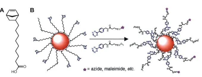
A-0 HO
B0
NI M, eN NH NNH*=azide, maleimide, etc. 0.
Figure 1.7 The structure of the click-ready carboxylic acid ligand synthesized by Cordero et
al. (A) and an overview of the solubilization method (B) (adapted and modified from Cordero et al.) This ligand allows transfer to aqueous phase by reacting the norbornene-terminated
native ligands with tetrazine-bearing PEG chains. Conjugation to biomolecules can be carried out using bifunctional PEGs as shown.
1.4 Conclusion and motivation for the original work
In the last several years, visible QDs have been employed as powerful probes for biological studies
in vitro and in vivo. QD probes have been used to measure pH, glucose concentration, and 02
concentration, and they have allowed the study of biomolecular processes like cellular transport
and the motion of individual cell membrane receptors.65,95"104-106 QD probes have also been used for antibody-mediated cell labeling and tracking in vivo, non-endocytic cell labeling, and are
sufficiently stable in vivo to be detected days after injection.7310, 2,03,107
Currently, many areas of biological research are moving increasingly towards in vivo studies, as
these models better capture the full range of behavior of cells and tissues in health and disease. As
the focus of research shifts to this setting, imaging techniques and probes must evolve to enable
studies. Visible QDs have been successful probes for in vivo and intravital imaging, but their
usefulness is ultimately limited by the optical properties of tissue. In response to this, NIR-emitting
QDs have adapted for use as biological probes. 36,108 However, the frontier for QD-based probes
is SWIR imaging because this wavelength range has minimal tissue interaction for optical imaging.
In this thesis, we set out to make SWIR-emissive QDs into biological probes by translating the
PIL ligand to SWIR QDs (Chapter 2) and by developing a new polymeric ligand designed to be
1.5 Bibliography
(1) Lim, Y. T.; Kim, S.; Nakayama, A.; Stott, N. E.; Bawendi, M. G.; Frangioni, J. V. Selection of Quantum Dot Wavelengths for Biomedical Assays and Imaging. Mol.
Imaging 2003, 2 (1), 50-64.
(2) Welsher, K.; Liu, Z.; Sherlock, S. P.; Robinson, J. T.; Chen, Z.; Daranciang, D.; Dai, H. A Route to Brightly Fluorescent Carbon Nanotubes for Near-Infrared Imaging in Mice. Nat.
Nanotechnol. 2009, 4 (11), 773-780.
(3) Bashkatov, A. N.; Genina, E. A.; Kochubey, V. I.; Tuchin, V. V. Optical Properties of Human Skin, Subcutaneous and Mucous Tissues in the Wavelength Range from 400 to 2000 Nm. J Phys. D. Appl. Phys. 2005, 38 (15), 2543-2555.
(4) BASHKATOV, A. N.; GENINA, E. A.; TUCHIN, V. V. OPTICAL PROPERTIES OF
SKIN, SUBCUTANEOUS, AND MUSCLE TISSUES: A REVIEW. J Innov. Opt. Health Sci. 2011, 04 (01), 9-38.
(5) Wilson, R. H.; Nadeau, K. P.; Jaworski, F. B.; Tromberg, B. J.; Durkin, A. J. Review of Short-Wave Infrared Spectroscopy and Imaging Methods for Biological Tissue
Characterization. J. Biomed. Opt. 2015, 20 (3), 030901.
(6) Sordillo, L. A.; Pu, Y.; Pratavieira, S.; Budansky, Y.; Alfano, R. R. Deep Optical Imaging of Tissue Using the Second and Third Near-Infrared Spectral Windows. J. Biomed. Opt. 2014, 19 (5), 056004.
(7) Diao, S.; Hong, G.; Antaris, A. L.; Blackburn, J. L.; Cheng, K.; Cheng, Z.; Dai, H. Biological Imaging without Autofluorescence in the Second Near-Infrared Region. Nano
Res. 2015, 8 (9), 3027-3034.
(8) Zhang, H.; Salo, D.; Kim, D. M.; Berezin, M. Y. Penetration Depth in Tissue-Mimicking Phantoms from Hyperspectral Imaging in SWIR in Transmission and Reflection
Geometry. 2016, 21 (12), 970311.
(9) Bruns, 0. T.; Bischof, T. S.; Harris, D. K.; Franke, D.; Shi, Y.; Riedemann, L.; Bartelt, A.; Jaworski, F. B.; Carr, J. A.; Rowlands, C. J.; et al. Next-Generation in Vivo Optical
Imaging with Short-Wave Infrared Quantum Dots. Nat. Biomed. Eng. 2017, 1 (4), 0056.
(10) Hong, G.; Diao, S.; Chang, J.; Antaris, A. L.; Chen, C.; Zhang, B.; Zhao, S.; Atochin, D.
N.; Huang, P. L.; Andreasson, K. I.; et al. Through-Skull Fluorescence Imaging of the
Brain in a New near-Infrared Window. Nat. Photonics 2014, 8 (9), 723-730.
(11) Antaris, A. L.; Chen, H.; Cheng, K.; Sun, Y.; Hong, G.; Qu, C.; Diao, S.; Deng, Z.; Hu, X.; Zhang, B.; et al. A Small-Molecule Dye for NIR-II Imaging. Nat. Mater. 2016, 15 (2), 235-242.
(12) Carr, J. A.; Franke, D.; Caram, J. R.; Perkinson, C. F.; Saif, M.; Askoxylakis, V.; Datta, M.; Fukumura, D.; Jain, R. K.; Bawendi, M. G.; et al. Shortwave Infrared Fluorescence Imaging with the Clinically Approved Near-Infrared Dye Indocyanine Green. Proc. Natl.
Acad Sci. 2018, 201718917.
(13) Cosco, E. D.; Caram, J. R.; Bruns, 0. T.; Franke, D.; Day, R. A.; Farr, E. P.; Bawendi, M.
G.; Sletten, E. M. Flavylium Polymethine Fluorophores for Near- and Shortwave Infrared
Imaging. Angew. Chemie -Int. Ed. 2017, 56, 13126-13129.
(14) Goswami, P. P.; Syed, A.; Beck, C. L.; Albright, T. R.; Mahoney, K. M.; Unash, R.; Smith, E. A.; Winter, A. H. BODIPY-Derived Photoremovable Protecting Groups Unmasked with Green Light. J Am. Chem. Soc. 2015, 137 (11), 3783-3786.
Surface Chemistry Determine Serum Protein Adsorption and Macrophage Uptake. J Am.
Chem. Soc. 2012, 134 (4), 2139-2147.
(16) Albanese, A.; Tang, P. S.; Chan, W. C. W. The Effect of Nanoparticle Size, Shape, and Surface Chemistry on Biological Systems. Annu. Rev. Biomed Eng. 2012, 14 (1), 1-16.
(17) Popovid, Z.; Liu, W.; Chauhan, V. P.; Lee, J.; Wong, C.; Greytak, A. B.; Insin, N.; Nocera, D. G.; Fukumura, D.; Jain, R. K.; et al. A Nanoparticle Size Series for In Vivo Fluorescence Imaging. Angew. Chemie Int. Ed 2010, 49 (46), 8649-8652.
(18) Tenzer, S.; Docter, D.; Rosfa, S.; Wlodarski, A.; Kuharev, J.; Rekik, A.; Knauer, S. K.; Bantz, C.; Nawroth, T.; Bier, C.; et al. Nanoparticle Size Is a Critical Physicochemical Determinant of the Human Blood Plasma Corona: A Comprehensive Quantitative Proteomic Analysis. A CS Nano 2011, 5 (9), 7155-7167.
(19) Sun, X.; Rossin, R.; Turner, J. L.; Becker, M. L.; Joralemon, M. J.; Welch, M. J.; Wooley,
K. L. An Assessment of the Effects of Shell Cross-Linked Nanoparticle Size, Core Composition, and Surface PEGylation on in Vivo Biodistribution. Biomacromolecules
2005, 6 (5), 2541-2554.
(20) Wu, X.; He, X.; Wang, K.; Xie, C.; Zhou, B.; Qing, Z. Ultrasmall Near-Infrared Gold Nanoclusters for Tumor Fluorescence Imaging in Vivo. Nanoscale 2010, 2 (10), 2244-2249.
(21) Lin, C. J.; Yang, T.; Lee, C.; Huang, S. H.; Sperling, R. A. Bioconjugation of Fluorescent Gold Nanoclusters toward Biological Labeling. 2009, 3 (2), 395-401.
(22) Huang, C. C.; Yang, Z.; Lee, K. H.; Chang, H. T. Synthesis of Highly Fluorescent Gold Nanoparticles for Sensing Mercury(II). Angew. Chemie -Int. Ed. 2007, 46 (36), 6824-6828.
(23) Chen, Y.; Montana, D. M.; Wei, H.; Cordero, J. M.; Schneider, M.; Le Gudvel, X.; Chen,
0.; Bruns, 0. T.; Bawendi, M. G. Shortwave Infrared in Vivo Imaging with Gold
Nanoclusters. Nano Lett. 2017, 17 (10), 6330-6334.
(24) Fischer, S.; Bronstein, N. D.; Swabeck, J. K.; Chan, E. M.; Alivisatos, A. P. Precise Tuning of Surface Quenching for Luminescence Enhancement in Core-Shell Lanthanide-Doped Nanocrystals. Nano Lett. 2016, 16 (11), 7241-7247.
(25) Dong, H.; Du, S. R.; Zheng, X. Y.; Lyu, G. M.; Sun, L. D.; Li, L. D.; Zhang, P. Z.; Zhang,
C.; Yan, C. H. Lanthanide Nanoparticles: From Design toward Bioimaging and Therapy. Chem. Rev. 2015, 115 (19), 10725-10815.
(26) Naczynski, D. J.; Tan, M. C.; Zevon, M.; Wall, B.; Kohl, J.; Kulesa, A.; Chen, S.; Roth, C. M.; Riman, R. E.; Moghe, P. V. Rare-Earth-Doped Biological Composites as in Vivo
Shortwave Infrared Reporters. Nat. Commun. 2013, 4, 1-10.
(27) Zhong, Y.; Ma, Z.; Zhu, S.; Yue, J.; Zhang, M.; Antaris, A. L.; Yuan, J.; Cui, R.; Wan, H.;
Zhou, Y.; et al. Boosting the Down-Shifting Luminescence of Rare-Earth Nanocrystals for Biological Imaging beyond 1500 Nm. Nat. Commun. 2017, 8 (1), 1-7.
(28) Payne, S. A.; Chase, L. L.; Smith, L. K.; Kway, W. L.; Wyers, W. F. Infrared Cross-Section Measurements for Crystals Doped with Er3+, Tm3+, and Ho3+. IEEE .1 Quantum Electron. 1992, 28 (11), 2619-2630.
(29) Zhao, X.; He, S.; Tan, M. C. Design of Infrared-Emitting Rare Earth Doped Nanoparticles
and Nanostructured Composites. J. Mater. Chem. C 2016, 4 (36), 8349-8372.
(30) Hong, G.; Lee, J. C.; Robinson, J. T.; Raaz, U.; Xie, L.; Huang, N. F.; Cooke, J. P.; Dai, H. Multifunctional in Vivo Vascular Imaging Using Near-Infrared II Fluorescence. Nat.
(31) Ghosh, D.; Bagley, A. F.; Jeong, Y.; Birrer, M. J.; Bhatia, S. N.; Belcher, A. M. Deep,
Noninvasive Imaging and Surgical Guidance of Submillimeter Tumors Using Targeted M13-Stabilized Single-Walled Carbon Nanotubes. Proc. Nati. Acad. Sci. 2014, 111 (38),
13948-13953.
(32) Bardhan, N. M.; Ghosh, D.; Belcher, A. M. Carbon Nanotubes as in Vivo Bacterial Probes. Nat. Commun. 2014, 5, 1--11.
(33) Hong, G.; Diao, S.; Antaris, A. L.; Dai, H. Carbon Nanomaterials for Biological Imaging
and Nanomedicinal Therapy. Chem. Rev. 2015.
(34) Sch6ppler, F.; Mann, C.; Hain, T. C.; Neubauer, F. M.; Privitera, G.; Bonaccorso, F.; Chu,
D.; Ferrari, A. C.; Hertel, T. Molar Extinction Coefficient of Single-Wall Carbon
Nanotubes. J. Phys. Chem. C 2011, 115 (30), 14682-14686.
(35) Sanchez, S. R.; Bachilo, S. M.; Kadria-Vili, Y.; Lin, C. W.; Weisman, R. B.
(N,m)-Specific Absorption Cross Sections of Single-Walled Carbon Nanotubes Measured by Variance Spectroscopy. Nano Lett. 2016, 16 (11), 6903-6909.
(36) Allen, P. M.; Liu, W.; Chauhan, V.; Lee, J.; Ting, A. Y.; Fukumura, D.; Jain, R. K.;
Bawendi, M. G. InAs(ZnCdS) Quantum Dots Optimized for Biological Imaging in the Near-Infrared. JACS Commun. 2010, No. 132, 470-471.
(37) Yu, P.; Beard, M. C.; Ellingson, R. J.; Fernere, S.; Curtis, C.; Drexler, J.; Luiszer, F.; Nozik, A. J. Absorption Cross-Section and Related Optical Properties of Colloidal InAs
Quantum Dots. J. Phys. Chem. B 2005, 109 (15), 7084-7087.
(38) Xie, R.; Peng, X. Synthetic Scheme for High-Quality InAs Nanocrystals Based on Self-Focusing and One-Pot Synthesis of InAs-Based Core-Shell Nanocrystals. Angew. Chemie -Int. Ed. 2008, 47 (40), 7677-7680.
(39) Franke, D.; Harris, D. K.; Chen, 0.; Bruns, 0. T.; Carr, J. A.; Wilson, M. W. B.; Bawendi,
M. G. Continuous Injection Synthesis of Indium Arsenide Quantum Dots Emissive in the Short-Wavelength Infrared. Nat. Commun. 2016, 7, 1-9.
(40) Moreels, I.; Lambert, K.; Muynck, D. De; Vanhaecke, F.; Poelman, D.; Martins, J. C.; Allan, G.; Hens, Z. Size-Dependent Optical Properties of Colloidal {PbS} Quantum Dots.
ACS Nano 2009, 3 (10), 3023-3030.
(41) Semonin, 0. E.; Johnson, J. C.; Luther, J. M.; Midgett, A. G.; Nozik, A. J.; Beard, M. C. Absolute Photoluminescence Quantum Yields of IR-26 Dye, PbS, and PbSe Quantum Dots. J. Phys. Chem. Lett. 2010, 1 (16), 2445-2450.
(42) Weidman, M. C.; Beck, M. E.; Hoffman, R. S.; Prins, F.; Tisdale, W. A. Monodisperse, Air-Stable PbS Nanocrystals via Precursor Stoichiometry Control. A CS Nano 2014, 8 (6),
6363-6371.
(43) Moreels, I.; Justo, Y.; De Geyter, B.; Haustraete, K.; Martins, J. C.; Hens, Z. Size-Tunable, Bright, and Stable PbS Quantum Dots: A Surface Chemistry Study. A CS Nano 2011, 5 (3), 2004-2012.
(44) Pietryga, J. M.; Werder, D. J.; Williams, D. J.; Casson, J. L.; Schaller, R. D.; Klimov, V.
I.; Hollingsworth, J. A. Utilizing the Lability of Lead Selenide to Produce
Heterostructured Nanocrystals with Bright, Stable Infrared Emission. J Am. Chem. Soc.
2008, 130 (14), 4879-4885.
(45) Nasilowski, M.; Nienhaus, L.; Bertram, S. N.; Bawendi, M. G. Colloidal Atomic Layer Deposition Growth of PbS/CdS Core/Shell Quantum Dots. Chem. Commun. 2017, 53 (5),
869-872.
A Bright and Biocompatible Fluorescent Nanoprobe in the Second near-Infrared Window. A CS Nano 2012, 6 (5), 3695-3 702.
(47) Hong, G.; Robinson, J. T.; Zhang, Y.; Diao, S.; Antaris, A. L.; Wang,
Q.;
Dai, H. In Vivo Fluorescence Imaging with Ag 2 S Quantum Dots in the Second Near-Infrared Region *Angew. Chemie 2012, 201206059, 9818-982 1.
(48) Zhang, Y.; Zhang, Y.; Hong, G.; He, W.; Zhou, K.; Yang, K.; Li, F.; Chen, G.; Liu, Z.; Dai, H.; et al. Biodistribution, Pharmacokinetics and Toxicology of Ag2S near-Infrared
Quantum Dots in Mice. Biomaterials 2013, 34 (14), 3639-3646.
(49) Hu, J.; Li, L.; Yang, W.; Manna, L.; Wang, L.-W.; Alivisatos, A. P. Linearly Polarized Emission from Colloidal Semiconductor Quantum Dots. Science (80-. ). 2001, 292 (2001),
2060-2063.
(50) Peng, X. Mechanisms for the Shape-Control and Shape-Evolution of Colloidal Semiconductor Nanocrystals. Adv. Mater. 2003, 15 (5), 459-463.
(51) Ithurria, S.; Dubertret, B. Quasi 2D Colloidal CdSe Platelets with Thicknesses Controlled
at the Atomic Level. J. Am. Chem. Soc. 2008, 130 (49), 16504-16505.
(52) Reiss, P.; Protiere, M.; Li, L. Core/Shell Semiconductor Nanocrystals. Small 2009, 5 (2),
154-168.
(53) Chen, 0.; Zhao, J.; Chauhan, V. P.; Cui, J.; Wong, C.; Harris, D. K.; Wei, H.; Han, H. S.; Fukumura, D.; Jain, R. K.; et al. Compact High-Quality CdSe-CdS Core-Shell
Nanocrystals with Narrow Emission Linewidths and Suppressed Blinking. Nat. Mater.
2013, 12 (5), 445-451.
(54) Pearson, G. Hard and Soft Acids and Bases. J. Am. Chem. Soc. 1963, 85 (22), 3533-3539. (55) Pearson, R. G. Hard and Soft Acids and Bases, HSAB, Part 1: Fundamental Principles. J.
Chem. Educ. 1968, 45 (9), 581.
(56) Pearson, R. G. Hard and Soft Acids and Bases, HSAB, Part 2. Surv.Progr. Chem. 1969, 5
(10), 1-52.
(57) Pearson, R. G. Absolute Electronegativity and Hardness: Application to Inorganic Chemistry. Inorg. Chem. 1988, 27 (4), 734-740.
(58) Green, M. L. H. A New Approach to the Formal Classification of Covalent Compounds of the Elements. J Organomet. Chem. 1995, 500 (1-2), 127-148.
(59) Fritzinger, B.; Capek, R. K.; Lambert, K.; Martins, C.; Hens, Z. Utilizing Self-Exchange To Address the Binding of Carboxylic Acid Ligands to CdSe Quantum Dots. Jacs 2010, No. 23, 10195-10201.
(60) Owen, J. S.; Park, J.; Trudeau, P.; Alivisatos, A. P. Reaction Chemistry and Ligand Exchange at Cadmium # Selenide Nanocrystal Surfaces Reaction Chemistry and Ligand Exchange at Cadmium-Selenide Nanocrystal Surfaces. Communication 2008,
12279-12281.
(61) Gomes, R.; Hassinen, A.; Szczygiel, A.; Zhao,
Q.;
Vantomme, A.; Martins, J. C.; Hens, Z. Binding of Phosphonic Acids to CdSe Quantum Dots: A Solution NMR Study. . Phys.Chem. Lett. 2011, 2 (3), 145-152.
(62) Morris-Cohen, A. J.; Donakowski, M. D.; Knowles, K. E.; Weiss, E. A. The Effect of a Common Purification Procedure on the Chemical Composition of the Surfaces of Cdse Quantum Dots Synthesized with Trioctylphosphine Oxide. J. Phys. Chem. C 2010, 114 (2), 897-906.
(63) Lokteva, I.; Radychev, N.; Witt, F.; Borchert, H.; Parisi, J.; Kolny-Olesiak, J. Surface Treatment of CdSe Nanoparticles for Application in Hybrid Solar Cells: The Effect of