cortex: insights from studies in primates, rodents, and birds
The MIT Faculty has made this article openly available. Please share
how this access benefits you. Your story matters.
Citation
Puig, M. Victoria, Jonas Rose, Robert Schmidt, and Nadja Freund.
“Dopamine Modulation of Learning and Memory in the Prefrontal
Cortex: Insights from Studies in Primates, Rodents, and Birds.”
Front. Neural Circuits. 8 (August 5, 2014).
As Published
http://dx.doi.org/10.3389/fncir.2014.00093
Publisher
Frontiers Research Foundation
Version
Final published version
Citable link
http://hdl.handle.net/1721.1/90936
Terms of Use
Creative Commons Attribution
Dopamine modulation of learning and memory in the
prefrontal cortex: insights from studies in primates,
rodents, and birds
M. Victoria Puig
1*, Jonas Rose
1,2*, Robert Schmidt
3and Nadja Freund
41
The Picower Institute for Learning and Memory, Department of Brain and Cognitive Sciences, Massachusetts Institute of Technology, Cambridge, MA, USA
2Animal Physiology, Institute of Neurobiology, University of Tübingen, Tübingen, Germany
3BrainLinks-BrainTools, Department of Biology, Bernstein Center Freiburg, University of Freiburg, Freiburg, Germany 4Department of Psychiatry and Psychotherapy, University of Tübingen, Tübingen, Germany
Edited by:
Guillermo Gonzalez-Burgos, University of Pittsburgh, USA
Reviewed by:
Onur Gunturkun, Ruhr University Bochum, Germany
Min Wang, Yale University, USA
*Correspondence:
M. Victoria Puig and Jonas Rose, The Picower Institute for Learning and Memory, Department of Brain and Cognitive Sciences, Massachusetts Institute of Technology, Cambridge, MA 02139, USA
e-mail: mvpuig@mit.edu
In this review, we provide a brief overview over the current knowledge about the role of
dopamine transmission in the prefrontal cortex during learning and memory. We discuss
work in humans, monkeys, rats, and birds in order to provide a basis for comparison
across species that might help identify crucial features and constraints of the dopaminergic
system in executive function. Computational models of dopamine function are introduced
to provide a framework for such a comparison. We also provide a brief evolutionary
perspective showing that the dopaminergic system is highly preserved across mammals.
Even birds, following a largely independent evolution of higher cognitive abilities, have
evolved a comparable dopaminergic system. Finally, we discuss the unique advantages and
challenges of using different animal models for advancing our understanding of dopamine
function in the healthy and diseased brain.
Keywords: prefrontal cortex, learning and memory, dopamine receptors, executive function, working memory, neuromodulation, evolution
INTRODUCTION
A major function of executive control is the flexible adaptation
to our ever-changing environment. The executive circuits of the
brain must, therefore, not only monitor and maintain current
behavioral goals but also incorporate new goals and rules. This
updating can come in the form of a quick integration of previously
acquired knowledge when, for example, a well-known stimulus
informs an animal of a change in reward contingencies. In many
cases, however, such updating requires new learning, for example
when a new stimulus is encountered for the first time. Executive
functions are commonly ascribed to the prefrontal cortex (PFC)
and frontostriatal networks. The function of these circuits relies
heavily on neuromodulation, in particular on dopamine (DA).
The aim of this review is to outline the contribution of DA and
its receptors in the PFC to learning and memory processes across
different species.
We will first introduce studies in the mammalian brain in the
sections on humans, non-human primates, and rodents. Due
to the challenges of investigating the role of DA transmission
in human PFC, we focus the human section on studies
utiliz-ing systemic injections of DA agents and impairments of DA
transmission in patients with a variety of neurological and
psy-chiatric disorders. The non-human primate and rodent sections
review behavioral studies conducted during local manipulations
of the DA system in the PFC. While the dopaminergic system
in different mammalian species follows largely the same
orga-nization, some conceptual and terminological differences can
make a comparison of data across species difficult (
Box 1). For
a comparative perspective, we will then outline behavioral studies
conducted in birds where local manipulations of the DA
sys-tem were implemented in a structure equivalent to the mammal
PFC, the nidopallium caudolaterale (NCL;
Jarvis et al., 2005
).
Such a comparison is of particular interest given the large
evo-lutionary gap between these species. The lines of birds and
mammals separated around 300 million years ago, long before
many of the cognitive functions attributed to the PFC evolved
(
Jarvis et al., 2005
;
Reiner et al., 2005
;
Jarvis, 2009
;
Rose et al.,
2009a
). In spite of this distance, birds and mammals (with
the exception of humans and apes) are largely on par when it
comes to cognitive abilities (
Emery and Clayton, 2004
;
Kirsch
et al., 2008
,
2009
). This implies a parallel or convergent
evolu-tion of cognievolu-tion between the species (
Emery and Clayton, 2004
;
Güntürkün, 2012
). As a result of this independent evolution, we
see stark differences in brain organization between birds and
mam-mals (
Jarvis et al., 2005
). Most notably, the avian telencephalon
does not show the laminar organization of the mammalian
cor-tex. However, other organizational principles were preserved or
evolved independently in both lines. This can be taken as a hint of
narrow neurobiological constraints in the evolution of a given
cognitive ability (
Colombo and Broadbent, 2000
;
Güntürkün,
2005a
).
ANATOMY OF THE DOPAMINE SYSTEM IN THE PREFRONTAL
CORTEX
The anatomy of the dopaminergic system is very similar between
all mammals and birds (for extensive review, see
Durstewitz et al.,
1998
,
1999b
;
Björklund and Dunnett, 2007
). DA neurons can be
identified by the expression of several catecholamine-synthesizing
BOX 1 | Conceptual/terminological differences between species.
When comparing the function of prefrontal DA across species it is important to clarify the terminology used in the different fields of research. As reviewed here, prefrontal DA plays an important role in learning and memory and an extensive body of literature is concerned with its role particularly in working memory (WM). In general, the term WM is strongly associated with its original definition by Baddeley and Hitch (1974), who famously proposed that systems for sensory storage (phonological loop, visuospatial sketchpad, and more recently, an episodic buffer) are governed by a central executive (Baddeley, 1992,2000). The gist of this definition is that an interconnected neural system allows the brief storage of information and, importantly, its manipulation.
In primates, a seminal contribution to the understanding of this system was the discovery ofFuster and Alexander (1971)of “delay cells” in the PFC. These neurons show increased activity during the delay period of WM tasks maintaining the memory of a stimulus. Consequently, in primates including humans, WM is often modeled as “active memory” (Zipser et al., 1993;Durstewitz, 2009), a sys-tem that holds information in memory by sustaining neural activity for a few critical seconds.
Research in rodents commonly uses a broader definition of WM, that refers to “a collection of processes that include the tempo-rary storage of information, as well as executive functions that mediate the manipulation and retrieval of trial-unique information to guide action after both short (seconds) and longer (minutes to hours) delays” (Phillips et al., 2004; see also:Mizumori et al., 1987;
Floresco and Phillips, 2001). Importantly, this definition includes a much larger range of delays (seconds to many hours) compared to what is typically used in humans and non-human primates (sec-onds). Consequently, in rodents, the definition of WM does not necessarily refer to active memory maintenance by delay cells but might rely on different mechanisms that could be classified as learn-ing mechanisms in primates. Thus, it is important to pay attention to the specific paradigms and definitions used when comparing results across species.
The definition of WM typically used in avian research was devel-oped in parallel to the definition in humans (Honig, 1978). Both concepts are largely comparable with the exception that no phono-logical loop is conceptualized in birds. The delay durations in avian research are largely comparable to those in the primate literature and active information maintenance by delay activity is generally assumed to be the key mechanism of WM (Miller et al., 1996;
Güntürkün, 2005a).
Taken together, there are fundamental terminological differences between species and it is important to keep these in mind when comparing results across species. In particular, the vast differ-ences in delay duration used in different paradigms could potentially engage distinct neural mechanisms – what is called WM in one species might be viewed as a learning mechanism in another.
enzymes, tyrosine hydroxylase (TH), aromatic amino acid
decar-boxylase (AADC), and dopamine-b-hydroxylase (DBH). With
modern immunohistochemical techniques it has been possible
to map out in detail the location of DA neurons and their
spe-cific projections. DA neurons originate in several neighboring
midbrain nuclei, being the substantia nigra pars compacta (SNc;
A9) and the ventral tegmental area (VTA; A10) the ones
pro-jecting to the forebrain. The total number of TH-positive cells
in VTA and SNc (bilateral count) is
∼20.000–30.000 in mice
and
∼40.000–45.000 in rats. This number increases
consider-ably in primates, 160.000–320.000 in monkeys and 400.000–
600.000 in young humans. DA neurons send afferents to many
target areas, including the several regions of the frontal
cor-tex, with the striatum being the most densely innervated
tar-get (
Björklund and Dunnett, 2007
;
Figure 1). PFC-projecting
DA neurons are intermingled in VTA and SNc both in
pri-mates and in rodents. However, the PFC in pripri-mates is much
more extensively innervated by midbrain DA afferents than in
rodents (
Thierry et al., 1973
;
Lindvall et al., 1978
;
Swanson, 1982
;
Descarries et al., 1987
;
Lewis and Sesack, 1997
;
Björklund and
Dunnett, 2007
).
Postsynaptically, DA exerts its actions within the PFC/NCL via
receptors grouped in two major families, D1-like receptors (D1
and D5 in mammals; D1A and D1B in birds) and D2-like receptors
(D2, D3, and D4 in mammals and birds), but D1-like receptors are
expressed to a greater extent than D2-like receptors (
Lidow et al.,
1991
;
Durstewitz et al., 1998
;
Seamans and Yang, 2004
;
de Almeida
et al., 2008
;
Santana et al., 2009
;
de Almeida and Mengod, 2010
).
In birds, the D1-like family is extended to include an additional
receptor (D1D;
Callier et al., 2003
;
Kubikova et al., 2010
). Both
families are G-protein-coupled receptors that exert slow changes
of activity in the cells and act as functional neuromodulators.
D1-like receptors show low affinity for DA, whereas D2-D1-like receptors
show higher affinity (
Seamans and Yang, 2004
). For the sake of
clarity, we will abbreviate D1-like and D2-like receptors as D1R
and D2R, respectively, and will point to a specific receptor subtype
whenever necessary.
Interestingly, dopaminergic signaling in the PFC depends on
brain maturation and the PFC is the brain structure that matures
last (
Gogtay and Thompson, 2010
). Analyses of human
post-mortem brain tissue reveal that the levels of mRNA expression
of the D2R and D5R subtypes in PFC are highest in neonates and
infants and decrease with age, whereas the D1R subtype mRNA
expression and protein levels increase with age and are highest
in adulthood (
Rothmond et al., 2012
). By contrast, both in rats
and non-human primates, densities of the D1R and D2R subtypes
peak during adolescence and decrease in adulthood (
Rosenberg
and Lewis, 1994
;
Andersen et al., 2000
). In songbirds, D1R and
D2R subtypes in the song nuclei increase with age and peak during
adolescence (
Kubikova et al., 2010
). The developmental patterns
of related brain regions in non-songbirds are still unclear.
NEUROPHYSIOLOGY OF DA NEURONS
“Classic” DA neurons show phasic activations (short duration
bursts of action potentials) following unpredicted reward
cod-ing a quantitative “prediction error” signal, namely the
differ-ence between received and predicted reward value. A reward
that is better than predicted elicits an activation (positive
pre-diction error response), a fully predicted reward draws no
response, and a reward that is worse than predicted induces
a decrease in activity (negative error response;
Schultz et al.,
1993
;
Schultz, 2007
,
2013
). These prediction error responses
of DA cells have been closely related to reinforcement
learn-ing models which assign a functional role of DA in modulatlearn-ing
cortico-striatal inputs through a reward-prediction error
teach-ing signal (
Schultz, 1997
,
2002
;
Morris et al., 2004
,
2006
;
Pan
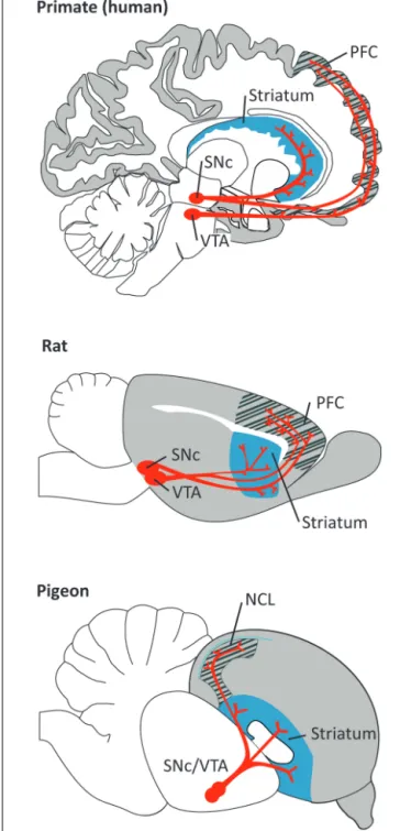
FIGURE 1 | Dopaminergic projections (in red) from the ventral tegmental area (VTA) and substantia nigra pars compacta (SNc) to the PFC/NCL and striatum in the brain of a primate (human), a rat, and a pigeon. Pallial (cortical) areas across species are shaded in gray, the
hatched area denotes the PFC/NCL, striatal areas are shaded in blue. Note that, in all species, DA neurons in both dopaminergic nuclei project to several subregions of the PFC/NCL and striatum.
et al., 2005
,
2008
). In fact, fast DA release consistent with these
reward predicting signals of DA neurons has been measured in
nucleus accumbens during associative learning (
Phillips et al.,
2003
;
Day et al., 2007
). Besides “classic” reward-prediction error
responses, phasic DA cell firing patterns also include responses
to salient and aversive sensory stimuli (
Horvitz, 2000
;
Joshua
et al., 2008
;
Brischoux et al., 2009
;
Matsumoto and Hikosaka,
2009
).
Dopamine neurons also exhibit tonic firing driven by
pacemaker-like membrane currents (
Grace and Bunney, 1984
;
Grace, 1991
;
Goto et al., 2007
). The functional relevance of this
tonic DA release is unknown. Transient suppression of tonic
spiking in DA neurons follows the omission of expected reward,
somehow implicating this spiking pattern in reward-based
learn-ing (
Tobler et al., 2003
). Recent work has shown that DA release
in the striatum increases gradually (ramps up) as rats expect
dis-tant reward, perhaps providing motivational drive (
Howe et al.,
2013
). However, these types of signals have not been described
in PFC.
Which of these DA signals reaches the PFC remains
cur-rently unclear. While phasic DA prediction error signals could
be used as a signal to transiently boost working
mem-ory (WM) of the corresponding stimuli (
Cohen et al., 2002
;
O’Reilly et al., 2002
), it has also been argued that mostly slower,
tonic DA signals are relevant in PFC. Moreover, the phasic
components of DA cell firing might be transmitted via
co-release of glutamate (
Seamans and Yang, 2004
;
Lavin et al.,
2005
;
Castner and Williams, 2007
;
Sheynikhovich et al., 2013
).
For computational models of DA function in PFC this has
two main consequences. Firstly, the timescales of tonic DA
would constrain functional roles to rather general cognitive
states such as arousal or attention. Secondly, DA function in
PFC circuits should be carefully contrasted with known
fea-tures of the putatively fast, phasic, signals of the nigrostriatal
system.
In general, heterogeneity among DA cells points to
addi-tional funcaddi-tional aspects that are not covered by classic
rein-forcement learning descriptions (
Berridge, 2007
;
Redgrave et al.,
2008
;
Bromberg-Martin et al., 2010
;
Morris et al., 2010
). While
functional roles of VTA and SNc neurons share common
properties (
Ilango et al., 2014
), overall evidence for
differ-ent functional groups among DA cells has been emerging
(
Brischoux et al., 2009
;
Matsumoto and Hikosaka, 2009
;
Lam-mel et al., 2012
;
Watabe-Uchida et al., 2012
). Moreover, the
heterogeneity in DA cell activity patterns is probably related
to heterogeneity in the anatomical pathways; DA neurons
con-tribute to reward or aversion depending on whether they are
activated from the laterodorsal tegmentum or the lateral
habe-nula, respectively (
Lammel et al., 2012
). For these reasons,
it has been difficult to dissociate the behavioral correlates of
DA release between the projection pathways to the striatum
and PFC.
HUMAN STUDIES
Investigating the direct role of DA signaling in human PFC during
learning and memory brings quite a few challenges and,
con-sequently, only few studies address this question. DA receptor
agonists and antagonists cannot be injected locally, restricted to
the PFC, and have to be administered systemically in humans.
Our knowledge about the role of DA transmission in the human
PFC, therefore, comes from studies combining imaging of the
brain with other manipulations such as systemic pharmacology
or transcranial magnetic stimulation, genetic profiling, and
from work in patients with neurological and psychiatric
disorders.
For instance, a recent fMRI study has revealed a connection
between context dependent WM and dopaminergic signaling in
human PFC (
D’Ardenne et al., 2012
). The authors first
iden-tified by fMRI that the dorsolateral PFC was involved in the
encoding of the context. Selective disruption of activity in this
region with transcranial magnetic stimulation adversely impacted
performance of the participants, causally implicating PFC in
con-text encoding. PFC activity during the task was then found to
correlate with phasic responses in the VTA and SNc. Based on
these results, the authors suggest that phasic DA signals
reg-ulate the encoding and updating of context representations in
the PFC.
In the 1970s, it was postulated that hypofrontality (i.e.,
decreased blood flow in the PFC) underlies mental disorders
and impaired cognitive function (
Ingvar and Franzén, 1974
).
In the context of schizophrenia, it was proposed that an excess
of DA in the mesolimbic system causes the positive symptoms
via hyperstimulation of D2R in the basal ganglia, whereas the
cognitive and negative symptoms follow insufficient D1R
acti-vation in the frontal cortex (
Abi-Dargham and Moore, 2003
;
Abi-Dargham, 2004
). We now know that DA hypofrontality by
itself cannot fully explain schizophrenia or other complex
men-tal disorders. Impairments in PFC dopaminergic signaling and
genetic profiling in these patients, however, have provided
valu-able information about the role of PFC DA in learning and
memory. For example, schizophrenia patients exhibit
imbal-ances in PFC dopaminergic signaling as determined by imaging
approaches (
Seeman, 1987
;
Okubo et al., 1997
;
Thompson et al.,
2014
), and show deficits in learning and WM (
Kalkstein et al.,
2010
) that correlate with genetic variations in DA related genes
(
Glatt et al., 2003
;
Vereczkei and Mirnics, 2011
). In Parkinson’s
disease (PD) patients, degeneration of neurons in the SNc results
in decreased phasic and tonic PFC DA levels (
Scatton et al., 1983
;
Moustafa and Gluck, 2011
), which could explain the cognitive
impairments present along with the motor deficits (
Narayanan
et al., 2013
). A more direct involvement of DA in PFC-dependent
memory processes was established in PD patients with and
with-out DA medication. In a spatial WM task, subjects had to find
tokens in boxes presented on a screen. Subjects that were off
the DA precursor levodopa (
L-DOPA) made more errors
(check-ing boxes that had already been opened) compared to when
they had received
L-DOPA, indicating that DA is required for
proper spatial WM performance. Surprisingly, visual learning
and memory was not affected by
L-DOPA in this task (
Lange
et al., 1992
). Similarly,
L-DOPA withdrawal did not affect the
performance of PD patients in an N-back task, where WM is
assessed when subjects are presented with a series of stimuli and
have to indicate when a stimulus is the same as the one n steps
back (
Mattay et al., 2002
). However, in PD patients
undergo-ing deep brain stimulation surgery, microstimulation of the SN
disrupts reinforcement learning in a two-alternative
probabil-ity learning task (
Ramayya et al., 2014
). Furthermore, research
conducted in attention deficit hyperactivity disorder (ADHD)
patients, who also display learning and memory deficits, have
also provided some insight into the role of DA in learning and
memory (
Brown, 2006
;
Alderson et al., 2013
). In these patients,
the size of the PFC is reduced (
Seidman et al., 2005
), and genes
involved in dopaminergic pathways are altered (
Gizer et al.,
2009
). Taken together, the results from work in
schizophre-nia, PD, and ADHD patients point to an abnormal DA
trans-mission as being responsible for behavioral deficiencies in
some learning and memory tasks that depend heavily on PFC
function.
Genetic studies have also provided valuable insight into
the contribution of the DA system in learning and
mem-ory. Individuals with the Val/Val catechol-O-methyltransferase
(COMT, enzyme that deactivates catecholamines) polymorphism
[Val(108/158)Met] exhibit higher COMT activity that correlates
with lower DA levels in the PFC (
Chen et al., 2004
), and have
a slightly higher risk of developing schizophrenia (
Sagud et al.,
2010
). Moreover, Val/Val carriers perform worse in the
Wiscon-sin card sorting test (WCST) compared to carriers of the Met
allele (
Egan et al., 2001
;
Malhotra et al., 2002
). The WCST
con-sists of a battery of cognitive tasks that include WM, sensitivity
to reinforcement, and behavioral flexibility. In addition, brain
imaging studies indicate that Val/Val carriers need greater PFC
activity to perform WM tasks (
Egan et al., 2001
;
de Frias et al.,
2010
). Stress may be another factor that should be taken into
con-sideration. Healthy human subjects under stress perform poorly
in WM tasks (
Olver et al., 2014
) and exhibit exacerbated levels
of PFC DA measured by positron emission tomography (PET;
Lataster et al., 2011
). In line with this finding, subjects with
the above mentioned Val/Val COMT alleles and corresponding
reduced levels of PFC DA perform better under stress during WM
(
Buckert et al., 2012
).
Early evidence for the involvement of D1R in WM processes
comes from work by
Müller et al. (1998)
that showed that
sys-temic injections of pergolide, a combined D1R/D2R agonist, but
not bromocriptine, a D2R agonist, facilitated WM performance
in a delayed matching task with delays of 2–16 s. These results
implicated D1R and not D2R in WM modulation. The important
role of D1R on WM is also suggested by the correlation between
the decrease of D1R binding in the lateral PFC and the decrease
in WM performance with age (
Bäckman et al., 2011
). However,
in another study, bromocriptine was shown to improve spatial
WM while the D2R antagonist haloperidol (a typical antipsychotic
drug) impaired it (
Luciana and Collins, 1997
). Other experiments,
though, did not report a general effect of bromocriptine on spatial
memory (
Kimberg et al., 1997
;
Müller et al., 1998
) nor binding of
the D2R agonist [11C]FLB457 correlated with performance on the
WCST (
Takahashi et al., 2008
).
Positron emission tomography studies in humans with the
radioactively marked D1R agonist [11C]SCH23390 have revealed
an inverted-U relationship between D1R binding in the PFC and
performance on the WCST (
Takahashi et al., 2008
). An
inverted-U relationship means that an optimal level of D1R activation is
required for best performance and, thus, levels below and above
this optimum impair performance. These experiments were meant
to confirm results provided by experimentation in monkeys (see
below). Further support for an inverted-U relationship between
D1R density and WM comes from patients with schizophrenia.
Deficits in WM have been associated with both decreased and
increased densities of PFC D1R in these patients (
Okubo et al.,
1997
;
Abi-Dargham and Moore, 2003
). Taken together,
recep-tor studies in humans point to an important role of PFC D1R
in WM with an optimal level of activation needed for best
per-formance. By contrast, the involvement of D2R needs further
elucidation.
NON-HUMAN PRIMATE STUDIES
The use of invasive approaches in monkeys has provided
valu-able insights into the crucial role of PFC DA and its receptors
in several higher-order executive functions. In fact, global
6-hydroxydopamine (6-OHDA) induced depletions of DA in the
lateral PFC of monkeys allowed to establish early on the
crit-ical role of DA in WM (
Brozoski et al., 1979
). Later, a series
of studies showed that there is an increase of extracellular DA
in the PFC during WM tasks (
Watanabe et al., 1997
) that exerts
its actions via local D1R (
Sawaguchi and Goldman-Rakic, 1991
,
1994
;
Williams and Goldman-Rakic, 1995
;
Murphy et al., 1996
;
Collins et al., 1998
;
Robbins, 2000
;
Seamans and Yang, 2004
;
Cast-ner and Williams, 2007
;
Arnsten et al., 2010
). More specifically,
local injections of D1R antagonists, but not D2R antagonists,
into the lateral PFC of monkeys caused deficits in oculomotor
delayed-response tasks; monkeys were less accurate in making
memory-guided saccades to remembered locations on the screen.
We note that the WM component of the task in these studies
was in the order of 1.5 to 6 s, comparable to the human
liter-ature. More recent work has evidenced that an optimal level of
D1R tone is required for adequate WM performance, and this
may be particularly vulnerable to changes in arousal state such
as fatigue or stress (
Arnsten et al., 2010
;
Arnsten, 2011
). Thus,
either too much (under stress) or too little (during fatigue) D1R
stimulation impairs performance following an inverted-U shaped
curve (
Arnsten et al., 1994
,
2010
;
Cai and Arnsten, 1997
;
Arnsten
and Goldman-Rakic, 1998
;
Goldman-Rakic et al., 2000
;
Williams
and Castner, 2006
;
Vijayraghavan et al., 2007
;
Arnsten, 2012
).
These reports in monkeys agree well with both the deleterious
effects of stress on WM performance and the inverted-U
relation-ship between D1R binding and cognitive capabilities reported in
human subjects. This inverted-U modulation of D1R also occurs
at the level of single PFC neurons engaged in WM. A D1R
ago-nist modulates persistent activity during memory delays following
an inverted-U response, whereby low levels of D1R stimulation
enhance spatial tuning whereas high levels reduce it (
Vijayragha-van et al., 2007
). By contrast, D2R have little effect on delay activity
and instead modulate the motor component of the task,
suggest-ing some contribution of PFC D2R to motor control function
(
Wang et al., 2004
). Systemic injections of D1R agonists and
antag-onists also alter the performance of monkeys during WM tasks, but
these studies have been reviewed elsewhere (
Castner and Williams,
2007
).
One general question is why detrimental effects of the “wrong”
DA concentration are present in the system in the first place. In
other words, what could be functional reasons for decreasing
WM performance? Speculatively, these could occur in
situa-tions in which the contribution of PFC to behavior is reduced
anyway. For example, in high stress, fight or flight mode,
behavioral control could be directed to subcortical areas to
empha-size speed (
Arnsten, 2012
;
Avery et al., 2013
). Alternatively, the
fine-tuning of DA concentration could be used to control the
“randomness” of behavior to emphasize exploitation or
explo-ration of certain behaviors (
Sutton, 1998
;
Doya, 2002
;
Parush
et al., 2011
;
Humphries et al., 2012
). Specifically, D1R activation
might push the PFC toward an exploitation mode by
protect-ing the WM content against distractors (
Durstewitz and Seamans,
2002
,
2008
). In contrast, based on both computational and
exper-imental approaches, D2R activation has been proposed to support
behavioral flexibility (exploration;
Floresco and Magyar, 2006
;
Durstewitz and Seamans, 2008
;
Puig and Miller, 2014
). As in
phys-iological situations selective stimulation of D1R or D2R seems
problematic, differences in receptor affinities may produce D2R
dominated states (very low and very high DA) and D1R
dom-inated states (intermediate DA). While these properties are also
well-suited to support the on- and offset of WM-related persistent
activity (
Box 2), it remains unclear whether the timescales of DA
modulation of the PFC firing are fast enough (
Cohen et al., 2002
;
O’Reilly et al., 2002
;
Seamans and Yang, 2004
;
Lavin et al., 2005
;
Sheynikhovich et al., 2013
).
The monkey lateral PFC has also been implicated in
associative stimulus-response learning (
Asaad et al., 1998
;
Pasupathy and Miller, 2005
;
Histed et al., 2009
;
Antzoulatos and
Miller, 2011
;
Puig and Miller, 2012
,
2014
). Reward-prediction
error responses of DA cells might be critically involved in these
learning processes (
Schultz, 1998
,
2007
,
2013
; see above).
Consis-tent with this role in reward prediction, phasic DA release occurs
in nucleus accumbens that is dynamically modified by associative
learning (
Phillips et al., 2003
;
Day et al., 2007
). Thus, it is plausible
that these DA signals also play a role in modulating PFC-dependent
learning. Indeed,
Puig and Miller (2012
,
2014)
have recently shown
that PFC D1R and D2R contribute to stimulus-response learning.
Monkeys performed an oculomotor delayed response task where
they learned by trial and error associations between visual cues and
saccades to a right or left target (
Figure 2A). Local microinjections
of both D1R and D2R antagonists (SCH23390 and eticlopride,
respectively) impaired the learning performance of the monkeys,
who made more errors and needed more correct trials to learn the
associations. The learning impairments correlated with a decrease
of neural information about the associations in single prefrontal
neurons during both the cue and memory delay (1 s) epochs of
the trial. Noteworthy, blocking D1R impaired learning more than
blocking D2R, whereas blocking D2R led to more perseverative
errors (Figures 2B,C). This suggests that PFC D1R contribute to
learning more than D2R, whereas the latter are more involved
in cognitive flexibility. These complementary roles of D1R and
D2R in PFC function agree well with the computational models
mentioned earlier that propose that D1R activation helps stabilize
new representations once an effective strategy has been identified
(exploitation) whereas D2R activation destabilizes PFC network
states favoring the exploration of new strategies (i.e., flexible
pro-cessing;
Durstewitz et al., 2000a
;
Seamans and Yang, 2004
;
Floresco
and Magyar, 2006
;
Durstewitz and Seamans, 2008
).
Contrary to the prominent role of DA in WM and
associa-tive learning, PFC DA does not influence familiar associations.
Blockade of D1R and D2R in the lateral PFC does not cause
BOX 2 | Computational perspectives on DA, WM, and PFC persistent activity.
Models of DA effects in the PFC can be categorized based on their biophysical details of description and their assumed DA release pat-terns. Furthermore, while the neuropsychological definitions of WM seem not always to be consistent across species (Box 1), computa-tional studies often focus on the mechanisms underlying persistent activity during delay periods.
An influential early model of DA action in the PFC (Durstewitz et al., 2000a; see also:Durstewitz et al., 1999a), bridged the gap between DA-induced conductance changes and functional roles. In small networks of multi-compartment models of pyramidal cells and interneurons, increased DA levels changed various intrinsic ionic as well as synaptic conductances. Through a differential effect on cells in high and low activity states, these changes lead to a better separation of the network response to target and distractor patterns. In particular, the network ability to maintain a robust rep-resentation of the target pattern for more than one second was improved by increased levels of DA. This feature could be a central function of DA release in PFC, to support persistent activity related to WM.
In a similar approach, increasing the dominance of feedback inhi-bition in the network resulted in an inverted-U shape function of DA concentration and persistent activity, suggesting a close rela-tion to well-known inverted-U shape relarela-tions between DA levels and behavioral performance (Seamans and Yang, 2004). Overall, the ability of DA to enhance persistent activity has been verified on dif-ferent modeling levels, ranging from detailed Hodgkin-Huxley-like compartmental models (Durstewitz et al., 2000b), over extended integrate-and-fire type descriptions (Brunel and Wang, 2001), to more abstract rate models (Chadderdon and Sporns, 2006). How-ever, it remains unclear which level of model detail is necessary to capture all relevant factors of the extremely complex cellular and synaptic effects of DA in the PFC (Seamans and Yang, 2004). It has been argued that the fundamental underlying principle of chang-ing the signal-to-noise ratio is the strengthenchang-ing of both excitatory and inhibitory transmission (Cohen et al., 2002); in some cases this is achieved through changes in ionic and synaptic conductance (Durstewitz et al., 2000a), and in others through simple changes in the gain of the neural activation function (Servan-Schreiber et al., 1990). Mechanistically, D1R and D2R have been argued to be essen-tial for changing the dynamics of PFC networks during WM. In the state space of PFC pyramidal and interneuron firing rates, baseline and persistent WM activity form two separate attractors. The level of DA controls the distance between these attractors as well as the structure of the underlying energy landscape, and thereby also the probability of noise to cause a switch between the two regimes (Durstewitz and Seamans, 2002). Still, besides the support of per-sistent activity, there are other aspects of DA function in PFC that might not be captured by the same principles.
While most previous modeling studies focused on the role of prefrontal DA on WM, a recent study emphasized that DA also affects long-term plasticity in the PFC (Sheynikhovich et al., 2013). Through a multi-compartment model of a PFC neuron (modified fromDurstewitz et al., 2000a) they demonstrated that DA can con-trol both the sign and amplitude of long-term plasticity. Potential functional roles of DA-mediated long-term plasticity in PFC could lie in the learning of complex high-dimensional representation of task rules and context (Mante et al., 2013;Rigotti et al., 2013). This would also expand the functional role from WM to a more fundamental role in shaping cognitive processes. The interaction of such struc-tural changes with the other roles of DA in changing PFC activity and oscillatory patterns during WM remains one important direction for future computational approaches.
any behavioral deficit in monkeys remembering highly
famil-iar stimulus-response associations (
Puig and Miller, 2012
,
2014
;
Figures 2A,D). This agrees with the hypothesis that DA is
essen-tial for the early stages of learning, but with extended training
DA appears to play a decreasing role. So there may be a transition
from goal-directed to habit-based instrumental performance likely
orchestrated by the basal ganglia (
Wickens et al., 2007
;
Graybiel,
2008
).
A series of investigations carried out by the groups of AC
Roberts and TW Robbins have shown in monkeys that DA
deple-tions in another region of the PFC, the orbitofrontal cortex (OFC),
disrupt conditioned reinforcement (i.e., when previously neutral
stimuli in the environment become associated with reward). After
DA depletions restricted to the OFC monkeys were insensitive to
conditioned reinforcers and persisted responding in the absence
of reward, resembling the compulsive responding of drug addicts
(
Walker et al., 2009
). The OFC is also critical for reversal learning,
the ability to switch responding to a previously non-reinforced
stimulus upon learning (
Robbins and Roberts, 2007
;
Kehagia et al.,
2010
). After excitotoxic lesions of the OFC monkeys were able
to learn novel stimulus-reward associations, but showed marked
perseverative deficits in their ability to reverse the associations
(
Clarke et al., 2008
). Interestingly, this was sensitive to serotonin
but not DA depletions (
Clarke et al., 2004
,
2005
,
2007
). In contrast,
DA, but not serotonin, depletions in the caudate nucleus disrupt
reversal learning, revealing striking neurochemical dissociations
between the DAergic and serotonergic neuromodulatory systems
in fronto-striatal circuits (
Clarke et al., 2011
,
2014
). The role of
specific DA receptors in these effects have not been explored, so
this important piece of information is missing. In this regard,
one study showed that systemic blockade of D2R, but not D1R,
impairs reversal learning in monkeys without affecting new
lean-ing (
Lee et al., 2007
). However, administration of drugs in this
study was systemic, making the specific contribution of PFC D1R
and D2R to the reported effects unclear.
RODENT STUDIES
Separate populations of PFC pyramidal neurons with unique
mor-phological and physiological properties have been identified in
mice that express only D1R or D2R (
Gee et al., 2012
;
Seong and
Carter, 2012
). This is similar to the well-established direct and
indirect pathways in the basal ganglia, that express D1R and
D2R, respectively (
Albin et al., 1989
;
Alexander and Crutcher,
1990
;
Smith et al., 1998
;
Gerfen and Surmeier, 2011
). In fact, a
recent study has demonstrated that selective (optogenetic)
activa-tion of D1R-expressing neurons in the striatum (direct pathway)
promotes reinforcement learning, whereas selective activation
of D2R-expressing neurons (indirect pathway) induces transient
punishment (
Kravitz et al., 2012
). However, the specific
contribu-tion of D1R- and D2R-expressing neurons in the PFC to learning
has yet to be elucidated.
Early work in rats demonstrated, as in monkeys, that
elevat-ing or depletelevat-ing DA in the PFC impaired spatial WM
perfor-mance (
Simon, 1981
;
Bubser and Schmidt, 1990
;
Murphy et al.,
1996
). In keeping with studies in monkeys, there is a phasic
release of DA into the PFC during delayed response tasks, the
magnitude of DA efflux being predictive of memory accuracy
FIGURE 2 | D1R and D2R in the monkey lateral PFC modulate associative learning but not highly familiar associations. (A) Delayed
associative learning and memory task. Animals fixated to start a trial. A cue object was followed by a brief memory delay and presentation of two target dots. Saccade to the target associated with the cue was rewarded with juice drops. Trials were blocked in pairs of novel cues (80% of trials) and pairs of familiar cues (20% of trials). When performance on novel trials reached the learning criteria (80% correct and 30 correct trials per novel cue), novel cues were replaced and a new block of trials started. Monkeys first completed several Baseline blocks (Bas; first green lines). Then, 3μl of either saline (controls; n= 20 sessions), a D1R antagonist (30 μg of SCH23390; n= 30 sessions), or a D2R antagonist (high concentration, 30μg of eticlopride, n = 10 sessions; low concentration, 1 μg of eticlopride, n= 26 sessions) were pressure-injected in the left lateral PFC (Inj, injection block). Drugs were injected after different numbers of baseline blocks in different sessions (S1–S2) to account for any confounds generated by a systematic behavior of the monkeys. We classified blocks as baseline, “early” (injection block and first two postinjection blocks), or “late”
(postinjection blocks 3–5). (B) Average learning rates across sessions. We measured the learning rate of each block of trials by fitting a sigmoid distribution to the performance of the monkeys on novel trials using a logistic regression model. Learning rates were the slopes of the fitted distributions. Learning rates decreased significantly after the injection of both D1R and D2R antagonists compared to baseline and post-saline blocks. The D2R antagonist reduced learning rates less than the D1R antagonist. (C) Average percent of perseverative errors (consecutive error trials of the same cue). Perseverative errors increased significantly after the injection of both D1R and D2R antagonists compared to baseline and post-saline blocks. The high concentration of the D2R antagonist elicited more perseveration than the other treatments. (D) Average percent correct of familiar trials during the baseline, early, and late blocks of trials. Dashed line depicts the 80% threshold used as part of the learning criteria. DA antagonists did not affect the performance of familiar associations. Shown are the mean and SEM. Two-way ANOVA for treatment and blocks as factors. *p< 0.05, **p < 0.01, ***p < 0.001, Tukey’s least significant difference post hoc test. Modified fromPuig and Miller (2012,2014).
(
Floresco and Phillips, 2001
;
Phillips et al., 2004
). Moreover, these
DA actions are mediated by D1R.
Zahrt et al. (1997)
reported
that overstimulation of PFC D1R with a D1R agonist induced
deleterious effects in spatial WM of rats performing a delayed
alternation task, an effect reversed by pretreatment with a D1R
antagonist. Rats were required to alternate between two arms to
obtain a reward, with a delay between trials of 5–30 s. Another
study using a comparable range of delays (0–16 s) found that
intra-PFC infusions of a D1R agonist, but not a D2R
antago-nist, could disrupt or facilitate performance in a task designed to
account for the contribution of attention to WM. Importantly,
this work suggested that different levels of DA may be required
for different cognitive processes (
Chudasama and Robbins, 2004
).
Seamans and Floresco used a delayed response variant of the
radial-arm maze task to demonstrate, also in rats, that other types
of “WM” with comparatively longer delays (in the order of 30 min
to several hours) are also sensitive to manipulations of PFC D1,
but not D2, receptors (
Seamans et al., 1998
;
Floresco and Phillips,
2001
;
Floresco and Magyar, 2006
;
Floresco, 2013
). We note that
some of these studies aimed at directly testing whether inadequate
activation of PFC D1R in rodents caused the same detrimental
effects on WM previously reported in monkeys, where memory
delays were in the order of few seconds. Thus, and as pointed
out previously (
Box 1), it seems like studies across species have
not reached a consensus in defining what “WM” is. However,
altogether, these studies implicate PFC D1R in different types of
“short-term” memory.
Also on par with primate studies, insufficient or excessive
activation of PFC D1R impairs the performance of rats in WM
tasks following an inverted-U shaped curve (
Seamans et al., 1998
;
Mizoguchi et al., 2009
;
Floresco, 2013
). Interestingly, this has been
recently extended to a more holistic view of the role of D1R/D2R
in cortico-striatal circuits. Transgenic mice with selective and
reversible overexpression of D2R in the striatum exhibit poor
WM abilities that correlate with exacerbated PFC D1R activation
(
Kellendonk et al., 2006
;
Li et al., 2011
). In contrast with the
mon-key literature, though, rodent work has suggested that PFC D2R
could play a role in WM.
Druzin et al. (2000)
reported that
intra-PFC infusions of a D2R agonist disrupt performance of rats in a
delayed-response task and that this D2R modulation of WM may
be linear (i.e., lower/higher levels of D2R activation are
associ-ated with better/poorer performance). Thus, PFC D2R could also
contribute to WM but following distinct principles of operation
than D1R (i.e., linear vs. an inverted-U modulation;
Williams
and Castner, 2006
;
Floresco, 2013
). So, perhaps the effects of the
D2R agonist bromocriptine observed in human studies can be
attributed in part to PFC D2R.
Furthermore, D4R may be key for emotional learning. In rats,
activation of D4, but not D1, receptor subtypes in the medial
PFC strongly potentiates the salience of emotional associative fear
memories. Furthermore, individual neurons in the medial PFC
actively encode emotional learning, and this depends on D4R
activation (
Laviolette et al., 2005
). Conversely, stimulation of D1R
and not D4R blocks the recall of previously learned emotionally
relevant information suggesting, again, that D1R help shape
mem-ories. So, PFC D1R and D4R may play discrete roles (memory vs.
learning) in the acquisition of emotional associations (
Lauzon
et al., 2009
).
D1R and D2R exert complex modulatory actions on the
activity of PFC neurons, as shown by in vitro recordings in
PFC slices of rodents (see for an extensive review
Seamans and
Yang, 2004
). Briefly, DA tends to enhance spiking via D1R
through Na
+, K
+, and Ca
2+currents (
Yang and Seamans, 1996
;
Gorelova and Yang, 2000
), an effect also observed in PFC slices
of monkeys (
Henze et al., 2000
;
González-Burgos et al., 2002
).
Conversely, DA decreases spiking via D2R, possibly through
modulation of glutamatergic receptors and Na
+conductances
(
Gulledge and Jaffe, 1998
,
2001
;
Gorelova and Yang, 2000
;
Tseng
and O’Donnell, 2004
). Moreover, stimulation of PFC D2R can
also induce an afterdepolarization mediated by L-type Ca
2+channels and NMDA receptors (
Gee et al., 2012
). Besides these
contributions of DA to the modulation of PFC activity, several
rodent studies have also provided evidence that PFC neurons
shape the activity of DA neurons. For example,
Takahashi et al.
(2011)
found that OFC inactivation impaired state-value
rep-resentations in VTA DA cell activity, in particular the effect of
the animals own action plan on the state value. Furthermore,
Jo et al. (2013)
showed that PFC inactivation increases the DA
response to reward-predicting stimuli. This matches a series of
computational modeling studies in which PFC becomes part of
the system that determines the value of the current state and
propagates this information to the DA system (e.g.,
Frank et al.,
2001
;
O’Reilly and Frank, 2006
;
Hazy et al., 2007
). Although
this supports a general role of PFC in shaping DA cell activity,
the specific contribution during behavior depends on the
cor-responding firing patterns of the PFC neurons that affect DA
cells.
BIRD STUDIES
Higher cognitive abilities evolved largely independently in birds
and mammals. This parallel evolution gave rise to several crucial
differences in neural organization. While avian and mammalian
striatum and pallium are homolog (derived from a common
ances-tor), there are considerable differences in the organization of the
pallium (
Jarvis et al., 2005
). For instance, the avian telencephalon
does not have a pallial commissure comparable to the mammalian
corpus callosum. The most notable difference, however, is the
lack of the typical cortical lamination in the avian pallium (
Jarvis
et al., 2005
). In other words, in spite of a shared evolutionary
ancestry and a similar functionality, the avian and mammalian
“cortex” look entirely different: what has evolved into layers in the
mammalian brain might have evolved into different regions in the
avian brain (
Jarvis et al., 2013
). Other organizational principles
were preserved or independently evolved. For instance, a recent
analysis of the avian connectome revealed a very similar network
organization between birds and mammals (
Shanahan et al., 2013
).
Using graph theory, the authors found that the telencephalon of
both species has a comparable organization into modular,
small-world networks with a connective core of hub nodes. The most
relevant here is the “prefrontal” hub. While the avian brain has
no homolog of the mammalian PFC, it has a functional analog
(structure with comparable functionality) – the NCL. A detailed
comparison between both structures has been provided elsewhere
(
Güntürkün, 2005a
,
b
;
Kirsch et al., 2008
). Briefly, PFC and NCL
are centers of multimodal integration that are closely connected to
all secondary sensory and motor regions (
Kröner and Güntürkün,
1999
).
Much like the PFC, the NCL is involved in WM as revealed
by lesion studies (
Mogensen and Divac, 1982
;
Güntürkün, 1997
)
and single cell recordings in pigeons during Go/Nogo tasks
(
Diekamp et al., 2002
). Recently, an elegant study demonstrated
that single neurons in the NCL of crows maintain memory
information in two versions of a delayed match to sample task
(DMS;
Veit et al., 2014
), the classical paradigm of WM research
in primates. The animals were trained to view a sample image
and indicate this image among similar images following a short
delay (1–2.3 s). Similar experiments revealed an involvement of
NCL in other cognitive functions such as categorization (
Kirsch
et al., 2009
), the integration of time-to-reward with reward
amount (
Kalenscher et al., 2005
), and executive control over
what information is maintained in WM (
Rose and Colombo,
2005
). Another hallmark of prefrontal function, the processing
of rules that guide behavior, was recently reported in the NCL
of crows (
Veit and Nieder, 2013
). The authors used the same
paradigm that was used in the original demonstration of such
processes in primate PFC, a modified DMS task (
Wallis et al.,
2001
). They report that single neurons in the NCL represent
behavioral rules that instruct the animals how to respond to
subsequent stimuli, a result that mirrors the original findings in
the PFC.
The NCL, as the PFC, is the prime cortical (pallial) target
of dopaminergic innervation (
Durstewitz et al., 1999b
). As in
mammals, these projections arise in VTA and SNc (
Waldmann
and Güntürkün, 1993
;
Figure 1). Dopaminergic projections to
the avian telencephalon show two distinct anatomical features
(
Wynne and Güntürkün, 1995
). One type, “en passant”
projec-tions, are also found in the mammalian brain. These axons travel
through the telencephalon, contacting a large number of
den-drites and somata of predominantly smaller target neurons. The
other type, “baskets,” has not been reported in the mammalian
brain. Here, individual fibers densely wrap around the somata
and initial dendrites of predominantly larger cells. Interestingly,
this type of innervation might be functionally comparable to
the pattern of innervation in the mammalian cortex. In
mam-mals, large pyramidal neurons lie mainly in deeper layers and
are targeted by DA terminals through their proximal (in primates
also distal) dendrites. The basket structures might be a way to
generate a similar innervation of larger cells in the absence of
cortical organization (
Durstewitz et al., 1999b
). Compared to the
mammalian PFC, the avian NCL contains members of both DA
receptor families, with a considerably lower density of D2
com-pared to D1 receptors (
Dietl and Palacios, 1988
;
Durstewitz et al.,
1998
).
Overall, the role of DA in the avian brain is largely
compara-ble to its role in the mammalian brain. DA is involved in motor
control and learning, and in birds it also contributes to the
acqui-sition and control of birdsong (
Rieke, 1980
,
1981
;
Güntürkün,
2005a
;
Fee and Goldberg, 2011
). Even though birdsong is a major
focus of avian research, here we will only briefly refer to this work.
It has been reviewed extensively elsewhere and the main focus
of the song literature is the role of DA in basal ganglia circuits
(
Kubikova et al., 2010
;
Fee and Goldberg, 2011
;
Simonyan et al.,
2012
). To our knowledge, no study has recorded avian
dopamin-ergic neurons during learning, so there is no direct evidence for
reward prediction error coding in avian DA neurons. However,
several studies provide indirect evidence for temporal
discount-ing (TD)-learndiscount-ing in birds. The only study that recorded from
single DA neurons in the VTA of songbirds showed that DA
neurons are strongly modulated by social context. The authors
interpret this result in the light of “approval” – positive
feed-back of the females that the male subjects sang to (
Yanagihara
and Hessler, 2006
). Later work confirmed that such social context
activity is involved in modulating the singing-related activation
of the song system (
Hara et al., 2007
). Further evidence comes
from behavioral studies. Pigeons learn a simple discrimination
task faster if they receive a larger reward for correct
discrimi-nation than with a smaller contingent reward. This difference
in learning rate can be predicted by different reward
predic-tion errors due to the different reward magnitudes (
Rose et al.,
2009b
). Furthermore, injections of D1R antagonists in the
stria-tum abolish this effect (
Rose et al., 2013
). Interestingly, the birds
are still able to learn the discrimination but the learning rate is no
longer modulated by the contingent reward magnitude. Learning
shows an average rate with a slight decrease in performance on
a large reward and a slight increase in performance with a small
reward.
As in the mammalian PFC, DA in the avian NCL is critically
involved in mechanisms of learning and memory. DA levels in
the PFC of monkeys increase during WM tasks (
Watanabe et al.,
1997
) and, consistently, microdialysis in the NCL of pigeons
show an increase in DA during a DMS task with a delay (4 s)
compared to the same task without a delay (
Karakuyu et al., 2007
).
Furthermore, injections of a D1R agonist (SKF81297) into the
NCL and striatum improve performance on a DMS task (
Herold
et al., 2008
). Interestingly, these injections were only beneficial
on days with low performance; if the animals performed well,
agonist injections disrupted performance. These findings are in
line with the mammalian literature showing that DA
modu-lates performance following an inverted-U shaped curve, where
too much or too little D1R activation is detrimental to
per-formance. It also complements nicely the reports showing that
humans with genetically lower levels of DA in PFC are less
sus-ceptible to the detrimental effects of stress on WM (see Human
Studies). In addition, and again in line with the mammalian
literature on WM, injections of a D1R antagonist (SCH23390)
into the NCL disrupt the ability of pigeons to focus their
atten-tion over longer periods of time and to ignore distracting stimuli
(
Rose et al., 2010
).
In a recent study,
Herold et al. (2012)
assessed the expression
of different DA receptor types in the NCL of pigeons trained on
different cognitive tasks. This approach allowed the dissociation
of changes in receptor expression due to WM (using a DMS task),
stimulus selection (a stimulus-response task), or general task
com-ponents such as reward and response selection. It is noteworthy
that the mammalian D1R family is extended in the avian brain.
In addition to D1A (D1) and D1B (D5) receptors, the avian brain
also contains the receptor D1D. The authors report that general
task components have no influence on D1R expression in the
NCL. However, WM components increase expression of D1B and
stimulus-response learning increases expression of D1A and D1D
receptors. None of the task components affected the expression
of D2R. These results demonstrate an involvement of DA
recep-tors in the NCL not only in WM but also in learning mechanisms
(
Herold et al., 2012
). In line with these results, microinjections of
a D1R antagonist (SCH23390) to the NCL of pigeons resulted in
severe disruptions of discrimination reversal learning (
Diekamp
et al., 2000
). This result is in contrast to the finding that DA in
caudate nucleus, but not the OFC, of monkeys is required for
reversal learning (see Non-Human Primate Studies;
Clarke et al.,
2004
,
2005
,
2007
,
2011
).
CONCLUSION
Despite decades of intense research, we are only now starting to
comprehend the specific roles of DA in several PFC-dependent
learning and memory processes. A main obstacle in understanding
the complex DA modulation of PFC function, both at
anatom-ical and physiologanatom-ical levels, is the outstanding heterogeneity
and specificity of the DA system itself. Therefore, a cross-species
comparison may contribute to identify general principles of DA
function in the PFC. Each model species discussed here provides
its unique advantages and challenges. Certainly, one of the main
goals of studying the dopaminergic system is to expand our
under-standing of the healthy and diseased human brain in order to
develop better treatments for neurological and psychiatric
disor-ders with abnormal DA transmission. Since this research poses
many technical constraints, non-human primates offer an
alter-native to study complex behavior and higher cognitive functions.
In contrast, rodents can be manipulated vastly with a variety
of genetic/optogenetic approaches, but their cognitive abilities
might not be sufficient to address higher cognitive functions of
humans. Finally, studying the avian brain offers an evolutionary
perspective that might help identify crucial features and
con-straints of the dopaminergic system. Indeed, the crucial role of
DA in executive function is highlighted by the fact that the
inde-pendent evolution of higher-order cognition in birds gave rise to
a largely comparable DA function – even in the absence of cortical
layering.
Some major findings have been consistently replicated in
dif-ferent species, establishing their robustness. First, elevating or
depleting DA levels in PFC impair performance in WM tasks.
Second, PFC DA modulates WM via D1R. The potential
involve-ment of D2R in WM is more controversial. Third, PFC D1R
modulate WM following an inverted-U shaped curve. That is,
an optimal level of D1R activation is required for adequate WM
performance, and this is sensitive to changes in arousal state
such as fatigue or stress. Recent studies in monkeys point to
interesting extensions of these findings, but still need to be
con-firmed in other species and in other paradigms. They showed that
the inverted-U curve modulation of WM may also occur at the
level of spiking in PFC neurons, and that both PFC D1R and
D2R play relevant roles in associative learning but not associative
memory.
Clearly, more work will be necessary to fully understand the role
of different receptor subtypes present in the avian and mammalian
brains in learning and memory processes. In order to succeed,
and as underscored in this review, researchers working on
dif-ferent disciplines and with difdif-ferent species will need to reach a
consensus in how to define different types of learning and
mem-ory processes, paying particular attention to WM-related concepts
and terminology (Box 1). Computational modeling could provide
such unified definitions and hypotheses that are testable across
species.
Importantly, recent investigations conducted in rodents have
highlighted the close interaction between D1 and D2 receptors
present in cortico-striatal circuits. In addition, separate
popula-tions of pyramidal neurons have been identified in the rat PFC
that preferentially express only D1R or D2R, similarly to the
D1R-expressing direct and D2R-D1R-expressing indirect pathways of the
basal ganglia. Although the specific contribution of these PFC
neuron populations to learning and memory has yet to be
elu-cidated, the use of genetic and invasive approaches in rodents is
proving to be an excellent source of information. However,
non-human primate models are better suited to gain deeper insights
into the role of DA in more sophisticated tasks that are closer to
the human cognitive repertoire. Unfortunately, genetic
manip-ulations and invasive approaches such as optogenetics are just
beginning to be developed in primates. A rapid advancement
in the development of techniques applicable to humans is
espe-cially necessary, since human studies on the role DA in learning
and memory have been particularly scarce. In this regard, it is
now possible to measure DA release with accurate timescales by
molecular fMRI (
Lee et al., 2014
). We hope that these emerging
technical advances in primates will allow a more detailed
under-standing of the roles of D1R and D2R in higher-order executive
function. This will be particularly important for the development
of adequate drug therapies for patients with disorders that show
disrupted prefrontal DA signaling such as schizophrenia, PD, and
ADHD.
ACKNOWLEDGMENTS
This work was supported by the BrainLinks-BrainTools Cluster
of Excellence funded by the German Research Foundation (DFG,
grant no. EXC 1086, to Robert Schmidt) and the Volkswagen
Foun-dation (Freigeist fellowship to Jonas Rose). We also acknowledge
support from the Deutsche Forschungsgemeinschaft and Open
Access Publishing Fund of Tuebingen University (to Jonas Rose
and Nadja Freund).
REFERENCES
Abi-Dargham, A. (2004). Do we still believe in the dopamine hypothesis? New data bring new evidence. Int. J. Neuropsychopharmacol. 7(Suppl. 1), S1–S5. doi: 10.1017/S1461145704004110
Abi-Dargham, A., and Moore, H. (2003). Prefrontal DA transmission at D1 receptors and the pathology of schizophrenia. Neuroscientist 9, 404–416. doi: 10.1177/1073858403252674
Albin, R. L., Young, A. B., and Penney, J. B. (1989). The functional anatomy of basal ganglia disorders. Trends Neurosci. 12, 366–375. doi: 10.1016/0166-2236(89)90074-X
Alderson, R. M., Kasper, L. J., Hudec, K. L., and Patros, C. H. G. (2013). Attention-deficit/hyperactivity disorder (ADHD) and working memory in adults: a meta-analytic review. Neuropsychology 27, 287–302. doi: 10.1037/a0032371
Alexander, G. E., and Crutcher, M. D. (1990). Functional architecture of basal ganglia circuits: neural substrates of parallel processing. Trends Neurosci. 13, 266–271. doi: 10.1016/0166-2236(90)90107-L
Andersen, S. L., Thompson, A. T., Rutstein, M., Hostetter, J. C., and Teicher, M. H. (2000). Dopamine receptor pruning in prefrontal cortex during the periadolescent period in rats. Synapse 37, 167–169. doi: 10.1002/1098-2396(200008)37:2<167::AID-SYN11>3.0.CO;2-B
Antzoulatos, E. G., and Miller, E. K. (2011). Differences between neural activity in prefrontal cortex and striatum during learning of novel abstract categories. Neuron 71, 243–249. doi: 10.1016/j.neuron.2011.05.040
Arnsten, A. F. (2012). Neuromodulation of thought: flexibilities and vulner-abilities in prefrontal cortical network synapses. Neuron 76, 223–239. doi: 10.1016/j.neuron.2012.08.038
Arnsten, A. F., Cai, J. X., Murphy, B. L., and Goldman-Rakic, P. S. (1994). Dopamine D1 receptor mechanisms in the cognitive performance of young adult and aged monkeys. Psychopharmacology (Berl.) 116, 143–151. doi: 10.1007/BF02245056 Arnsten, A. F., and Goldman-Rakic, P. S. (1998). Noise stress impairs prefrontal
cortical cognitive function in monkeys: evidence for a hyperdopaminergic mechanism. Arch. Gen. Psychiatry 55, 362–368. doi: 10.1001/archpsyc.55. 4.362
Arnsten, A. F. T. (2011). Catecholamine influences on dorsolateral prefrontal cortical networks. Biol. Psychiatry 69, e89–e99. doi: 10.1016/j.biopsych.2011. 01.027
Arnsten, A. F. T., Paspalas, C. D., Gamo, N. J., Yang, Y., and Wang, M. (2010). Dynamic network connectivity: a new form of neuroplasticity. Trends Cogn. Sci. 14, 365–375. doi: 10.1016/j.tics.2010.05.003
Asaad, W. F., Rainer, G., and Miller, E. K. (1998). Neural activity in the pri-mate prefrontal cortex during associative learning. Neuron 21, 1399–1407. doi: 10.1016/S0896-6273(00)80658-3
Avery, M. C., Dutt, N., and Krichmar, J. L. (2013). A large-scale neural network model of the influence of neuromodulatory levels on working mem-ory and behavior. Front. Comput. Neurosci. 7:133. doi: 10.3389/fncom.2013. 00133
Bäckman, L., Karlsson, S., Fischer, H., Karlsson, P., Brehmer, Y., Rieckmann, A., et al. (2011). Dopamine D1 receptors and age differences in brain activation during working memory. Neurobiol. Aging 32, 1849–1856. doi: 10.1016/j.neurobiolaging.2009.10.018
Baddeley, A. (1992). Working memory. Science 255, 556–559. doi: 10.1126/sci-ence.1736359
Baddeley, A. (2000). The episodic buffer: a new component of working memory? Trends Cogn. Sci. 4, 417–423. doi.org/10.1016/S1364-6613(00)01538-2