659
0364-3190/03/0400–0659/0 © 2003 Plenum Publishing Corporation
Low- and High-Affinity Reactions in Rapid
Neurotransmission*
Yves Dunant
1,2and Alain Bloc
1(Accepted July 30, 2002)
Until 1950–1960, most physiologists were reluctant to accept chemical neurotransmission. They believed that chemical reactions were much too slow to account for the speed of synaptic processes. The first breakthrough was to discover the impressive velocity of acetylcholinesterase. Then, nicotinic receptors provided an example of complex ultrarapid reactions: fast activation at a low ligand affinity, then desensitization if the ligand is not rapidly removed. Here, we describe synaptic transmission as a chain of low-affinity rapid reactions, assisted by many slower regula-tory processes. For starting quantal acetylcholine release, mediatophores are activated by high Ca2⫹concentrations, but they desensitize at a higher affinity if Ca2⫹remains present. Several mechanisms concur to the rapid removal of Ca2⫹from the submembrane microdomains. Among
them, the Ca2⫹/H⫹antiport is a typical low-affinity, high-speed process that certainly contributes to the rapidity of neurotransmission.
KEY WORDS: Rapid neurotransmission; mediatophore; synaptic vesicles; calcium clearance; Ca2⫹/H⫹
antiport; exocytosis.
Rapidity at the Expense of a Low Affinity
Neurotransmission in rapid synapses is a flash-like process: a spark of calcium in the nerve ending, a spark of transmitter in the cleft, and the impulse is over. Transmission must therefore involve a chain of chemi-cal reactions with ultrarapid kinetics. However, as no-ticed by Bernard Katz (1) “time is gained at the expense of sensitivity.” This implies that fast reactions should operate at a low-affinity because the time constant for the ligand dissociation has to be short. Neuromuscular nicotinic receptors open for 1 ms or so in response to an abrupt elevation of acetylcholine (ACh) to submillimo-lar concentrations. If the transmitter is not rapidly elim-inated from the cleft, the receptors desensitize and
transmission is perturbed and even blocked. Accord-ingly, neuromuscular acetylcholinesterase hydrolysizes very quickly ACh at a high concentration. Such kinetics are perfectly adapted for the discontinuous working of rapid synapses where nerve impulses can be transmitted at high frequencies, up to 100 Hz or more. These rapid postsynaptic mechanisms are regulated by a host of reactions that do not need to be as fast and can operate at higher affinities: allosteric actions on receptors, phos-phorylations, dephosphos-phorylations, up or down receptor regulations, slow modulations of the local membrane potential, etc.
While these kinetics aspects of transmission are well accepted for the postsynaptic side, much less is known on the presynaptic side. By analogy, one can expect that also the presynaptic nerve terminals uti-lize a chain of fast, low-affinity reactions, regulated by a variety of slower reactions. The object of the pres-ent article is to focus on ultrarapid, low-affinity reac-tions in the processes starting and arresting transmitter release.
* Special issue dedicated to Dr. Stanislav Tuˇcek.
1Départment de Pharmacologie, Université de Genève, Centre
Médical Universitaire, CH-1211 Genève-4, Switzerland.
2Address reprint requests to: Pharmacologie, C.M.U. CH 1211
Genève-4, Switzerland. Tel: ⫹⫹ 41 22 702 54 32; Fax: ⫹⫹ 41 22 702 54 52; E-mail: [email protected]
a “Ca2⫹sensor.” Its kinetics faithfully mimic those of
transmitter release in natural synapses. Moreover, cells transfected with mediatophore cDNA and pre-filled with ACh become able to generate ACh quanta in a Ca2⫹-dependent manner upon electrical stimula-Calcium Activation of Transmitter Release
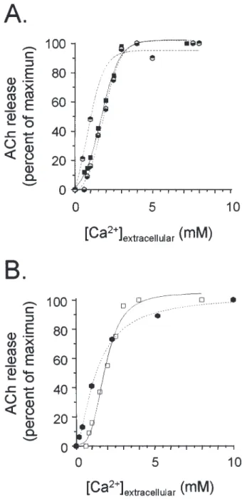
It has long been known that transmitter release is a Ca2⫹-dependent process (2,3), although other fac-tors, particularly depolarization per se, may play a role (4,5). At neuromuscular and nerve-electroplaque junc-tions, quantal ACh release is activated by millimolar concentrations of Ca2⫹in a highly cooperative process (Hill coefficient ⫽ 3–4) (6–8). Investigations on other rapid synapses have provided similar values. When ACh release is elicited from synaptosomes by using a calcium ionophore, the efficient Ca2⫹ concentrations are in the same range but the Hill coefficient is close to 1, probably because of a slower Ca2⫹entry through the ionophore (Fig. 1).
A molecule proposed to be the crucial Ca2⫹sensor for quantal transmitter release should faithfully meet the above kinetics. SNARE proteins, particularly synapto-tagmin, were candidates, but recent research indicated that if the SNAREs are required for vesicle docking they do not support the very last Ca2⫹-dependent reac-tion, either vesicle fusion (9–11) or transmitter release (12). To our knowledge, only one molecule adequately fulfils the requested criteria. It is mediatophore, an oligomer of a 15–16 kD proteolipid subunit, present at the active zone of presynaptic cholinergic terminals. Reconstituted in various systems, mediatophore sup-ports Ca2⫹-dependent and quantal ACh release (13–15). The 16-kD proteolipid forming mediatophore is also a subunit of the membrane sector of V-ATPase, and is found in some invertebrate gap junctions (16). The same very conserved proteolipid was recently identified in yeast as responsible for the very last Ca2⫹-sensitive step of membrane fusion (10).
Figure 1 compares the Ca2⫹-dependency of ACh release using synapses in situ, synaptosomes, and me-diatophore expressed in different systems. Release in intact synapses was monitored either by recording the amplitude of the electroplaque potential or, biochemi-cally, by measuring transmitter output in response to a brief train of impulses. The Ca2⫹-dependency was prac-tically identical with both methods (K0.5 ⫽ 1.55 mM
Ca2⫹and Hill coefficient ⫽ 3.93). In Xenopus oocytes primed with Torpedo mediatophore mRNA and depo-larized using high KCl, the Ca2⫹-dependency was close to that of the intact synapse. On the other hand, when the ionophore A 32187 was used, the slope was less steep (Hill coefficient ⫽ 1–2), but, strikingly, the Ca2⫹-dependency remained the same in the native synaptosomes and in proteoliposomes equipped with mediatophore (17,18). Therefore, reconstituted in oocyte membrane or proteoliposomes, the mediatophore pro-teolipid behaves both as an “ACh translocator” and as
Fig. 1. Ca2⫹-dependency of mediatophore-reconstituted ACh release
faithfully mimics Ca2⫹-dependency of physiological transmission in
intact synapses. Graph A: an identical Ca2⫹-dependency is seen for
the electrophysiological recording of synaptic transmission in the
Tor-pedo electric organ (䊏), the biochemical measurement of the amount
of transmitter released in intact synapses ( ), and ACh released in re-sponse to KCl depolarization from oocytes primed with mediatophore mRNA ( ). (From ref. 8,18.) Graph B: ACh release elicited by using A23187 from Torpedo synaptosomes (ⵧ) or from mediatophore-con-taining proteoliposomes ( ). (From ref. 17.)
tion (13,15). It is worthy of note that the 15-kD prote-olipid interacts with SNARE proteins and, indirectly, with voltage-gated calcium channels (19,20).
Calcium Desensitization of Transmitter Release
While a sudden elevation of intraterminal Ca2⫹ triggers transmitter release, a prolonged exposure to Ca2⫹ provokes a process called “fatigue,” “desensiti-zation” or “adaptation” of transmission (21–23). The phenomenon, investigated in more detail by using cholinergic synaptosomes, is very similar to the de-sensitization affecting ionotropic receptors (or to inac-tivation of certain ion channels). Compared to release activation, desensitization requires lower Ca2⫹ con-centrations but develops more slowly. Surprisingly, desensitization was shown to be an intrinsic property of mediatophore as well. When incorporated into lipo-somes, mediatophore exhibits the two typical charac-teristics of the physiological release: low-affinity and rapid activation on one side, high-affinity and slow de-sensitization on the other side (24). In addition, the pharmacological profile of ACh release performed by the reconstituted mediatophore is similar to that of na-tive terminals (25). The effects of Zn2⫹also reinforce the view that mediatophore is the key mechanism of physiological release. When Zn2⫹ is introduced via A 23187 either in nerve terminals or in mediatophore containing liposomes, it mimics the effects of Ca2⫹by causing both activation and desensitization of ACh release (26).
Therefore, mediatophore appears to be the key molecule in the ultrarapid reactions triggering quantal transmitter release. It is striking that the kinetics of such a presynaptic proteolipid homo-oligomer resem-ble so much those of ionotropic receptors and of many ion channels. In the mentioned experiments, medi-atophore in the plasmalemma supported quantal ACh release directly from a cytosolic pool of neurotrans-mitter (27). However, the same or a very similar pro-teolipid is probably at work in other secreting systems: (i) mediatophore can form fusion channels by apposi-tion of two proteolipid rings between a vesicle and the plasma membrane; and (ii) the rings can eventually en-large, giving rise to full fusion and exocytosis (28).
Calcium Buffering in Nerve Terminals
The entry of Ca2⫹through voltage-gated channels is obviously an extremely rapid reaction at a biochem-ical time scale, though a short delay is observed be-tween the onset of depolarization and the Ca2⫹ signal (29). Substantial amounts of calcium enter the nerve terminals of rapid synapses upon activity (30,31).
However, free Ca2⫹sparks very briefly in the cytosol,
only in restricted “microdomains” situated at the inner mouth of Ca2⫹channels (32). There, Ca2⫹
concentra-tion explosively reaches a submillimolar concentraconcentra-tion. Ca2⫹ is subsequently reduced, first by a very fast
process accounting for the largest part of Ca2⫹
buffer-ing, then by slower mechanisms that are sensitive to thapsigargin, an inhibitor of Ca2⫹-ATPase pumps (33).
The fall of presynaptic free Ca2⫹level has been
attrib-uted to a variety of processes: buffering by cytosolic Ca2⫹-binding proteins (mostly of the EF-hand family),
Na⫹/Ca2⫹ exchange at the presynaptic membrane,
Ca2⫹-ATPase pumping in organelles of the nerve
ter-minals and also of perisynaptic glial cells (33–37). Among them, the calcium ATPase pumps work at sub-micromolar Ca2⫹ ranges; they are thus typical
high-affinity transporters, chiefly located in endoplasmic reticulum but also present in other organelles and at the plasma membrane. The Ca2⫹-ATPase pumps of nerve
terminals and periterminal glial cells can be blocked by thapsigargin, cyclopiazonic acid, 2,5-diterbutyl-1, 4-benzohydroquinone or vanadate, resulting in signif-icant alterations of transmitter release. In different cells and under different experimental conditions, re-lease is either increased or prolonged in time, or even inhibited, probably because of the above-described desensitization (33,35,38).
High and Low-Affinity Calcium Sequestration in Synaptic Vesicle
Docked at the active zone of nerve terminals (39), synaptic vesicles are in a strategic position for seques-trating calcium. It has long been demonstrated that cholinergic and other vesicles have calcium binding capabilities and are able to accumulate Ca2⫹ by an
ATP-dependent mechanism (40,41). More recently, Gonçalves et al. (42) discovered that calcium is taken up by synaptic vesicles via two distinct processes: a Ca2⫹-ATPase pump and Ca2⫹/H⫹antiport.
The vesicular Ca2⫹ pump has a high affinity for
Ca2⫹(K
0.5⫽ 0.6 M) and is inhibited by vanadate. It
displays a maximum velocity at 25 M Ca2⫹ and
pH 7.4, while larger Ca2⫹ concentrations inhibit the
pump. The vesicular Ca2⫹pump differs from the
retic-ulum Ca2⫹pump by having distinct kinetics: the
retic-ulum pump has a still higher affinity for Ca2⫹(K 0.5⫽
0.017 M) and is inhibited at 25 M Ca2⫹, that is, at
a concentration corresponding to the maximum veloc-ity of the vesicular pump.
As for Ca2⫹/H⫹ the antiport, it displays a lower
affinity (K0.5⫽ 217 M) with a maximum activity at
depends on the ⌬pH through the vesicular membrane. Its relies on the activity of V-ATPase (inhibition by bafilomycin). The existence of these two calcium up-take mechanisms was demonstrated on isolated brain vesicles by using a battery of different techniques giving convergent results: uptake of 45Ca, activity
of Ca2⫹-ATPase and V-ATPase, dissipation of the
vesicular proton gradient, release of protons from the vesicles (42).
Transient Calcium Accumulation in Synaptic Vesicles and Extrusion from Nerve Terminals
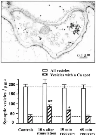
After a brief tetanic nerve stimulation, calcium spots became visible inside synaptic vesicles and were identified by using electron microscopic imaging. At the nerve-electroplaque junction, the total number of vesicles did not change after the stimulation, but the number of vesicles containing a calcium spot signifi-cantly increased (Fig. 2). The effect was transient and returned to control values in parallel to the decline of the total amount of calcium accumulated in the termi-nals upon activity (43,44). Transient Ca accumulation in synaptic vesicles after nerve activity was also demonstrated in mammalian sympathetic ganglion (45) and hippocampus CA1 synapses (46). Vesicular Ca accumulation was accompanied by a similar in-crease in the number of Ca spots in the reticulum of pre- and postsynaptic structures.
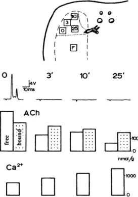
Ancient observations suggested that the more synaptic vesicles accumulated calcium, the more they loose vesicular acetylcholine (Fig. 3). During the course of a 10-Hz stimulation of the nerves to the elec-tric organ in vivo, transmission was exhausted after 3 min or so. At this stage, the free (cytoplasmic) pool of ACh had been significantly reduced and renewed, but not that of vesicle-bound ACh (see review in 27). If nerve stimulation was prolonged in spite of trans-mission failure, calcium progressively accumulated in nerve terminals while the level of vesicular ACh correlatively decreased (31,47).
Two mechanisms can be proposed as an explana-tion for the expulsion of calcium from vesicles. First, calcium could return from the vesicle to the cytosol. It may then be expelled out of the terminal by plas-malemmal Na⫹/Ca2⫹ antiports and Ca2⫹ ATPase
pumps. In a second scenario, the vesicles loaded with calcium may fuse with the membrane and extrude cal-cium by exocytosis. To test this, synapses were sub-mitted to ultrarapid freezing and the membranes were examined for vesicle openings in freeze-fracture repli-cas. No increase in the density of vesicle openings was
found during the very moment of synaptic transmission (48), but the number of openings clearly rose during the first few minutes following a brief tetanic stimula-tion (44). Similarly, vesicle openings were found to peak after a period of activity in various synapses (see references in 49).
What happens to the vesicular ACh when cal-cium accumulates in the organelle either via the Ca2⫹/H⫹antiport or the Ca2⫹-ATPase pump? One can suspect that ACh is displaced from the binding sites provided by the intravesicular proteoglycan matrix (50,51); unbound ACh will then escape from the vesi-cle either by inversion of the vesicular ACh trans-porter (ACh/H⫹ antiport) or via the Vo sector of V-ATPase (which, being also the mediatophore prote-olipid, can pass ACh upon Ca2⫹ activation). In this view, vesicular ACh would be delivered from the vesicle to the cytosol, just at the microdomain of the Fig. 2. Transient accumulation of calcium in synaptic vesicles after
a brief period of activity. After a 100-Hz nerve stimulation, frag-ments of electric organ were fixed at a low temperature in a calcium-free medium containing oxalate. Calcium was revealed with pyroantimioniate and identified by using electron spectroscopy im-aging. Calcium spots were found in vesicles and other structures. After stimulation, the total number of vesicles did not significantly change, while the number of calcium-containing vesicles transiently doubled. (From refs. 43,44.)
active zone, preventing local exhaustion of the rapidly turning over free ACh pool.
An alternative process could occur in those se-creting systems where the transmitter is released through a fusion pore in a “kiss and run” manner. Cal-cium entering the docked vesicles will displace from the matrix a given amount of transmitter, which will escape through the fusion pore.
CONCLUSION
We propose that transmission in rapid synapses is supported by a chain of rapid processes working at a high speed, but at the expense of a low affinity. Among them are the action potentials, presynaptic Ca2⫹ entry, Ca2⫹ activation of transmitter release through mediatophore, Ca2⫹/H⫹exchange in vesicles,
ionotropic receptor activation, and transmitter removal from the cleft. In some of these processes, activation is followed by desensitization when the ligand (or the signal) persists. This chain of rapid reactions is as-sisted by a large number of slower processes, often working with higher affinities, and which are vital for the regulations, fine tuning, and maintenance of the synapse. The latter category encompasses metabotropic pre- and postsynaptic receptors, glial elements, Ca2⫹ -and V-ATPases, SNARE proteins, ion channels mod-ulating membrane potential, plasmalemmal and vesicu-lar transporters, enzymes involved in neurotransmitter synthesis and storage, and in the energy supply of the synapses, etc. Alteration of any of these regulatory functions is of course expected to modify the main core of rapid reactions, and thereby synaptic trans-mission. A typical example is found in reconstitution experiments. Co-transfection of choline acetyltrans-ferase and/or vesicular ACh transporter together with mediatophore do not change the kinetics of ACh re-lease or the quantal size, but increases the rere-lease effi-ciency by allowing a larger number of quanta to be generated (15,52).
At fast synapses, this complex network of rapid and slow mechanisms accomplishes the most fascinat-ing task one can imagine: to use chemical reactions for the millisecond transmission of nerve impulses.
ACKNOWLEDGMENTS
This work was supported by the Swiss FNRS (Grant No 31.57135.99)
REFERENCES
1. Katz, B. 1988. Looking back at the neuromuscular junction. Pages 3–9, in Seilin, L. C., et al. (eds.), Neuromuscular junc-tion, Elsevier Science, Amsterdam.
2. Harvey, A. M. and MacIntosh, F. C. 1940. Calcium and synap-tic transmission in a sympathesynap-tic ganglion. J. Physiol. Lond. 97:408–416.
3. Katz, B. 1969. The Release of Neural Transmitter Substances. University Press, Liverpool.
4. Zhang, C. and Zhou, Z. 2002. Ca(2⫹)-independent but voltage-dependent secretion in mammalian dorsal root ganglion neu-rons. Nat. Neurosci. 5:425–430.
5. Parnas, H., Segel, L., Dudel, J., and Parnas, I. 2000. Autorecep-tors, membrane potential and the regulation of transmitter re-lease. Trends Neurosci. 23:60–68.
6. Dodge, F. A. and Rahamimoff, R. 1967. Co-operative action of calcium ions in transmitter release at the neuromuscular junc-tion. J. Physiol. Lond. 193:419–432.
7. Dunant, Y., Eder, L., and Servetiadis-Hirt, L. 1980. Acetyl-choline release evoked by single or a few nerve impulses in the electric organ of Torpedo. J. Physiol. Lond. 298:185–203.
Fig. 3. In vivo experiment illustrating calcium-acetylcholine exchange
in synaptic vesicles. The upper drawing is the silhouette of a Torpedo, indicating the electrodes for nerve stimulation and the places where small pieces of electric organ were excized for tissue analysis at the times indicated during the course of stimulation. F ⫽ final control in an unstimulated place. The nerves were stimulated at 10 Hz for the indicated times. The first line shows records of the electrical dis-charge, the second line amounts of free and vesicle-bound ACh, and the third one intracellular calcium. After 3 min, transmission was ex-hausted because of failure in ACh release. At this stage there was a de-crease in the free pool of ACh but not in the vesicle-bound ACh, and some calcium accumulated in nerve terminals. When stimulation was prolonged in spite of transmitter failure, vesicular ACh progressively declined while calcium continued to accumulate. (From ref. 31.)
8. Muller, D., Loctin, F., and Dunant, Y. 1987. Inhibition of evoked acetylcholine release: Two different mechanisms in the Torpedo electric organ. Eur. J. Pharmacol. 133:225–234. 9. Shoji-Kasai, Y., Yoshida, A., Sato, K., Hoshino, T., Ogura, A.,
Kondo, S., Fujimoto, Y., Kuwahara, R., Kato, R., and Taka-hashi, M. 1992. Neurotransmitter release from synaptotagmin-deficient clonal variants of PC12 cells. Science 256:1821– 1823.
10. Peters, C., Bayer, M. J., Bühler, S., Andersen, J. S., Mann, M., and Mayer, A. 2001. Trans-complex formation by proteolipid channels in the terminal phase of membrane fusion. Nature 409:581–588.
11. Bruns, D. and Jahn, R. 2002. Molecular determinants of exo-cytosis. Pflugers Arch. 443:333–338.
12. Morel, N., Dunant, Y., and Israel, M. 2001. Neurotransmitter re-lease through the V0 sector of V-ATPase. J. Neurochem. 79:485–488.
13. Falk-Vairant, J., Corrèges, P., Eder-Colli, L., Salem, N., Roulet, E., Bloc, A., Meunier, F., Lesbats, B., Loctin, F., Synguelakis, M., Israël, M., and Dunant, Y. 1996. Quantal acetylcholine release induced by mediatophore transfection. Proc. Natl. Acad. Sci. USA 93:5203–5207.
14. Dunant, Y. and Israël, M. 1998. In vitro reconstitution of neuro-transmitter release. Neurochem. Res. 23:709–718.
15. Bloc, A., Roulet, E., Loctin, F., and Dunant, Y. 1997. Acetyl-choline release from mouse neuroblastoma cells co-transfected with mediatophore and choline acetyltransferase cDNAs. NATO ASI Series 100:175–182.
16. Finbow, M. E. and Pitts, J. D. 1998. Structure of the ductin channel. Biosci. Rep. 18:287–297.
17. Birman, S., Israël, M., Lesbats, B., and Morel, N. 1986. Solubi-lization and partial purification of a presynaptic membrane pro-tein ensuring calcium-dependent acetylcholine release from proteoliposomes. J. Neurochem. 47:433– 444.
18. Cavalli, A., Eder-Colli, L., Dunant, Y., Loctin, F., and Morel, N. 1991. Release of acetylcholine from Xenopus oocytes injected with nRNAs from cholinergic neurons. EMBO J. 10:1671– 1675.
19. Galli, T., McPherson, P. S., and De Camilli, P. 1996. The Vo sector of the V-ATPase, synaptobrevin, and synaptophysin are associated on synaptic vesicles in a Triton X-100-resistant, freeze-thawing sensitive, complex. J. Biol. Chem. 271:2193– 2198.
20. Shiff, G., Synguelakis, M., and Morel, N. 1996. Association of syntaxin with SNAP 25 and VAMP (synaptobrevin) in Torpedo synaptosomes. Neurochem. Int. 29:659–667.
21. Katz, B. and Miledi, R. B. 1969. Tetrodotoxin-resistant electric activity in presynaptic terminals. J. Physiol. Lond. 203:459– 487.
22. Adams, D. J., Takeda, K., and Umbach, J. A. 1985. Inhibitors of calcium buffering depress evoked transmitter release at the squid giant synapse. J. Physiol. Lond. 369:145–159.
23. Hsu, S. F., Augustine, G. J., and Jackson, M. B. 1996. Adapta-tion of Ca(2⫹)-triggered exocytosis in presynaptic terminals. Neuron 17:501–512.
24. Israël, M., Meunier, F. M., Morel, N., and Lesbats, B. 1987. Calcium-induced desensitization of acetylcholine release from synaptosomes or proteoliposomes equiped with mediatophore, a presynaptic membrane protein. J. Neurochem. 49:975–982. 25. Morot-Gaudry-Talarmain, Y., Diebler, M.-F., Robba, M.,
Lancelot, J.-C., Lesbats, B., and Israël, M. 1989. Effect of cetiedil analogs on acetylcholine and choline fluxes in synapto-somes and vesicles. Eur. J. Pharmacol. 166:427– 433.
26. Dunant, Y., Loctin, F., Vallée, J.-P., Parducz, A., Lesbats, B., and Israël, M. 1996. Activation and desensitization of acetyl-choline release by zinc in Torpedo nerve terminals. Pflügers Arch. 432:853–858.
27. Dunant, Y. and Israël, M. 2000. Neurotransmitter release in rapid synapses. Biochimie 82:289–302.
28. Mayer, A. 2001. What drives membrane fusion in eukaryotes? Trends. Biochem. Sci. 26:717–723.
29. Llinas, R., Steinberg, I. Z., and Walton, K. 1981. Relationship between presynaptic calcium current and postsynaptic potential in squid giant synapse. Biophys. J. 33:323–352.
30. Blaustein, M. P. 1971. Preganglionic stimulation increases cal-cium uptake by sympathetic ganglia. Science 172:391–393. 31. Babel-Guérin, E. 1974. Métabolisme du calcium et libération de
l’acétylcholine dans l’organe électrique de la Torpille. J. Neuro-chem. 23:525–532.
32. Llinas, R., Sugimori, M., and Silver, R. B. 1992. Microdomains of high calcium concentration in a presynaptic terminal. Science USA 256:677–679.
33. Castonguay, A. and Robitaille, R. 2001. Differential regula-tion of transmitter release by presynaptic and glial Ca2⫹ in-ternal stores at the neuromuscular synapse. J. Neurosci. 21: 1911–1922.
34. McGraw, C. F., Somlyo, A. V., and Blaustein, M. P. 1980. Lo-calization of calcium in presynaptic nerve terminals: An ultra-structure and electron microprobe analysis. J. Cell Biol. 85:228– 241.
35. Kostyuk, P. and Verkhratsky, A. 1994. Calcium stores in neu-rons and glia. Neuroscience 63:381–404.
36. Neher, E. 1998. Vesicle pools and Ca2⫹microdomains: New
tools for understanding their roles in neurotransmitter release. Neuron 20:389–399.
37. Marsal, J., Esquerda, J. E., Fiol, C., Solsona, C., and Tomas, J. 1980. Calcium fluxes in isolated pure cholinergic nerve endings from the electric organ of Torpedo marmorata. J. Physiol. Paris 76:443–457.
38. Fossier, P., Baux, G., Trudeau, L. E., and Tauc, L. 1992. In-volvement of Ca2⫹ uptake by a reticulum-like store in the con-trol of transmitter release. Neuroscience 50:427–434.
39. Couteaux, R. and Pécot-Dechavassine, M. 1973. Données ultra-structurales et cytochimiques sur le mécanisme de libération de l’acétylcholine dans la transmission synaptique. Arch. Ital. Biol. 3:231–262.
40. Israël, M., Manaranche, R., Marsal, J., Meunier, F. M., Morel, N., Frachon, P., and Lesbats, B. 1980. ATP-dependent calcium up-take by cholinergic synaptic vesicles isolated from Torpedo electric organ. J. Membr. Biol. 54:115–126.
41. Michaelson, D. M., Ophir, I., and Angel, I. 1980. ATP-stimulated Ca2⫹ transport into cholinergic Torpedo synaptic vesicles. J. Neurochem. 35:116–124.
42. Gonçalves, P. P., Meireles, S. M., Gravato, C., and Vale, M. G. 1998. Ca2⫹-H⫹-Antiport activity in synaptic vesicles isolated
from sheep brain cortex. Neurosci. Lett. 247:87–90.
43. Parducz, A. and Dunant, Y. 1993. Transient increase in cal-cium in synaptic vesicles after stimulation. Neuroscience 52: 27–33.
44. Parducz, A., Loctín, F., Babel-Guérin, E., and Dunant, Y. 1994. Exo-endocycytotic images following tetanic stimulation at a cholinergic synapse: A role in calcium extrusion? Neuroscience 62:93–103.
45. Parducz, A., Toldi, J., Joo, F., Siklos, L., and Wolff, J. R. 1987. Transient increase of calcium in pre- and postsynaptic or-ganelles of rat superior cervical ganglion after tetanizing stimu-lation. Neuroscience 23:1057–1061.
46. Buchs, P. A., Stoppini, L., Parducz, A., Siklos, L., and Muller, D. 1994. A new cytochemical method for the ultrastructural local-ization of calcium in the central nervous system. J. Neurosci. Meth. 54:83–93.
47. Dunant, Y., Babel-Guérin, E., and Droz, B. 1980. Calcium me-tabolism and acetylcholine release at the nerve-electroplaque junction. J. Physiol. Paris 76:471–478.
48. Muller, D., Garcia-Segura, L. M., Parducz, A., and Dunant, Y. 1987. Brief occurrence of a population of large intramembrane particles in the presynaptic membrane during transmission of a nerve impulse. Proc. Natl. Acad. Sci. USA 84:590–594.
49. Dunant, Y. 2000. Quantal acetylcholine release: Vesicle fusion or intramembrane particles? Microscopy Res. Tech. 49:38–46. 50. Uvnas, B. 1973. An attempt to explain nervous transmitter
release as due to nerve impulse-induced ion exchange. Acta Physiol. Scand. 87:168–175.
51. Rahamimoff, R. and Fernandez, J. M. 1997. Pre- and postfusion regulation of transmitter release. Neuron 18:17–27.
52. Malo, M., Diebler, M. F., Prado de Carvalho, L., Meunier, F. M., Dunant, Y., Bloc, A., Stinnakre, J., Tomasi, M., Tchelingerian, J., Couraud, P. O., and Israël, M. 1999. Evoked acetylcholine release by immortalized brain endothelial cells genetically modified to express choline acetyltransferase and/or the vesicular acetylcholine transporter. J. Neurochem. 73:1483– 1491.