Use of the Enterprise™ Intracranial Stent for
Revascularization of Large Vessel Occlusions in
Acute Stroke
Zsolt Kulcsár
1, 2, Christophe Bonvin
3, Karl-Olof Lovblad
4, Benjamin Gory
1, Hasan Yilmaz
1,
Roman Sztajzel
3, Daniel Rufenacht
1, 2Received: September 2, 2009; revision accepted: December 9, 2009 Published Online: February 28, 2010
1 Neurointerventional Division, Geneva University Hospital, Switzerland, 2
Neurozentrum, Klinik Hirslanden, Zurich, Switzerland, 3 Neurology Division, Geneva University Hospital, Switzerland, 4
Neuroradiology Division, Geneva University Hospital, Switzerland.
Abstract
Background and Purpose: Major cerebral thromboembolism often resists recanalization with currently available
tech-niques. The authors present their initial experience with a self-expanding stent for use in intracranial vascular reconstruc-tion, permitting immediate recanalization of acute thromboembolic occlusions of the anterior circulation.
Patients and Methods: Patients treated with the Cordis Enterprise™ self-expanding intracranial stent system for acute
thromboembolic occlusion of the major anterior cerebral arteries were included. Treatment comprised systemic and in-traarterial thrombolysis, mechanical thrombectomy, and stent placement. Stent deployment, recanalization rate by means of Thrombolysis In Cerebral Infarction (TICI) scores and the clinical outcome were all assessed.
Results: Six patients presenting with acute carotid T (n = 2) or proximal middle cerebral artery occlusion (n = 4) were
treated. The mean National Institutes of Health Stroke Scale (NIHSS) score at presentation was 14; the mean age was 57 years. Successful stent deployment and immediate recanalization were achieved in all six with a TICI score of ≥ 2. Neither distal emboli nor any procedure-related complications were encountered. One patient developed symptomatic intracerebral hemorrhage and two patients needed decompressive craniectomy after treatment. The mean NIHSS score at 10 days was 10, but only one patient showed a complete recovery at 3 months.
Conclusion: Intracranial placement of the Enterprise™ self-expanding stent has proven to be feasible and efficient in
achieving immediate recanalization of occluded main cerebral arteries. The use of antiplatelet therapy after treatment may, however, increase the risk of reperfusion intracerebral hemorrhage.
Key Words: Acute stroke · Intracranial stenting · Revascularization · Thrombolysis · Thrombectomy
Clin Neuroradiol 2010;20:54–60
10.1007/s00062-010-9024-x
Einsatz des Enterprise-stent-systems zur Rekanalisierung großer intrakranieller Arterien beim akuten Schlaganfall
Zusammenfassung
Hintergrund und Ziel: Die Wiedereröffnung großer thromboembolisch verschlossener Arterien gelingt mit bisherigen
endo-vaskulären Techniken häufig nicht. Die Autoren berichten über ihre Erfahrungen mit einem selbstexpandierenden Stentsystem zur Wiederherstellung des Blutflusses bei Patienten mit akutem thrombembolischem Verschluss in der vorderen Hirnzirkula-tion.
Patienten und Methodik: Patienten mit Verschluss einer großen Arterie in der vorderen Gerhirnkreislauf, die mit einem
Enterprise-Stentsystem (Cordis Enterprise™) behandelt wurden, wurde in diese Studie eingeschlossen. Die Behandlung beinhaltete darüber hinaus eine systemische und intraarterielle Lyse sowie eine mechanische Thrombektomie. Die
Stent-Introduction
Thromboembolic occlusions of the major cerebral teries of the anterior circulation, the internal carotid ar-tery (ICA) and the proximal middle cerebral arar-tery (MCA), are conditions that almost always result in se-vere neurologic compromise. Outcomes are usually poor, with a high mortality rate. Such lesions are often refractory to intravenous and/or intraarterial thrombol-ysis [1–3]. Furthermore, early reocclusion after intrave-nous thrombolysis is not infrequent, and is associated with clinical deterioration and poor outcome [4]. The impact of rapid recanalization on improvement of clini-cal outcome is increasingly recognized [5, 6].
Additional therapeutic options for patients with major vessel occlusion include mechanical thrombecto-my. However, for acute stroke patients failing pharma-ceutical and mechanical thrombolysis, few alternatives remain. As a complementary treatment, with the advent of self-expanding stents suitable for intracranial deliv-ery, stent application for rapid recanalization of acute intracranial vessel occlusions has resulted in high angio-graphic success rates [7–12]. We present our initial ex-perience with the off-label use of the Enterprise™ (Codman & Shurtleff, Inc., Raynham, MA, USA) intra-cranial self-expanding stent for vascular reconstruction in acute stroke.
Patients and Methods
From the stroke database of our institution, we retro-spectively identified and analyzed those patients, who, by suffering a major anterior circulation acute stroke, were treated with intracranial placement of the Enter-prise™ stent, as an off-label use. Between November 2007 and September 2008, altogether six such patients were treated. These patients were diagnosed by
com-puted tomography (CT) and CT angiography to have a major anterior circulation occlusion. Not responding to initial intravenous recombinant tissue plasminogen ac-tivator (rtPA) or presenting in a time window excluding access to such treatment by policy of our local stroke center, we requested consent to intraarterial recanaliza-tion procedures. After angiographic confirmarecanaliza-tion of persistent occlusion and failure or impossibility to pro-ceed with mechanical thrombectomy, intracranial vas-cular reconstruction was performed by stent placement with the Enterprise™ system (Figure 1).
Retrospectively, a database was created with the following characteristics: demographics (age, sex, car-diovascular risk factors), clinical (etiology of stroke, time of symptom onset, baseline and 10-day National Institutes of Health Stroke Scale [NIHSS] scores, symp-tom onset-to-treatment time, time to revascularization), radiographic and angiographic data (location and ex-tension of clot, pharmacological treatment, and me-chanical devices used before and after stenting). The degree of collateral flow was assessed according to the Angiographic Collateral Flow Grading System, where grade 0 means no collateral flow and grade 4 means complete and rapid collateral flow [13]. The degree of recanalization was classified based on the Thrombolysis In Cerebral Infarction (TICI) perfusion categories for reporting purposes [13]. Clinical outcome was assessed by modified Rankin Scale (mRS) at 3 months.
Procedural Technique
Under general anesthesia, a standard 7- or 8-F trans-femoral approach was used to access the thrombosed vessel. Initial parenchymography [14] and selective study of the concerned carotid artery allowed for assess-ment of the occlusion site, as well as of collateral supply.
applikation, die Rekanalisierung unter Verwendung der TICI-Scores (Thrombolysis In Cerebral Infarctions) Scores und der klinische Verlauf wurden analysiert und bewertet.
Ergebnisse: Sechs Patienten wurden eingeschlossen. Zwei Patienten hatten einen Karotis-T-Verschluss und vier Patienten
einen Verschluss der A. cerebri media. Der durchschnittliche NIHSS-Score (National Institutes of Health Stroke Scale) Scores betrug bei Aufnahme 14; das mittlere Alter lag bei 57 Jahren. Die Stentapplikation war bei allen Patienten erfolg-reich, und eine Rekanalisierung mit einem TICI-Score ≥ 2 konnte in allen Fällen erzielt werden. Distale Emboli und proze-durale Komplikationen traten nicht auf. Im Verlauf erlitt ein Patient eine symptomatische intrazerebrale Blutung, zwei weitere Patienten mussten zur Druckentlastung kraniektomiert werden. Der mittlere NIHSS-Score nach 10 Tagen betrug 10; nur ein Patient zeigte eine komplette Restitution nach 3 Monaten.
Schlussfolgerung: Die intrakranielle Applikation des Enterprise-Stentsystems (Cordis Enterprise™) zur Behandlung
groß-er artgroß-eriellgroß-er Vgroß-erschlüsse im vordgroß-eren Gehirnkreislauf ist möglich und sichgroß-er durchführbar. In diesgroß-er Sgroß-erie zeigt sich eine hohe Effizienz mit Reperfusion bei allen Patienten. Die Gabe von Thrombozytenfunktionshemmern erhöht möglicherweise das Risiko einer großen Reperfusionsblutung.
Mechanical thrombectomy attempts were made in three cases, having been combined with intraarterial throm-bolysis in two patients. In the remaining three cases, due to a combination of anatomic considerations, advanced time and occlusion type, stent positioning was performed
alone. The patients received peripro-cedural heparin for systemic antico-agulation with a bolus dose of 2,000– 5,000 IU followed by additional 1,000 IU every 60 min. Immediately before or after stent deployment, intrave-nous aspirin was administered (250 or 500 mg) and intravenous tirofiban infusion was started adjusted to pa-tients’ body weight.
For the performance of intracra-nial stenting, a 0.014" microwire-guided 0.021" inner diameter-sized
Prowler® Select™ Plus microcatheter
(Codman & Shurtleff, Inc.) was navi-gated beyond the site of occlusion, followed by deploying a self-expand-ing Enterprise™ stent. The length of the stent was chosen to completely cover the occluded vascular segment, and reached from 22 to 37 mm. The decision regarding the duration of treatment with tirofiban and an initi-ation of clopidogrel was made based on the postprocedural brain CT find-ings, and by taking the risks of hem-orrhagic transformation into consid-eration.
Results
Four female and two male patients with a mean age of 57 ± 13 years presenting with severe acute stroke of the anterior circulation were treated with the Enterprise™ self-expanding stent system between November 2007 and September 2008 at our stroke center. The mean
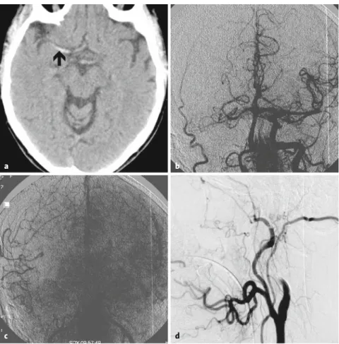
Figures 1a to 1d. Unenhanced brain CT scan of patient #1 with hyperdense MCA sign (arrow; a).
Parenchymography with contrast injection from the aortic arch demonstrating occlusion of the right ICA and MCA with a significant perfusion delay of the right hemisphere (b, c). Lateral view of right common carotid injection showing occlusion of the ICA (d).
a b
c d
Table 1. Characteristics of patients undergoing intracranial stenting. F: female; L: left; M: male; NIHSS: National Institutes of Health Stroke Scale;
R: right; T: carotid T.
Patient # Age (years)/ Admission Stroke Time elapsed Thrombus Grade of sex NIHSS score etiology Symptom onset Angiography location collateral
to angiography to stent deploy- (side) circulation
(h:min) ment (min)
1 61/F 12 Atrial fibrillation 3:29 101 T + M1 (R) 3 2 48/F 16 Carotid dissection 4:48 51 M1 (L) 1 3 43/M 15 Cryptogenic 3:41 42 T + M1 (R) 1 4 49/F 16 Carotid dissection 4:13 35 T + M1 (L) 2 5 66/M 17 Cryptogenic 2:13 103 T + M1 (R) 1 6 76/F 9 Cardioembolic 2:47 120 M1 (L) 2
baseline NIHSS score at presenta-tion was 14 ± 3. After an initial CT, one patient was treated with intrave-nous rtPA, receiving half of the body-weight-adjusted dose without clinical improvement. The mean time to first angiography was 3 h and 31 min. The baseline demographic data, the clot location, and the grad-ing of collateral circulation are listed in Table 1.
Unsuccessful mechanical throm-bectomy was attempted in one case
with the MERCI Retrieval System®
(Concentric Medical Inc., Mountain View, CA, USA). The Penumbra System™ (PS; Penumbra Inc.,
Al-ameda, CA, USA) was used prior to stenting in two cases. In one of these patients, partial recanalization of the M1 segment was achieved via thromboaspiration; however, the reperfusion catheter could not be intro-duced in the occluded upper trunk of the MCA bifurca-tion. The branch was then catheterized with the
Prowl-er® Plus microcatheter, and after unsuccessful local
rtPA infusion, it could be recanalized by stent place-ment. In the second case, no recanalization could be achieved with a combination of PS and intraarterial ly-sis, so stent placement was used to reopen the occluded carotid bifurcation. In the other four cases, the perform-ing physician decided upon immediate stentperform-ing. The oc-cluded segment was recanalized in all cases by a self-ex-panding Enterprise™ stent of 4.5 mm in diameter and 22, 28 or 37 mm in length chosen depending on the length of the occlusion. In all cases, one stent allowed for covering the concerned vascular segments ranging from the MCA bifurcation to the supraclinoid or cav-ernous carotid segment, or covering the M2 and M1 seg-ments. The stent placement allowed for immediate re-vascularization of the occluded segments. The mean time to stent deployment needed from the first angio-graphic study was 75 ± 34 min, including diagnostic runs and mechanical maneuvers.
After stent deployment, immediate recanalization was observed in all six patients; however, the revascu-larized vessels exhibited remaining luminal irregulari-ties. This angiographic aspect was further improved upon administration of intravenous aspirin and tirofi-ban. By the end of the procedure, two patients demon-strated a recanalization grade of 2a, two of 2b, and two
of 3 according to the TICI grading system (Figure 2). No procedure-related complications were encountered.
Brain CT scans 24–36 h post treatment demonstrat-ed that three out of six patients developdemonstrat-ed an infarct af-fecting more than two thirds of the MCA territory, and one patient developed a capsular, space-occupying in-tracerebral hematoma. Two patients became candidates for decompressive craniectomy because of a space-oc-cupying edema developed a few days after stroke onset, and one of them required repeated decompression be-cause of a postoperative intracerebral hematoma that occurred 1.5 h after craniectomy. The mean NIHSS score at 10 days was 10 (range 0–17). Two patients ex-hibited an improvement of > 2 points on the NIHSS. One patient showed a complete recovery with an mRS of 0, and two patients were dead at the 3-month fol-low-up. One of them, although improved from an initial NIHSS score of 9 to 3 after thromboaspiration, died of a malignancy before the 3-month follow-up (Table 2). The stroke-related mortality was 16%.
Discussion
Current reported clinical experience reveals that occlu-sions of the intracranial ICA and the M1 segment of the MCA are highly resistant not just to intravenous throm-bolysis [15], but also to intraarterial pharmacological and mechanical recanalization efforts [16, 17]. It is well known, that carotid T and complete M1 segment occlu-sions have the highest morbidity and mortality rates with less favorable clinical outcome [3, 18–20]. It has also been demonstrated that early recanalization is strongly related with better clinical outcome [5, 6, 16].
Figures 2a and 2b. Anterior (a) and lateral (b) view of the recanalized right ICA and MCA after
stent deployment. Note the residual luminal irregularities still present after stent deployment (arrow) and the opacification of the lenticulostriate arteries (arrowhead).
The pooled analysis of the two MERCI trials has shown that best results were achieved with combined throm-bectomy and thrombolytic maneuvers in patients with ICA occlusions, however, with recanalization rates lim-ited to less than two thirds of patients [21]. Another me-chanical thrombectomy device, the PS, has also shown promising results in terms of revascularization and clini-cal outcome, with recanalization results ranging be-tween 82% and 100% [22–24]. Despite the improving revascularization results, obviously not many options remain for the relatively high percentage of patients where the currently accepted mechanical and chemical thrombolytic attempts fail.
One solution for this group is recanalization using intracranial stents. After the advent of the intracranially trackable balloon-mounted stents and, later, the self-ex-panding intracranial stents, attempts were made to use these devices in recanalization procedures of intracra-nial arterial occlusions. There are few case reports and series demonstrating that these devices are effective in the recanalization of vascular occlusion with a relatively high angiographic success rate [7–12, 25–27]. In the mul-ticenter cases series of Levy et al., who treated 18 such patients, the authors reported a 79% recanalization rate of TICI 2/3 [25]. In this case series, only three out of seven patients presenting with involvement of the ICA could be recanalized with stent placement, but vessel reconstruction to a TICI 2/3 degree could be achieved in all patients showing solitary M1 occlusions. Significant clinical improvement of > 4 points on the NIHSS was observed only in seven out of 14 patients, where a TICI 2/3 grade recanalization could be achieved [25]. In a re-cently published series comprising twelve acute stroke patients, the authors reported on the use of the Wing-span™ stent for revascularization. In this series, five pa-tients had an occlusion of the M1 or M2 segment of the
MCA. Four of them had a partial recanalization of TIMI 2 or 3 degree, but only one patient showed a fa-vorable outcome at 3 months [12]. These results demon-strate that revascularization efforts with available intra-cranial stents are not always successful. In a very recent multicenter study, the authors evaluated the use of the Enterprise™ stent as a salvage revascularization tool, after routine interventions aiming at recanalization had been unsuccessful. In this series, a TIMI 2/3 recanaliza-tion rate of 100% was achieved with 75% of patients showing at least 4-point improvement on the NIHSS [28].
The Enterprise™ self-expanding vascular recon-struction system was designed specifically for brain an-eurysm repair. The stent has a closed-cell design with flared ends, which are both aspects we consider of ad-vantage when it comes to trapping a clot toward the ves-sel wall. Given its maximum available length of 37 mm, this device is long enough to cover extensive lesions without the need for multiple stent deployment. The stent was deployed in all of our cases starting from the MCA bifurcation to the supraclinoid or cavernous por-tion of the ICA covering the whole occluded segment.
The stent delivery Prowler® Select™ Plus microcatheter
was easy and atraumatic to navigate in the cerebral vas-culature and the stent was easy to deliver even in pa-tients with tortuous precerebral and intracranial vessel anatomy. By its radial force, the stent was able to par-tially reconstruct the occluded vessels immediately after deployment, and the residual luminal irregularities fur-ther improved with the administration of aspirin and ti-rofiban. The mean time to stent deployment and thus to recanalization was relatively short. The stent delivery and deployment did not produce any visible clot frag-mentation, and there were no signs of late embolization in patients not suffering a major infarct. This was
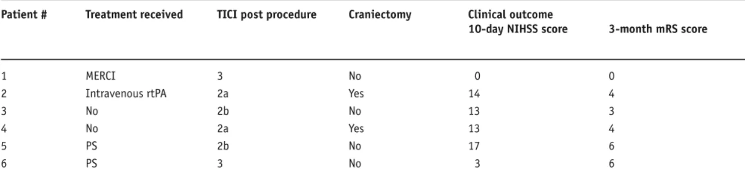
impos-Table 2. Procedure and postprocedure data. mRS: modified Ranking Scale; NIHSS: National Institutes of Health Stroke Scale; PS: Penumbra™
Sys-tem; rtPA: recombinant tissue plasminogen activator; TICI: Thrombolysis In Cerebral Infarction.
Patient # Treatment received TICI post procedure Craniectomy Clinical outcome
10-day NIHSS score 3-month mRS score
1 MERCI 3 No 0 0
2 Intravenous rtPA 2a Yes 14 4
3 No 2b No 13 3
4 No 2a Yes 13 4
5 PS 2b No 17 6
sible to exclude in patients developing an infarct of more than two thirds of the MCA territory. In two cases, the stent caged the origin of an M2 branch, which remained occluded by clot at the immediate follow-up angiograms (patients with a TICI 2a recanalization). We suggest the immediate recanalization of the carotid artery and MCA in all of our patients to be attributable to the fact, that the stent was long enough to cover extended occlusions, and by its closed-cell design and radial force was able to compress and trap the thrombus.
There are, however, two major concerns using in-tracranial stents for acute recanalization in ischemic stroke. One of them is the fear of side branch and perfo-rator occlusion; opening the stent inside the thrombus might cause “snow-plowing” of the clot into side branch-es or perforating arteribranch-es. In complete carotid and/or M1 segment occlusions, the arising branches covered by thrombus are already occluded, so if it were a case of pushing the thrombus further along these vessels, the situation would not get any worse. On the contrary, we believe that the anatomic vascular configuration forces the intracranial microdevices to track in close proximity to the vessel wall, i.e., along the outer curve (in the case of the carotid artery and the M1 segment, where the posterior communicating, the anterior choroidal and the lenticulostriate arteries arise). Based on this con-cept, the microcatheter with the stent will be navigated between the vessel wall and the thrombus and not inside the clot itself. With deployment, the stent will push away the clot from the origin of the perforators and trap it against the opposite wall at the inner curve. We could indeed demonstrate the reappearance of the lenticulo-striate arteries in all of our cases (see Figure 2a). The reperfusion of perforators was also demonstrated in a similar patient series [12].
The other concern is related to the need for anti-platelet therapy after stent deployment, in view of the risk of intracerebral hemorrhage (ICH). On the one hand, the risk of combined antiplatelet therapy in acute stroke is not extensively studied and thus not recom-mended by the current guidelines [29]. The risk of ICH may be further increased if tPAs were concomitantly used [29, 30]. However, stent deployment with antiag-gregant therapy was occasionally used in combination with plasminogen activators in the above-mentioned case series [8–10]. On the other hand, the use of anti-platelet therapy might constitute a problem in cases when patients require neurosurgical intervention such as decompressive craniectomy or hematoma
evacua-tion. In our case series, there was a relatively high inci-dence of ICH after revascularization. One patient de-veloped a space-occupying hematoma 15 h after stent implantation, which did not cause a worsening in his NIHSS score of 17. Another patient suffered a symp-tomatic ICH 5 days after recanalization, soon after a decompressive craniectomy. Both patients were receiv-ing oral aspirin and clopidogrel treatment.
Despite successful recanalization, there were only two patients showing an improvement of > 4 points on the NIHSS. The explanation for the relatively poor out-come of our patient series may rely on the quality of collateral supply of the suffering brain. Although recan-alized within a 6-h time window, all four patients with poor clinical outcomes had insufficient collateral circu-lation of grade 1 or 2, according to the angiographic as-sessment (Table 1).
A low patient number limits this study, as does its retrospective nature. It is hoped that a prospective study with a larger sample size might result in more clarity in order to determine the efficacy of self-expanding stents for acute intracranial vascular thrombosis.
Conclusion
In the setting of acute ischemic stroke caused by throm-boembolic occlusion of the intracranial ICA or com-plete M1 segment of the MCA, where chances of recan-alization by the currently accepted methods are limited, intracranial vessel reconstruction by self-expanding stents seems to be feasible and angiographically effi-cient. Our initial experience with the Enterprise™ self-expanding stent used for intracranial vascular re-construction showed a high potential to reach immedi-ate recanalization of acute major thromboembolic oc-clusions. These results indicate that primary stenting might have a role in the treatment of acute strokes, where currently accepted modalities fail. The criteria for this kind of treatment should, however, be well de-lineated to overcome the difficulties and possible com-plications arising with the use of antiplatelet therapies in cases where huge infarction develops despite success-ful recanalization. The collateral circulation through corticocortical anastomoses seems to have a crucial im-portance in achieving better clinical outcomes after re-vascularization.
Acknowledgment
We gratefully acknowledge the invaluable contribution of Dr. Isabel Wanke and Dr. John Mangiardi to manuscript preparation.
Conflict of Interest Statement
The authors declare that there is no actual or potential conflict of interest in relation to this article.
References
1. Kim YS, Garami Z, Mikulik R, Molina CA, Alexandrov AV, for the CLOT-BUST Collaborators. Early recanalization rates and clinical outcomes in patients with tandem internal carotid artery/middle cerebral artery occlusion and isolated middle cerebral artery occlusion. Stroke 2005; 36: 869–71.
2. Arnold M, Nedeltchev K, Mattle HP, Loher TJ, Stepper F, Schroth G, Brekenfeld C, Sturzenegger M, Remonda L. Intra-arterial thrombolysis in 24 consecutive patients with internal carotid artery T occlusions. J Neurol Neurosurg Psychiatry 2003;74:739–42.
3. Zaidat OO, Suarez JI, Santillan C, Sunshine JL, Tarr RW, Paras VH, Selman WR, Landis DM. Response to intra-arterial and combined intravenous and intra-arterial thrombolytic therapy in patients with distal internal carotid artery occlusion. Stroke 2002;33:1821–6, comment 1827. 4. Rubiera M, Alvarez-Sabin J, Ribo M, Montaner J, Santamarina E,
Arenil-las JF, Huertas R, Delgado P, Purroy F, Molina CA. Predictors of early arterial reocclusion after tissue plasminogen activator-induced recan-alization in acute ischemic stroke. Stroke 2005;36:1452–6.
5. Rha J-H, Saver JL. The impact of recanalization on ischemic stroke outcome: a meta-analysis. Stroke 2007;38:967–73.
6. Zangerle A, Kiechl S, Spiegel M, Furtner M, Knoflach M, Werner P, Mair A, Wille G, Schmidauer C, Gautsch K, Gotwald T, Felber S, Poewe W, Willeit J. Recanalization after thrombolysis in stroke patients: predic-tors and prognostic implications. Neurology 2007;68:39–44. 7. Gupta R, Vora NA, Horowitz MB, Tayal AH, Hammer MD, Uchino K, Levy
EI, Wechsler LR, Jovin TG. Multimodal reperfusion therapy for acute ischemic stroke: factors predicting vessel recanalization. Stroke 2006; 37:986–90.
8. Levy EI, Ecker RD, Hanel RA, Sauvageau E, Wehman JC, Guterman LR, Hopkins LN. Acute M2 bifurcation stenting for cerebral infarction: les-sons learned from the heart: technical case report. Neurosurgery 2006;58:E588, discussion E588.
9. Levy EI, Ecker RD, Horowitz MB, Gupta R, Hanel RA, Sauvageau E, Jovin TG, Guterman LR, Hopkins LN. Stent-assisted intracranial recanaliza-tion for acute stroke: early results. Neurosurgery 2006;58:458–63, discussion 458–63.
10. Sauvageau E, Samuelson RM, Levy EI, Jeziorski AM, Mehta RA, Hopkins LN. Middle cerebral artery stenting for acute ischemic stroke after unsuc-cessful Merci retrieval. Neurosurgery 2007;60:701–6, discussion 706. 11. Levy EI, Mehta R, Gupta R, Hanel RA, Chamczuk AJ, Fiorella D, Woo HH,
Albuquerque FC, Jovin TG, Horowitz MB, Hopkins LN. Self-expanding stents for recanalization of acute cerebrovascular occlusions. AJNR Am J Neuroradiol 2007;28:816–22.
12. Brekenfeld C, Schroth G, Mattle HP, Do DD, Remonda L, Mordasini P, Arnold M, Nedeltchev K, Meier N, Gralla J. Stent placement in acute cerebral artery occlusion: use of a self-expandable intracranial stent for acute stroke treatment. Stroke 2009;40:847–52.
13. Higashida RT, Furlan AJ, Roberts H, Tomsick T, Connors B, Barr J, Dillon W, Warach S, Broderick J, Tilley B, Sacks D. Trial design and reporting standards for intra-arterial cerebral thrombolysis for acute ischemic stroke. Stroke 2003;34:e109–37.
14. Théron J, Nelson M, Alachkar F, Mazia D. Dynamic digitized cerebral parenchymography. Neuroradiology 1992;34:361–4.
15. Saqqur M, Uchino K, Demchuk AM, Molina CA, Garami Z, Calleja S, Akhtar N, Orouk FO, Salam A, Shuaib A, Alexandrov AV. Site of arterial occlusion identified by transcranial Doppler predicts the response to intravenous thrombolysis for stroke. Stroke 2007;38:948–54. 16. Furlan A, Higashida R, Wechsler L, Gent M, Rowley H, Kase C, Pessin M,
Ahuja A, Callahan F, Clark WM, Silver F, Rivera F. Intra-arterial prouro-kinase for acute ischemic stroke. The PROACT II study: a randomized controlled trial. Prolyse in Acute Cerebral Thromboembolism. JAMA 1999;282:2003–11.
17. Smith WS, Sung G, Starkman S, Saver JL, Kidwell CS, Gobin YP, Lutsep HL, Nesbit GM, Grobelny T, Rymer MM, Silverman IE, Higashida RT, Budzik RF, Marks MP. Safety and efficacy of mechanical embolectomy
in acute ischemic stroke: results of the MERCI trial. Stroke 2005; 36:1432–8.
18. Sims JR, Rordorf G, Smith EE, Koroshetz WJ, Lev MH, Buonanno F, Schwamm LH. Arterial occlusion revealed by CT angiography predicts NIH Stroke Score and acute outcomes after IV tPA treatment. AJNR Am J Neuroradiol 2005;26:246–51.
19. Linfante I, Llinas RH, Selim M, Chaves C, Kumar S, Parker RA, Caplan LR, Schlaug G. Clinical and vascular outcome in internal carotid artery versus middle cerebral artery occlusions after intravenous tissue plas-minogen activator. Stroke 2002;33:2066–71.
20. Rubiera M, Ribo M, Delgado-Mederos R, Santamarina E, Delgado P, Montaner J, Alvarez-Sabin J, Molina CA. Tandem internal carotid ar-tery/middle cerebral artery occlusion: an independent predictor of poor outcome after systemic thrombolysis. Stroke 2006;37:2301–5. 21. Flint AC, Duckwiler GR, Budzik RF, Liebeskind DS, Smith WS. Mechanical
thrombectomy of intracranial internal carotid occlusion: pooled results of the MERCI and Multi MERCI Part I trials. Stroke 2007;38:1274–80. 22. Grunwald IQ, Walter S, Papanagiotou P, Krick C, Hartmann K,
Dauter-mann A, Fassbender K, Haass A, Bolar LJ, Reith W, Roth C. Revascular-ization in acute ischaemic stroke using the Penumbra System: the first single center experience. Eur J Neurol 2009;16:1210–6.
23. Penumbra Pivotal Stroke Trial Investigators. The Penumbra Pivotal Stroke Trial: safety and effectiveness of a new generation of mechani-cal devices for clot removal in intracranial large vessel occlusive dis-ease. Stroke 2009;40:2761–8.
24. Kulcsár Z, Bonvin C, Pereira VM, Altrichter S, Yilmaz H, Lovblad KO, Sztajzel R, Rüfenacht DA. Penumbra™ System: a novel mechanical thrombectomy device for large vessel occlusions in acute stroke. AJNR Am J Neuroradiol:in press (Epub 2009 Dec 17).
25. Levy EI, Sauvageau E, Hanel RA, Parikh R, Hopkins LN. Self-expanding versus balloon-mounted stents for vessel recanalization following em-bolic occlusion in the canine model: technical feasibility study. AJNR Am J Neuroradiol 2006;27:2069–72.
26. Fitzsimmons BF, Becske T, Nelson PK. Rapid stent-supported revasculariza-tion in acute ischemic stroke. AJNR Am J Neuroradiol 2006;27:1132–4. 27. Brekenfeld C, Tinguely P, Schroth G, Arnold M, El-Koussy M, Nedeltchev
K, Byrne JV, Gralla J. Percutaneous transluminal angioplasty and stent placement in acute vessel occlusion: evaluation of new methods for interventional stroke treatment. AJNR Am J Neuroradiol 2009;30: 1165–72.
28. Mocco J, Hanel RA, Sharma J, Hauck EF, Snyder KV, Natarajan SK, Lin-fante I, Siddiqui AH, Hopkins LN, Boulos AS, Levy EI. Use of a vascular reconstruction device to salvage acute ischemic occlusions refractory to traditional endovascular recanalization methods. J Neurosurg:in press (Epub 2009 Sep 18).
29. Adams HP Jr, del Zoppo G, Alberts MJ, Bhatt DL, Brass L, Furlan A, Grubb RL, Higashida RT, Jauch EC, Kidwell C, Lyden PD, Morgenstern LB, Qureshi AI, Rosenwasser RH, Scott PA, Wijdicks EFM. Guidelines for the early management of adults with ischemic stroke: a guideline from the Amer-ican Heart Association/AmerAmer-ican Stroke Association Stroke Council, Clinical Cardiology Council, Cardiovascular Radiology and Intervention Council, and the Atherosclerotic Peripheral Vascular Disease and Quality of Care Outcomes in Research Interdisciplinary Working Groups: The American Academy of Neurology affirms the value of this guideline as an educational tool for neurologists. Stroke 2007;38:1655–711.
30. Ciccone A, Motto C, Aritzu E, Piana A, Candelise L, on behalf of the MAST-I Collaborative Group. Risk of aspirin use plus thrombolysis after acute isch-aemic stroke: a further MAST-I analysis. Lancet 1998;352:880.
Address for Correspondence
Zsolt Kulcsár, MD Neurozentrum Klinik Hirslanden Witellikerstraße 40 8032 Zurich Switzerland Phone (+41/44)387-2858, Fax -2861 e-mail: kulcsarzsolt22@gmail.com