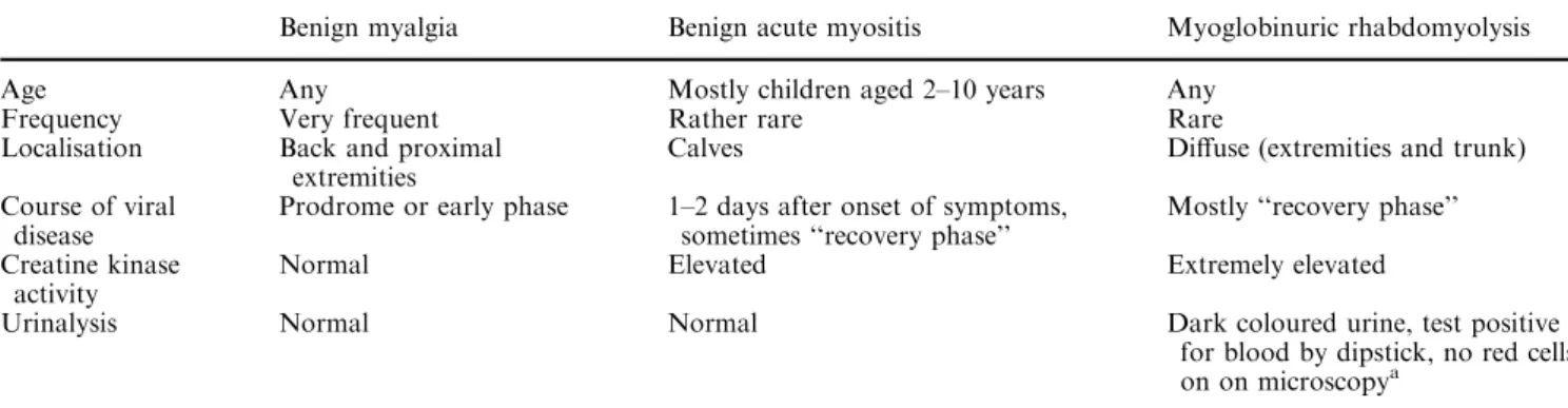
An 8-year-old boy with a 4-day history of fever, cough and malaise, and a 2-day history of painful calves and difficulty walking
2
0
0
Texte intégral
Figure
Documents relatifs