TECHNICAL NOTE
Confirmation of natural gas explosion from methane
quantification by headspace gas chromatography
–mass
spectrometry (HS-GC-MS) in postmortem samples:
a case report
V. Varlet&M. Augsburger
Received: 23 January 2012 / Accepted: 12 June 2012 / Published online: 21 June 2012 # Springer-Verlag 2012
Abstract A new analytical approach for measuring methane in tissues is presented. For the first time, the use of in
situ-produced, stably labelled CDH3provides a reliable and precise
methane quantification. This method was applied to postmortem samples obtained from two victims to help determine the explo-sion origin. There was evidence of methane in the adipose tissue (82 nmol/g) and cardiac blood (1.3 nmol/g) of one victim, which corresponded to a lethal methane outburst. These results are discussed in the context of the available literature to define an analysis protocol for application in the event of a gas explosion.
Keywords Methane . Explosion . HS-GC-MS
Introduction
Although methane does not have major toxic effects on organisms, the gas has two characteristics that can lead to death. First, significant quantities of methane can compete with and deplete oxygen levels. Thus, oxygen is not suffi-ciently distributed within an organism, causing hypoxia (diz-ziness and fainting) and, ultimately, lethal anoxia (suffocation
and neuronal death) [1]. This property has been exploited in
suicides with natural gas [2] and has played a role in accidental
deaths due to sewer shaft falls [3], drainage pit work [4],
handling decaying material [5] or working in mines [6,7].
Secondly, methane is extremely flammable and may form explosive mixtures with air at concentrations between 5 and
15 % by volume [8]. Therefore, methane combustion in mines
[9,10], during tunnel urban works [11] or domestic incidents
[12], can lead to lethal explosions through the conversion of
methane chemical energy into mechanical and thermal energy
[13–15]. Conversely, only one suicide case has been reported
that involved methane explosivity as a deliberate lethal agent
[16]. The highly flammable and potentially explosive
proper-ties of methane make it an extremely noxious agent.
In mining industry accidents, methane determination in postmortem tissues is necessary to identify the cause of death. Indeed, the chemical combustion of methane can be lethal for several reasons, including extremely high temperatures (up to
2,650 °C) [15], mechanical effects (blast and pressure
follow-ing explosion) or asphyxia due to oxygen depletion [6].
Con-sequently, it is very difficult to establish a lethal methane concentration. Local concentration (especially in the lungs) can be more important in asphyxia cases, when the victim inhales methane for a long period of time, than in cases of death due to gas explosion (thermal, mechanical effects). Moreover, in cases of asphyxia following an explosion, con-tinued respiration during survival period allows methane to distribute into the organs, and this interval is always unknown, which complicates the interpretation of the implication of various methane concentrations in organs. The initial ambient air composition can provide information regarding methane concentration for estimation of the survival period. Finally, to avoid methane release from the body and to exclude methane generation due to decomposition, minimal delay between death and sample collections is of great importance.
From an analytical point of view, these measurements are performed via injections of gaseous samples into a gas chromatograph (GC) equipped with a flame ionisation
de-tector (FID) [17, 18] or a thermal conductivity detector
(TCD). Mass spectrometry (MS) has recently been used as
V. Varlet (*)
:
M. AugsburgerForensic Chemistry and Toxicology Unit,
University Centre of Legal Medicine Lausanne-Geneva, 1011 Lausanne, Switzerland
a confirmatory tool, but quantification by GC-MS has not been previously performed. The main drawback of GC-FID and GC-TCD methods is the absence of an internal standard. Indeed, the quantifications were performed with external
calibration with standard gaseous methane [6], pentane
[12] or a mixture of methane/argon [4]. Methanol has also
been used as an internal standard [19]. However, taking into
account the high reactivity of Grignard reagents such as
methylmagnesium chloride (CH3MgCl) towards water, it
becomes possible to generate gaseous methane. By using
deuterated water (D2O) instead of H2O, an internal standard
(CDH3) can be generated. The control of concentration is
performed by the stoichiometry of the reaction and the volume in which the reaction takes place.
The aim of this study was to develop a new method of
methane quantification by headspace gas chromatography–
mass spectrometry (HS-GC-MS) using a labelled stable iso-tope of methane generated in situ. The procedure was applied to postmortem samples from victims of a gas explosion.
Materials and methods
Reagents
Methylmagnesium chloride 3.0 M in tetrahydrofuran (THF) was from Sigma-Aldrich (Saint Louis, USA). Deuterated water was obtained from Cambridge Isotope Laboratories, Inc. (Andover, USA). Certified methane was obtained from Carbagas (Lausanne, Switzerland).
Standard generation
The methodology for generating standards in situ was
de-scribed previously [20]. Methane (CH4) and deuterated
methane (CDH3) were generated separately in 20-mL
head-space vials. The reactions of Grignard reagent with water and deuterated water are given below:
CH3MgClþ H2O! CH4þ MgClOH ð1Þ
CH3MgClþ D2O! CDH3þ MgClOD ð2Þ
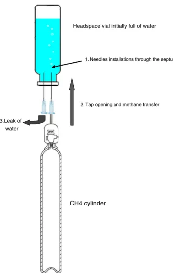
Due to the high reactivity of these reactions, it is important to proceed quickly (methylmagnesium chloride reacts with water in ambient air) and safely (in a fume hood). Grignard reagent and water were added without any contact between them in an aluminium cap with no septa or holes and introduced into a
headspace vial (Fig.1). The vial was rapidly and hermetically
closed and then vortexed to allow methane generation. Precise
volumes of gas (CH4and CDH3) were sampled (automatically
or manually) with a gas syringe through the vial septum and directly introduced into the GC injector.
Fig. 1 Design of methane generation in situ
Headspace vial initially full of water
Gasmethanecanister
2. Tap opening and methane transfer
3.Leak of water
1. Needles installations through the septum
CH4 cylinder
Control samples
Certified methane (99.995 % purity) was used to make control samples. Known volumes of methane were diluted in headspace vials previously saturated with nitrogen: 0.053,
0.105 and 0.210μmol/mL were used (Fig.2). The methane
yield was compared against these external controls (relative
bias <25 % for each concentration,n03).
Calibration curve
The amounts of Grignard reagent and water were calculated to
produce concentrations of 0.5μmol/mL of headspace in the vial:
3.3μL CH3MgCl (3.0 M in THF) and 20μL water (or
deuter-ated water) were introduced into a 20-mL headspace vial. A
calibration curve (R2
0.9756,n04 for each point) was built with
five concentrations: 0.025, 0.05, 0.1, 0.25 and 0.5μmol/mL
corresponding to 50, 100, 200, 400 and 1,000 μL of injected
gaseous sample from the standard vial (obtained by
reac-tion1). For unknown sample measurement, a 1,000-μL
gas-eous sample was taken. Before each injection, a volume of
100μL (corresponding to 0.5 μmol/mL) was taken from the
internal standard vial (obtained by reaction2).
GC-MS analysis
An Agilent 6890N GC (Agilent Technologies, Palo Alto, CA) combined with a headspace gas autosampler and equipped with a HP Molecular sieve 5-Å PLOT capillary column (30 m×
0.32 mm, 30μm) from Restek (Bellefonte, USA) was used. A
CP Porabond Q column was also used. The temperature program was 40 °C held for 5 min. The injector (splitless mode) was set at 100 °C, and the interface MS temperature was 230 °C. Helium was employed as a carrier gas at a flow rate of 1.9 mL/min.
The detection was performed with an Agilent 5973 mass spectrometer (Agilent Technologies, Palo Alto, CA), operat-ing in an electron ionisation mode at 70 eV. Selected ion monitoring mode was used to acquire the methane signal at
m/z 16 and 17 for CDH3.
Case report
A 31-year-old woman and her 5-year-old daughter were killed when there was an explosion in their apartment. Both victims underwent autopsy in our laboratory a few hours after the explosion. Only brain and cardiac blood were sampled from the woman, and cardiac blood and adipose tissue were collected from the daughter for the purpose of gas analysis. The objective was to assess methane exposure to confirm the hypothesis of a natural gas explosion. The lungs were too damaged to be useful for analysis.
Results and discussion
Only a handful of studies have assessed methane concentra-tions in postmortem tissue. A minimal methane concentration
of 6μL/g or 0.25 μmol/mL of cardiac blood was reported in a
study performed on 22 victims of a gas outburst accident in a
coal mine [6]. The methane concentration in cardiac blood
was 14.1±5.3μL/g or 0.59 μmol/mL (n022). This result is in
Table 1 Concentration of methane in tissues of the two victims and HbCO saturation
Woman (31 years old) Girl (5 years old) Methane concentration (μmol/g)
Cardiac blood ND 1.3×10⁻³a Brain ND Adipose tissue 82×10−3 HbCO (%) Peripheral blood 5 19.5 ND not detected aApproximate value Abundance Retention time CH4 CDH3 O2 CO2 Retention time Fig. 3 Chromatograms of methane and deuterated methane in adipose tissue (daughter)
agreement with results obtained from rats subjected to various
methane/oxygen concentrations in a closed space [7]. A lethal
methane concentration of 19μL/g (0.79 μmol/mL) was found
in rat blood (n05) after 2 min under 100 % CH4. A lethal
methane concentration of 25μL/g (1.04 μmol/mL) was found
in rat blood (n05) after 20–25 min of exposure to a CH4
atmosphere increasing from 0 to 100 % at 1 L/min. Finally, a
lethal methane concentration of 29μL/g (1.21 μmol/mL) was
found in rats blood (n05) after 80–85 min under an oxygen/
CH4atmosphere mixture (2:8,v/v%) increasing to 100 % of
methane after 1 h. The initial composition of ambient air is therefore very important for interpreting blood methane con-centration. The lung is the best tissue for diagnosing acute methane exposure. A minimal lung methane concentration
above 160μL/g (6.67 μmol/mL) was measured in coal miners
(n02) [6] and seems consistent with the results obtained in rats
[7]: 163±48 μL/g (6.8 μmol/mL) under oxygen/CH4
atmo-sphere mixture and 442±107 μL/g (18.4 μmol/mL) under
100 % methane. However, brain or adipose tissues are also useful because methane is soluble in fat.
The methane concentration in the tissue from the two
autopsied victims is presented in Table1. Methane was only
detected in samples from the daughter, with the highest levels
in the adipose tissue (Fig.3). It is interesting to note that only
the daughter’s tissue showed significant HbCO saturation
(19.5 % in cardiac blood compared to 5 % in the mother). These results indicate that the daughter received a higher exposure to methane and CO gases than the mother before the explosion or that the mother passed away shortly after the explosion because her methane concentration values were not sufficient for death from anoxia.
The observed methane concentrations were considerably
below the 0.25μmol/g value that is considered indicative of
“methane death” caused by asphyxia due to oxygen depletion
[6]. However, this cutoff value should be only used for gas
outbursts in mines or confined, underground spaces. More-over, this value was established without information regarding the true role of methane. Methane concentration variations in the miners’ tissues show that the causes of death and survival periods were different. Some of the workers could have died due to asphyxia before the explosion, and others may have died after the explosion due to anoxia or explosion-related injuries. Nevertheless, it can be assumed that a methane
con-centration greater than 0.25μmol/g, even in the absence of an
explosion, is an indicator of“methane-related death”.
The difficulty in determining lethal methane tions can be illustrated by the different methane concentra-tions obtained in different tissues according to gas exposure (Tab. 2). It is necessary to identify four interconnected parameters before interpreting methane concentrations:
1. Initial ambient air composition. The results compiled in
Table2[4,11] indicate a very low methane exposure. In Table
2 Methane concentrations in various tissues and or gans after several types of accidents (adapted from [ 4 , 6 , 7 , 11 ]) Met hane exposur e Met hane conce ntration (μ mol/g ) W ork in pit [ 4 ] U rban tunnel exp losion [ 11 ] Coal mine explosion [ 6 ] Rats unde r 100 % CH 4 [ 7 ] Rats under 0→ 100 % CH 4 (1 L/min) [ 7 ] Rats unde r C H4 /02 (8:2 ) (1 h )→ 100 % C H4 [ 7 ] Blo od 12 × 1 0 − 3±1 1×1 0 − 3(n 0 3) 0.59 ± 0.2 (n 0 22) 0.80 ± 1 6 × 10 − 3(n 0 5) 1.04 ± 5 0 × 10 − 3(n 0 5) 1.21 ± 7 5 × 10 − 3(n 0 5) Br ain 15 × 1 0 − 3±1 4 × 1 0 − 3(n 0 3) 2 × 10 − 3(n 0 1) 0.89 (n 0 2) 1.00 ± 8 8 × 10 − 3(n 0 5) 1.09 ± 6 7 × 10 − 3(n 0 5) 1.00 ± 0.1 (n 0 5) Li ver 7.3 × 1 0 − 3 ± 7.0 × 1 0 − 3 (n 0 3) 12 × 1 0 − 3 ±1 8× 1 0 − 3 (n 0 3) 0.40 (n 0 2) 0.59 ± 5 4 × 10 − 3 (n 0 5) 0.67 ± 3 4 × 10 − 3 (n 0 5) 0.92 ± 0.14 (n 0 5) K idney 14 × 1 0 − 3± 7.9 × 1 0 − 3(n 0 3) 0.17 (n 0 2) 0.96 ± 6 3 × 10 − 3(n 0 5) 0.92 ± 8 8 × 10 − 3(n 0 5) 1.00 ± 0.2 (n 0 5) H eart 19 × 1 0 − 3±2 0 × 1 0 − 3(n 0 3) 0.48 (n 0 2) 1.1 ± 0.14 (n 0 5) 0.83 ± 3 4 × 10 − 3(n 0 5) 0.92 ± 5 9 × 10 − 3(n 0 5) Lu ng 0.13 ± 0.2 (n 0 3) 14 × 1 0 − 3± 6.0 × 1 0 − 3(n 0 3) 9.27 (n 0 2) 19 ± 4.5 (n 0 5) 7.96 ± 3.4 (n 0 5) 6.79 ± 0.2 (n 0 5) Fa t 7.4 × 1 0 − 3 ± 6.0 × 1 0 − 3 (n 0 3) 16 × 1 0 − 3 ±1 4× 1 0 − 3 (n 0 4) 0.14 (n 0 2) 0.71 ± 0.1 (n 0 5) 0.67 ± 5 0 × 10 − 3 (n 0 5) 1.00 ± 0.2 (n 0 5) V itreous 7.4 × 1 0 − 3± 4.7 × 1 0 − 3(n 0 3) Musc le 4.9 × 1 0 − 3± 2.3 × 1 0 − 3(n 0 3) Med ulla oblon gata 0.51 (n 0 2) Sple en 0.20 (n 0 2) Pa ncreas 0.16 (n 0 2) The last three columns show the results of a study on methane asphyxia on rats
the first case, there was no explosion, and the cause of
death was CO2intoxication leading to lethal asphyxia of
three workers in a draining pit [4]. In the second case,
there was a gas outburst following a rapidly increasing methane leak past the explosion limit of 5 % and a
simultaneous decrease of O2[11]. The main causes of
death were wounds caused by the explosion and CO intoxication. However, in both cases, the methane con-centrations were too low to cause death due to asphyxia.
2. Room geometry. Results compiled in Table 2 [6, 7]
illustrate the importance of room geometry and gas flow rates on methane concentrations. Mixtures of methane with oxygen influence survival time because the meth-ane is diluted. A large methmeth-ane leak without ventilation can rapidly transform the composition of ambient air until the explosive threshold is reached. As methane is lighter than air, it collects at the top of a room. Depend-ing on the room volume, the time to reach 5 % methane can vary, and this influences the final methane concen-trations in victims’ organs.
3. Delay of gas exposure before and after explosion (if an
explosion occurs). The results listed in Table 2 [7]
demonstrate that the survival period is strongly related to ambient air composition, which is itself related to room geometry. Increased oxygen could allow a longer time of methane exposure, which could explain the greater distribution of methane in the body and its accumulation in fat-rich tissues (e.g. adipose tissue and brain). The delay and magnitude of methane exposure (initial ambient air composition) can be deduced from the methane concentrations obtained in the different samples and the room geometry.
4. Analysed sample. As illustrated in Table 2, methane
concentration can be influenced by the nature of the sample, the length of methane exposure and the victim’s metabolism. Usually, the lungs are the first organs im-pacted by methane, but if the methane concentration in the ambient air is sufficiently low, the concentration can decrease in the lungs and increase in the heart, blood and especially in lipophilic organs, such as brain and adipose tissue. Conversely, methane is not stored in appreciable quantities in spleen, pancreas or kidney, which are further along in the detoxification pathway. Therefore, a high methane concentration in lipophilic tissue and a low concentration in the lungs could mean that the victim did not die rapidly and could have been asphyxiated. Similarly, a high methane concentration in the lungs and low concentrations in other tissues could
indicate a rapid, lethal methane explosion [11].
In our case (Table1), the values of methane concentrations
in cardiac blood and adipose tissue were between those mea-sured in samples from a sudden lethal methane explosion (no
asphyxia) [11] and those measured in samples following
asphyxia due to a methane explosion [6]. The fact that no
methane was measured in the mother seems to indicate a relatively sudden and lethal methane explosion.
From an analytical point of view, employing deuterated methane as an internal standard before assessing the un-known samples allows troubleshooting that can prevent the loss of valuable samples. Leaks or analytical discrepancies should affect methane and deuterated methane equally, which results in more reliable measurements.
Conclusion
The labelled stable isotope of methane allows precise iden-tification and quaniden-tification. Further work is necessary to fully validate the analytical method of methane measure-ment by HS-GC-MS. These findings are important for establishing a sampling protocol following explosions. Car-diac blood and lungs provide the best samples to monitor methane after a gas outburst, but injuries sustained during the explosion may render these tissues unusable. Therefore, because methane is lipophilic, brain and adipose tissue can be analysed instead. In the reported cases, the absence of HbCO saturation and the weak methane concentration in cardiac blood seem to indicate a rapid natural gas explosion. The presence of methane in the adipose tissue of one of the victims confirmed that natural gas was the lethal explosive agent.
References
1. Lareng L, Francois RC, Virenque C, Bertin M, Bertrand M, Brouchet A (1969) Anoxia, the cause of asphyxias due to non-burned natural gas. An experimental study of the asphyxia with Lacq’s natural gas, methane and nitrogen. Presse Med 77:349–351 2. Akhgari M, Elham B (2010) Deaths involving natural gas
inhala-tion. Toxicol Ind Health 26:345–347
3. Byard RW, Wilson GW (1992) Death scene gas analysis in sus-pected methane asphyxia. Am J Forensic Med Pathol 13:69–71 4. Manning TJ, Ziminski K, Hyman A, Figueroa G, Lukash L (1981)
Methane deaths? Was it the cause? Am J Forensic Med Pathol 2:333–336
5. Cherian MA, Richmond I (2000) Fatal methane and cyanide poi-soning as a result of handling industrial fish: a case report and review of the literature. J Clin Pathol 53:794–795
6. Terazawa K, Takatori T, Tomii S, Nakano K (1985) Methane asphyxia. Coal mine accident investigation of distribution of gas. Am J Forensic Med Pathol 6:211–214
7. Watanabe T, Morita M (1998) Asphyxia due to oxygen deficiency by gaseous substances. Forensic Sci Int 96:47–59
8. Laursen E, Hempel-Jorgensen I, Lassen E (1995) Landfill gas. Ugeskr Laeger 157:6585–6586
9. Allister C, Hamilton GM (1983) Cardowan coal mine explosion: experience of a mass burns incident. Br Med J 287:403–405
10. Kobek M, Jankowski Z, Chowaniec C, Jabłoski C, Gaszczyk-Ozarowski Z (2009) Assessment of the cause and mode of death of victims of a mass industrial accident in the Halemba coal mine. Forensic Sci Int Suppl Ser 1:83–87
11. Nagao M, Takatori T, Oono T, Iwase H, Iwadate K, Yamada Y, Nakajima M (1997) Death due to a methane gas explosion in a tunnel on urban reclaimed land. Am J Forensic Med Pathol 18:135–139
12. Park J, Min JS, Heo S, Lim MA, Park SW (2005) Quantification of propane in biological materials by head-space GC. Forensic Sci Int 151:165–170
13. Suzutani T, Ishibashi H, Takatori T (1979) Medico-legal studies on the deaths from coal-mine accidents 3. Causes of death. Hokkaido J Med Sci 54:479–486
14. Takatori T, Tomii S, Terazawa K (1981) Medicolegal studies on death from coal-mine accident by gas spurt. Jpn J Legal Med 35:462–467
15. Skowronek R, Chowaniec C (2009) The role, objectives and usefulness of medico-legal determinations in post-accidental
procedures in traumatic deaths in hard coal-mining industry. Arch Med Sad Krym LIX:101–111, In Polish
16. El Demellawy D, Fernandes J (2007) Suicide by explosion of natural gas: case report and review of literature. Am J Forensic Med Pathol 28:48–52
17. Sigrist T, Sutter K, Germann U (1998) Methane in cadaver blood —homicide by natural gas or postmortem formation. Arch Kriminol 201:24–30, In German
18. Takatori T, Terazawa K (1980) A case report: determination of methane gas in cadaveric tissues from a coal-mine accident by gas chromatography. Hokkaido J Med Sci 55:363–365
19. Yablochkin VD (2004) Forensic and chemical determination of methane in cadaveric samples. Sud Med Ekspert 47:36–38, In Russian
20. Varlet V, Lagroy De Croutte E, Augsburger M, Mangin P (2012) Accuracy profile validation of a new method for carbon monoxide measurement in the human blood using headspace-gas chromatog-raphy–mass spectrometry (HS-GC-MS). J Chromatogr B 880:125– 131
![Table 2 [4, 11] indicate a very low methane exposure. In T able 2 Methane concentrations in various tissues and or gans after several types of accidents (adapted from [ 4 , 6 , 7 , 11 ]) Met hane exposur e Met hane conce ntration ( μ mol/g ) W ork in pit [](https://thumb-eu.123doks.com/thumbv2/123doknet/14841228.625174/4.892.478.790.117.1067/indicate-methane-exposure-methane-concentrations-various-accidents-ntration.webp)
