HAL Id: hal-01774687
https://hal.archives-ouvertes.fr/hal-01774687
Submitted on 7 May 2018
HAL is a multi-disciplinary open access
archive for the deposit and dissemination of
sci-entific research documents, whether they are
pub-lished or not. The documents may come from
teaching and research institutions in France or
abroad, or from public or private research centers.
L’archive ouverte pluridisciplinaire HAL, est
destinée au dépôt et à la diffusion de documents
scientifiques de niveau recherche, publiés ou non,
émanant des établissements d’enseignement et de
recherche français ou étrangers, des laboratoires
publics ou privés.
AN UNCOMMON FORM OF A COMMON DISEASE
Nawal El Houmami, Samuel Vernaz, Philippe Minodier, Jean-Luc Jouve,
Didier Raoult, Pierre-Edouard Fournier
To cite this version:
Nawal El Houmami, Samuel Vernaz, Philippe Minodier, Jean-Luc Jouve, Didier Raoult, et al.. AN
UNCOMMON FORM OF A COMMON DISEASE. Journal of Paediatrics and Child Health, Wiley,
2017, 53 (7), pp.727-728. �10.1111/jpc.13552�. �hal-01774687�
bearing was allowed on the left leg post-operatively, with partial weight bearing on the right leg due to the initial varus angulation. The patient progressed well with physiotherapy and his symptoms of thyrotoxicosis were controlled with pharmacotherapy.
A bone mineral density (BMD) scan was performed post-oper-atively. BMD was measured at the lumbar spine (0.660 g/cm2, Z score −2.4), total body (0.759 g/cm2, Z score −3.1) and right
radius (0.369 g/cm2). The patient’s chronological age was 13 years 11 months, while a bone age between 14 and 15 years was deter-mined by bilateral hand radiographs per the Greulich and Pyle standards. The patient weighed 43 kg, his height was 167.5 cm, with a body mass index of 15.3.
The effects of thyroid hormones on bone remodelling are well described in adults, with increased bone resorption, decreased BMD and an increased incidence of hip and long bone fractures.1–3However, for children, fragility fractures secondary to Graves’ disease are a rare occurrence. The literature describing such fractures in children is limited to two case reports, including a distal radius fracture3and an oblique non-displaced femur
frac-ture.4 Both reports describe a mechanism of injury consistent
with the isolated injury, while the patient presented here sus-tained these fractures after minimal trauma, having fallen from standing height. Of note, 3 months following bilateral neck of femur fractures, the patient also sustained a fracture of the right distal tibia when transferring from a wheelchair, again a mechan-ism of low energy. This report highlights the significant risk of insufficiency fractures in paediatric patients with Graves’ disease even with minimal trauma. We recommend that all paediatric patients with Graves’ disease be assessed by their endocrinologist with regards to their risk of insufficiency fracture and that a high index of suspicion for fracture be maintained even for seemingly minor musculoskeletal complaints.
Dr Roseanna Hoswell1
Dr Matthew L Broadhead1,2
Dr Sandeep Tewari1 1Department of Orthopaedics
John Hunter Hospital
2School of Medicine and Public Health
Faculty of Health and Medicine University of Newcastle Newcastle, New South Wales Australia Conflict of interest: None declared.
References
1 Lucidarme N, Ruiz JC, Czernichow P, Leger J. Reduced bone mineral density at diagnosis and bone mineral recovery during treatment in children with Graves’ disease. J. Pediatr. 2000; 137: 56–62.
2 Numbenjapon N, Costin G, Gilsanz V, Pitukcheewanont P. Low cortical bone density measured by computed tomography in children and ado-lescents with untreated hyperthyroidism. J. Pediatr. 2007;150: 527–30. 3 Sarezky MD, Corwin DJ, Harrison VS, Jacobstein C. Hyperthyroidism presenting with pathologic fractures. Pediatrics 2016; 137: e20150169.
4 Cheruvu S, Alverson BK, Quintos JB. Femoral fracture as a rare presen-tation of prepubertal graves disease. J. Pediatr. 2013;162: 429–30.
Dear Editor,
AN UNCOMMON FORM OF A COMMON DISEASE
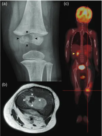
An 18-month-old boy was referred to our tertiary institution with a 2-month history of a left-sided intermittent painful limp. He had been living on Mayotte, an island located in the Indian Ocean, where he had been vaccinated with Bacillus Calmette-Guérin at birth. Physical examination showed no fever, mild tenderness and swelling of the left knee, slightly limited in range of motion. Routine laboratory tests were within normal ranges. Plain radiographs showed a lytic lesion in the epiphysis of the distal femur (Fig. 1a) with no chest abnormality. Magnetic resonance imaging confirmed a large empty signal area within the epiphyseal ossification centre with no transphyseal spread (Fig. 1b). Suspecting a primary subacute epiphyseal osteomyelitis,1open surgical drainage and
curettage of the osseous lesion were performed. Given the patient’s age, bone specimens were inoculated in Bactec PED Plus (Becton Dickinson, Sparks, NV, USA) culture vials to
Fig. 1 (a) Plain anteroposterior radiograph of the left knee shows an oste-olytic lesion (black arrowheads) at the medial condyle of femoral epiphysis. (b) Axial T1-weighted magnetic resonance image of the left knee shows a large empty signal area in the epiphysis (white arrowheads) surrounded by oedema, and enlarged popliteal nodes. (c) Whole-body 18F-fluorodeoxy-D -glucose (FDG)-hybrid positron emission tomography–computed tomogra-phy shows an increased accumulation of FDG in the left distal femoral epiphysis (red target lines) and right adrenal gland area (yellow arrowhead).
Journal of Paediatrics and Child Health53 (2017) 724–728
© 2017 Paediatrics and Child Health Division (The Royal Australasian College of Physicians)
727 Letters to the Editor
search forKingella kingae in addition to routine microbiological cultures.2 Broad-spectrum polymerase chain reaction (PCR)
targeting the universal 16S rRNA gene and real-time PCR tar-geting the cpn60 gene of K. kingae were also performed from fresh bone materials. As Mayotte is an endemic tuberculosis area, the search for mycobacteria was done by streaking bone specimens onto specific medium and by using real-time PCR assays targeting Mycobacterium species.3 Of these testings, the
real-time PCR assay targeting M. tuberculosis was positive. His-tologically, lesion specimens showed a non-specific inflamma-tory granuloma. The tuberculin skin test and Interferon-gamma release assay were not contributive. Human immuno-deficiency virus antibody testing was negative. A whole-body 18F-fluorodeoxy-D-glucose-hybrid positron emission tomography–computed tomography revealed a disseminated mycobacterial disease spread to the left popliteal, inguinal lymph nodes and right adrenal gland area (Fig. 1c). Subse-quently, the patient progressively improved with rifampicin, isoniazid, pyrazinamide and ethambutol for 3 months, followed by rifampicin and isoniazid for 9 months. Three years later, complete healing of the tuberculous lesions with no growth disturbance was observed.
Childhood tuberculosis is a growing public health issue world-wide.4The current shifting in population migration led to the
re-emergence of uncommon forms of tuberculosis that are poorly recognised in toddlers, especially in low tuberculosis bur-den areas whereK. kingae is the leading cause of osteoarticular infections in children aged 6–36 months.2,3 An increase of
awareness of clinicians and the use of appropriate imaging and molecular tools are required to establish the diagnosis of tuber-culosis at an early stage and avoid long-term complications.
Informed consent was obtained from the child’s parents who authorised us to report the case.
Dr Nawal El Houmami1,2
Dr Samuel Vernaz3
Dr Philippe Minodier4
Professor Jean-Luc Jouve2
Professor Didier Raoult1
Professor Pierre-Edouard Fournier1 1Aix-Marseille University, Research Unit on Emerging and Infectious
Diseases (URMITE), UMR 63, INSERM 1095, CNRS 7278, IRD 198, IHU Mediterranean Infection, Public Hospitals of Marseille
2Department of Pediatric Orthopedics
La Timone Children’s Hospital
4Department of Pediatric Emergency Medicine
University North Hospital Marseille and3Department of Pediatrics
Mamoudzou Hospital Mayotte France Conflict of interest: None declared.
References
1 Yoo WJ, Choi IH, Yun YH et al. Primary epiphyseal osteomyelitis caused by mycobacterium species in otherwise healthy toddlers. J. Bone Joint Surg. Am. 2014;96: e145.
2 Yagupsky P. Kingella kingae: From medical rarity to an emerging pae-diatric pathogen. Lancet Infect. Dis. 2004;4: 358–67.
3 El Houmami N, Minodier P, Bouvier C et al. Primary subacute epiphy-seal osteomyelitis caused by Mycobacterium species in young chil-dren: A modern diagnostic approach. Eur. J. Clin. Microbiol. Infect. Dis. 2017;36: 771–7.
4 Dodd PJ, Gardiner E, Coghlan R, Seddon JA. Burden of childhood tuber-culosis in 22 high-burden countries: A mathematical modelling study. Lancet Glob. Health 2014;2: e453–9.
728 Journal of Paediatrics and Child Health53 (2017) 724–728
© 2017 Paediatrics and Child Health Division (The Royal Australasian College of Physicians)