HAL Id: hal-01455011
https://hal.archives-ouvertes.fr/hal-01455011
Submitted on 3 Dec 2019
HAL is a multi-disciplinary open access
archive for the deposit and dissemination of
sci-entific research documents, whether they are
pub-lished or not. The documents may come from
teaching and research institutions in France or
abroad, or from public or private research centers.
L’archive ouverte pluridisciplinaire HAL, est
destinée au dépôt et à la diffusion de documents
scientifiques de niveau recherche, publiés ou non,
émanant des établissements d’enseignement et de
recherche français ou étrangers, des laboratoires
publics ou privés.
Distributed under a Creative Commons Attribution| 4.0 International License
Biologic Ionic Selectivity
Khaoula Boukari, Guillaume Paris, Tijani Gharbi, Sébastien Balme,
Jean-Marc Janot, Fabien Picaud
To cite this version:
Khaoula Boukari, Guillaume Paris, Tijani Gharbi, Sébastien Balme, Jean-Marc Janot, et al.. Confined
Nystatin Polyenes in Nanopore Induce Biologic Ionic Selectivity. Journal of Nanomaterials, Hindawi
Publishing Corporation, 2016, 2016, 2671383 (9 p.). �10.1155/2016/2671383�. �hal-01455011�
Research Article
Confined Nystatin Polyenes in Nanopore Induce
Biologic Ionic Selectivity
Khaoula Boukari,
1,2Guillaume Paris,
1Tijani Gharbi,
1Sébastien Balme,
3Jean-Marc Janot,
3and Fabien Picaud
11Laboratoire de Nanom´edecine, Imagerie et Th´erapeutique, EA 4662, Universit´e de Bourgogne-Franche-Comt´e,
Centre Hospitalier Universitaire de Besanc¸on, 16 route de Gray, 25030 Besanc¸on Cedex, France
2CINaM, CNRS UMR 7325, Aix-Marseille University, Campus de Luminy, 13288 Marseille Cedex 9, France
3Institut Europ´een des Membranes, ENSCM, CNRS UMR 5635, Universit´e de Montpellier, Place Eug`ene Bataillon,
34095 Montpellier Cedex 5, France
Correspondence should be addressed to Fabien Picaud; [email protected] Received 29 March 2016; Accepted 22 May 2016
Academic Editor: David Cornu
Copyright © 2016 Khaoula Boukari et al. This is an open access article distributed under the Creative Commons Attribution License, which permits unrestricted use, distribution, and reproduction in any medium, provided the original work is properly cited.
Antifungal polyenes such as nystatin (or amphotericin B) molecules play an important role in regulating ions permeability through membrane cell. The creation of self-assembled nanopores into the fungal lipid membranes permits the leakage and the selectivity of ions (i.e., blockage of divalent cations) that cause the cell death. These abilities are thus of first interest to promote new biomimetic membranes with improved ionic properties. In the present work, we will use molecular dynamic simulations to interpret recent experimental data that showed the transfer of the nystatin action inside artificial nanopore in terms of ion permeability and selectivity. We will demonstrate that nystatin polyenes can be stabilized in a hydrophobic carbon nanotube, even at high concentration. The high potential interaction between the polyenes and the hydrophobic pore wall ensures the apparition of a hole inside the biomimetic nanopore that changes its intrinsic properties. The probability ratios of cation versus anion show interesting reproducibility of experimental measurements and, to a certain extent, opened the way for transferring biological properties in synthetic membranes.
1. Introduction
Nystatin (NYS) is a membrane-active polyene produced by
Streptomyces noursei strains [1]. These antifungal molecules
are amphiphilic [2, 3]. They are composed of a polar head, a hydrophilic chain, and a hydrophobic chain. This amphiphilic character leads to their self-assembly in biological mem-branes. Indeed, polyenes can incorporate inside the lipid membrane and form a barrel [4] where all hydrophobic chains are turned on the external part of the barrel and face the lipids, while the hydrophilic chains form the inner part of the channel [3, 5]. This configuration allows the barrel inter-calation inside the lipid membrane (depending on the sterol molecules [6]) and plays an important role in the ion perme-ability and selectivity (especially to monovalent ions [2, 5]). The mechanism responsible for the barrel formation is still
under discussion but may follow the diagram illustrated in Figure 1. However, its ionic properties are involved in the antifungal action of the polyenes [7, 8].
Solid state nanopores appear now as excellent tools to mimic biological channel properties because of their com-bined well defined geometries (shape and dimensions) and mechanical robustness. But controlling the inner nanopore diameter is still very challenging since it necessitates con-trolling diameter range from subnanometers’ to hundreds of nanometers’ scale, depending on the applications. As already mentioned, solid state nanopores do not exhibit selectivity or activity without functionalization. Both the stability and the property of the solid state nanopores are highly dependent on the experimental conditions of formation (e.g., track etched nanopore) as well as used material. For several years, some applications were developed using solid state nanopores such Hindawi Publishing Corporation
Journal of Nanomaterials
Volume 2016, Article ID 2671383, 9 pages http://dx.doi.org/10.1155/2016/2671383
Nystatin molecule Phospholipid molecule
OH group
Amphiphilic domain
Polar head Polar head
Hydrophobic carbon domain
Figure 1: Schematic formation of the nystatin pore inside lipid membrane.
as DNA sensing [9], control of the molecular transport, or fabrication of performing nanofluidic devices [10–13]. Some precise chemical treatments allow now controlling the diam-eter range of nanopores [14, 15] and the properties [16]. How-ever, if we want to combine the high selectivity of biological channel and the robustness of solid state nanopore, one route is the development of hybrid biological/solid state nanopores.
This proof of concept was done with𝛼-hemolysin for DNA
sensing application [14, 17] and with gramicidin A for higher ionic permeability and selectivity devices.
The transfer of the biological properties of polyene to artificial membranes which could thus exhibit the same behavior as in the cell could be envisaged but still remains an important challenge. This transfer could find applications in water desalination [18] or dialysis [19, 20] with low energy cost. Recently, Balme et al. demonstrated experimentally that insertion of NYS polyenes inside solid state nanopore leads to very interesting ionic selective properties. We will try here to understand using molecular dynamic simulations the forma-tion and the properties of such experimental systems [20, 21]. After a short description of the simulation method, we will present the progressive filling of the nanopore and the conse-quence to the properties of the polyenes in terms of geometry and energy. Then, we will analyze conductance of the hybrid nanopore and the role of the dipole moment, created after the polyenes incorporation in the ionic selectivity.
2. Computational Method and Model
The model for nystatin (NYS) was carried out using the 3D structure of the DB00681 model in the DrugBank database (Figure 2). To obtain the missing potential parameters, we have followed the protocol described by Norrby and Brandt [22] by constructing the Hessian matrix for further use in the force field parametrization. This matrix was obtained via ab
initio quantum calculations using Gaussian 09 package
soft-ware [23]. In order to check the stability of polyene, nystatin was equilibrated for 2 nS in a water box of 40× 30 × 30 ˚A3 (Figure 2). Then, NYS was transferred into a solid nanopore modeled by a carbon nanotube and denoted by CNT. Its diameter was chosen to be sufficient to be compared with experiments [20, 21], that is, 7 nm, while its length was equal to 5 nm. The nanopore was filled progressively by 50 polyenes, water, and K+and Cl−ions at 0.1 M concentration. The final
system contained 43702 atoms in a periodic box of 80× 75 ×
80 ˚A3with 10914 water molecules and 28 KCl ions in order to conserve the neutrality of the system.
Molecular Dynamic (MD) simulations and minimization were performed using NAMD 2.7b2 package [24]. A tem-perature of 300 K (Langevin dynamics) and a pressure of 1 atm (Langevin piston) of the system were kept constant during simulation. The long range electrostatic forces were evaluated by using the classical particle mesh Ewald (PME)
Journal of Nanomaterials 3
Figure 2: Structure of nystatin polyene.
method with a grid spacing of 1.2 and a fourth-order spline interpolation. The integration time step was equal to 1 fs.
To calculate the conductance in the hybrid membrane, electric field was imposed to the equilibrated system. The constant increase of the electric field value assures a voltage ramp which allows deducting the resulting ionic current. To do this, 25 nS of MD simulations was performed for each electric field. The analyses of the ionic displacements inside the hybrid nanopore were carefully done in order to calculate the ionic current [25] using
𝐼 (𝑡) = 𝐿1 𝑦Δ𝑡 𝑁 ∑ 𝑖=1 𝑞𝑖(𝑦𝑖(𝑡 + Δ𝑡) − 𝑦𝑖(𝑡)) , (1)
whereΔ𝑡 represents the time between each frame, 𝐿𝑦is the
pore length (5 nm), and𝑦𝑖is the absolute position of the ion with charge𝑞𝑖along the pore axis.
3. Results and Discussion
In order to create the hybrid nanopore, CNT was pro-gressively filled with the nystatin molecules. To do this, ten NYS molecules were incorporated in the center of the nanopore and stabilized by MD simulations. Then, once completely relaxed, ten other polyenes were added and so on, until 50 NYS molecules. For all systems, we observed a total aggregation of the molecules around the inner surface of the nanotube. This behavior was already reported for gramicidin [26] and amphotericin [20] molecules. The strong interaction of the hydrophobic chain of the polyenes with the hydrophobic CNT wall is at the origin of this behavior. It allows the polyenes recovering at best similar biological conditions. The control of the stability of each progressive filling was checked by the Root Mean Square Deviation (RMSD) of the NYS structure. Ten other NYS molecules were added once the RMSD value becomes roughly constant.
Figure 3 presents the different RMSD superimposed as a function of the time simulation. The results show that after 120 nS of simulation time the whole system is perfectly stabilized. The deformation of the NYS skeleton is limited due to its confinement in the solid nanopore.
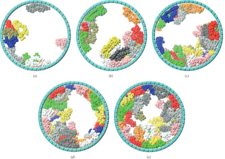
The different systems, modeled with 10, 20, 30, 40, and 50 nystatin molecules, are reported in Figure 4. As clearly shown
0 2 4 6 8 10 12 RMS D (Å) 100 200 300 400 0 t (ns) 10 NYS 20 NYS 30 NYS 40 NYS 50 NYS
Figure 3: RMSD per added polyene for the progressive filling of the nanopore by the 50 nystatin molecules. For each concentration, the total RMSD was divided by 10 in order to calculate it as a function of the added NYS.
in these simulations snapshots, the polyenes tend to rapidly reach the pore wall and adsorb on it in order to optimize their interactions. After reaching a minimal concentration (40 NYS molecules were necessary to fully recover the pore wall), nystatin molecules were completely organized around the inner surface of the tube and a channel was formed in the tube center (see Figures 4(d) and 4(e) after insertion of 40 and 50 NYS molecules). It should be noted that the first 30 polyenes were more structurally perturbed (higher RMSD per NYS) than the following 20 ones, due to their adsorption on the CNT wall. Beyond 40 NYS molecules, the RMSD shows a slower increase since the latter NYS molecules are surrounded by other polyenes instead of CNT rigid wall. The snapshots showing the successive molecular arrangements during the simulation demonstrate the formation of a channel in the nanopore that could allow the ion diffusion. The approximate size of the different channels could be estimated using geometric arguments. To do this, we have divided the length of the pore in small portion of length 0.1 nm. The minimal distance between each atom of faced NYS was then calculated to determine the diameter of the pore for the last two systems. The minimum value obtained, when 40 or 50 NYS molecules were confined, oscillated between 1.43 nm and 0.79 nm. These values are significantly smaller than those of the CNT nanopore one (7 nm). The important decrease of the pore size, due to the presence of NYS molecules, could naturally affect the ions behavior through the application of an electric field. However, while confined in conditions far from biologic ones, the NYS molecules self-organize in the CNT to create a big channel. This behavior is similar in the fungal cell and allows the leakage of ions. The analysis of the different energy contributions is necessary to really understand the role of each type of molecules in the NYS behavior.
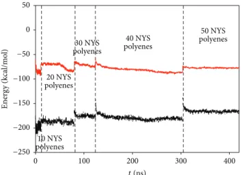
Figure 5 shows the van der Waals and electrostatic interactions per NYS molecule as a function of the simulation time. As shown on this figure, the total energy of the system
(a) (b) (c)
(d) (e)
Figure 4: Snapshots of the simulation at (a) 30 nS (10 nystatin molecules), (b) 81 nS (20 nystatin molecules), (c) 129 nS (30 nystatin molecules), (d) 310 nS (40 nystatin molecules), and (e) 420 nS (50 nystatin molecules). Carbon nanotube and nystatin polyenes are presented by cyan and different colors, respectively.
decreases until 20 encapsulated NYS molecules (from−273
to −276 kcal/mol). Then, the total energy per molecule
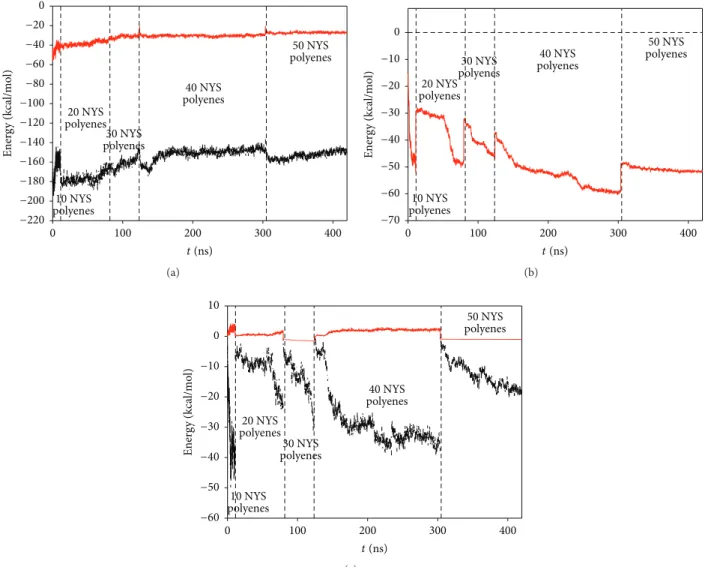
increases slowly when the 20 following polyenes fill the CNT (−270 kcal/mol). At the final stage of the simulations (50 NYS molecules), the value of the energy is clearly higher (−242 kcal/mol), indicating a strong energy loss per molecule due to the 50 NYS molecules intense confinement. In order to explain this, we have separated in Figure 6 the different energy contributions responsible for the stability of the NYS molecules inside the nanopore. From the results, two main reasons can be given to explain the energy loss. First, the progressive insertion of polyenes inside the pore expels the water molecules in the reservoirs. This leads to the increase of the water/polyene energy contribution. Second, the completion of CNT inner surface was reached for 30 inserted molecules. The following ones can only interact with NYS wall which leads also to the slow increase of the CNT-NYS energy contribution. Nevertheless, the biomimetic system can be considered as stable since the lateral interaction between each NYS molecule should compensate the energy variation observed in the last system.
In order to confirm this, we have evaluated the average number of hydrogen bonds (Figure 7) formed between all
polyenes during the last stage of the simulations (from𝑡 =
340 nS to 𝑡 = 420 nS). A hydrogen bond was taken into account when the donor/acceptor distance was lower than 0.33 nm and the donor/hydrogen/acceptor angle was below
90∘. The H-bond average was computed as the total number
of H-bonds created in the system between NYS molecules during the simulation and divided by the frame number. An average of 14 H-bonds was found when NYS molecules were enclosed into the nanopore. This leads to an important cohesion of the molecules where they are confined and can be compared to value obtained for amphotericin when embedded inside membrane cell [6] (almost 10 H-bonds with sterol molecules). Of course, the number of H-bonds evolves during the simulation, starting from some unities at the beginning of the filling to 14 near the completion of the nanopore (50 confined NYS polyenes). However, we can conclude that, progressively, the formation of this hydrogen bond lattice stabilizes the polyenes inside the hydrophobic nanopore and compensates the energy loss of the polyene with the other external components of the system.
Journal of Nanomaterials 5 −250 −200 −150 −100 −50 0 50 Ener g y (k cal/mo l) 100 200 300 400 0 t (ns) 50 NYS polyenes 40 NYS polyenes 30 NYS polyenes 20 NYS polyenes 10 NYS polyenes
Figure 5: Total interaction energy of the polyenes with their neighbors (per nystatin polyene) as a function of time simulation (black curve for electrostatic interaction, red curve for van der Waals interaction).
In biological membrane NYS channel is well known to be permeable only to both monovalent cation and anion. This property should be studied in CNT in order to elucidate the role of the NYS confinement and self-assembly in the solid state nanopore on the permeation properties. To this end,
membrane conductance𝐺memwas determined through the
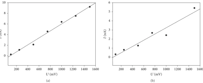
current/voltage curve as a function of the NYS concentration. Figure 8 shows the different currents as a function of voltage obtained with the two last biomimetic systems (with 40 and 50 NYS molecules) at 0.1 M of KCl.
As shown in Figure 8,𝐼 = 𝑓(𝑉) points are almost aligned. The linear regression of the curves can thus be used to determine the conductance for these two systems. When NYS occupied the inner part of the nanopore, the conductance was equal to 6.56 nS. In the second case, where 50 NYS molecules filled the CNT, the conductance decreased to 3.44 nS. It should be noticed that the conductance calculated for the bare nanopore leads to a value of 168 nS. This conductance decrease can be directly assigned to the NYS confinement inside the membrane pore. Indeed, the inner diameter of the nanopore is first changed due to the polyene presence as well as the nature of the molecules that conducted ions (CNT ver-sus NYS). When bare nanopore is considered, hydrophobic carbon wall limits the ions diffusion through the pore. On the other cases, ions face NYS polyenes when they diffuse. The consequent change of the membrane conductance is the combination of these two factors. Besides, by calculating the ratio between the conductance for 40 and that for 50 confined NYS polyenes, we retrieve almost the ratio of the channel radii. Note that we did not perform conductance calculations for different salts or different salt concentrations. However, the strong decrease of the membrane conductance in presence of confined polyenes is a sufficient proof of the functional behavior of our biomimetic nanopore. In addition, the recent experiments conducted on functional nanopore for the same concentration of NaCl or KCl did not show strong conductance differences between the two salts [20, 21]. While
the morphology of the organized polyenes inside the solid state nanopore was completely different to that observed in biologic lipid membrane, the resulting conductance behaves similarly with no difference between monovalent salts. This corroborates the partial biologic behavior of our func-tional nanopores. For divalent cations, conduction was also observed to a less extent while it was currently admitted that polyenes blocked the passage of divalent cations through a fungal cell. This can be due to the geometry adopted by the polyenes in CNT, which is far from its geometry in lipid cell. In order to estimate the selectivity of the biomimetic nanopore demonstrated in [20, 21], we analyzed the dielectric dipole of the confined molecules as a function of time, omitting ions that could be present inside the nanopore during the calculations. This should help us to determine the probability of ion passage through the nanopore and, then, compare it to the permeation ratio measured in experiments. The evolution of the dipole moment for two biomimetic systems (40 and 50 NYS molecules) shows important vari-ations in the radial plane of the CNT. However, contrary to confined amphotericin B in similar CNT [20, 21], one of these components does not vanish during the simulation as shown in Figure 9. This can be interpreted as coming from the specific NYS organization inside the nanopore. While amphotericin B molecules were mainly organized homo-geneously around the nanopore, nystatin polyenes create a channel which is quite twisted. This geometry can thus not compensate every component of the radial dipolar moment. The (𝑥, 𝑧) radial dipolar moment components vary around −6 ± 30 D, −60 ± 30 D (40 NYS molecules), and −4 ± 24 D, −50 ± 20 D (50 NYS molecules), respectively. The component parallel to the nanopore axis fluctuates around a positive
value of60±30 D and 40±30 D, respectively (see red arrow in
Figure 9). This imposes an important orientation of the total dipolar moment along the nanopore axis (similar to a spin) which should impose an ionic selectivity along the pore.
The interaction between the dipolar moment along the pore axis and ion (cation or anion) inside the CNT nanopore can be calculated supposing that this ion, schematized by its own van der Waals radius, is in front of the CNT center. Following a Boltzmann distribution the probability ratio of finding K+ and Ca2+ versus Cl− ions and finally K+ versus Na+ ions can be plotted as a function of time (Figures 10(a) and 10(b) for 40 and 50 confined NYS polyenes, resp.). From these results, it appears clearly that the probability of finding chlorine ions inside the channel is always higher compared to cations. This is due to a more favorable interaction with the dipolar moment imposed by the confined NYS polyenes. The divalent cations are poorly encouraged to diffuse through the NYS channel due to their higher positive charges (Figure 10, red lines). The value of𝑃K/𝑃Naratio (green curves) cannot be explained by the charge of the cations but only by the van der Waals radius between the two cations which involves a higher affinity to the membrane for potassium ions.
The change of the dipolar moment value between the two CNT fillings (40 and 50 NYS molecules) leads to low variation in the probability ratio.𝑃K/𝑃Na ratio was modified
−220 −200 −180 −160 −140 −120 −100 −80 −60 −40 −20 0 Ener g y (k cal/mo l) 100 200 300 400 0 t (ns) 50 NYS polyenes 40 NYS polyenes 30 NYS polyenes 20 NYS polyenes 10 NYS polyenes (a) −70 −60 −50 −40 −30 −20 −10 0 Ener g y (k cal/mo l) 100 200 300 400 0 t (ns) 50 NYS polyenes 40 NYS polyenes 30 NYS polyenes 20 NYS polyenes 10 NYS polyenes (b) −60 −50 −40 −30 −20 −10 0 10 Ener g y (k cal/mo l) 100 200 300 400 0 t (ns) 50 NYS polyenes 40 NYS polyenes 30 NYS polyenes 20 NYS polyenes 10 NYS polyenes (c)
Figure 6: Energy contribution per nystatin polyene as a function of time simulation for (a) water molecules-NYS and (b) carbon atoms of the nanopore-NYS and (c) ions-NYS interactions.
0 10 20 30 40 % o f f ra m e 12 13 14 15 16 11 H-bond number
Figure 7: Average intermolecular hydrogen bonds number between all confined nystatin polyenes (solid line). The dashed line is an approximated Gaussian fitted curve of the simulation one.
to 0.01. The most important variation was observed for
𝑃K/𝑃Cl which increased from 0.12 to 0.3. The biomimetic
channels are thus extremely sensitive to monovalent ions and tend to promote anion diffusion compared to cation one. Nevertheless, the biologic properties observed in lipid
membrane were not fully retrieved. Indeed, 𝑃Ca/𝑃Cl ratio
can reach 0.01 which means that almost 1% of divalent cation could pass through the functional nanopore while 0% are awaited. The polyene disorder observed along the solid nanopore (fully different from the self-assembled channel observed in lipid membrane) and the lack of sterol molecules could explain the small differences observed between our biomimetic systems and the living cells.
However, our simulations agree well with the experiments recently published [20, 21]. In these papers, Balme et al. demonstrated that it was possible to incorporate polyenes (amphotericin and nystatin) inside polycarbonate nanopore membrane with interesting ionic selective properties. While
Journal of Nanomaterials 7 0 2 4 6 8 10 I (nA) 400 600 800 1000 1200 1400 1600 200 U (mV) (a) 0 1 2 3 4 5 6 I (nA) 800 600 400 1000 1200 1400 1600 200 U (mV) (b)
Figure 8:𝐼 = 𝑓(𝑉) curves obtained for systems with (a) 40 confined NYS polyenes and (b) 50 confined NYS polyenes. The biomimetic membrane conductance was equal to (a) 6.56 nS and (b) 3.44 nS, respectively.
(a) (b)
Figure 9: Dipolar moment of nystatin inside the nanopore for (a) 40 confined NYS polyenes and (b) 50 confined NYS polyenes. The red arrow presented the total dipolar moment. Carbon nanotube and nystatin polyenes are presented by cyan and different colors, respectively.
complete divalent cation selectivity was not retrieved (as in our simulations), these experiments showed that anions could cross the biomimetic membrane compared to cations. At salt concentration 0.1 M, the probability ratios 𝑃cat/𝑃Cl obtained are 0.3 and 0.5 for Na+ and K+, respectively, and 0.03 for divalent Ca2+. 𝑃K/𝑃Na ratio was found to be equal to 1.25. Compared to our simulated systems, it appears that the experimental biomimetic channel is closer to the system simulated with 40 confined NYS polyenes, which corresponds to a channel of diameter 1.43 nm. Indeed, in this case, the radius of the channel is larger and more symmetric than in the case where 50 NYS molecules were confined. This induced a more important directive dipolar moment that could help to increase selectivity.
4. Conclusion
Thanks to molecular dynamics simulations, we have con-ducted a theoretical study of the confinement of nystatin inside a carbon nanotube of 7 nm diameter. 50 NYS polyenes can fill the inner part of tube forming a hole in the center. The number of confined polyenes strongly influences the conduc-tance of the biomimetic nanopore due to the modification of the ionic diffusing path. The resulting dipolar moment influ-ences also the selectivity of the biomimetic membrane, with a strong preference for the anionic permeation compared to cationic one while divalent cations are almost blocked at the entry of the pore. Moreover, we have demonstrated here that the creation of the biomimetic channel reproduced selectivity
10 20 30 40 50 60 0 t (ns) 10 20 30 40 50 60 0 t (ns) 10 20 30 40 50 60 0 t (ns) PK + /PCl − 0.00 0.05 0.10 0.15 0.20 0.000 0.002 0.004 0.006 0.008 0.010 PCa 2+ /PCl − 1.05 1.10 1.15 1.20 1.25 1.30 PK + /PNa + (a) 20 40 60 80 100 120 140 160 0 t (ns) 20 40 60 80 100 120 140 160 0 t (ns) 20 40 60 80 100 120 140 160 0 t (ns) PK + /PCl − 0.0 0.1 0.2 0.3 0.4 0.5 0.00 0.05 0.10 0.15 0.20 PCa 2+ /PCl − 1.06 1.08 1.10 1.12 1.14 1.16 1.18 1.20 PK + /PNa + (b)
Figure 10: Ratio of the thermal equilibrium probability of K+(black line) and Ca2+(red line) over Cl−and of K+over Na+(green line), as a function of time for (a) 40 confined NYS polyenes and (b) 50 confined NYS polyenes.
ratios close to those demonstrated experimentally [20, 21]. The functional nanopore exhibits interesting ionic properties that mimic partially the biologic behavior of NYS polyenes inside biological membrane. The organization of the polyenes
inside the solid state nanopore is found to be completely different from that in lipid membrane. However, we succeed in modifying the solid state nanopore properties according to the confined molecules one.
Journal of Nanomaterials 9
Competing Interests
The authors declare that there are no competing interests regarding the publication of this paper.
Acknowledgments
Computations have been performed on the supercomputer facilities of the M´esocentre de calcul de Franche-Comt´e. This work was supported by the French Research Program ANR-BLANC, Project TRANSION (ANR-2012-BS08-0023).
References
[1] T. Brautaset, O. N. Sekurova, H. Sletta et al., “Biosynthesis of the polyene antifungal antibiotic nystatin in Streptomyces noursei ATCC 11455: analysis of the gene cluster and deduction of the biosynthetic pathway,” Chemistry & Biology, vol. 7, no. 6, pp. 395–403, 2000.
[2] N. Akaike and N. Harata, “Nystatin perforated patch recording and its applications to analyses of intracellular mechanisms,”
Japanese Journal of Physiology, vol. 44, no. 5, pp. 433–473, 1994.
[3] A. Cass, A. Finkelstein, and V. Krespi, “The ion permeability induced in thin lipid membranes by the polyene antibiotics nystatin and amphotericin B,” The Journal of General Physiology, vol. 56, no. 1, pp. 100–124, 1970.
[4] J. Czub, A. Neumann, E. Borowski, and M. Baginski, “Influence of a lipid bilayer on the conformational behavior of ampho-tericin B derivatives—a molecular dynamics study,” Biophysical
Chemistry, vol. 141, no. 1, pp. 105–116, 2009.
[5] M. P. Borisova, R. A. Brutyan, and L. N. Ermishkin, “Mecha-nism of anion-cation selectivity of amphotericin B channels,”
The Journal of Membrane Biology, vol. 90, no. 1, pp. 13–20, 1986.
[6] K. Boukari, S. Balme, J. Janot, and F. Picaud, “Towards new insights in the sterol/amphotericin nanochannels formation: a molecular dynamic simulation study,” The Journal of Membrane
Biology, vol. 249, no. 3, pp. 261–270, 2016.
[7] J. Bolard, “How do the polyene macrolide antibiotics affect the cellular membrane properties?” Biochimica et Biophysica Acta
(BBA)—Reviews on Biomembranes, vol. 864, no. 3-4, pp. 257–
304, 1986.
[8] J. Bolard, M. Seigneuret, and G. Boudet, “Interaction between phospholipid bilayer membranes and the polyene antibiotic amphotericin B. Lipid state and cholesterol content depen-dence,” Biochimica et Biophysica Acta, vol. 599, no. 1, pp. 280– 293, 1980.
[9] G. F. Schneider and C. Dekker, “DNA sequencing with nano-pores,” Nature Biotechnology, vol. 30, no. 4, pp. 326–328, 2012. [10] C. Y. Lee, W. Choi, J.-H. Han, and M. S. Strano, “Coherence
resonance in a single-walled carbon nanotube ion channel,”
Science, vol. 329, no. 5997, pp. 1320–1324, 2010.
[11] P. Pang, J. He, J. H. Park, P. S. Krsti´c, and S. Lindsay, “Origin of giant ionic currents in carbon nanotube channels,” ACS Nano, vol. 5, no. 9, pp. 7277–7283, 2011.
[12] A. Plecis, R. B. Schoch, and P. Renaud, “Ionic transport pheno-mena in nanofluidics: experimental and theoretical study of the exclusion-enrichment effect on a chip,” Nano Letters, vol. 5, no. 6, pp. 1147–1155, 2005.
[13] R. B. Schoch, J. Han, and P. Renaud, “Transport phenomena in nanofluidics,” Reviews of Modern Physics, vol. 80, no. 3, pp. 839– 883, 2008.
[14] S. Cabello-Aguilar, S. Balme, A. A. Chaaya et al., “Slow trans-location of polynucleotides and their discrimination by 𝛼-hemolysin inside a single track-etched nanopore designed by atomic layer deposition,” Nanoscale, vol. 5, no. 20, pp. 9582– 9586, 2013.
[15] C. Marichy, M. Bechelany, and N. Pinna, “Atomic layer deposi-tion of nanostructured materials for energy and environmental applications,” Advanced Materials, vol. 24, no. 8, pp. 1017–1032, 2012.
[16] M. Lepoitevin, G. Nguyen, M. Bechelany, E. Balanzat, J.-M. Janot, and S. Balme, “Combining a sensor and a pH-gated nano-pore based on an avidin-biotin system,” Chemical
Communica-tions, vol. 51, no. 27, pp. 5994–5997, 2015.
[17] A. R. Hall, A. Scott, D. Rotem, K. K. Mehta, H. Bayley, and C. Dekker, “Hybrid pore formation by directed insertion of 𝛼-haemolysin into solid-state nanopores,” Nature Nanotechnology, vol. 5, no. 12, pp. 874–877, 2010.
[18] M. T. Myint, A. Ghassem, and N. Nirmalakhandan, “Low energy/cost desalination: Low dose and low mean ion resident time in concentrate stream of electro-dialysis reversal,” Water
Science and Technology, vol. 63, no. 9, pp. 1855–1863, 2011.
[19] J. Agar, “Solar-assisted haemodialysis,” Clinical Journal of the
American Society of Nephrology, vol. 7, no. 2, pp. 310–314, 2012.
[20] D. Thiele, S. Kraszewski, S. Balme, F. Picaud, J.-M. Janot, and P. D´ejardin, “Structure and ionic selectivity of a hybrid polyene/ artificial polymer solid state membrane,” Soft Matter, vol. 9, no. 3, pp. 684–691, 2013.
[21] S. Balme, D. Thiele, S. Kraszewski, F. Picaud, J.-M. Janot, and P. D´ejardin, “Ionic selectivity of nystatin A1 confined in nano-porous track-etched polymer membrane,” IET
Nanobiotechnol-ogy, vol. 8, no. 3, pp. 138–142, 2014.
[22] P.-O. Norrby and P. Brandt, “Deriving force field parameters for coordination complexes,” Coordination Chemistry Reviews, vol. 212, no. 1, pp. 79–109, 2001.
[23] M. J. Frisch, G. W. Trucks, H. B. Schlegel et al., Gaussian 09, Gaussian, Wallingford, UK, 2009.
[24] J. C. Phillips, R. Braun, W. Wang et al., “Scalable molecular dynamics with NAMD,” Journal of Computational Chemistry, vol. 26, no. 16, pp. 1781–1802, 2005.
[25] J. M. Xue, X. Q. Zou, Y. B. Xie, and Y. G. Wang, “Molecular dynamics simulations on the ionic current through charged nanopores,” Journal of Physics D: Applied Physics, vol. 42, no. 10, Article ID 105308, 2009.
[26] S. Balme, J.-M. Janot, L. Berardo et al., “New bioinspired membrane made of a biological ion channel confined into the cylindrical nanopore of a solid-state polymer,” Nano Letters, vol. 11, no. 2, pp. 712–716, 2011.
Submit your manuscripts at
http://www.hindawi.com
Scientifica
Hindawi Publishing Corporation
http://www.hindawi.com Volume 2014
Hindawi Publishing Corporation
http://www.hindawi.com Volume 2014
Ceramics
Journal ofNanoparticles
Journal of Hindawi Publishing Corporationhttp://www.hindawi.com Volume 2014
Hindawi Publishing Corporation
http://www.hindawi.com Volume 2014
International Journal of
Biomaterials
Hindawi Publishing Corporation
http://www.hindawi.com Volume 2014
Nanoscience
Journal ofTextiles
Hindawi Publishing Corporation
http://www.hindawi.com Volume 2014
Journal of
Crystallography
Journal of Hindawi Publishing Corporationhttp://www.hindawi.com Volume 2014
The Scientific
World Journal
Hindawi Publishing Corporation
http://www.hindawi.com Volume 2014
Hindawi Publishing Corporation
http://www.hindawi.com Volume 2014
Coatings
Journal ofAdvances in
Materials Science and Engineering Hindawi Publishing Corporation
http://www.hindawi.com Volume 2014
Hindawi Publishing Corporation
http://www.hindawi.com Volume 2014
Metallurgy
Journal ofHindawi Publishing Corporation
http://www.hindawi.com Volume 2014
BioMed
Research International
Materials
Journal of Hindawi Publishing Corporationhttp://www.hindawi.com Volume 2014
N
a
no
ma
te
ria
ls
Hindawi Publishing Corporation
http://www.hindawi.com Volume 2014
Journal of