HAL Id: cea-02454709
https://hal-cea.archives-ouvertes.fr/cea-02454709
Submitted on 24 Jan 2020HAL is a multi-disciplinary open access
archive for the deposit and dissemination of sci-entific research documents, whether they are pub-lished or not. The documents may come from teaching and research institutions in France or abroad, or from public or private research centers.
L’archive ouverte pluridisciplinaire HAL, est destinée au dépôt et à la diffusion de documents scientifiques de niveau recherche, publiés ou non, émanant des établissements d’enseignement et de recherche français ou étrangers, des laboratoires publics ou privés.
Tailoring Structure and Surface Chemistry of Hollow
Allophane Nanospheres for Optimization of Aggregation
by Facile Methyl Modification
Peixin Du, Antoine Thill, Peng Yuan, Shun Wang, Dong Liu, Frédéric
Gobeaux, Liangliang Deng, Yaran Song
To cite this version:
Peixin Du, Antoine Thill, Peng Yuan, Shun Wang, Dong Liu, et al.. Tailoring Structure and Surface Chemistry of Hollow Allophane Nanospheres for Optimization of Aggregation by Facile Methyl Mod-ification. Applied Surface Science, Elsevier, 2020, 510, pp.145453. �10.1016/j.apsusc.2020.145453�. �cea-02454709�
Tailoring Structure and Surface Chemistry of Hollow Allophane Nanospheres for Optimization of 1
Aggregation by Facile Methyl Modification 2
3
Peixin Du1,2, Antoine Thill3, Peng Yuan1,2*, Shun Wang1,2, Dong Liu1,2, Frédéric Gobeaux3, Liangliang Deng1,2,
4
Yaran Song1,2
5 6
1. CAS Key Laboratory of Mineralogy and Metallogeny/Guangdong Provincial Key Laboratory of Mineral 7
Physics and Materials, Guangzhou Institute of Geochemistry, Chinese Academy of Sciences, 511 Kehua Street, 8
Guangzhou 510640, China 9
2. University of Chinese Academy of Sciences, 19 Yuquan Road, Beijing 100049, China 10
3. LIONS, NIMBE, CEA, CNRS, Université Paris-Saclay, CEA-Saclay, 91191 Gif sur Yvette, France 11
12
*Email: yuanpeng@gig.ac.cn; Tel: +86 20 85290341
13 14
Abstract
15
Allophane, an earth-abundant and easy-to-be-synthesized hollow nanospherical material, 16
readily loses its unique pore structure via irreversible aggregation of particles upon drying, which 17
mainly results from capillary stress in the unsaturated inner cavity. To tackle this problem, we 18
develop a strategy for tailoring the capillary stress and thus the aggregation state of allophane by 19
introducing methyl moieties onto the inner surface during preparation. Combined spectroscopic 20
results verified the formation of methyl-allophane with methyl groups only on its inner surface. The 21
presence of a reflection at approximately 33 Å in the X-ray diffraction pattern, ascribed to the 22
interference between particles, indicated an increased structural order in methyl-allophane. The 23
thermal analysis data revealed a decrease of the inner-surface hydrophilcity. The Brunauer-Emmett-24
Teller (BET) specific surface area increased from 269 to 523 m2/g after methyl modification. An 25
aggregation model, in contrast with that of allophane, was proposed based on the microscopic and 26
small-angle X-ray scattering results to explain these observed changes. This work exhibited that 27
substitution of silanol by methyl on the inner surface of allophane leads to improvement of structural 28
order by eliminating the presence of oligomeric silicates and decreases the hydrophilicity, resulting 29
in the reduction of the capillary stress in the inner cavity and thus the inhibition of irreversible 30
aggregation of particles during drying. The insight into the mechanisms underneath the above 31
mentioned changes upon methyl modification unraveled in this work is helpful for addressing the 32
common aggregation issue of other nanomaterials. 33
Keywords
34
allophane; hollow nanosphere; structure; surface chemistry; aggregation; methyl. 35
1. Introduction
37
Hollow nanospheres, classified as zero-dimension (0D) nanomaterials, are attracting 38
increasing research attention due to their unique structures (e.g., an inner cavity foraccommodating 39
guests) and tunable inner/outer surface properties. These materials hold great promise in fields such 40
as energy storage and conversion [1, 2], drug delivery and controlled release [3], and photo- and 41
electro-catalysis [4, 5]. To date, a range of hollow spherical materials have been fabricated using 42
noble metals (e.g., Pt), oxides (e.g., TiO2, SiO2, LiMn2O4), and carbon materials, among others
[6-43
8]. However, most such materials have submicron sizes [6, 9, 10] and can only be obtained through 44
complicated and expensive templating strategies [11]; solid hollow nanospheres with a diameter of 45
a few nanometres have been rarely reported [12-14]. This largely hinders the potential applications 46
of hollow spherical materials in dimension-selected areas. 47
Allophane (1−2SiO2Al2O35−6H2O, denoted Allo hereafter), a hydroxyaluminosilicate
48
mineral that is widely distributed in soils of volcanic origin [15], is a naturally occurring hollow 49
nanospherical material with a diameter of 3.5−5.0 nm and a wall thickness of 0.6−1.0 nm. The wall 50
of Allo consists of a curved gibbsite-like sheet with Al2–µOH groups that are, on the inner side,
51
substituted by orthosilicate groups. This local structure with a Si/Al molar ratio of 0.5 also occurs 52
in imogolite (Imo, a nanotubular polymorph of Allo) and thus is called an imogolite local structure 53
(ImoLS) [16]. Unlike Imo whose Si/Al molar ratio is 0.5, Allo has a Si/Al molar ratio of 0.5−1.0, 54
and the excess Si is attached to the ImoLS in the form of oligomeric silicate, contributing to the low 55
structural order of Allo. Several perforations with size of approximately 0.35 nm, formed by 56
connection of vacancies, exist in the wall of Allo [17, 18] and these perforations serve as passages 57
for small molecules such as H2O.
58
Allo originating from soils possesses a unique pore structure formed by loose stacking of 59
particles, probably in a configuration of interleaved strings [19], which endows the Allo-rich soils 60
with a low bulk density (≤ 0.85 g/cm3), a high specific surface area (SSA, 15−90 m2/g [20]) and a
61
large adsorption capacity towards guests [17]. However, natural Allo-rich soils are commonly multi-62
component, and purification procedures (e.g., removal of organic matter with H2O2 and
particle-63
size separation by sedimentation/centrifugation) are required to obtain highly pure Allo. During 64
purification, the unique pore structure of Allo is readily destroyed via irreversible aggregation (also 65
referred to as shrinking [21]) of particles upon drying, which largely decreases the porosity and SSA 66
of Allo. Synthesized high-purity Allo can be produced in large amounts but readily undergoes 67
irreversible aggregation during drying even if freeze-drying is used [22, 23]. This irreversible 68
aggregation results mainly from capillary stress in the inner cavity of unsaturated Allo [21], which 69
represents the net interparticle stress that tends to pull one particle to another [24]. This capillary 70
stress mainly results from the ultrasmall particle size of Allo with only short-range order in three 71
dimensions [15, 25] and the highly hydrophilic inner surface [26] due to the prevailing Si–OH 72
therein. 73
In this work, tailoring the capillary stress and thus the aggregation state of Allo is achieved by 74
introducing methyl moieties onto the inner surface during preparation. A combination of techniques, 75
such as X-ray diffraction (XRD), X-ray absorption near-edge structure (XANES) spectroscopy, 76
cryo-transmission electron microscopy (Cryo-TEM), small-angle X-ray scattering (SAXS), thermal 77
analysis and N2 physisorption, have been used to detect the detailed methyl-modification-induced
78
changes of structure, surface chemistry, inner-cavity capillary stress and aggregation state in the 79
products and the mechanisms underneath these changes. This work simply uses 80
triethoxymethylsilane (TEMS) to replace tetraethyl orthosilicate (TEOS), the most frequently used 81
Si source for the synthesis of Allo. The hydrolysis and condensation of the Al and methyl-bearing 82
Si precursors resulted in the formation of nanosized fragments [16, 27, 28] having a general formula 83
of (OH)3Al2O3SiOCH3, and these fragments assembled into methyl-allophane (denoted mAllo
84
hereafter) via oriented attachment and internal reorganization when they were small enough [29]. 85
Si–CH3 groups are on the inner surface and Al-OH groups on the outer surface of the resultant
86
mAllo (Fig. 1a), which differs with the structure of Allo of which Si-OH groups are on the inner 87
surface (Fig.1b). Such a change regulates the surface properties of mAllo and effectively inhibits 88
the irreversible aggregation of mAllo particles during drying. 89
91
Fig. 1 Schematic diagram of the structures of mAllo (a) and Allo (b). Si−CH3 groups on the inner surface of mAllo
92
replace Si−OH groups (and oligomeric silicate at Si/Al molar ratios > 0.5) on the inner surface of Allo, resulting in 93
their different aggregation states in dried powder. 94
2. Experimental section
95
2.1 Materials and methods 96
Orthosilicate sodium (Na4SiO4) was purchased from Alfa Aesar, USA. Aluminium chloride
97
hexahydrate (AlCl3·6H2O), TEOS and TEMS were provided by Aldrich, USA. All chemicals and
98
reagents used in this study were of analytical grade and used as received. Ultrapure water with a 99
resistivity of 18.25 MΩ·cm was used throughout the experiments. Methyl-imogolite (mImo) used 100
for comparison in the SAXS analysis is the same as that previously reported [30]. 101
The experimental procedure for synthesizing mAllo is as follows: 0.1 M AlCl3 and TEMS
102
were mixed at an initial Si/Al molar ratio of 0.75. To the resulting solution, 0.2 M NaOH was 103
added at a rate of 1.0 mL min-1 until a value of OH/Al = 2 was achieved under continuous
104
stirring. After being stirred for another hour, the resulting dispersion was aged for one night 105
and then heated to and maintained at 100°C for 48 h. The dispersion was coagulated by adding 106
diluted ammonia to approximately pH 7, centrifuged at 11000 rpm for 5 min, and dialysed for 107
4 days to remove the ethanol, Na+ and Cl−. An aliquot of the dispersion was stored for scanning
transmission electron microscopy (STEM), atomic force microscopy (AFM), cryo-TEM and 109
SAXS characterization, and the remainder was freeze-dried. The obtained solids were labelled 110
as mAllo. For comparison, Allo was synthesized by following the above procedure except 111
TEOS was used instead of TEMS as a Si source. 112
2.2 Characterization techniques 113
XRD analysis was performed on a Bruker D8 Advance diffractometer (Manheim, 114
Germany) with a Ni filter and Cu Kα radiation (λ = 0.154 nm) generated at 40 kV and 40 mA. 115
The specimens were scanned from 2° to 70° (2θ) with a step size of 0.02° and a measuring time 116
of 0.8 s per step. 117
Fourier transform infrared (FTIR) spectra were recorded using a Bruker Vertex 70 IR 118
spectrometer (Manheim, Germany) at room temperature. The specimens were prepared by 119
mixing 0.9 mg of sample and 80 mg of KBr, followed by pressing the mixture into pellets. A 120
pure KBr wafer was measured and used as the background. All of the spectra were collected 121
over 64 scans in the range of 4000–400 cm−1 at a resolution of 4 cm−1.
122
Solid state 27Al magic-angle-spinning nuclear magnetic resonance (MAS NMR) and 29Si MAS 123
NMR spectra were recorded using a Bruker AVANCE III 600 spectrometer in a static magnetic field 124
of 14.1 T at resonance frequencies of 156.4 and 119.2 MHz, respectively. 27Al MAS NMR spectra
125
were recorded on a 4 mm probe by the small-flip angle technique with a pulse length of 0.5 μs (< 126
π/12), a recycle delay of 1 s and a spinning rate of 14 kHz. 29Si MAS NMR spectra with high-power
127
proton decoupling were recorded on a 4 mm probe with a spinning rate of 10 kHz, a π/4 pulse length 128
of 2.6 μs, and a recycle delay of 40 s. The chemical shifts of 27
Al and 29Si were referenced to 1 M 129
Al(NO3)3 and tetramethylsilane (TMS), respectively.
130
C K-edge XANES spectroscopy was recorded at the soft X-ray spectroscopy (4B7B) 131
endstation of the Beijing Synchrotron Radiation Facility (BSRF). All the spectra were recorded at 132
room temperature with a resolution of 0.2 eV in total electron yield (TEY) detection mode. The 133
spectra were normalized to the incident photon flux. 134
The STEM images were collected on an FEI Talos F200S field-emission transmission 135
electron microscope operating at an accelerating voltage of 200 kV. The specimens were 136
prepared by dropping two droplets of the dispersion onto a carbon-coated copper grid. 137
AFM characterization was performed on a Bruker Multimode 8 scanning probe 138
microscope with a silicon tip on a nitride lever. The ScanAsyst-air mode was used to protect 139
the samples. To prepare the specimens, a mica sheet was cleaved (yielding a smooth negatively 140
charged surface) and then dipped into the dispersion for 30 s. Then, two cleaning steps in water 141
were performed to eliminate the excess materials. All of the specimens were dried in air for 142
one week before the AFM measurements. 143
Cryo-TEM experiments were performed using a JEOL 2010 FEG microscope operating 144
at 200 kV at a low temperature (−180°C). Images were recorded with a Gatan camera. Drops 145
of the dispersions were deposited on copper grids covered with a holey carbon film (Quantifoil 146
R2/2) previously treated by glow discharging. The excess liquid on the grids was blotted with 147
filter paper, and the grids were quickly immersed in liquid ethane to form a thin vitreous ice 148
film. The whole process was conducted using a Vitrobot apparatus (FEI Company). 149
SAXS analysis was performed using a Xeuss 2.0 apparatus (Xenocs) under vacuum at a 150
wavelength of 1.542 Å. The scattering vector, defined as q = kd − ki (the wave vectors of the
151
incident and scattered beams) and with a modulus of q = 4π sin θ / λ (λ is the incident 152
wavelength and 2θ is the scattering angle), ranged from 0.03 to 1.2 Å−1 and was attained with
153
a single sample-to-detector distance. The sample-to-detector distance was calibrated with 154
tetradecanol and the detector count was normalized by empty beam measurements. Samples 155
were sealed in glass capillaries (diameter of 1.5 mm, wall thickness of 0.1 mm, WJM-Glas). 156
Standard procedures were applied to obtain the differential scattering cross section per unit 157
volume (called hereafter intensity in cm−1) as a function of q [31]. 158
Thermogravimetric (TG) and differential scanning calorimetry (DSC) analyses were 159
performed using a Netzsch STA 409PC instrument (Selb, Germany). Approximately 10 mg of 160
sample was heated in a corundum crucible from 30 to 1000°C at a rate of 10°C/min in a N2
161
atmosphere (60 cm3/min). 162
Nitrogen adsorption-desorption isotherms were measured on a Micromeritics ASAP 2020 163
system (Micromeritics Co., Norcross, USA) at liquid nitrogen temperature (–196°C). Before the 164
measurement, the samples were outgassed at 200°C for 12 h. The specific surface area (SSA) value 165
was calculated using the multiple-point Brunauer-Emmett-Teller (BET) method, and the total pore 166
volume (Vtotal) was evaluated from the N2 uptake at a relative pressure of approximately 0.97. The
167
t-plot method was used to calculate the microporous specific surface area (SSAmicro) and the
168
micropore volume (Vmicro). The pore size distribution (PSD), ranging from 0.45 to 10 nm, was
169
analysed using non-local density functional theory (NLDFT). 170
3. Results and discussion
171
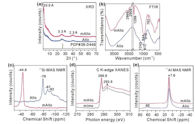
3.1 Structural changes at molecule or local scales 172
The structural changes induced by methyl modification were characterized via a combination 173
of techniques. Two reflections at 3.3 and 2.3 Å that are characteristic of ImoLS were observed in 174
the XRD pattern of mAllo (Fig.2a), and the overall FTIR spectrum of mAllo (Fig.2b) resembled 175
that of Allo, both indicating that mAllo has a structure similar to that of Allo. Bands ascribed to Si– 176
CH3 [30] (including the asymmetric and symmetric stretching vibrations at 2969 and 2909 cm−1 and
177
the bending vibration at 1269 cm−1) and to –Si–C [32] (bending vibration at 782 cm−1) appeared in 178
the FTIR spectrum of mAllo (Fig. 2b). A new resonance at −44.8 ppm that is ascribed to 179
T3(6Al)≡Si−CH3 [33], instead of the resonance at −79 ppm that is ascribed to T3(6Al)≡Si−OH [34]
180
and is characteristic of ImoLS [16], was observed in the 29Si MAS NMR spectrum of mAllo (Fig. 181
2c). Moreover, the C K-edge XANES spectrum of mAllo (Fig. 2d) exhibited peaks at 288.9 and 182
293.5 eV, which might be attributed to the σ*(C-H) and σ*(C-Si) resonances, respectively. This 183
result is almost identical to that for mImo (Fig. 2d), the structure of which has been well 184
characterized [35], indicating the same chemical environments for C atoms (i.e., T3(6Al)≡Si−CH3)
185
in mAllo and mImo. From these results, methyl groups are assumed to be anchored to the inner 186
surface of mAllo. The 27Al MAS NMR spectrum of mAllo (Fig.2e) was dominated by a resonance
187
at 7.6 ppm that is ascribed to six-coordinated Al (AlVI), which is similar to the case of Allo,
188
indicating the same chemical environments for the Al atoms (i.e., a curved gibbsite-like sheet 189
serving as the outer framework [26]) in mAllo and Allo. The above mentioned results indicate that 190
methyl modification occurs only on the inner surface of Allo, exhibiting little effect on the outer 191
Al–O octahedral sheet. 192
A shoulder band at 1243 cm−1, which appeared in the FTIR spectrum of Allo and is ascribed to 193
the oligomeric silicate on the inner surface, was absent in the spectrum of mAllo (Fig.2b). This is 194
due to the presence of Si-CH3 groups, which prevents the attachment of oligomeric silicate, resulting
195
in a higher structural order in mAllo than in Allo. This assumption is further supported by the change 196
of the band at approximately 1000 cm−1 originating from the framework vibrations, of which the 197
high-wavenumber branch was much sharper in mAllo than in Allo (Fig. 2b). Moreover, the 198
resonance at 60 ppm in the 27Al NMR spectrum of Allo, which is ascribed to four-coordinated Al
199
(AlIV), was absent in the spectrum of mAllo (Fig. 2e). A similar change occurred for the broad
200
resonance at approximately −90 ppm in the 29Si NMR spectra (Fig.2c), which is ascribed to a 201
significant fraction of less ordered Si species with 0−5 next-nearest neighbour Al atoms [28]. These 202
changes also indicate the presence of a more ordered structure in mAllo than in Allo. 203
204
Fig. 2 XRD patterns (a), FTIR spectra (b), 29Si MAS NMR spectra (c), C K-edge XANES spectra (d) and 27Al MAS
205
NMR spectra (e) of mAllo (a-e) and Allo (a-c, e)/mImo (d). 206
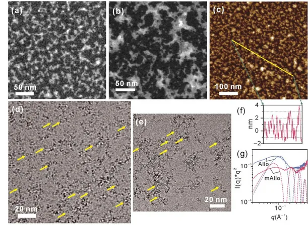
3.2 Changes of aggregation state and particle size 207
Microscopic observations showed distinct differences in aggregation behaviors between mAllo 208
and Allo. Very small nanoparticles were observed in the STEM image of mAllo (Fig.3a). These 209
nanoparticles mainly occurred as interlaced strings, which is in contrast with the case of Allo where 210
large aggregates dominated (Fig. 3b). To avoid the possible damage resulting from long-term 211
exposure to an electron beam during STEM observation, AFM that operates under ambient 212
conditions was used to further characterize mAllo. A similar configuration of interlaced strings was 213
shown in the AFM images of mAllo (Fig.3c), albeit with different specimen preparation methods 214
for STEM and AFM. This configuration might result from the interaction between particles in the 215
same string. This assumption was further supported by the XRD and FTIR results. On the XRD 216
pattern of mAllo (Fig. 2a), a very-small-angle peak was observed. This new reflection is ascribed 217
to the interference between particles arising after drying, indicating that the particles in the same 218
string are closely linked to each other. The band at approximately 1000 cm−1 in the FTIR spectrum 219
of Allo that arose from the Si−O stretching vibration was split into two bands at 944 and 910 cm−1 220
in the spectrum of mAllo (Fig. 2b). This splitting phenomenon into two bands also occurred in a 221
more pronounced way in the FTIR spectra of Imo and mImo and are characteristic of a tubular 222
morphology [29, 36]. However, the STEM and AFM results precluded the occurrence of any 223
imogolite-like nanotubes that were produced as a byproduct (Fig. 3a,c). A possible explanation is 224
that a moderate interparticle stress in the same string induced by capillary stress makes mAllo 225
exhibit a similar FTIR adsorption feature as that of tubular Imo, while in the case of Allo, the 226
presence of a strong interparticle stress prevents the formation of strings. 227
Cryo-TEM and SAXS have been used to characterize mAllo and Allo particles in dispersions 228
(without potential drying artifacts). The hollow nanospherical structure of these nanoparticles, 229
which is the most definitive feature of the Allo structure [17], was observed in the cryo-TEM images 230
(highlighted by arrows in Fig.3d,e). These hollow nanospheres were better dispersed (some of them 231
were even isolated) compared to those observed in the STEM or AFM images. Unlike Allo particles 232
that were gathered in aggregates having an average dimension of appropriately 100 nm (Fig.3e), 233
mAllo particles were homogeneously distributed (Fig. 3d). Notably, some proto-234
(methyl-)imogolite-like fragments were probably present in the products (Fig.3d,e), although they 235
can hardly be differentiated from the nanospheres (mAllo or Allo) and quantified using cryo-TEM 236
due to their very small sizes. The SAXS measurements of both Allo and mAllo revealed an increase 237
of the intensity at the lowest scattering angles (Fig. 3g). The smooth increase of the intensity is 238
characteristic of disordered (fractal) aggregates in dispersions. This is in good agreement with the 239
cryo-TEM observations where both samples displayed aggregates (Fig. 3d,e). From these above 240
microscopic and SAXS results, it is found that hollow nanospheres prevail in both Allo and mAllo 241
and that methyl modification induces changes in the arrangements of these nanospheres in both 242
liquid and dried powder. 243
244
Fig. 3 STEM images of mAllo (a) and Allo (b), AFM image of mAllo (c) and cryo-TEM images of mAllo (d) and 245
Allo (e). (f) Cross-section profile of the yellow line in (c), showing average particle sizes of 3−5 nm. (g) SAXS 246
curves of mAllo and Allo. Arrows in (d) and (e) highlight the presence of hollow nanospheres. The dash line in (g) 247
represents the expected scattering curves for monodispersed core-shell spheres with external (inner) diameters of 248
4.8 (3.6) and 3.6 (2.4) nm, respectively. 249
Thanks to the high z-axis resolution of AFM, an average particle size of 3−5 nm (with 250
appropriately 3 nm being dominant) of the mAllo particles was recorded (Fig.3f). This average size 251
agrees with the previously reported values of Allo obtained by electron microscopy [17]. This result 252
indicates that the overall particle size of Allo remained in the same range upon methyl modification. 253
Similar SAXS curves with weak oscillations were observed for both samples (Fig. 3g). The 254
positions of these oscillations enable to access the average characteristic dimensions of mAllo and 255
Allo through comparison with the oscillation positions of modeled core-shell particles [30, 36]. The 256
dimensions that yield oscillations at the closest positions indicate an inner/external diameter of 257
3.6/4.8 nm for mAllo and 2.4/3.6 nm for Allo (Fig. 3g). Notably, in addition to mAllo particles with 258
sizes of 3−5 nm, a few of particles with z-scans of less than 1 nm were observed (Fig. 3f). It is likely 259
that these particles are proto-methyl-imogolite-like fragments that adsorb flat on mica sheets, as 260
previously reported [16]. Despite not being fully quantitative, the proportion of the particles with 261
different sizes, summarized from the repeated section analyses of the AFM images, indicates that 262
hollow nanosphere is the dominating morphology of the nanoparticles. 263
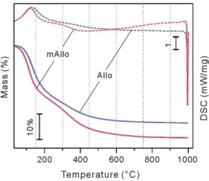
3.3 Changes of surface hydrophilicity 264
These structural changes result in modification of the inner-surface hydrophilicity of the 265
products. As exhibited in the TG curves (Fig.4), for the mass loss below 150°C, which is mainly 266
attributed to the desorption of physisorbed water [26], a lower temperature was required for mAllo 267
than for Allo to lose the same mass. The corresponding endothermic peak in the DSC curves also 268
occurred at a lower temperature for mAllo than for Allo (Fig. 4). These results indicate that the 269
physisorbed water in mAllo is removable at a lower temperature than that in Allo; that is, the inner 270
surface of mAllo is less hydrophilic than that of Allo. 271
272
Fig. 4 TG-DSC curves of mAllo and Allo. 273
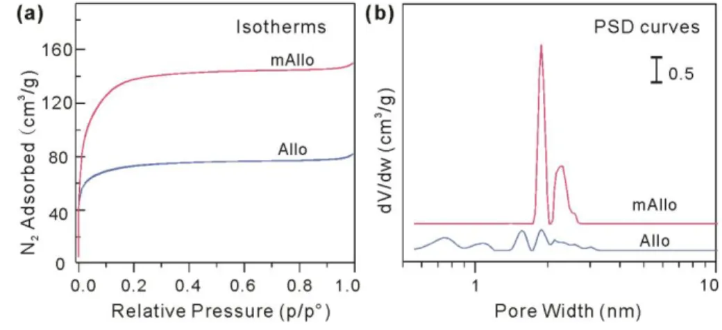
3.4 Changes of porosity and specific surface area 274
According to the International Union of Pure and Applied Chemistry (IUPAC) 275
recommendation [37], the N2 physisorption isotherms of mAllo and Allo are characterized as type I
276
(Fig. 5a), indicating that both materials are dominated by micropores. The SSAand Vtotal of mAllo
277
(523 m2/g and 0.2273 cm3/g) were much higher than those of Allo (269 m2/g and 0.1222 cm3/g)
278
(Table 1). As discussed above, methyl modification results in limited changes in the particle sizes 279
and outer structural framework of Allo. The high SSA and porosity of mAllo arise from the 280
configuration of interlaced strings. In addition, the PSD curves derived using a NLDFT method (Fig. 281
5b) exhibited a distinct difference. Specifically, micropores with wide-ranging diameters were 282
observed in Allo, while pores with diameters of approximately 2 nm dominated in mAllo. This 283
difference might be because the pores in Allo originated from cavities between aggregates formed 284
by the irreversible aggregation of particles, while the pores in mAllo arose from the network formed 285
by the interleaving of strings of particles (as schematically shown in Fig.1). 286
287
Fig. 5 N2 adsorption/desorption isotherms (a) and PSD curves (b) of mAllo and Allo.
288
Table 1 Textural data of mAllo and Allo obtained from N2 adsorption analysis.
289
SSA (m2/g) SSAmicro (m2/g) Vtotal (cm3/g) Vmicro (cm3/g)
Allo 269.5 267.2 0.1222 0.1156
mAllo 523.1 519.6 0.2273 0.2191
3.5 Mechanisms underneath the above mentioned changes 290
As discussed above, the introduction of Si–CH3 to the inner surface of mAllo makes this
291
surface less hydrophilic than that of Allo. The weakened surface hydrophilicity decreases the 292
capillary stress in the inner cavity of mAllo and thus inhibits the irreversible aggregation of particles 293
during drying, resulting in higher SSA and porosity values. On the other hand, the absence of Si– 294
OH on the inner surface of mAllo prevents the formation of oligomeric silicate therein and thus 295
increases the structural order and total pore volume of this material. Notably, the simultaneous 296
absence of oligomeric silicate and AlIV in mAllo might provide insight into the probable location of 297
AlIV in the Allo structure; i.e., AlIV might be mainly located in oligomeric silicate by substituting Si
on the inner surface, which sheds light on the long-standing question of whether AlIV is located
299
around the perforations on the wall [38] or in the oligomeric silicate upon substituting Si atoms [39] 300
in the Allo structure. This assumption is further supported by the fact that scarcely any AlIV occurs 301
in Imo consisting of only ImoLS [28]. 302
4. Conclusion
303
Tailoring the structure and surface chemistry and thus the aggregation state of hollow Allo 304
nanospheres has been achieved by a simple strategy of methyl modification. The as-obtained mAllo 305
is a novel hollow nanospherical material with a higher structural order, a much less hydrophilic 306
inner surface and a larger porosity and SSA than Allo, which allow its promising applications in a 307
variety of fields such as selective gas adsorption. This facile inner-surface methyl-modification 308
method and the basic knowledge on the mechanisms underneath the changes of the structure, surface 309
chemistry, inner-cavity capillary stress and aggregation state of Allo nanospheres reported in this 310
work is of great significance for dealing with the common aggregation issue of other nanomaterials. 311
Conflicts of interest
312
There are no conflicts to declare. 313
Acknowledgements
314
This work is supported by the Science and Technology Planning Project of Guangdong Province, 315
China (2017B020237003), the National Natural Science Foundation of China (41672042 and 316
41972045), Youth Innovation Promotion Association CAS for the excellent members (2016-81-01), 317
the China Postdoctoral Science Foundation (2018M640831) and the Open Funds of the Beijing 318
Synchrotron Radiation Facility (2017-BEPC-PT-001065). Cryo-TEM observations were made, 319
thanks to “Investissementsd'Avenir” LabEx PALM (ANR-10-LABX-0039-PALM). This is a 320
contribution (No. xxxx) from GIGCAS. 321
References
322
[1] M. Sasidharan, K. Nakashima, N. Gunawardhana, T. Yokoi, M. Inoue, S.-i. Yusa, M. Yoshio, T. 323
Tatsumi, Novel titania hollow nanospheres of size 28 ± 1 nm using soft-templates and their application 324
for lithium-ion rechargeable batteries, Chem. Commun., 47 (2011) 6921-6923. 325
[2] M.Q. Zhu, S.M. Li, J.H. Liu, B. Li, Promoting polysulfide conversion by V2O3 hollow sphere for
326
enhanced lithium-sulfur battery, Appl. Surf. Sci., 473 (2019) 1002-1008. 327
[3] Y. Hu, Y. Ding, D. Ding, M. Sun, L. Zhang, X. Jiang, C. Yang, Hollow chitosan/poly (acrylic acid) 328
nanospheres as drug carriers, Biomacromolecules, 8 (2007) 1069-1076. 329
[4] H.P. Liang, H.M. Zhang, J.S. Hu, Y.G. Guo, L.J. Wan, C.L. Bai, Pt hollow nanospheres: facile 330
synthesis and enhanced electrocatalysts, Angew. Chem. Int. Edit., 43 (2004) 1540-1543. 331
[5] M. Tian, X.L. Cui, C.X. Dong, Z.P. Dong, Palladium nanoparticles dispersed on the hollow 332
aluminosilicate microsphere@hierarchical gamma-AlOOH as an excellent catalyst for the hydrogenation 333
of nitroarenes under ambient conditions, Appl. Surf. Sci., 390 (2016) 100-106. 334
[6] F. Xu, Z. Tang, S. Huang, L. Chen, Y. Liang, W. Mai, H. Zhong, R. Fu, D. Wu, Facile synthesis of 335
ultrahigh-surface-area hollow carbon nanospheres for enhanced adsorption and energy storage, Nat. 336
Commun., 6 (2015) 7221. 337
[7] W.J. Wu, W.T. Qi, Y.F. Zhao, X. Tang, Y.F. Qiu, D.W. Su, H.B. Fan, G.X. Wang, Hollow CeO2 spheres
338
conformally coated with graphitic carbon for highperformance supercapacitor electrodes, Appl. Surf. 339
Sci., 463 (2019) 244-252. 340
[8] A.E. Awadallah, W. Ahmed, M.R.N. El-Din, A.A. Aboul-Enein, Novel aluminosilicate hollow sphere 341
as a catalyst support for methane decomposition to COx-free hydrogen production, Appl. Surf. Sci., 287 342
(2013) 415-422. 343
[9] G. Zheng, S.W. Lee, Z. Liang, H.-W. Lee, K. Yan, H. Yao, H. Wang, W. Li, S. Chu, Y. Cui, 344
Interconnected hollow carbon nanospheres for stable lithium metal anodes, Nature Nanotechnology, 9 345
(2014) 618-623. 346
[10] Z. Zhou, J. Gu, X. Qiao, H. Wu, H. Fu, L. Wang, H. Li, L. Ma, Double protected lanthanide 347
fluorescence core@shell colloidal hybrid for the selective and sensitive detection of ClO−, Sensors 348
Actuators B: Chem., 282 (2019) 437-442. 349
[11] X.W. Lou, L.A. Archer, Z. Yang, Hollow micro-/nanostructures: synthesis and applications, Adv. 350
Mater., 20 (2008) 3987-4019. 351
[12] B.H. Bac, Y. Song, M.H. Kim, Y.-B. Lee, I.M. Kang, Single-walled hollow nanospheres assembled 352
from the aluminogermanate precursors, Chem. Commun., (2009) 5740-5742. 353
[13] M.B. Tahir, G. Nabi, N.R. Khalid, W.S. Khan, Synthesis of Nanostructured Based WO3 Materials
354
for Photocatalytic Applications, J. Inorg. Organomet. P., 28 (2018) 777-782. 355
[14] Y. Wu, H. Wang, W. Tu, Y. Liu, Y.Z. Tan, X. Yuan, J.W. Chew, Quasi-polymeric construction of 356
stable perovskite-type LaFeO3/g-C3N4 heterostructured photocatalyst for improved Z-scheme 357
photocatalytic activity via solid p-n heterojunction interfacial effect, J. Hazard. Mater., 347 (2018) 412-358
422. 359
[15] C. Levard, E. Doelsch, I. Basile-Doelsch, Z. Abidin, H. Miche, A. Masion, J. Rose, D. Borschneck, 360
J.Y. Bottero, Structure and distribution of allophanes, imogolite and proto-imogolite in volcanic soils, 361
Geoderma, 183 (2012) 100-108. 362
[16] P. Du, P. Yuan, A. Thill, F. Annabi-Bergaya, D. Liu, S. Wang, Insights into the formation mechanism 363
of imogolite from a full-range observation of its sol-gel growth, Appl. Clay Sci., 150 (2017) 115–124. 364
[17] R. Parfitt, Allophane and imogolite: role in soil biogeochemical processes, Clay Miner., 44 (2009) 365
135-155. 366
[18] B. Creton, D. Bougeard, K.S. Smirnov, J. Guilment, O. Poncelet, Structural model and computer 367
modeling study of allophane, J. Phys. Chem. C, 112 (2008) 358-364. 368
[19] Y. Adachi, J. Karube, Application of a scaling law to the analysis of allophane aggregates, Colloid 369
Surface A, 151 (1999) 43-47. 370
[20] S. Filimonova, S. Kaufhold, F.E. Wagner, W. Häusler, I. Kögel-Knabner, The role of allophane nano-371
structure and Fe oxide speciation for hosting soil organic matter in an allophanic Andosol, Geochim. 372
Cosmochim. Ac., 180 (2016) 284-302. 373
[21] T. Woignier, J. Primera, L. Duffours, P. Dieudonné, A. Raada, Preservation of the allophanic soils 374
structure by supercritical drying, Micropor. Mesopor. Mater., 109 (2008) 370-375. 375
[22] F. Ohashi, S.-I. Wada, M. Suzuki, M. Maeda, S. Tomura, Synthetic allophane from high-376
concentration solutions: nanoengineering of the porous solid, Clay Miner., 37 (2002) 451-456. 377
[23] J. Castello, J.J. Gaumet, J.F. Muller, S. Derousseaux, J. Guilment, O. Poncelet, Laser ablation of 378
aluminosilicates: Comparison between allophane and mixed alumina/silicas by Fourier Transform-Ion 379
Cyclotron Resonance-Mass Spectrometry, Appl. Surf. Sci., 253 (2007) 7773-7778. 380
[24] W.J. Likos, N. Lu, Hysteresis of capillary stress in unsaturated granular soil, J Eng Mech-Asce, 130 381
(2004) 646-655. 382
[25] D. Wang, A. Fernandez-Martinez, Order from disorder, Science, 337 (2012) 812-813. 383
[26] P. Du, P. Yuan, D. Liu, S. Wang, H. Song, H. Guo, Calcination-induced changes in structure, 384
morphology, and porosity of allophane, Appl. Clay Sci., 158 (2018) 211-218. 385
[27] V. Farmer, A. Fraser, J. Tait, Synthesis of imogolite: a tubular aluminum silicate polymer, J. Chem. 386
Soc., Chem. Commun., (1977) 462-463. 387
[28] G.I. Yucelen, R.P. Choudhury, A. Vyalikh, U. Scheler, H.W. Beckham, S. Nair, Formation of single-388
walled aluminosilicate nanotubes from molecular precursors and curved nanoscale intermediates, J. Am. 389
Chem. Soc., 133 (2011) 5397-5412. 390
[29] A. Thill, P. Picot, L. Belloni, A mechanism for the sphere/tube shape transition of nanoparticles with 391
an imogolite local structure (imogolite and allophane), Appl. Clay Sci., 141 (2017) 308-315. 392
[30] Y. Liao, P. Picot, J.-B. Brubach, P. Roy, S. Le Caër, A. Thill, Self-supporting thin films of imogolite 393
and imogolite-like nanotubes for infrared spectroscopy, Appl. Clay Sci., 164 (2017) 58-67. 394
[31] P. Lindner, T. Zemb, Neutrons, X-rays and Light: Scattering Methods Applied to Soft Condensed 395
Matter, Elsevier, 2002. 396
[32] M. Boyer, E. Paineau, M. Bacia-Verloop, A. Thill, Aqueous dispersion state of amphiphilic hybrid 397
aluminosilicate nanotubes, Appl. Clay Sci., 96 (2014) 45-49. 398
[33] D.-Y. Kang, N.A. Brunelli, G.I. Yucelen, A. Venkatasubramanian, J. Zang, J. Leisen, P.J. Hesketh, 399
C.W. Jones, S. Nair, Direct synthesis of single-walled aminoaluminosilicate nanotubes with enhanced 400
molecular adsorption selectivity, Nat. Commun., 5 (2014) 3342. 401
[34] P.F. Barron, M.A. Wilson, A.S. Campbell, R.L. Frost, Detection of imogolite in soils using solid-402
state Si-29 NMR, Nature, 299 (1982) 616-618. 403
[35] I. Bottero, B. Bonelli, S.E. Ashbrook, P.A. Wright, W.Z. Zhou, M. Tagliabue, M. Armandi, E. 404
Garrone, Synthesis and characterization of hybrid organic/inorganic nanotubes of the imogolite type and 405
their behaviour towards methane adsorption, Phys. Chem. Chem. Phys., 13 (2011) 744-750. 406
[36] M.S. Amara, S. Rouzière, E. Paineau, M. Bacia-Verloop, A. Thill, P. Launois, Hexagonalization of 407
aluminogermanate imogolite nanotubes organized into closed-packed bundles, J. Phys. Chem. C, 118 408
(2014) 9299-9306. 409
[37] M. Thommes, K. Kaneko, A.V. Neimark, J.P. Olivier, F. Rodriguez-Reinoso, J. Rouquerol, K.S. 410
Sing, Physisorption of gases, with special reference to the evaluation of surface area and pore size 411
distribution (IUPAC Technical Report), Pure Appl. Chem., 87 (2015) 1051-1069. 412
[38] H. Shimizu, T. Watanabe, T. Henmi, A. Masuda, H. Saito, Studies on allophane and imogolite by 413
high-resolution solid-state 29Si- and 27Al-NMR and ESR, Geochem. J., 22 (1988) 23-31.
414
[39] B. Goodman, J. Russell, B. Montez, E. Oldfield, R. Kirkpatrick, Structural studies of imogolite and 415
allophanes by aluminum-27 and silicon-29 nuclear magnetic resonance spectroscopy, Phys. Chem. 416
Miner., 12 (1985) 342-346. 417