0099-2240/06/$08.00⫹0 doi:10.1128/AEM.00863-06
Copyright © 2006, American Society for Microbiology. All Rights Reserved.
Virus-Bacterium Interactions in Water and Sediment of
West African Inland Aquatic Systems
Yvan Bettarel,
1* Marc Bouvy,
2Claire Dumont,
3and Te
´lesphore Sime-Ngando
3UR 167 CYROCO, Centre IRD de Bel Air, Route des Hydrocarbures, BP 1386, Dakar, Senegal1; UR 167 CYROCO,
Universite´ Montpellier II, Case 093, 34095 Montpellier Cedex 5, France2; and Universite´ Blaise Pascal,
Laboratoire de Biologie des Protistes, UMR CNRS 6023, 63177 Aubie`re Cedex, France3 Received 12 April 2006/Accepted 18 May 2006
The ecology of virioplankton in tropical aquatic ecosystems is poorly documented, and in particular, there are no references concerning African continental waters in the literature. In this study, we examined virus-bacterium interactions in the pelagic and benthic zones of seven contrasting shallow inland waters in Senegal, including one hypersaline lake. SYBR Gold-stained samples revealed that in the surface layers of the sites, the numbers of viruses were in the same range as the numbers of viruses reported previously for productive temperate systems. Despite high bacterial production rates, the percentages of visibly infected cells (as determined by transmission electron microscopy) were similar to the lowest percentages (range, 0.3 to 1.1%; mean, 0.5%) found previously at pelagic freshwater or marine sites, presumably because of the local environ-mental and climatic conditions. Since the percentages of lysogenic bacteria were consistently less than 8% for pelagic and benthic samples, lysogeny did not appear to be a dominant strategy for virus propagation at these sites. In the benthic samples, viruses were highly concentrated, but paradoxically, no bacteria were visibly infected. This suggests that sediment provides good conditions for virus preservation but ironically is an unfavorable environment for proliferation. In addition, given the comparable size distributions of viruses in the water and sediment samples, our results support the paradigm that aquatic viruses are ubiquitous and may have moved between the two compartments of the shallow systems examined. Overall, this study provides additional information about the relevance of viruses in tropical areas and indicates that the intensity of virus-bacterium interactions in benthic habitats may lower than the intensity in the adjacent bodies of water.
The interest of plankto-ecologists in viruses has not faded since virus particles were shown to be at least as numerous as prokaryotes in fresh and marine waters (5, 49). To date, more than 100 sites have been investigated for the presence of vi-ruses, and studies conducted worldwide have come to the same conclusion: with concentrations in most cases exceeding 107
particles per ml, viruses are actually the most abundant bio-logical entities in aquatic systems (25, 53, 57, 69, 72).
Apart from being ubiquitous and numerous, viruses also play fundamental roles in structuring the microbial food webs, primarily by killing microbes (25), by governing microbial di-versity and diversification (70), and, to a lesser extent, by being a potential food source for protists (10, 27). Natural phages are now presumed to be the largest reservoir of uncharacterized genetic diversity on Earth (31, 57), and theories regarding their role in shaping the diversity of prokaryotes have been pro-posed (61, 71). The term “viriosphere” has even been coined, illustrating the stake that an increasing number of scientists have in virus ecology (58).
Given the responsiveness of planktonic viruses and microbes to environmental conditions and more specifically to solar ra-diation and temperature (41, 48), investigating their relevance at all latitudes is fundamental. Geographically, most of the aquatic viruses that have been ecologically examined so far have been located in temperate regions. Studies of the
occur-rence of viruses in polar zones are rare (29, 39), and to our knowledge, tropical waters have been investigated only once previously, by Peduzzi and Schiemer (46).
During the past two decades, biophysicochemical data pro-vided by multidisciplinary studies have consistently revealed that limnology in temperate zones is somewhat different from limnology in tropical systems (2, 59). For instance, because of the typically high bacterial growth rates mostly resulting from the temperatures in tropical regions, trophic pathways within microbial food webs are not readily homologous in the two zones (13, 62). The same conclusion is expected to be reached regarding virus-mediated processes. However, in the only study conducted with tropical waters (two freshwater reservoirs in Sri Lanka), the levels of viral abundance and activity prelimi-narily appeared to be in the same range as the levels reported for temperate regions (46). Thus, there is a severe lack of data concerning virioplankton “performance” at tropical latitudes, including the African continent, whose waters have not been studied previously.
Over the last few decades, West African inland waters have typically been diverted for irrigation and cattle ranching to promote development of the local human population. Thou-sands of reservoirs of different sizes have been built in most sub-Saharan countries, predominantly for agricultural and do-mestic purposes. The presence of dense populations and the by-products of these agricultuoriented sites commonly re-sult in rapid eutrophication of waters that are often identified as highly productive (12).
The objective of the present work was to examine virus-bacterium interactions at additional tropical sites where virus
* Corresponding author. Mailing address: Institut de Recherche pour le Developpement (IRD), Centre de Bel Air, UR 167 CYROCO, BP 1386, Dakar, Senegal. Phone: 221 849 33 07. Fax: 221 832 16 75. E-mail: bettarel@ird.sn.
infectivity and decay may be affected by the large amount of local solar irradiation and by the typically elevated tempera-tures. The levels and activities of viruses and their bacterial hosts were determined for the surface waters and sediments of inland aquatic systems of Senegal, which is the westernmost country in continental Africa. Six contrasting freshwater sites located in the basin of the Senegal River covering a wide, representative spectrum of productivity in African aquatic en-vironments, as well as one hypersaline lake, were sampled in November 2004. Below we discuss the effect of viruses on bacterial mortality in shallow freshwaters and speculate about the origin of viruses in the superficial sediment layers.
MATERIALS AND METHODS
Study sites and environmental context.Samples were collected on 3, 4, and 5 November 2004, at the end of the flood season. Samples were obtained from
seven contrasting aquatic systems in a⬃20,000-km2
area delimited in the south by the Cape Vert Peninsula and in the north by the Senegal River (Fig. 1). The seven sites were selected to cover a wide range of productivity and salinity in Senegal continental aquatic systems. The sites were designated as follows. Lake
Retba (surface area,⬃400 ha) is located near Dakar (30 km), and it has shallow
lagoon-like features and a maximum depth of 3 m and is close (400 m) to the
Atlantic Ocean. Lake Retba is a hypersaline lake with a maximum salt
concen-tration of 463 g liter⫺1. Djoudj is a pond located in the National Ornithological
Park of Senegal. This site is a habitat for thousands of migratory birds from October to April and thus receives considerable amounts of bird excretion that makes it highly productive. Djeuss Stream is a narrow defluent of the Senegal River that occasionally provides water to Bango Reservoir. Diama Dam is lo-cated on the Senegal River, 27 km upstream of Saint Louis City. This dam was built in 1985 to prevent saltwater input into the drinking water reservoirs fed by the Senegal River. Bango Reservoir is located 30 km from St. Louis City and is the main drinking water reservoir for this city. Finally, Lake Guiers is a large shallow lake that is 50 km long; the average width is 7 km, the surface area is 240
km2
, and the volume is approximately 390⫻ 106
m3
. This lake receives water from the Senegal River and is a drinking water reservoir for the city of Dakar. Samples were obtained from two different sites in this lake; N⬘Gnith (NG) is located in the central zone near the pumping station, and Keur Momar Sarr (KMS) is located in the southern part, near the gate that regulates the water flow and level of the lake. Nutrient and chlorophyll concentrations at all sites would be considered eutrophic to hypereutrophic according to Carlson’s criteria (16), compared to lakes in temperate regions.
Sampling.Triplicate samples were collected from the sediments and subsur-face waters at the seven sampling sites. The water samples used for nutrient and chlorophyll analysis, as well as for determining bacterial and viral parameters, were obtained with acid-cleaned sterile bottles at locations 0.5 m below the surface. The samples used for measurement of dissolved inorganic nutrients
(NO3-N, NH4-N, PO4-P) were filtered through Whatman GF/F fiberglass filters,
stored at⫺20°C, and analyzed as described by Strickland and Parsons (56). Chlorophyll a concentrations were determined fluorometrically following filtra-tion of samples onto Whatman GF/F fiberglass filters, storage in liquid nitrogen, and methanol extraction (72). Oxygen concentrations were determined in situ using a YSI probe with temperature correction. For bacterial and viral
param-eters, samples were fixed with prefiltered (0.02m) buffered formaldehyde (final
concentration, 2%), stored at 4°C in the dark, and immediately analyzed when they arrived at the laboratory.
Sediment cores were taken using a corer (Uwitec no. 016001) equipped with polyvinyl chloride tubes having an inside diameter of 60 mm. Cores were pro-cessed immediately after collection, using three subsamples taken with 5-ml sterile syringes. The top centimeter of the core layer was carefully extracted and used for bacterial, viral, and chlorophyll analyses. Approximately 100 g (wet weight) of sediment was collected for determination of sediment structure and composition (42).
Enumeration of viruses and microorganisms.Amounts of planktonic bac-teria were determined by standard techniques using the fluorochrome 4⬘,6⬘-di-amidino-2-phenylindole (DAPI) and epifluorescence microscopy (EM) (47). The numbers of virus-like particles (VLPs) in triplicate 50- to 200-l samples were
determined after retention of the particles on 0.02-m-pore-size membranes
(Anodisc) and staining with SYBR Gold (17). On each slide, 300 to 600 bacteria and VLPs were counted in 15 to 20 fields.
Concurrently, viruses were counted and their sizes were determined using transmission electron microscopy (TEM) (6). The viruses in 5-ml aliquots of formalin-fixed samples were harvested by ultracentrifugation onto grids (400-mesh Cu electron microscope grids with carbon-coated Formvar film) by using a
Centrikon TST 41.14 swing-out rotor at 120,000⫻ g for 2 h. The grids were then
stained for 30 s with uranyl acetate (2%, wt/wt), and viruses were counted and measured with a JEOL model 1200EX TEM operated at 80 kV and a
magnifi-cation of⫻40,000. The following three classes of virus capsid size were examined
to characterize viral populations:⬍60 nm, 60 to 95 nm, ⬎95 nm.
Benthic viruses and bacteria were extracted as recommended by Danovaro et al. (19) and were analyzed using the procedures described above. Briefly, aliquots of the fixed sediment samples (1 ml) were diluted with tetrasodium pyrophos-phate (4 ml; final concentration, 10 mM) and incubated for 20 min at 4°C. Samples were then sonicated three times (100 W for 1 min) and diluted 200- to
1,000-fold with filtered (0.02m) formaldehyde (final concentration, 2%). This
procedure has been shown to extract the majority of VLPs and bacteria from sediment (26), except silty sediment, in which both VLPs and bacteria are thought to be less efficiently extractable (26, 34).
Bacterial production.For both sediments and overlaying waters, heterotrophic
bacterial production (BP) was determined by the [methyl-3H]thymidine
incor-poration method (38). For water samples, duplicates and one control (zero time)
were incubated with [methyl-3H]thymidine (47 Ci mmol⫺1; Amersham) in the
dark at the in situ temperature. The incubation time was 15 min, and the final thymidine concentration was 20 nM (saturation conditions); we assumed that isotope dilution could be prevented at this concentration (50). Radioactivity was counted using the liquid scintillation procedure. Bacterial production was calcu-lated from the radioactivity incorporated into a trichloroacetic acid precipitate,
using a conversion factor of 2⫻ 1018cells produced per mol of incorporated
thymidine (4).
For the sediments, two replicates and one control were incubated with labeled thymidine used under saturation conditions at a final concentration of 1,000 nM (30). Subsamples (0.5 g) of wet sediment were incubated in 10-ml centrifugation tubes at the in situ temperature. Incubation was stopped with formaldehyde after
30 min. Samples were then centrifuged (8,500⫻ g) for 20 min, the supernatants
were discarded, and the pellets were washed three times with 5 ml of 80% ethanol. Finally, the pellets were washed twice with 5 ml of ice-cold trichloro-acetic acid (5%), filtered onto 0.2-m-pore-size membrane filters, transferred to vials with 3 ml of 2 N NaOH, and heated for 2 h in a water bath at 100°C. After cooling, 1 ml of each supernatant was transferred to a scintillation vial, and scintillation cocktail was added. Bacterial production was estimated using the same conversion factor that was used for surface waters. This conversion factor has been used by several authors for lake sediment samples (30, 38).
Viral infection of prokaryotes.Benthic bacteria were detached from the sed-iment by treatment with sodium pyrophosphate and sonication, as described above. The bacteria in duplicate 8-ml aliquots of formalin-fixed samples were
harvested by ultracentrifugation at 70,000⫻ g for 20 min onto 400-mesh Cu
grids, stained for 30 s with uranyl acetate (2%, wt/wt), and examined at a
magnification of⫻40,000 by using a TEM operated at 80 kV to distinguish
between virus-infected and uninfected bacteria (66). The extraction procedure for benthic bacteria resulted in reliable observations by TEM since individual bacterial cells were clearly identifiable based on morphology and refringence. At least 600 bacterial cells were inspected per grid, and the number of infected bacteria encountered ranged from 15 to 20. To estimate virus-induced bacterial mortality (VIBM), the frequency of infected cells (FIC) was calculated from the frequency of visibly infected cells (FVIC) (expressed as a percentage) using the
formula (67): FIC⫽ 7.11 ⫻ FVIC. FIC was then converted to VIBM as
de-scribed by Binder (11): VIBM⫽ (FIC ⫹ 0.6FIC2
)/(1⫺ 1.2FIC).
Viral production was estimated by multiplying the lysed bacterial production
(BP⫻ VIBM) by the burst size (BS) (i.e., the mean number of viruses in the
infected cells which were filled with phages) (9, 66).
Fraction of lysogenic bacteria.We used the approaches of Weinbauer et al. (68) and Mei and Danovaro (43) to initiate prophage induction in benthic and
pelagic bacteria, respectively. Mitomycin C (final concentration, 1g ml⫺1;
catalog no. M-0503; Sigma Chemical Co.) was added to duplicate 10-ml water or
sediment samples (1 g of sediment with 9 ml of prefiltered [0.02m] overlaying
water). Untreated samples served as controls. Both types of samples were for-malin fixed after they were incubated for 12 h (43, 68). Prophage induction was calculated by determining the difference between the viral abundance in mito-mycin C-treated incubation mixtures (Vm) and the viral abundance in control incubation mixtures (Vc). The fraction of lysogenic bacteria cells (FLC)
(ex-pressed as a percentage) was calculated as follows: FLC⫽ 100[(Vm ⫺ Vc)/(BS ⫻
BACt0)], where BS is the burst size (number of virus particles bacterium⫺1) and
BACtois the bacterial abundance at the start of the experiment (i.e., before the
addition of mitomycin C) (68).
Statistical analyses.Data were log transformed to satisfy the requirements of normality and homogeneity of variance necessary for parametric analyses. Dif-ferences in the means were tested using a t test. Simple relationships between original data sets were tested by Pearson correlation analysis. All statistical analyses were performed using the SigmaStat 2.0 software.
RESULTS
Environmental conditions. With temperatures between 22 and 29°C and maximum depths of 5 m, the seven aquatic sites sampled were typical of shallow tropical systems (Table 1). The depths of light penetration which were determined using a Secchi disk were low, ranging from 19 cm in Lake Retba to 90 cm in the southern part of Lake Guiers (KMS). In the water
TABLE 1. Geographical coordinates and physicochemical parameters of the seven inland aquatic systems in November 2004
Site Latitude north Longitude west Maximum depth (m) Temp (°C)a Secchi depth (cm) Oxygen (mg liter⫺1) PO4⫺ (M) NO3 ⫺⫹ NO 2⫺ (M) NH4 ⫹ (M) Chlorophyll Pelagic (g liter⫺1) (g cmBenthic⫺3) Lake Retba 14°49⬘30⬙ 17°14⬘30⬙ 0.5 28.9 (4.2) 19 2.1 7.2 1.4 1.0 8.7 1.9 Djoudj Pond 16°24⬘95⬙ 16°18⬘18⬙ 3.1 22.0 (0.1) 25 4.8 2.3 12.8 20.9 49.5 1.9 Djeuss Stream 16°06⬘10⬙ 16°23⬘25⬙ 1.6 23.6 (0.1) 56 4.0 0.6 0.4 1.0 10.1 1.0 Diama Dam 16°12⬘51⬙ 16°24⬘59⬙ 4.1 23.9 (0.1) 27 5.8 0.5 7.4 1.0 2.7 2.7 Bango Reservoir 16°04⬘03⬙ 16°27⬘04⬙ 3.4 25.2 (0.4) 62 6.8 0.1 0.2 0.8 15.0 1.4 Lake Guiers (NG) 16°11⬘90⬙ 15°54420⬙ 3.0 23.5 (0.3) 81 7.4 0.1 0.1 0.1 36.2 1.0 Lake Guiers (KMS) 15°56⬘15⬙ 15°56⬘56⬙ 1.2 23.4 (0.1) 90 6.0 0.1 0.3 1.1 25.4 1.0
column, the levels of dissolved oxygen ranged from 1.4 to 7.4 mg liter⫺1, and the lowest values were obtained for hypersaline Lake Retba. Among the freshwater sites, the most nutrient-rich site was Djoudj Stream, where the levels of dissolved inorganic nitrogen (nitrate, nitrite, and ammonium), as well as the chlorophyll concentrations, were highest. This was also true for orthophosphate when Lake Retba, in which the PO4
concentration was 3 to 72 times higher than the PO4
concen-trations at the other sites, was excluded. At this single hyper-saline site, the shallow water column (50 cm) favored high temperatures (28.9°C) (Table 1). Overall, no significant rela-tionships between benthic and pelagic chlorophyll concentra-tions were found (P⬎ 0.05; n ⫽ 7).
Sediment characteristics.The sites investigated were rela-tively comparable and were characterized by sediments con-sisting mostly of sand (fine and coarse), which accounted for 30 to 98% of the cores. An exception was Bango Reservoir, where the sediments were dominated by clay (41%) and, to a lesser extent, silt (29%) (Table 2).
Size classes of viruses. Viruses smaller than 60 nm were clearly dominant at all sites studied (Table 3). They accounted for 68% and 59% of the total community in the overlaying waters and in the sediments, respectively. Viruses larger than 95 nm were relatively underrepresented in both the pelagic and benthic compartments (means, 3% and 6%, respectively). Re-gardless of the site, the fractions of⬍60-nm viruses were com-parable for the sediment (mean, 59.3%; coefficient of variation [CV], 10.5%) and the water column (mean, 68.1%; CV, 9.6%). The same was true for the middle size class (60 to 95 nm), which on average accounted for 32.3% (CV, 9.4%) of the total abundance. Conversely, the contribution of large viruses (⬎90 nm) was distinctly greater in the benthic environment (mean, 6.4%; CV, 66.9%) than in the pelagic environment (mean, 3.1%; CV, 117.9%).
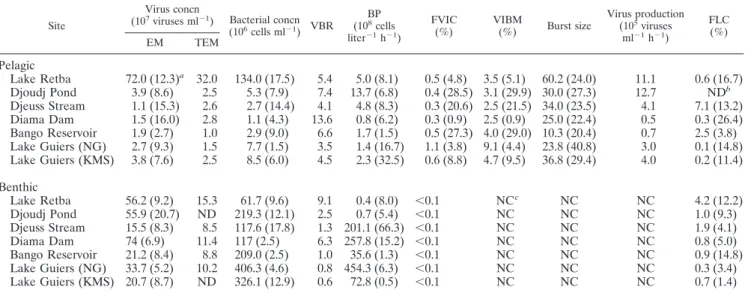
Standing stocks. In the overlaying waters, enumeration of viruses by EM and TEM did not reveal significant differences (P ⫽ 0.62, as determined by a t test). The epifluorescence microscopy estimates of pelagic viral concentrations ranged from 1.1⫻ 107VLPs ml⫺1(Djeuss Stream) to 72.0⫻ 107VLPs
ml⫺1(Lake Retba) (Table 4). Viruses were 3 to 14 times more abundant than bacteria. The highest densities were found at Lake Retba, where the concentrations of viruses and bacteria were more than 1 order of magnitude greater than the con-centrations at the other sites. Viral abundance was highly cor-related with bacterial abundance and dissolved phosphate con-centrations (Table 5).
Epifluorescence microscopy counts revealed that benthic ruses were 5 to 50 times more abundant than planktonic vi-ruses. The average benthic-to-pelagic ratios as calculated by EM and TEM were 15.3 and 4.7, respectively. The most viriobenthos-rich site (Diama Dam; mean, 7.4⫻ 108VLPs ml⫺1) was different
from the site at which the greatest densities in the water column were found (Lake Retba) (Table 4). The virus concentrations in the sediment were highly correlated with benthic chlorophyll contents (r⫽ 0.92; P ⬍ 0.01).
The mean virus-to-bacterium ratio (VBR) was significantly higher in the water column (mean, 6.5; CV, 49.8%) than in the sediment (mean, 3.1; CV, 99.3%). Benthic bacteria were more numerous than viruses at both sites in Lake Guiers. The pat-tern of bacterial abundance in the overlaying waters was rad-ically different from the pattern in the sediments. For example, Lake Retba was the site at which the concentration of plank-tonic bacteria was highest and, paradoxically, the site at which the concentration of benthic bacteria was lowest. Although the viral and bacterial concentrations were highly correlated with each other in the water column, this was not the case for the sediment (Table 5).
BP.The mean value for BP was 35-fold higher for the sed-iments (146.1⫻ 108cells liter⫺1h⫺1) than for the overlaying
waters (4.2 ⫻ 108 cells liter⫺1 h⫺1). However, benthic BP
varied greatly among sites. The values were almost negligible in the sediments from Djoudj Pond and Lake Retba, but par-adoxically, the water columns of these two bodies of water exhibited the highest pelagic BP (Table 4). Generally, pelagic BP was significantly and positively correlated with nitrogen content (NO2⫺, NO3⫺, and NH4⫹) (Table 5).
Lytic infection.In the water column, the FVIC ranged from 0.3 to 1.1% (mean,⫽ 0.5%). The maximum values were ob-tained for the two stations in Lake Guiers (mean for NG, 1.1% [CV, 3.8%]; mean for KMS 0.6% [CV, 8.8%]) (Table 4). The derived bacterial mortality rates due to lytic viruses were al-ways less than 10% of the BP. Noticeably, the level of small viruses (⬍60 nm) was negatively correlated with the FVIC in the water column (Table 5).
In the various sediment samples, none of the 5,840 bacteria that were inspected were found to be infected. Therefore, we could not calculate VIBM, viral production, and BS for benthic habitats (Table 4).
The burst size in the pelagic samples varied substantially, ranging from 10.3 (Bango) to 60.2 (Retba) with an average of
TABLE 3. Distribution of three virus size classes in the surface waters and sediments at the seven study sites in November 2004
Site
% of virus size classes
Water Sediment ⬍60 nm 60–95 nm ⬎95nm ⬍60nm 60–95 nm ⬎95nm Lake Retba 73.3 21.9 4.8 55.7 36.8 7.5 Djoudj Pond 71.5 27.8 0.7 NDa ND ND Djeuss Stream 64.3 32.8 2.9 64.8 33.2 2 Diama Dam 75.4 24.5 0.1 54.1 31.7 4.2 Bango Reservoir 68.4 30.8 0.8 53.2 32.5 14.3 Lake Guiers (NG) 54.2 34.6 11.2 68.5 27.3 4.2 Lake Guiers (KMS) 69.5 29.4 1.1 ND ND ND aND, not determined.
TABLE 2. Composition of sediments at the seven study sites
Site
Sediment composition (%) Clay
(⬍2 m) (2–50Siltm) (50–200Fine sandm) Coarse sand(0.2–2 mm)
Lake Retba 7.8 1.8 41.0 49.4 Djoudj Pond 12.3 8.1 40.8 38.8 Djeuss Stream 21.8 27.8 33.7 16.7 Diama Dam 0.9 1.4 57.2 40.5 Bango Reservoir 41.2 28.8 13.8 16.2 Lake Guiers (NG) 2.5 3.9 66.9 26.7 Lake Guiers (KMS) 1.8 1.4 24.7 72.1
31.4 (CV, 45.1%) (Table 3). Burst size was positively and significantly correlated with viral, bacterial, and phosphorus concentrations (Table 5).
The pelagic viral production ranged from 0.7 to 12.7⫻ 105
viruses ml⫺1 h⫺1 (mean, 5.2 ⫻ 105 viruses ml⫺1 h⫺1; CV,
87.0%). The highest values were obtained for Djouj Pond, while the lowest values were obtained for Diama Dam along with negligible bacterial production (Table 4).
Lysogenic infection.In the water column, the FLC was be-tween 0.1% (Lake Guiers NG) and 7.1% (Djeuss Stream) (Table 4). On average, 1.8% (CV, 139.3%) of pelagic bacteria were estimated to be lysogenic. FLC did not correlate with any of the parameters examined.
Since no infected bacteria were detected in the sediment, we estimated the fraction of lysogenic benthic bacteria by using a BS of 38, which was determined by a TEM examination of sediment samples from Lake Hallwil by Filippini et al. (21). To our knowledge, this value is the sole previously published value for TEM-measured BS for benthic bacteria and is relatively
close to the mean pelagic BS (31.4) calculated in this study. Overall, with only 1.4% of the bacteria being lysogenic (range, 0.3 to 4.2%), virulent viruses distinctly seemed to prevail in the viriobenthos.
DISCUSSION
To our knowledge, this study was the first study dealing with aquatic viral ecology in Africa. We examined the viral com-partment in benthic and pelagic environments of seven con-trasting continental sites located in Senegal (West Africa) for evidence of differences and similarities in virus-bacterium in-teractions, compared to the interactions widely described for temperate regions. Overall, despite the characteristically ele-vated microbial activities observed in tropical freshwaters, we hypothesize that viral control of bacterial populations in these waters may not be as relevant as the control that has been found in temperate systems.
TABLE 4. Mean viral and microbial parameters for the pelagic and benthic zones of the seven study sites in November 2004
Site
Virus concn
(107viruses ml⫺1) Bacterial concn
(106 cells ml⫺1) VBR BP (108 cells liter⫺1h⫺1) FVIC (%) VIBM (%) Burst size Virus production (105 viruses ml⫺1h⫺1) FLC (%) EM TEM Pelagic Lake Retba 72.0 (12.3)a 32.0 134.0 (17.5) 5.4 5.0 (8.1) 0.5 (4.8) 3.5 (5.1) 60.2 (24.0) 11.1 0.6 (16.7) Djoudj Pond 3.9 (8.6) 2.5 5.3 (7.9) 7.4 13.7 (6.8) 0.4 (28.5) 3.1 (29.9) 30.0 (27.3) 12.7 NDb Djeuss Stream 1.1 (15.3) 2.6 2.7 (14.4) 4.1 4.8 (8.3) 0.3 (20.6) 2.5 (21.5) 34.0 (23.5) 4.1 7.1 (13.2) Diama Dam 1.5 (16.0) 2.8 1.1 (4.3) 13.6 0.8 (6.2) 0.3 (0.9) 2.5 (0.9) 25.0 (22.4) 0.5 0.3 (26.4) Bango Reservoir 1.9 (2.7) 1.0 2.9 (9.0) 6.6 1.7 (1.5) 0.5 (27.3) 4.0 (29.0) 10.3 (20.4) 0.7 2.5 (3.8) Lake Guiers (NG) 2.7 (9.3) 1.5 7.7 (1.5) 3.5 1.4 (16.7) 1.1 (3.8) 9.1 (4.4) 23.8 (40.8) 3.0 0.1 (14.8) Lake Guiers (KMS) 3.8 (7.6) 2.5 8.5 (6.0) 4.5 2.3 (32.5) 0.6 (8.8) 4.7 (9.5) 36.8 (29.4) 4.0 0.2 (11.4) Benthic Lake Retba 56.2 (9.2) 15.3 61.7 (9.6) 9.1 0.4 (8.0) ⬍0.1 NCc NC NC 4.2 (12.2) Djoudj Pond 55.9 (20.7) ND 219.3 (12.1) 2.5 0.7 (5.4) ⬍0.1 NC NC NC 1.0 (9.3) Djeuss Stream 15.5 (8.3) 8.5 117.6 (17.8) 1.3 201.1 (66.3) ⬍0.1 NC NC NC 1.9 (4.1) Diama Dam 74 (6.9) 11.4 117 (2.5) 6.3 257.8 (15.2) ⬍0.1 NC NC NC 0.8 (5.0) Bango Reservoir 21.2 (8.4) 8.8 209.0 (2.5) 1.0 35.6 (1.3) ⬍0.1 NC NC NC 0.9 (14.8) Lake Guiers (NG) 33.7 (5.2) 10.2 406.3 (4.6) 0.8 454.3 (6.3) ⬍0.1 NC NC NC 0.3 (3.4) Lake Guiers (KMS) 20.7 (8.7) ND 326.1 (12.9) 0.6 72.8 (0.5) ⬍0.1 NC NC NC 0.7 (1.4)
aThe values in parentheses are coefficients of variation (expressed as percentages).
bND, not determined.
cNC, could not be calculated.
TABLE 5. Correlations of basic parameters estimated for the surface water of the seven study sitesa
Parameter
Correlationb
[virus]-TEM [bacteria] [HNF] BS VP ⬍60-nm
viruses [oxygen] [PO4⫺] [NO2⫺⫹ NO3⫺] [NH4⫹]
[virus]-EM 0.99*** 0.99*** 0.99*** 0.83** 0.68* ⫺0.77** 0.96*** [bacteria] 0.99*** 0.84** 0.68* ⫺0.76* 0.95*** [HNF] 0.82* 0.68* ⫺0.77* 0.96*** BP 0.71* 0.93** FVIC ⫺0.78* BS ⫺0.85** 0.829* VP ⫺0.74* 0.824*
aIn the sediment, only EM counts of viruses were correlated with benthic chlorophyll concentrations (r⫽ 0.92; P ⬍ 0.001). [virus]-EM, virus concentration
determined by EM; [bacteria], concentration of bacteria; [HNF], concentration of heterotrophic nanoflagellates; VP, viral lytic production; [virus]-TEM, virus
concentration determined by TEM; [oxygen], oxygen concentration; [PO4⫺], PO4⫺concentration; [NO2⫺⫹ NO3⫺], concentration of NO2⫺plus NO3⫺; [NH4⫹], NH4⫹
concentration.
Viruses in surface waters. (i) Abundance and lytic activity.
Both EM and TEM counts of pelagic viruses (range, 1.1⫻ 107
to 72.0⫻ 107VLPs ml⫺1) are in the classical range (107to 108
VLPs ml⫺1) for temperate productive systems, such as estuar-ies or eutrophic lakes (53, 69). At all study sites, the water temperature was higher than 25°C. In tropical regions, such temperatures strongly favor bacterial activity (13), and this was also the case in this study, in which the heterotrophic bacterial production was about 1 order of magnitude higher than the production commonly found in temperate regions. One may therefore expect higher levels of viruses in tropical inland wa-ters than in temperate inland wawa-ters. Apart from the hypersa-line Lake Retba (see below), the absence of particularly high densities of planktonic viruses was associated with relatively low VBRs (3.5 to 13.7; mean, 6.5), which were similar to the lowest values recorded for temperate lakes (36). Samples were collected from the surface layers, where virus decay attribut-able to virucidal solar radiation is thought to occur at high rates (35, 41). Although the VBR is not a strong indicator of virus-host interactions, we suspect that the low values obtained for six of the seven study sites may indicate that the level of viral attack is low as a result of a low probability of contact between the viral pathogens and their bacterial hosts.
In this single-time study, the FVIC at the various sites were indeed extremely low (range, 0.3 to 1.1%; mean, 0.5%) and were among the lowest FVIC recorded for both marine and freshwater systems (69). As samples were collected from the surface layer at the sites (depth,⬃50 cm), the virucidal prop-erties of solar radiation, especially UV wavelengths, were thus presumed to significantly affect the viral stocks and infectivity, which may help explain why not only the VBR but also the FVIC were so low. In the only previous report on virioplankton in tropical freshwaters (Sri Lanka) (46), the significance of viruses appeared to be somewhat different. Although the viral densities (3.1⫻ 107to 7.7⫻ 107VLPs ml⫺1) were comparable
to those obtained in this study (except the value for Lake Retba), the FVIC (range, 1.6 to 4.4%; mean, 3.0%; n⫽ 14) were clearly higher (sixfold higher than the values estimated in the present study). Our water samples were collected at a depth of 50 cm or less, while Peduzzi and Schiemer (46) col-lected their samples at depths between 1 and 25 m. At such depths, viruses may be protected from solar radiation more than they were in our surface water samples.
At all of the sites studied, the VIBM, as empirically derived from the FVIC, was not as high as 10% of the bacterial pro-duction. Such low levels of viral lysis are rare, as values in temperate lake ecosystems predominantly fluctuate between 10 and 50% (7, 8, 9, 52, 64, 66). Nevertheless, one has to keep in mind that the latent period (the time from infection to the appearance of mature phages) depends greatly on bacterial growth. In this case, because of high bacterial production, the VIBM may be of greater importance if this aspect is included in the calculation. One important issue is to examine whether the apparent poor contribution that viruses make to the con-trol of bacterioplankton is associated with strong protistan or metazoan predation pressure.
(ii) Significance of lysogeny.The low infection rates deter-mined in this study may also be explained by high proportions of lysogenic bacteria, but this did not appear to be the case. A maximum of 7.1% lysogens was found at the Djeuss Stream
site, while the values were less than 2.5% at the five other freshwater sites. In the only study of lysogeny conducted with freshwater (Lake Superior, United States), the workers re-ported similar values (0.1 to 7.4%) (60). Lysogeny is thought to be a strategy for virus propagation in systems with poor growth conditions for the bacterial hosts that would be unfavorable for their replication (25, 69). In tropical waters, temperature and nutrient availability are generally optimal for sustaining ele-vated growth rates of bacterioplankton. Lysogeny may there-fore be circumstantial in tropical systems, although this hy-pothesis needs to be explored further.
(iii) Size of viruses.The majority (68%) of pelagic viruses in this study were less than 60 nm in diameter. In temperate freshwater, viral head sizes are also usually in the size range from 30 to 60 nm (69). Similar results were reported for estu-arine and mestu-arine waters at different latitudes, including the French Atlantic coast (3), the North Atlantic Ocean (5), Southern California (18), the Adriatic Sea (65), Norwegian fjords (5), and the Alboran Sea (1), indicating that there is relative homogeneity in viral capsid size on a global scale. Interestingly, we found a negative correlation between FVIC and the percentage of small dominant viruses (⬍60 nm). Thus, when the possibility of infection is low, small viruses, for un-determined reasons, may be more competitive than large vi-ruses for infection of bacterial hosts.
(iv) Hypersaline Lake Retba. To our knowledge, with an average salt concentration of 480 g per liter of water, Lake Retba is certainly one of the most saline environments that have been investigated from a microbiological perspective to date (55). The level of viruses (mean, 7.2⫻ 108VLPs ml⫺1) is
also among the highest levels recorded for natural aquatic systems (72). However, such concentrations are not unusual in hypersaline environments; for example, the values typically range from 0.1⫻ 108to 10⫻ 108VLPs ml⫺1in the Dead Sea
(45), in Mono Lake (15), in solar salterns (28), and in a deep hypersaline anoxic basin of the Mediterranean Sea (20). The results of previous studies seem to indicate that such extreme environments may be reservoirs for long-term preservation of planktonic viruses, although the reasons for this remain un-clear. In Lake Retba, as well as in the two other hypersaline environments for which the viral impact on bacterial produc-tion has been estimated (15, 28), the high virus-host contact rates (as a consequence of high viral and bacterial densities) were paradoxically coupled with modest viral infection rates. Brum et al. (15) hypothesized that significant subpopulations of prokaryotes were very likely resistant to the cooccurring viruses. In Lake Retba, as in other hypersaline environments, oxygen availability is typically low because of poor solubility. However, low levels of oxygen in water generally provide ad-vantageous conditions for enhanced lytic activity in freshwater systems (9, 66). Inexplicably, in Lake Retba, none of the large square halophilic bacteria that dominate the bacterioplankton assemblage showed signs of infection, whereas only unidenti-fied cocci had intracellular phages (as determined by TEM analyses).
Excluding Lake Retba, the mean BS for the six other fresh-water sites was 26.7. This value is very close to the value (mean, 24) that Wommack and Colwell (72) calculated from an inven-tory of BS from the literature in temperate zones. In Lake Retba, the BS was noticeably larger (average 60.2). Large burst
sizes have also been reported for the hypersaline Mono Lake (mean, 100) by Brum et al. (15). Since the proportion of small planktonic viruses (⬍60 nm) was not higher in Lake Retba than in the other continental sites, the bacterial biovolume, which is typically larger in hypersaline environments (55), may contain a larger number of viruses.
Viruses in the sediments: virus-bacterium interactions.The current view that viruses are more abundant (up to 3 orders of magnitude) in aquatic sediments than in the overlaying water columns (22, 33, 40, 43) was confirmed at our study sites. Mei and Danovaro (43) recently calculated using previously pub-lished data that the average ratio of benthic viral abundance to pelagic viral abundance was 20 in both marine and freshwater systems. In our study, the counts determined by EM resulted in a ratio of 15.3. However, the origin of high abundance of viriobenthos is still uncertain. On several occasions, it has been suggested that the mechanical import of viruses from overlay-ing waters due to (i) sinkoverlay-ing of free viral particles and (ii) settling of viruses adsorbed to suspended material does not contribute significantly to benthic viral production (23, 43, 44). However, surprisingly, none of the nearly 6,000 bacteria in-spected by TEM was infected by phages, regardless of the site. If benthic bacteria are not perceptibly susceptible to viral in-fection, then the high concentrations of viruses in the sediment may be due to sedimentation, accumulation, and persistence of viruses that originated from the open-water environment. Such a virtual absence of phage-infected cells in the benthos was also reported based on TEM analyses of three benthic micro-habitats in a shallow wetland system (21). Filippini et al. found only a single infected bacterium in 4,269 cells from sediment samples that were inspected. They also reported negligible infection rates (FVIC,⬍0.1%) in two other nonpelagic micro-habitats (plant biofilm and litter) despite high levels of free viruses. In another freshwater system, Lake Ku¨hwo¨rter, an oxbow lake, viruses were also shown to have little effect on bacterioplankton production in the sediment (22). Conversely, in marine environments, benthic viruses have often been re-ported to be as efficient bacterial killers as planktonic viruses (26, 34, 43). In these studies, viral production and activity were determined mainly by dilution approaches, which are known to dramatically stimulate benthic heterotrophic activity and de facto viral activity (32). Concurrently, the Wu¨rgler bag incu-bation technique used by Glud and Middelboe (26) can be used only for anoxic sediment and therefore may not be used for measurement of viral production in all benthic habitats. Among the different methods used to quantify virus-mediated processes, TEM observations of whole cells provide direct ev-idence of phage infection. In all sediment samples, bacteria were clearly identifiable on the basis of staining quality, cell transparency, and membrane refringence. In addition, given that phages could be detected within pelagic cells, it is reason-able to believe that the absence of visibly infected prokaryotes in the sediment may not be due to methodological bias. Nev-ertheless, there are uncertainties since the sonication proce-dure used to extract bacteria from sediment may result in disruption of cell membranes and may be more effective with weakened infected cells.
Even though it is recognized that bacteria are very active in sediments (30, 38), the reasons why they may be weakly sus-ceptible to viral infection are unclear. Viral proliferation in
sediments may be limited by physical or biochemical condi-tions that prevent high encounter rates (21, 23). Viruses ad-sorbed on sediment particles may also not be infectious for bacterioplankton (34), and attachment to sediment particles may also alter bacterial physiology and to an unknown extent may render cells more resistant to viral attack (22). In addition, a prevalence of temperate benthic viruses as opposed to viru-lent benthic viruses not only would elucidate the absence of intracellular mature phages but also would protect the host from a novel attack (21). However, this was not the case in this study since less than 5% of benthic bacteria were shown to be lysogenic. Similar negligible fractions of lysogenic bacteria (⬍3.5%) were reported previously for benthic habitats of the Mediterranean Sea (43). Alternatively, the highly diverse pro-karyote communities in the sediment (63) may also not be the best vectors for virus propagation (21).
If the benthic environment does not provide favorable ditions for viral proliferation, then the consistently high con-centrations of viriobenthos implies that viruses may be pro-duced in the water column. Although this hypothesis is controversial and has no clear support, interestingly, the dis-tributions of two of the three virus size classes (⬍60 nm and 60 to 95 nm) were similar in the sediments and the overlaying waters at all sites studied, suggesting that the viruses may have had the same origin. Nevertheless, no evidence supports this hypothesis given that many types of viruses are now known to be ubiquitous and capable of moving between different envi-ronments (14, 51, 57). Thus, in this scenario, a fraction of free or adsorbed planktonic viruses may be transported through water fluxes to the bottom, where the sediment functions as a filter. The typical shallow depth of these environments (⬍5 m) may facilitate the movement of virioplankton between the two compartments. Then, entrapped and concentrated in the ma-trix and the biofilm of the sediment, as suggested by Flood and Ashbolt (24), viruses may encounter conditions suitable for preservation by resisting decay, which is primarily caused by UV radiation and protease activities (20).
Finally, our preliminary analysis of virioplankton ecology in shallow inland waters of West Africa revealed the consistently low impact of viruses on prokaryote mortality in this region. The reasons that some bacterioplankton may be less suscepti-ble to phage infection at these latitudes need to be elucidated. Are tropical viruses poorly adapted to the high levels of UV irradiance? Conversely, is the development of resistance to viral attack at such a large scale realistic? Overall, our results indicate the need to increase sampling efforts for a wide range of latitudes as viral processes may be very variable.
ACKNOWLEDGMENTS
This work was part of a study dedicated to the Senegal River Estuary and was supported by the IRD Research Unit “CYROCO” (cyanobac-terial occurrence in tropical regions).
We thank Maı¨mouna MBoup and Daniel Corbin for their technical assistance. We also appreciate the helpful comments of R. Arfi, D. W. Coats, and E. Verling.
REFERENCES
1. Alonso, M., F. Jimenez-Gomez, J. Rodriguez, and J. Borrego. 2001. Distri-bution of virus-like particles in an oligotrophic marine environment (Al-borean Sea, western Mediterranean). Microb. Ecol. 42:407–415.
2. Arfi, R., M. Bouvy, P. Cecchi, D. Corbin, and M. Pagano. 2003. Environ-mental conditions and phytoplankton assemblages in two shallow reservoirs of Ivory Coast (West Africa). Arch. Hydrobiol. 156:511–534.
3. Auguet, J.-C., H. Montanie´, and P. Lebaron. 2006. Structure of virioplankton in the Charente Estuary (France): transmission electron microscopy versus pulsed field gel electrophoresis. Microb. Ecol. 51:197–208.
4. Bell, R. T. 1993. Estimating production of heterotrophic bacterioplankton via incorporation of tritiated thymidine, p. 495–503. In P. F. Kemp, B. F. Sherr, E. B. Sherr, and J. J. Cole (ed.), Handbook of methods in aquatic microbial ecology. Lewis Publishers, Boca Raton, Fla.
5. Bergh, Ø., K. Y. Børsheim, G. Bratbak, and M. Heldal. 1989. High abun-dance of viruses found in aquatic environments. Nature 340:467–468. 6. Bettarel, Y., T. Sime-Ngando, C. Amblard, and H. Laveran. 2000. A
com-parison of methods for counting viruses in aquatic systems. Appl. Environ. Microbiol. 66:2283–2289.
7. Bettarel, Y., J. R. Dolan, K. Hornak, R. Lemee, M. Masin, M.-L. Pedrotti, E.
Rochell-Newell, K. Simek, and T. Sime-Ngando.2002. Strong, weak, and missing links in a microbial community of the N.W. Mediterranean Sea. FEMS Microbiol. Ecol. 42:451–462.
8. Bettarel, Y., C. Amblard, T. Sime-Ngando, J.-F. Carrias, D. Sargos, F.
Garabetian, and P. Lavandier.2003. Viral lysis versus flagellate grazing as factors regulating bacterial abundance in a oligomesotrophic lake: a short-term study. Microb. Ecol. 45:119–127.
9. Bettarel, Y., T. Sime-Ngando, C. Amblard, and J. Dolan. 2004. Viral activity in two contrasting lake ecosystems. Appl. Environ. Microbiol. 70:2941–2951. 10. Bettarel, Y., T. Sime-Ngando, M. Bouvy, R. Arfi, and C. Amblard. 2005. Low consumption of virus-sized particles by heterotrophic nanoflagellates in two lakes of the French Massif Central. Aquat. Microb. Ecol. 39:205–209. 11. Binder, B. 1999. Reconsidering the relationship between virally induced
bacterial mortality and frequency of infected cells. Aquat. Microb. Ecol.
18:207–215.
12. Bouvy, M., R. Arfi, P. Cecchi, D. Corbin, M. Pagano, L. Saint-Jean, and S.
Thomas. 1998. Trophic coupling between bacterial and phytoplanktonic compartments in shallow tropical reservoirs (Ivory Coast, West Africa). Aquat. Microb. Ecol. 15:25–37.
13. Bouvy, M., M. Troussellier, P. Got, and R. Arfi. 2004. Bacterioplankton responses to bottom-up and top-down controls in a West African reservoir (Selingue, Mali). Aquat. Microb. Ecol. 34:301–307.
14. Breitbart, M., and F. Rohwer. 2005. Here a virus, there a virus, everywhere the same virus? Trends Microbiol. 13:278–284.
15. Brum, J. R., G. F. Steward, S. C. Jiang, and R. Jellison. 2005. Spatial and temporal variability of prokaryotes, viruses, and viral infections of pro-karyotes in an alkaline, hypersaline lake. Aquat. Microb. Ecol. 41:261–270. 16. Carlson, R. E. 1977. A trophic state index for lakes. Limnol. Oceanogr.
22:361–369.
17. Chen, F., J.-R. Lu, B. Binder, B., Y.-C. Liu, and R. E. Hodson. 2001. Appli-cation of digital image analysis and flow cytometry to enumerate marine viruses stained with SYBR Gold. Appl. Environ. Microbiol. 67:539–545. 18. Cochlan, W. P., J. Wikner, G. F. Steward, D. C. Smith, and F. Azam. 1993.
Spatial distribution of viruses, bacteria and chlorophyll a in neritic, oceanic and estuarine environments. Mar. Ecol. Prog. Ser. 92:77–87.
19. Danovaro, R., A. Dell’anno, A. Trucco, M. Serresi, and S. Vanucci. 2001. Determination of virus abundance in marine sediments. Appl. Environ. Microbiol. 67:1384–1387.
20. Danovaro, R., C. Corinaldesi, A., Dell’Anno, M., Fabiano, and C. Corselli. 2005. Viruses, prokaryotes and DNA in the sediments of a deep-hypersaline anoxic basin (DHAB) of the Mediterranean Sea. Environ. Microbiol. 7:586– 592.
21. Filippini, M., N. Buesing, Y. Bettarel, T. Sime-Ngando, and M. Gessner. 2006. Infection paradox: high abundance but low impact of freshwater benthic viruses. Appl. Environ. Microbiol. 72:4893–4898.
22. Fischer, U. R., C. Wieltschnig, A. K. T. Kirschner, and B. Velimirov. 2003. Does virus-induced lysis contribute significantly to bacterial mortality in the oxygenated sediment layer of shallow oxbow lakes? Appl. Environ. Micro-biol. 69:5281–5289.
23. Fischer, U. R., W. Weisz, C. Wieltschnig, A. K. T. Kirschner, and B. Velimirov. 2004. Benthic and pelagic viral decay experiments: a model-based analysis and its applicability. Appl. Environ. Microbiol. 70:6706–6713.
24. Flood, J. A., and N. J. Ashbolt. 1999. Virus-sized particles can be entrapped and concentrated one hundred fold within wetland biofilms. Adv. Environ. Res. 3:1225–1228.
25. Fuhrman, J. A. 1999. Marine viruses and their biogeochemical and ecolog-ical effects. Nature 399:541–548.
26. Glud, R. N., and M. Middelboe. 2004. Virus and bacteria dynamics of a coastal sediment: implication for benthic carbon cycling. Limnol. Oceanogr.
49:2073–2081.
27. Gonza`lez, J. M., and C. A. Suttle.1993. Grazing by marine nanoflagellates on
viruses and virus-sized particles: ingestion and digestion. Mar. Ecol. Prog. Ser. 94:1–10.
28. Guixa-Boixareu, N., J. I. Calderon-Paz, M. Heldal, G. Bratbak, and C.
Pedros-Alio.1996. Viral lysis and bacterivory as prokaryotic loss factors along a salinity gradient. Aquat. Microb. Ecol. 11:215–227.
29. Guixa-Boixareu, N., D. Vaque´, J. M. Gasol, J. Sanchez-Camara, and C.
Pedros-Alio.2002. Viral distribution and activity in Antarctic waters. Deep-Sea Res. II 49:827–845.
30. Haglund, A.-L., P. Lantz, E. To¨rnblom, and L. Tranvik.2003. Depth
distri-bution of active bacteria and bacterial activity in lake sediment. FEMS Microbiol. Ecol. 46:31–38.
31. Hambly, E., and C. A. Suttle. 2005. The viriosphere, diversity, and genetic exchange within phage communities. Curr. Opin. Microbiol. 8:444–450. 32. Hansen, J. W., B. Thampdrup, and B. B. Jorgensen. 2000. Anoxic incubation
of sediment in gas-tight plastic bags: a method for biogeochemical process studies. Mar. Ecol. Prog. Ser. 208:273–282.
33. Hewson, I., J. M. O’Neil, J. A. Fuhrman, and W. C. Dennison. 2001. Virus-like particle distribution and abundance in sediments and overlaying waters along eutrophication gradients in two subtropical estuaries. Limnol. Ocean-ogr. 46:1734–1746.
34. Hewson, I., and J. A. Fuhrman. 2003. Viriobenthos production and virio-plankton sorptive scavenging by suspended sediment particles in coastal and pelagic water. Microb. Ecol. 46:337–347.
35. Hofer, J. S., and R. Sommaruga. 2001. Seasonal dynamics of viruses in an alpine lake: importance of filamentous forms. Aquat. Microb. Ecol. 26:1–11. 36. Jacquet, S., I. Domaizon, S. Personnic, A. S. Pradeep Ram, M. Heldal, S.
Duhamel, S., and T. Sime-Ngando.2005. Estimates of protozoan- and viral-mediated mortality of bacterioplankton in Lake Bourget (France). Freshw. Biol. 50:627–645.
37. Reference deleted.
38. Kirschner, A. K. T., and B. Velimirov. 1999. Benthic bacterial secondary production measured via simultaneous 3H-thymidine and 14C-leucine incor-poration, and its implication for the carbon cycle of a shallow macrophyte-dominated backwater system. Limnol. Oceanogr. 44:1871–1881.
39. Madan, N. J., W. A. Marshall, and J. Laybourn-Parry. 2005. Virus and microbial loop dynamics over an annual cycle in three contrasting Antarctic lakes. Freshw. Biol. 50:1291–1300.
40. Maranger, R., and D. F. Bird. 1996. High concentrations of viruses in the
sediments of Lac Gilbert, Que´bec. Microb. Ecol. 31:141–151.
41. Maranger, R., P. A. Del Gorgio, and D. F. Bird. 2002. Accumulation of damaged bacteria and viruses in lake exposed to solar radiation. Aquat. Microb. Ecol. 28:213–227.
42. Mathieu, C., and F. Pieltain. 2003. Analyse chimique des sols. Tec/Doc, Cachan, France.
43. Mei, M. L., and R. Danovaro. 2004. Virus production and life strategies in aquatic sediments. Limnol. Oceanogr. 49:459–470.
44. Middelboe, M., R. N. Glud, and K. Finster. 2003. Distribution of viruses and bacteria in relation to diagenetic activity in an estuarine sediment. Limnol. Oceanogr. 48:1447–1456.
45. Oren, A., G. Bratbak, and M. Heldal. 1997. Occurrence of virus-like particles in the Dead Sea. Extremophiles 1:143–149.
46. Peduzzi, P., and F. Schiemer. 2004. Bacteria and viruses in the water column of tropical freshwater reservoirs. Environ. Microbiol. 6:707–715.
47. Porter, K. G., and Y. S. Feig. 1980. The use of DAPI for identification and enumeration of bacteria and and blue-green algae. Limnol. Oceanogr. 25: 943–9480.
48. Pradeep Ram, A. S., D. Boucher, T. Sime-Ngando, D. Debroas, and J.-C.
Romagoux.2005. Phage bacteriolysis, prostistan bacterivory potential, and bacterial production in a freshwater reservoir: coupling with temperature. Microb. Ecol. 50:64–72.
49. Proctor, L. M., and J. A. Fuhrman. 1990. Viral mortality of marine bacteria and cyanobacteria. Nature 343:60–62.
50. Robarts, R. D., and T. Zohary. 1993. Fact or fiction—bacterial growth rates and production as determined by (methyl-3H)-thymidine? Adv. Microb. Ecol. 13:371–425.
51. Sano, E., S. Carlson, L. Wegley, and F. Rohwer. 2004. Movement of viruses between biomes. Appl. Environ. Microbiol. 70:5842–5846.
52. Sˇimek, K., J. Pernthaler, M. G. Weinbauer, K. Hornˇa´k, J. R. Dolan, J.
Nedoma, M. Masˇı´n, and R. Amann.2001. Changes in bacterial community composition and dynamics and viral mortality rates associated with enhanced flagellate grazing in a mesoeutrophic reservoir. Appl. Environ. Microbiol.
67:2723–2733.
53. Reference deleted.
54. Sime-Ngando, T., and A. S. Pradeep Ram. 2005. Grazers effects on pro-karyotes and viruses in a freshwater microcosm experiment. Aquat. Microb. Ecol. 41:115–124.
55. Sorokin, D. Y., and J. G. Kuenen. 2005. Haloalkaliphilic sulphur-oxidizing bacteria in soda lakes. FEMS Microbiol. Rev. 29:685–702.
56. Strickland, J. D. H., and T. R. Parson. 1968. A practical handbook of seawater analysis. Fisheries Research Board of Canada bulletin. Fisheries Research Board of Canada, Ottawa, Ontario, Canada.
57. Suttle, C. A. 2005. Viruses in the sea. Nature 437:356–361.
58. Suttle, C. A. 2005. The viriosphere: the greatest biological diversity on Earth and driver of global processes. Environ. Microbiol. 7:481–482.
59. Talling, J. F., and J. Lemoalle. 1998. Ecological Dynamics of tropical inland waters. Cambridge University Press, Cambridge, United Kingdom. 60. Tapper, M. A., and R. E. Hicks. 1998. Temperate viruses and lysogeny in
Lake Superior bacterioplankton. Limnol. Oceanogr. 43:95–103.
bacterial growth rate, abundance, diversity and carbon demand. Aquat. Mi-crob. Ecol. 13:19–27.
62. Torre´ton, J.-P., J. Page`s, and V. Talbot. 2002. Bacterioplankton and phyto-plankton biomass and production in Tuamotu atoll lagoons. Aquat. Microb. Ecol. 28:267–277.
63. Torsvik, V., L. Øvreas, and T. F. Thingstad. 2002. Prokaryotic diversity-magnitude, dynamics, and controlling factors. Science 296:1064–1066. 64. Vrede, K., U. Stensdotter, and E. S. Lindstrom. 2003. Viral and
bacterio-plankton dynamics in two lakes with different humic contents. Microb. Ecol.
46:406–415.
65. Weinbauer, M. G., and P. Peduzzi. 1994. Frequency, size and distribution of bacteriophages in different marine bacterial morphotypes. Mar. Ecol. Prog. Ser. 108:11–20.
66. Weinbauer, M. G., and M. G. Ho¨fle.1998. Significance of virus lysis and
flagellate grazing as factors controlling bacterioplankton production in a eutrophic lake. Appl. Environ. Microbiol. 64:431–438.
67. Weinbauer, M. G., C. Winter, and M. G. Ho¨fle.2002. Reconsidering
trans-mission electron microscopy based estimates of viral infection of bacterio-plankton using conversion factors derived from natural communities. Aquat. Microb. Ecol. 27:103–110.
68. Weinbauer, M. G., I. Brettar, and M. G. Ho¨fle.2003. Lysogeny and
virus-induced mortality of bacterioplankton in surface, deep, and anoxic marine waters. Limnol. Oceanogr. 48:1457–1465.
69. Weinbauer, M. G. 2004. Ecology of prokaryotic viruses. FEMS Microbiol. Rev. 28:127–181.
70. Weinbauer, M. G., and F. Rassoulzadegan. 2004. Are viruses driving micro-bial diversification and diversity? Environ. Microbiol. 6:1–11.
71. Winter, C., A. Smit, G. J. Herndl, and M. G. Weinbauer. 2005. Linking bacterial richness with abundance and prokaryotic activity. Limnol. Ocean-ogr. 50:968–977.
72. Wommack, K. E., and R. R. Colwell. 2000. Virioplankton: viruses in aquatic ecosystems. Microbiol. Mol. Biol. Rev. 64:69–114.