HAL Id: hal-02623219
https://hal.inrae.fr/hal-02623219
Submitted on 26 May 2020HAL is a multi-disciplinary open access archive for the deposit and dissemination of sci-entific research documents, whether they are pub-lished or not. The documents may come from teaching and research institutions in France or abroad, or from public or private research centers.
L’archive ouverte pluridisciplinaire HAL, est destinée au dépôt et à la diffusion de documents scientifiques de niveau recherche, publiés ou non, émanant des établissements d’enseignement et de recherche français ou étrangers, des laboratoires publics ou privés.
The microvine, a model to study the effect of
temperature on grapevine latent bud development and
fruitfulness
Frédérico Alcantara Novelli Dias, Laurent Torregrosa, Nathalie Luchaire, Cléa
Houel, Anne Pellegrino
To cite this version:
Frédérico Alcantara Novelli Dias, Laurent Torregrosa, Nathalie Luchaire, Cléa Houel, Anne Pellegrino. The microvine, a model to study the effect of temperature on grapevine latent bud development and fruitfulness. OENO One, Institut des Sciences de la Vigne et du Vin (Université de Bordeaux), 2019, 53 (3), pp.393-407. �10.20870/oeno-one.2019.53.3.2313�. �hal-02623219�
The microvine, a model to study the effect of temperature
on grapevine latent bud development and fruitfulness
Frederico A. N. Dias1,2, Laurent Torregrosa2, Nathalie Luchaire3, Clea Houel2 and Anne Pellegrino3 1Agriculture Department, Federal University of Lavras, CP 3037, 37200-000 Lavras- G, Brazil
2AGAP, Montpellier University, CIRAD, Ibra, Montpellier SupAgro, 2 place Pierre Viala, 34060 Montpellier,
France
3LEPSE, Montpellier University, Inra, Montpellier SupAgro, 2 place Pierre Viala, 34060 Montpellier, France.
Corresponding author:[email protected]
Aim: The success of inflorescence primordia initiation and differentiation within latent buds (i.e. bud fruitfulness) is a critical issue for grapevine yield sustainability under climate change. The aim of the present study was to track the timing and rate of inflorescence development in latent buds along the cane and to quantify their responses to elevated day/night (D/N) temperatures.
Methods and results: The experiments were conducted under controlled conditions, using the microvine model, which is suitable for establishment in small areas. Two imagery methods for analyzing bud anatomy were assessed: light microscopy and x-ray microtomography. Light microscopy was laborious, but it was the most accurate method for investigating organogenesis in the primordial shoot of the latent bud. In plants grown in a greenhouse (D/N, 25°C/15°C), the number of phytomer primordia in latent buds increased linearly from the apical to the basal buds on the cane. A maximum of six phytomers and two inflorescence primordia were observed beneath the 20th bud position that is, slightly fewer than usually reported with macrovines. The first and second inflorescences started to differentiate at the 14th and 18th bud position, respectively. Temperature increases in the growth chamber (D/N, 20–30°C/15–25°C) only slightly changed the final number of preformed phytomers and the probability of inflorescence primordia differentiation per bud. However, elevated temperature sharply accelerated and thereby shortened development of the latent bud primordial shoot, resulting in differentiation of the first inflorescence primordia straight from the fifth bud position. Based on the spatiotemporal conversion of bud position into thermal time, the first inflorescence started to differentiate 332 growing degree days (°Cd) (or 41 days) after bud emergence at D/N 20°C/15°C, and only 98°Cd (or 5 days) after bud emergence at D/N 35°C/25°C. Finally, the number of preformed phytomers was shown to correlate with primary bud length and cane diameter, independent of temperature. These easily measured variables may be used as indicators of bud developmental stage and potential bud fruitfulness in further studies using the microvine.
Conclusions: The microvine appears to be suitable for parameterizing a developmental model of grapevine latent buds under controlled environmental conditions and when evaluating the response to elevated D/N temperatures. Significance and impact of the study: The precise description of the timing and rate of differentiation of phytomers and inflorescences opens new perspectives for understanding the molecular processes underlying the response of bud fruitfulness to environmental constraints.
uitfulness, latent bud, microscopy, microtomography, microvine, phenology
A B S T R A C T
K E Y W O R D S
Received: 27 Septembre 2018 yAccepted: 19 May 2019 yPublished: 8 July 2019 DOI: 10.20870/oeno-one.2019.53.3.2409
V I N E A N D W I N E OPEN ACCESS JOURNAL
INTRODUCTION
Major issues for the wine industry include the need for new insights into relations between the genes and traits that help determine grapevine yield and quality, and how they are affected by climate change. To guarantee sustainable grapevine production in the future, a better understanding is required of grapevine responses to the predicted elevated temperatures and water deficit (Torregrosa et al., 2017; Ollat et al., 2018). However, the studies that are needed are difficult to conduct due to the extended period (two consecutive growing seasons) required for the development of reproductive organs along the proleptic shoots (Pratt, 1971; Carmona et al., 2008).
Of the different yield components, the number of inflorescences deserves special attention, because they account for up to 80% of season-to-season variation in grapevine yield (Carmona et al., 2008; Guilpart et al., 2014). Inflorescence initiation starts within the latent buds during the predormancy period of the preceding harvest cycle (Morrison, 1991). The primordia of inflorescences develop along the cane in an acropetal manner (Pratt, 1971; Srinivasan and Mullins, 1981). When two inflorescence primordia develop within the buds, they have been observed to initiate and differentiate successively and to cease development about 1 month after bloom for the six basal nodes (Vasconcelos et al., 2009; Eltom et al., 2015). The transition in colour (from green to yellowish brown) on the cane, due to periderm formation, coincides with the entrance of latent buds into dormancy; the initiation of new nodes and new inflorescence primordia on the bud’s primordial shoot then stops (Lavee and May, 1997). The predormancy period is therefore critical for the potential number of inflorescence primordia in the buds, also referred to as potential bud fruitfulness. After release from dormancy, the inflorescences differentiate further, developing new inflorescence branches and flower dichasia (Fernandez et al., 2010).
The rate of inflorescence development within latent buds generally increases along the cane from the proximal buds to the distal buds, to reach a maximum at the 7th to 10th bud position from the base of the canes (Clingeleffer, 1989; Eltom et al., 2014). The primordial shoot of the latent bud generally holds 6–12 phytomers and 1–3 inflorescence primordia, which develop
from the fourth phytomer position (Pratt, 1971; Carmona et al., 2008; Vasconcelos et al., 2009). However, bud fruitfulness relies greatly on the cultivar, crop management, and environmental conditions during development of the inflorescence primordia (Pratt, 1971; Srinivasan and Mullins, 1981; McLoughlin et al., 2011). The initiation and differentiation of inflorescence primordia depends on the carbohydrate status of the plant and on the accumulation of starch within the developing buds (Jones et al., 2013; Eltom et al., 2015). Cane mass is positively correlated with cane starch content, therefore cane mass (or cane vigour) can be linked to potential bud fruitfulness (Jones et al., 2013; Eltom et al., 2014). Reduced light intensity has been shown to lower potential bud fruitfulness, probably due to lower photosynthetic activity and the accumulation of carbohydrate in the cane (Buttrose, 1974; Sánchez and Dokoozlian, 2005). In addition to the effect of light on bud fruitfulness (Guilpart et al., 2014; Li-Mallet et al., 2016), temperature has proved to be a major climatic factor to consider (Pouget, 1981), especially in the context of global warming. Temperature increases during the pre bloom and full-bloom phases tend to reduce the number of non-fruitful buds and increase the number of inflorescence primordia per bud in many cultivars (Srinivasan and Mullins, 1981; Jones et al., 2013; Molitor and Keller, 2016). Below 20°C, severe reduction in bud fruitfulness (in Riesling and Syrah) or unfruitful latent buds (in Muscat Gordo and Sultana) have been observed by Buttrose (1970). Elevated temperatures, up to 30°C (depending on the cultivars), increased the number of inflorescence primordia per bud and their individual weight, whereas the reverse effect was observed above that temperature threshold (Dunn, 2005).
Although the positive effects of warm temperature (in the range 20–30°C) on final bud fruitfulness have been described in several reports (Petrie and Clingeleffer, 2005; Sánchez and Dokoozlian, 2005; Molitor and Keller, 2016), little is known about the progress of vegetative and reproductive organogenesis within latent buds and their specific sensitivities to elevated temperatures during bud development. Most of the relevant studies were performed outdoors, where the effects of temperature fluctuations cannot be investigated in isolation from the effects of simultaneous fluctuations in other physical parameters. The large size of the grapevine plants and the
reproductive development over two seasons limit the possible use of a growth chamber, which would enable the effects of light, temperature or water availability to be studied separately. Another limitation of bud development studies is the fact that the phenotyping methods used generally, such as binocular microscopy observations, are destructive and time-consuming (Dry, 2000; Sánchez and Dokoolian, 2005; Cox et al., 2012; Jones et al., 2013). Non-destructive approaches based on tomography have recently been investigated in plants, including grapevines. Such methods allow three-dimensional images of objects to be reconstructed from hundreds of two-dimensional images. Thus, tomography has been used to describe the anatomy and wood density of tree species (Fromm et al., 2001; Stuppy et al., 2003; Steppe et al., 2004). It has also been used in the grapevine, in vivo, to visualize the vessel network and functional responses to water deficit (Brodersen et al., 2011; Milien et al., 2012).Therefore, tomography may be suitable for phenotyping grapevine latent buds in a non-destructive way.
The microvine is a natural grapevine mutant characterized by dwarf stature and continuous flowering. This phenotype has several advantages for research, including minimal experimental space requirements, short reproduction time and production of prolific flowering shoots (Boss and Thomas, 2002; Chaib et al., 2010; Houel et al., 2015).The microvine phenotype results from an alteration of the plant’s sensitivity to gibberellins, owing to a single mutated DNA base in the grapevine gibberellin-insensitive gene (VvGAI1). Although the VvGAI1 gene is highly expressed in vegetative organs, no expression has been observed in reproductive organs, where another isogene (VvGAI2) is expressed, thus resulting in functional proteins. Therefore, the plant’s reproductive development responses to temperature are not directly affected by the microvine mutation.
Several experiments have been performed on microvines under fully controlled environmental conditions to quantify the effects of abiotic stress on shoot and berry development simultaneously (Rienth et al., 2014; Luchaire et al., 2017). The present study was performed to parameterize the microvine as a plant model to study the development of latent buds under controlled
temperature conditions. We used this model to study the effect of temperature elevation on the timing and rate of phytomer and inflorescence development within latent buds. We also evaluated the pros and cons of the destructive light microscopy method and the non-destructive x-ray microtomography method for characterizing changes in bud organogenesis due to temperature fluctuations.
MATERIAL AND METHODS
1. Plant material and growing conditions
The research was carried out at the Montpellier SupAgro–INRA campus, France, in 2013. Fourteen plants of the ML1 microvine line (Luchaire et al., 2017) were potted in 3-L pots filled with Neuhauss Humin substrata N2 (Klasmann-Deilmann, Bourgoin Jallieu, France) and 15 g of Osmocote exact standard fertilizer (Everris, Limas, France). Maximum evapotranspiration was supported by supplying each plant with 75–400 mL of water per day, depending on plant leaf area. The microvines were initially pruned to two-bud spurs. When they reached five unfolded leaves, a unique proleptic axis (the main cane axis) was retained. All plants were grown over 75 days in a greenhouse under a controlled temperature treatment (T0), corresponding to day/night (D/N) temperatures 25°C/15°C (Supplementary table 1). At the end of this period, plants held 26–37 unfolded phytomers.
Five plants were harvested in the greenhouse; this group had been subjected to T0 alone. The nine remaining plants were transferred to growth chambers to undergo additional temperature treatments for 27–29 days before also being harvested. The plants were separated into three groups of three. Each group was grown in a growth chamber under one of three temperature treatments, corresponding to D/N temperatures of 20°C/15°C (T1), 30°C/15°C (T2) and 30°C/25°C (T3) (see Supplementary table 1). Air temperature and relative humidity were measured with a capacitive hygrometer (HMP35AVaisala; Oy, Helsinki, Finland). Photosynthetic photon flux density (PPFD) was measured with a PPFD sensor (190SB; LI-COR, Lincoln, NB,USA). Climatic data were recorded every 5 min (in the growth chamber) to every 30 min (in the greenhouse) and stored in a datalogger (CR10 Wiring Panel; Campbell Scientific, Shepshed, Leicestershire, UK). For
all treatments, the minimum and maximum air temperatures were close to the target values. Mean vapor pressure deficit was c.1 KPa in both greenhouse and growth chamber (see Supplementary table 1). Daily photosynthetically active radiation ranged from 18.5 mol/m2in the
greenhouse to 27 mol/m2 in the growth
chambers, with10-h and 14-h photoperiods, respectively (see Supplementary table 1).
At harvest, leaves, flowers and fruit were removed from the main cane axes. The canes
were then stored in closed plastic bags at 4°C until imagery analysis of latent buds could be carried out, 1 week after the cane-sampling time.
2. Measurement of bud morphological traits
For each of the temperature treatments (T0–T3), the development of primary bud axes within latent buds was measured using two methods: microscopy and x-ray microtomography. All latent buds along the main cane axes were analyzed by microscopy, except for (i) the first
Frederico A. N. Dias et al.
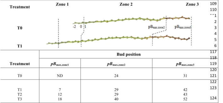
TABLE 1. Latent bud position and delimited zones along the cane
The cane was divided into three zones: zone 1, with newly developing buds during temperature treatments T1, T2 or T3 or after temperature treatment T0 in the greenhouse; and zones 2 and 3, with buds that were not lignified or lignified, respectively, after temperature treatment T0 followed by T1, T2 or T3. ND, no data; pBmax.zone1, pBmax.zone2and pBmax.zone3, maximum bud position
of zones 1, 2 and 3, respectively. Buds at positions – 2 to 0 were excluded from imagery analyses.
" "
"
& & & & & & & & & & & " $ $ $ $ $ $ #$'" 0 $ $ $ $ $ $ ##$" 8 $ $ $ $ ###" ! $ $ $ $ $ $ $ ##(" - $ $ $ $ $ $ ##)" 0 $ $ $ $ $ $ $ $ ##*" ' $ $ $ $ $ ##!" & $ $ $ $ ##+" = $ $ $ $ $ $ $ $ ##%" * $ $ $ $ ##&" 6 $ $ $ $ $ ##'" 0 $ $ $ $ $ $ $ #($" & $ $ $ $ $ $ $ $ #(#" = $ $ $ #((" " #()" " & #(*"
Treatment Zone 1 Zone 2 Zone 3
T0
T1
Treatment
Bud position
pBmax.zone1 pBmax.zone2 pBmax.zone3
T0 ND 24 31 T1 T2 T3 7 12 18 29 29 40 42 43 52
SUPPLEMENTARY TABLE 1. Environmental conditions and duration of each temperature treatment.
The target day/night (D/N) temperatures and the measured maximum, minimum and mean air temperatures (Tmax, Tminand Tmean, respectively),
mean vapour pressure deficit (VPDmean) and mean photosynthetically active radiation (PARd mean) during each treatment are shown.Temperature
treatments T1–T3 were imposed in growth chambers after an initial phase of plant growth in a greenhouse under temperature treatment T0. Data are expressed as means ±standard deviations.
Experimental conditions Temperature treatment Target D/N temperatures (°C) Treatment duration (days)
Tmax(°C) Tmin(°C) Tmean(°C)
VPDmean (kPa) PARd_mean (mol/m2) 26.2 13.9 19.9 1.09 18.3 ±1.1 ± 0.4 ± 0.7 ± 0.18 ± 4.4 21.0 14.6 18.1 0.94 26.2 ± 1.3 ± 1.7 ± 0.6 ± 0.05 ± 1.1 30.3 13.8 23.2 1.07 26.3 ± 0.1 ± 0.2 ± 0.2 ± 0.11 ± 2.8 31.0 22.4 27.4 1.09 27.0 ± 0.2 ± 0.2 ± 0.1 ± 0.04 ± 4.1 Greenhouse T0 25/15 75 Growth chamber T1 20/15 29 Growth chamber T2 30/15 29 Growth chamber T3 30/25 27
three distal buds, which were too small for imagery analysis (Table 1), and (ii)four buds beside this distal zone (one bud each out of 10 from the apex), which were selected for x-ray microtomography analysis.
2.1. Light microscopy
Latent buds were dissected longitudinally in the phyllotaxis plane of the primary bud axes, which corresponds to the dorsal–ventral separation plane of the bearing cane (Supplementary figure 1) (May, 2000). The two sides of the dissected buds were examined by using a stereomicroscope (Stemi 2000-C; Zeiss, Jena, Germany) at a magnification of 6.5´ to 50´, with a cold light source (15 V/150 W) and no filter (KL 1500 compact model; Schott, Mainz, Germany). Images were obtained using a Spot Insight Color digital camera (3.2.0 model; Diagnostic Instruments, Sterling Heights, Michigan, USA). A microscale (with graduations of 0.1 mm) was included in the images.
2.2. X-ray microtomography
For each treatment (T0–T3), high-resolution two-dimensional images of four intact buds were obtained with a Skyscan microtomography (1076 model, Kontich, Belgium; Figure 1A). The samples were scanned at the Montpellier RIO Imaging centre (France; http://www. mri.cnrs.fr/). The parameters were set for low-density objects (i.e. 40 kV, 250 µA and no filter). The resolution was 9 µm, with a step rotation of 0.1°.
Raw images in two dimensions (16 bits) were reconstituted using NRecon software (SkyScan, Kontich), as described by Milien et al. (2012). When the two-dimensional images were outside the phyllotaxis plane of the primary bud, with consequent impairment of accurate measurement of bud morphology, a three-dimensional image was reconstructed using Imaris software (Bitplane, Zurich, Switzerland).The images were then reduced to 8 bits using ImageJ software
" " " " ' ' ' ' ' ' ' ' ' ' ' ' ' ' " $ $ $ $ $ $ $ $ $ $ $ $ $ $ $ $ $ $ $ $ " $ $ $ $ " " " "
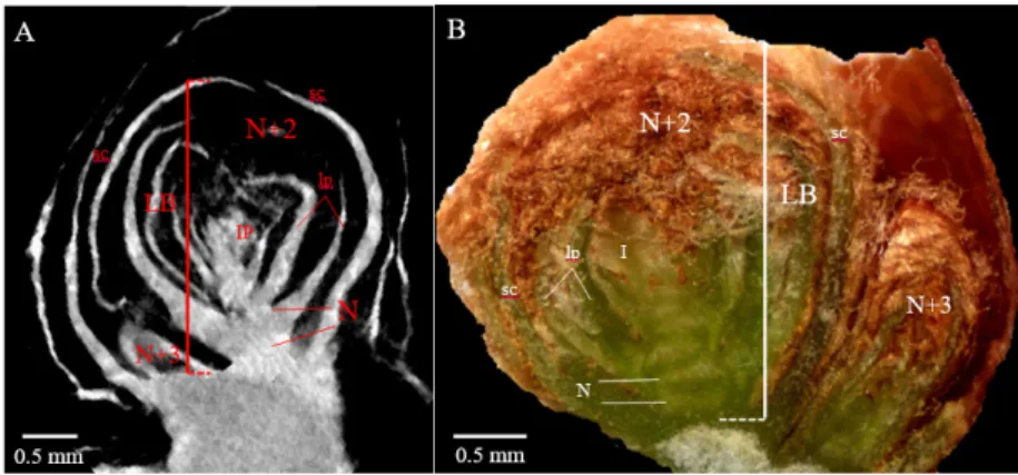
FIGURE 1. Latent bud morphological parameters measured by x-ray microtomography (A) and
stereomicroscopy (B).
IP, inflorescence primordium; LB, length of primary bud; lp, leaf primordium; N, node; N+2, primary-bud primordial shoot; N+3, secondary-bud primordial shoot; sc, bud scale.
SUPPLEMENTARY FIGURE 1. Typical microvine bud anatomy.
Orthogonal view of the bud (A), lateral view of the bud (B) and longitudinal section of the bud along its phyllotaxis plane (C). a, Phyllotaxis plane of the cane axis; b, phyllotaxis plane of the latent bud; N+2, primary-bud primordial shoot; N+3, secondary-bud primordial shoot.
(Rasband, 1997–2011), and to reduce the image size, only the central region of bud was selected.
3. Primary bud development and fruitfulness parameters
Five morphological parameters were measured on the primary buds (N+2 in Figure 1) (Supplementary table 2; see also Supplementary figure 1). The length of the primary bud (LB) corresponded with the distance between the base of the bud’s primordial shoot (the first basal preformed node) and the most external scale of the latent bud, following a longitudinal axis parallel to the bud’s primordial shoot. The number of preformed phytomers along the bud’s primordial shoot was determined from the number of nodes (nN). The number of inflorescence primordia per primordial shoot (nIP) was assessed, together with their individual position from the first basal preformed node (pIP). Lastly, the cane diameter was measured every 10 buds from the cane apex, in the middle part of the internode.
For microscopy, LB was measured using ImageJ software, whereas the other parameters (nN, nIP, and pIP) were determined from direct observations under the microscope (Figure 1B). For microtomography, all bud parameters (see
Figure 1A) were visualized and/or measured with ImageJ, using the two- or three-dimensional image reconstructions of the buds.
4. Bud development zones and calculations
4.1. Bud zone delimitations along the cane Bud positions from the cane apex were determined, excluding the distal zone with the three youngest buds (bud positions–2 to 0; see Table 1), which were too small (< 1 mm long) for imagery analysis. Three zones along the main cane were then delimited to separate the contrasting patterns of bud development under the different temperature treatments. Zone 1 included the newly developed buds during the temperature treatments in the growth chamber (i.e. T1, T2 or T3), plus the three youngest buds after the temperature treatment T0 in the greenhouse.
Buds located beneath zone 1 started developing before the temperature treatments in the growth chamber. They were separated into two additional zones, based on the transition of cane colour from green to yellowish brown (indicating the onset of periderm formation): zone 2 and zone 3 included the non-lignified and lignified buds, respectively, after the periods in the
Frederico A. N. Dias et al.
TABLE 2. Conversion of bud position into thermal time
Bi, bud positioni;pBmax.zone1, maximum bud position of zone 1.
" " " " " " " " " " " " " " " "
& & & & & & & & & "
Treatment Bud zone(s) !"#$%&'(
T0 2 and 3 !""!"#!!! ! !"!!!"!! Equation 1 T1–T3 1 !""!"#!!! ! !"!!!"!! Equation 2 T1–T3 2 and 3 !""!"#!!! ! !"!"#!!"#$!!!!"!! ! !!!!"!! !"!"#!!"#$!!!!!!"!! Equation 3 %&'(%!)*#+#),#-!". % %&'(%!)*#+#),%)5%2),3%41% # " # # # " " " " "
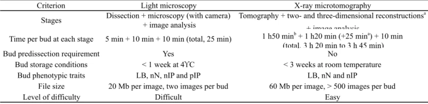
SUPPLEMENTARY TABLE 2. Comparison of light microscopy and x-ray microtomography for
analysis of latent bud anatomy.
LB, length of primary bud; nIP, number of inflorescence primordia per primordial shoot; nN, number of nodes; pIP,
inflorescence position on the bud primordial shoot. a Optional phyllotaxis plane reconstruction. b One to five buds simultaneously.
Criterion Light microscopy X-ray microtomography
Stages Dissection + microscopy (with camera) + image analysis
Tomography + two- and three-dimensional reconstructionsa
+ image analysis
Time per bud at each stage 5 min + 10 min + 10 min (total, 25 min) 1 h50 minb + 1 h20 min (+25 mina) + 10 min (total, 3 h 20 min to 3 h 45 min)
Bud predissection requirement Yes No
Bud storage conditions < 1 week at 4!C < 3 weeks at room temperature Bud phenotypic traits LB, nN, nIP and pIP LB, nN and nIP
File size 20 Mb per image, two images per bud 60 Mb per image, > 500 images per bud
greenhouse and growth chamber. The bud positions delimiting each zone (pBmax.zone1,
pBmax.zone2 and pBmax.zone3) are indicated in
Table 1.
4.2 Probability of inflorescence primordia development
The probability of the primordial shoot within latent buds holding 0, 1 or 2 inflorescence primordia was calculated for all treatments. These probabilities were determined per class of bud developmental stage (i.e. the number of pre initiated phytomer primordia) and for non-lignified buds of similar ages (bud position 1 or 2, see below) for all temperature treatments. 4.3 Conversion of bud position into thermal time The phyllochron was calculated from the reverse of the slope of a simple linear regression between the number of unfolded leaves on the cane and the cumulated thermal time after budburst, as explained by Luchaire et al. (2017). The phyllochron in the greenhouse averaged 21.5 growing degree days (°Cd) (T0, data not shown). It increased to 26.1°Cd (P < 5%) under growth chamber conditions, but it was similar for each of the temperature treatments T1–T3 (P > 5%). Each bud position (i) was then converted into the number of cumulated growing degree days after its emergence (GDDbud_i), using the specific phyllochron for the buds developed in the greenhouse and for the buds developed in the growth chamber (equations 1–3) (table 2).
5. Statistical analyses
Statistical analyses were performed using R (R2.13.2, R Foundation for Statistical Computing, Vienna, Austria). One-way ANOVA
was used for comparisons of means. When significant differences were found (P < 5%), mean values were separated using the Tukey’s honestly significant difference test. Simple linear models were fitted with R using the conventional least squares method. The quality of the fitted lines obtained was evaluated by calculating the root mean square error.
RESULTS
1. Light microscopy versus microtomography
The light microscopy method was relatively fast (25 min per bud) and did not require any expensive equipment (see Supplementary
table 2). This method was suitable for determining primary bud length, the number of phytomers and inflorescence primordia, and the position of the inflorescence on the bud’s primordial shoots. However, a disadvantage of microscopy was the need for accuracy of bud dissection, which had to be performed precisely following the phyllotaxis plane of the primary axis of the latent bud (see Supplementary figure 1). Inaccurate bud dissection automatically led to loss of the sample. Furthermore, cane storage should not exceed 1 week, because longer storage impairs bud dissection, due to changes in organ firmness. X-ray microtomography is a non-destructive or minimally invasive imagery method that requires specific and expensive equipment. This approach is potentially more appropriate than light microscopy when the plant material is rare, or when it requires special labour for histological observations (Smith et al., 2009). Additionally, the lack of need for bud dissection and the possibility of storing the cane samples for up to 3 weeks made microtomography more convenient than light microscopy. However, scanning and image reconstruction are time-consuming, because analysis of one to five buds required at least 3 h (for scanning and two-dimensional reconstruction) versus 1 h for microscopy (see Supplementary table 2). When the bud phyllotaxis plane needed to be reconstructed from three-dimensional images, the processing time for x-ray microtomography was even longer (over 25 min per bud).
Similarly to the light microscopy method, microtomography provided accurate evaluations of primary bud length, number of phytomers and number of inflorescence primordia per bud. However, it was less effective than microscopy for discerning the position of the inflorescence primordia on the main axis (see Figure 1A).
2. Development of the latent bud primordial shoot in microvines
After the T0 treatment under control temperatures (D/N, 25°C/15°C), the number of preformed phytomers on the primordial shoots of primary buds was counted, from the distal buds of the cane to the proximal buds (i.e. from position 1 to pBmax.zone3; see Table 1). The number of preformed phytomers in latent buds increased linearly from the 1st to the 20th bud position, after which it reached a maximum of c.
5.4 on average (Figure 2A). Therefore, the number of phytomers in buds was maximal four nodes above the onset of periderm formation (pBmax.zone2= 24; see Table 1).
Conversion of bud position into growing degree days, using the cane phyllochron (equation 1), enabled assessment of the timing and rate of phytomer differentiation under controlled temperatures (D/N, 25°C/15°C). The number of phytomers was maximal (5.4 on average) 430°Cd after bud emergence (bud position from 1 to 20). Based on those values, the bud’s primordial shoot phyllochron was 79°Cd. It was thus 3.7 times longer than the cane phyllochron (21.5°Cd).
When analysing the effects of temperature treatments (T1–T3) on latent bud developmental traits, only buds that were assumed to pursue their development during the temperature treatments were selected. These were the newly developing latent buds of zone 1 and the non-lignified buds of zone 2 after T0 (see Table 1). Buds beyond the20th position in zone 2 afterT0 were, however, excluded, because no supplemental preformed phytomers were observed below this position (see Table 1 and Figure 2A). Lastly, latent bud traits were examined at bud positions 1–27 after T1 (see Table 1). The same zone was retained for
treatments T2 and T3, to compare buds of the same age at all temperature treatments.
Development of latent bud primordial shoots under T1 treatment (D/N, 20°C/15°C) in growth chambers was similar, in terms of bud position, to that under controlled temperatures (D/N, 25°/15°C) in the greenhouse (Figure 2B). However, differentiation of phytomers ceased earlier, at position 16 under T1 versus position 20 under T0. The bud’s primordial shoot reached a maximum of about 4.1preformed phytomers instead of 5.4 under T0. Conversion of bud position into growing degree days (equations 2 and 3) indicated that primordial shoot development ceased 378°Cd after bud emergence. Its phyllochron was slightly longer than that of the one calculated under control temperatures in the greenhouse (+13°Cd), ranging up to 92°Cd.
Temperature increase during the day (T2: D/N, 30°C/15°C) sharply accelerated the differen-tiation of phytomers (Figure 2C). No additional change was observed compared with T2 when night temperature was also increased (T3: D/N, 30°C/25°C) (Figure 2D). For both treatments (T2 and T3), bud proximal shoot development ceased beyond the fifth bud position, with a maximum of about 4.5 preformed phytomers, similar to T1. The number of preformed phytomers was thus
Frederico A. N. Dias et al.
# #
#
#
FIGURE 2. Relations between number of phytomers on the latent bud primordial shoot and bud position
on the cane or cumulated growing degree days (GDD) after bud emergence for theT0 temperature treatment in the greenhouse (day/night, D/N, 25°C/15°C; A) and the T1, T2 and T3 temperature treatments in the growth chamber (D/N, 20°C/15°C, 30°C/15°C and 30°C/25°C; B, C and D, respectively).
The number of phytomers on the primordial shoot was determined by microscopy and microtomography. Each point is the mean of 2–5 buds for microcopy and corresponds to one unique bud for microtomography. Bars indicate standard errors.
The equations, parameters and root mean square errors (RMSEs) of the fitted lines are the number of phytomers: a ´ bud position + b for bud position < c, otherwise number of phytomers = a ´ b + c
A: a = 0.237; b = 0.712; c = 19.9; a ´ b + c = 5.4; RMSE =0.71. B: a = 0.236; b = 0.708; c = 16.1; a ´ b + c = 4.1; RMSE =0.57. C: a = 0.568; b = 1.703; c = 5.1; a ´ b + c = 4.6; RMSE =0.56. D: a = 0.592; b = 1.776; c = 4.6; a ´ b + c = 4.5; RMSE =0.51.
maximum of two inflorescence primordia (data not shown).
Using the pattern of phytomer development on the primordial shoot of buds (Figure 2A), the first and second inflorescences started to differentiate under controlled temperatures (D/N, 25°C/15°C) from the 14th and 18th bud positions, respectively. By converting those positions into growing degree days, the first inflorescence started to differentiate 301°Cd after bud emergence. This was followed by the second inflorescence primordia differentiation 91°Cd later.
Buds that developed in growth chambers (under T1, T2 and T3) were overall less fruitful, for a given class of bud phytomers, than the buds that developed in the greenhouse and only under T0 (Figure 3B, C). However, the differences were significant only between T0 (D/N, 25°C/15°C) and T2 (D/N, 30°C/15°C) (Figures 3B, C).Under growth chamber conditions, the elevated temperature during the night under T3 (D/N, 30°C/25°C) tended to favour bud fruitfulness, compared with T2 (P < 5%). In contrast, fruitfulness under T2 and T3 did not differ from that under T1.Therefore, the probability of inflorescence primordia differentiation was very little affected by the temperature treatments. However, the lower bud primordial shoot phyllochron under elevated temperatures (92°Cd maximal 133°Cd and 120°Cd after bud
emergence under T2 and T3, respectively. Lastly, the phyllochron of the bud’s primordial shoot was much shorter compared with under T0 and T1, ranging from 28.9°Cd for T2 to 26.7°Cd for T3.
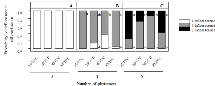
3. Microvine latent bud fruitfulness
Bud fruitfulness was assessed for all growing conditions and temperature treatments (i.e. T0, T1, T2 and T3) on the restricted zone delimited above, based on phytomer development. Bud positions 1–27 from the cane apex (i.e. zone 1 and part of zone 2; see Table 1) were assessed. Under controlled temperatures (D/N, 25°C/15°C), the first and second anlagen differentiated mainly at the fourth and fifth preformed phytomers of the bud’s primordial shoot, and to a lesser extent at the fifth and sixth phytomers (data not shown). Accordingly, no inflorescence primordia were observed on buds holding fewer than four phytomers (Figure 3A).The probability of first anlagen differentiation was maximal when buds carried four preformed phytomers (Figure 3B).When five phytomers were preformed, the probability of differentiation of a second inflorescence primordia reached 0.75 (Figure 3C). All buds carrying six preformed phytomers held a
# # # # # # # &!#
FIGURE 3. Probability of inflorescence primordia differentiation (latent buds holding 0, 1 or 2
inflorescence primordia) per class of bud development (3, 4 or 5 preformed phytomers on the primordial shoot; A, B and C, respectively) for the T0 temperature treatment in the greenhouse (day/night, D/N, 25°C/15°C) and the T1, T2 or T3 temperature treatments in the growth chamber (20°C/15°C, 30°C/15°C and 30°C/25°C).
Only data from buds at the 1st to 27th positions (zone 1 and part of zone 2, respectively) were included in the analysis. Each value is the mean of data from three to six buds.
for T1 versus 28°Cd on average for T2 and T3) accelerated inflorescence differentiation significantly. The first inflorescence started to differentiate at the 14th bud position under T1, similarly to under T0, which corresponded to 332°Cd after bud emergence. When the temperature increased, the first inflorescence differentiated 10 buds earlier, at the 4th bud position, under both T2 and T3. On a thermal time basis, inflorescence differentiation started at 105°Cd and 98°Cd after bud emergence for T2 and T3, respectively.
4. Relation between bud development and morphological parameters
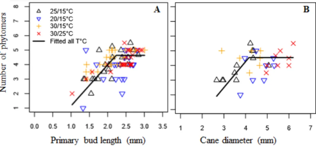
In the present work, we studied relations between latent bud length or cane diameter beneath the bud and the number of preformed phytomers, a parameter that has been shown to be linked to bud fruitfulness (Figure 4). For this analysis, we included all non–fully developed buds (i.e. those in the 1st to 27th bud positions, zone 1 and part of zone 2) in all experiments and temperature treatments (T0–T3).
Unfruitful developing latent buds (i.e. those with three or fewer preformed phytomers) could be associated with bud length less than 1.6 mm (Figure 4A) or cane diameter less than 3.3 mm (Figure 4B). The maximum numbers of phytomers and inflorescence primordia (up to 4.5 preformed phytomers, and 1 or 2 inflorescences) were reached when bud length was more than 2.2 mm or cane diameter more
than 4.2 mm. The intermediate class of bud length and cane diameter therefore corresponded to developing buds holding about one inflorescence (i.e. those with 3 to 4.5 preformed phytomers).
DISCUSSION
1. What are the benefits of x-ray microtomography and microscopy
when characterizing the morphological traits of latent buds?
Contrast in the microtomography image is produced by the attenuation of x-ray photons on the tissues. It depends on x-ray radiation energy, tissue thickness, density and atomic composition (Milien et al., 2012). Notably, differences in oxygen and carbon content in water and cellulose lead to high pixel contrasts in grayscale. Although in our study the microtomography parameters were optimized for latent bud tissue density and sample size, the images lacked contrast. The 9-µm image resolution, together with the slight difference in density and composition between bud tissues, therefore appeared as limiting factors impairing clear observation of some bud morphological traits. In other studies, in which x-ray microtomography technology was used to study the vascular network of grapevine, similar limitations were found at a higher resolution (4.5 µm) (Brodersen et al., 2011; Milien et al., 2012). According to the authors, the method was suitable for tracking different stem tissues (i.e.
Frederico A. N. Dias et al.
# # ' ' ' ' ' ' ' ' ' ' ' ' ' ' ' ' ' ' ' ' ' ' ' # ' ' ' ' ' ' ' ' ' ' ' ' ' ' ' ' ' ' ' ' ' ' ' # ' ' ' ' ' ' ' ' ' ' ' ' ' ' ' ' ' # % % % % % % % % % % % % % % % % % % % % % % % % % % % % % % % # % % % % # # # # $ # # # # # # #
FIGURE 4. Relation between primary bud length (A) and cane diameter (B)
and the number of preformed phytomers on the bud primordial shoot.
Only data from buds at the 1st to 27th positions (zone 1 and part of zone 2, respectively) were included in the analysis. Each value represents one bud or phytomer. The equation, parameters and root mean square errors (RMSEs) of the fitted lines are the number of phytomers: a ´ primary bud length (or cane diameter) + b for bud position (or cane diameter) < c, otherwise number of phytomers = a ´ b + c.
xylem, phloem, pith, annual rings and rays), because they were spatially separated. Conversely, the difference between the green colour intensity of preformed nodes and internodes on microscopic images facilitated detection of the numbers of preformed nodes and the position of inflorescence primordia.
Lastly, both image methods were useful for evaluating bud developmental stage and fruitfulness. Light microscopy provided additional information about the position of inflorescence primordia along the bud’s primordial shoot. This information might be useful for further studies comparing the potential fruitfulness of latent bud main axes and actual fruitfulness after budburst. Light microscopy was also faster and cheaper than microtomography, but it required technical expertise for accurate bud dissection. The emerging imagery technologies could overcome the current limitations of microtomography. Indeed, synchrotron radiation x-ray tomography microscopy is expected to provide high-quality anatomical images up to the cell resolution (0.45 µm) (Smith et al., 2009; Dhondt et al., 2010). These new technologies, allied to three-dimensional image reconstruction software, are therefore potentially promising methods for obtaining new insights into anlagen formation and ramification within latent buds.
2. Is the microvine an accurate model for studying latent bud development?
The primordial shoot of the microvine bud was less developed than that of the macrovine, with a maximum number of preformed phytomers ranging from four to six in the microvine versus 6–12 in the macrovine (Pratt, 1971; Carmona et al., 2008; Vasconcelos et al., 2009). As the first inflorescence primordia started to differentiate from the fourth phytomer in the microvine, similarly to in the macrovine, bud fruitfulness was at the lower limit of the range generally observed for macrovines (i.e. 1 or 2 inflorescence primordia). The maximum number of phytomers and inflorescence primordia of latent buds has been shown to vary along the cane in macrovines, with the most distal buds being generally less developed (Huglin, 1958; Pratt, 1971; Sinirvasan and Mullins, 1981). In the present study, bud primordial shoot development was maximal and stable before the 20th bud position (from the cane apex) under
controlled temperatures in the greenhouse (D/N, 25°C/15°C).
Microvine ML1is a natural gai (gibberellic acid insensitive) mutant of Vitis vinifera L. obtained by somatic regeneration of the L1 cell layer of Pinot Meunier (Boss and Thomas, 2002; Franks et al., 2002; Luchaire et al., 2017). The modified GAI1 protein resulting from the translation of Vvgai1 is no longer able to interact with gibberellins, because their DELLA domain is altered, and this effect is expressed as the dwarf phenotype. Because gibberellins are known to promote cell division in meristematic tissues (Hansen et al., 1999), the insensitivity of the microvine to gibberellins could be responsible for an early interruption of the development of latent bud primordial shoots, thus preventing formation of a third inflorescence. Therefore, the limited bud development was consistent with the properties of the microvine.
The spatiotemporal conversion of bud position into growing degree days allowed us to deepen our knowledge of the different phases of bud development. Specifically, development of latent bud proximal shoots was shown to last 430°Cd after bud emergence, which corresponded to 43 days after bud emergence under control temperature conditions (D/N, 25°C/15°C). The first and second inflorescences started to differentiate 301°Cd (or 30 days) and 392°Cd (or 39 days), respectively, after bud emergence. Periderm formation, 516°Cd (or 52 days) after bud emergence, probably marked the end of bud development and the start of the dormancy period (Pouget, 1963; Lavee and May, 1997). In accordance with our results, Vasconcelos et al. (2009) reported that the initiation and differentiation of two successive inflorescences within latent buds of Chenin blanc occurred over a 45-day period.
3. What is the effect of elevated temperatures on microvine bud fruitfulness?
Bud development responses to elevated temperatures were assessed in growth chamber conditions, where bud fruitfulness tended to be lower than under greenhouse conditions. This was probably due to the lower light intensity in the growth chamber, which was not compensated by the higher photoperiod and daily-cumulated radiation. Lower bud fruitfulness under reduced light intensity has been found in several cultivars by Sanchez and
Dokoozlian (2005).
Gibberellins mediate thermoperiodism, the ability of plants to detect the diurnal temperature change and modify their growth and development accordingly (Arana et al., 2011). It would therefore have been possible to observe differences in response during development of microvine latent buds under fluctuating temperatures. However, the results of a previous study on the microvine have shown that its specific mutation does not affect the phyllochron of the cane, which was similar to that of other grapevine cultivars such as Grenache and Syrah (Luchaire et al., 2017).
In the present study, latent bud fruitfulness of microvines was shown to be positively affected by temperature elevation, in a similar way to in macrovines. The comparison of buds at similar stages between the different temperature treatments showed that higher fruitfulness under warm conditions was only slightly linked to a higher probability of inflorescence differentiation in the buds; rather, it was due to acceleration of development of the bud’s primordial shoot. Indeed, the phyllochron was shown to be 3-fold shorter under warm temperature treatments (D/N, 30°C/15°C and 30°C/25°C) than under a cool treatment (D/N, 20°C/15°C). As a result, the first inflorescence started to differentiate 332°Cd (or 41 days) after bud emergence under cool conditions, in contrast to 101°Cd (or 6 days) on average under warm conditions. Many studies have also reported positive effects of elevated temperatures during the prebloom and bloom period on bud fruitfulness and subsequent yield in the following growing season (Pouget, 1981; Sánchez and Dokoozlian, 2005; Watt et al., 2008; Molitor and Keller, 2016).Similar to our results, a temperature increase of 4°C has been shown to accelerate the initiation and differentiation of the anlagen for Chardonnay by 2–3 weeks and to favour its level of branching (Watt et al., 2008). Therefore, the model of microvine bud development parameterized in the present study may be useful to better targetthe specific period of fruitfulness sensitivity to temperature in further experiments.
4. Is there any proxy to monitor bud development?
The laboriousness of light microscopy, and the high cost and current limitations of
microtomography for studying the effects of temperature on bud development, lead us to seek other options for the analysis of internal anatomy of grapevines. A link between phytomer diameter and bud fruitfulness has been reported for macrovines (Huglin, 1958; Sanchez and Dokoozlian, 2005; Eltom et al., 2015).
The three classes of latent buds described in the present work, that is latent buds with less than 1.6 mm of bud length or less than 3.3 mm of cane diameter, buds that reached more than 2.2 mm of bud length or cane diameter more than 4.2 mm and those intermediate buds with bud length between 1.6 to 2.2 mm or cane diameter between 3.3 and 4.2 mm may be assessed at first glance to describe latent buds based on their potential developmental stage and fruitfulness. Such estimates might be useful for improved targeting of buds for imagery analysis, after a period of growth under different temperature treatments. Moreover, such preselection may be necessary when the integrity of buds needs to be preserved for further molecular studies. However, these classes may not be relevant under extreme temperature or high light fluctuation, which have been shown to strongly affect bud development (Buttrose, 1969; Buttrose, 1970).
CONCLUSION
Latent bud development and fruitfulness in microvines were analysed through light microscopy and microtomography imagery methods. At the current operability of these technologies, light microscopy proved to be more accurate for assessing latent bud organogenesis and anatomy. The pattern of phytomer and inflorescence primordia development in microvine latent buds (in terms of rate and timing) was shown to be similar to that of macrovines, although it ceased earlier, thus resulting in a slight lower fruitfulness. Elevated temperature increased the potential fruitfulness of latent buds, mostly through acceleration of the development of the bud’s primordial shoot. In contrast, the probability of inflorescence primordia differentiation was little changed.
The present study therefore provides new insight into the characterization of bud developmental phases along the cane and over time, which may be useful for ecophysiological modelling approaches. Easy-to-measure and
destructive proxies, such as bud length and cane diameter beneath the bud, proved potentially useful for approximating bud developmental stage. Finally, the microvine, which was found to be more convenient than the macrovine for performing experiments under fully controlled environments, proved to be suitable for studying the effect of temperature on bud development and fruitfulness. This grapevine model seems promising and may improve our understanding of the physiological and molecular mechanisms regulating bud organogenesis under fluctuating environments.
REFERENCES
Arana M.V., Marín-de la Rosa N., Maloof J.N., Blázquez M.A. and Alabadí D., 2011. Circadian oscillation of gibberellin signaling in Arabidopsis. Proceedings of the National Academy of Sciences, 108, 9292.
Boss P.K. and Thomas M.R., 2002. Association of dwarfism and floral induction with a grape ‘green revolution’ mutation. Nature, 416(6883), 847–850. doi:10.1038/416847a
Brodersen C.R., Lee E.F., Choat B., Jansen S., Phillips R.J., Shackel K.A., McElrone A.J. and Matthews M.A., 2011. Automated analysis of three-dimensional xylem networks using high-resolution computed tomography. New Phytologist, 191(4), 1168–1179.
Buttrose M.S., 1969. Fruitfulness in grapevines: Effects of light intensity and temperature. Botanical Gazette,130(3), 166–173.
Buttrose M.S., 1970. Fruitfulness in grapevines: The response of different cultivars to light, temperature and day length. Vitis, 9, 121–125.
Buttrose M.S., 1974. Climatic factors and fruitfulness in grapevines. Horticultural Abstracts, 44, 319–326. Carmona M.J., Chaib J., Martinez-Zapater J.M. and Thomas M.R., 2008. A molecular genetic perspective of reproductive development in grapevine. Journal of Experimental Botany, 59(10), 2579–2596. doi:10. 1093/jxb/ern160
Chaïb J., Torregrosa L., Mackenzie D., Corena P., Bouquet A. and Thomas M., 2010. The grape microvine – a model system for rapid forward and reverse genetics of grapevines. Plant Journal, 62(6), 1083–1092. doi:10.1111/j.1365-313X.2010.04219.x Clingeleffer P.R., 1989. Effect of varying node number per bearer on yield and juice composition of Cabernet Sauvignon grapevines. Australian Journal of Experimental Agriculture, 29, 701–705.
Cox C.M., Favero A.C., Dry P.R., McCarthy M.G. and Collins C., 2012. Rootstock effects on primary bud necrosis, bud fertility, and carbohydrate storage
in Shiraz. American Journal of Enology and Viticulture, 63, 277–283.
Dhondt S., Vanhaeren H., Van Loo D., Cnudde V. and Inze D., 2010. Plant structure visualization by high-resolution X-ray computed tomography. Trends in Plant Science, 15(8), 419–422. doi:10.1016/ j.tplants.2010.05.002
Dry P.R., 2000. Canopy management for fruitfulness. Australian Journal of Grape and Wine Research, 6(2), 109–115. doi:10.1111/j.1755-0238.2000.tb 00168.x
Dunn G.M., 2005. Transforming flower to fruit. ASVO Seminar, 10, 116–124.
Eltom M., Winefield C.S. and Trought M.C.T., 2014. Effect of pruning system, cane size and season on inflorescence primordia initiation and inflorescence architecture of Vitis vinifera L. Sauvignon Blanc. Australian Journal of Grape and Wine Research, 20, 459–464. doi:10.1111/ajgw.12097
Eltom M., Winefield C. and Trought M.C.T., 2015. Effects of shoot girdling and/or periodic leaf removal on inflorescence primordia initiation and development in Vitis vinifera L. cv. Sauvignon Blanc. Australian Journal of Grape and Wine Research, 21, 118–122. doi:10.1111/ajgw.12113
Fernandez L., Torregrosa L., Segura V., Bouquet A. and Martinez-Zapater J-M., 2010. Transposon-induced gene activation as a mechanism generating cluster shape somatic variation in grapevine. Plant Journal, 61, 545–557. doi:10.1111/j.1365-313X. 2009.04090.x
Franks T., Botta R. and Thomas M., 2002. Chimerism in grapevines: Implications for cultivar identity, ancestry and genetic improvement RID A-3117-2010. Theoretical and Applied Genetics, 104, 192–199.
Fromm J.H., Sautter I., Matthies D., Kremer J., Schumacher P. and Ganter C., 2001. Xylem water content and wood density in spruce and oak trees detected by high-resolution computed tomography. Plant Physiology, 127(2), 416–425.
Guilpart N., Metay A. and Gary, C., 2014. Grapevine bud fertility and number of berries per bunch are determined by water and nitrogen stress around flowering in the previous year. European Journal of Agronomy, 54, 9–20. doi:10.1016/j.eja.2013.11.002 Hansen E., Olsen J.E. and Junttila O., 1999. Gibberellins and subapical cell divisions in relation to bud set and bud break in Salix pentandra. Journal of Plant Growth Regulation, 18, 167–170.
Houel C., Chatbanyong R., Doligez A., Rienth M., Foria S., Luchaire N., Roux C., Adivèze A., Lopez G., Farnos M., Pellegrino A., This P., Romieu C. and Torregrosa L., 2015. Identification of stable QTLs for vegetative and reproductive traits in the microvine (Vitis vinifera L.) using the 18 K
Infinium chip. BMC Plant Biology, 15(205), 1–19. doi: 10.1186/s12870-015-0588-0
Huglin P., 1958. Recherches sur les bourgeons de la vigne: Initiation florale et développement végétatif. Annales de l’Amélioration des Plantes, 8, 113–272. Jones J.E., Lee G. and Wilson S.J., 2013. A statistical model to estimate bud fruitfulness in Pinot noir. American Journal of Enology and Viticulture, 64(2), 274–279. doi:10.5344/ajev.2013.12086
Lavee S. and May P. 1997. Dormancy of grapevine buds –Facts and speculation. Australian Journal of Grape and Wine Research, 3(10), 31–46. doi:10. 1111/j.1755-0238.1997.tb00114.x
Li-Mallet A., Rabot A. and Geny L., 2016. Factors controlling inflorescence primordia formation of grapevine: Their role in latent bud fruitfulness? A review. Canadian Journal of Botany, 94(3), 1–17. doi:10.1139/cjb-2015-0108
Luchaire N., Rienth M., Romieu C., Nehe A., Chatbanyong R., Houel C., Ageorges A., Gibon Y., Turc O., Muller B., Torregrosa L. and Pellegrino A., 2017. Microvine: A new model to study grapevine growth and developmental patterns and their responses to elevated temperature. American Journal of Enology and Viticulture, 68(3), 283–292. doi:10.5344/ajev.2017.16066
May P., 2000. From bud to berry, with special reference to inflorescence and bunch morphology in Vitis vinifera L. Australian Journal of Grape and Wine Research, 6(2), 82–98. doi:10.1111/j.1755-0238.2000.tb00166.x
McLoughlin S.J., Petrie P.R. and Dry P.R., 2011. Impact of bud position and bearer length on the yield components in mechanically pruned Cabernet-Sauvignon (Vitis vinifera L.). Australian Journal of Grape and Wine Research,17, 129–135
Milien M., Renault-Spilmonta A.S., Cookson S.J., Sarrazina A. and Verdeilc J.L., 2012. Visualization of the 3D structure of the graft union of grapevine using X-ray tomography. Scientia Horticulturae, 144, 130–140.
Molitor D. and Keller M., 2016. Yield of Müller-Thurgau and Riesling grapevines is altered by meteorological conditions in the current and the previous growing seasons. OENO One, 50(4), 245–258. doi:10.20870/oeno-one.2016.50.4.1071 Morrison J.C., 1991. Bud development in Vitis vinifera L. Botanical Gazette, 152, 304–315.
Ollat N., Marguerit E., Lecourieux F., Destrac-Irvine A., Barrieu F., Dai Z., Duchêne E., Gambetta G., Gomes E., Lecourieux D., van Leeuwen C., Simonneau T., Torregrosa L., Vivin P. and Delrot S., 2018. Grapevine adaptation to abiotic stresses: An overview. XIIth International Conference on Grape Breeding and Genetics, 16–20 July, Bordeaux, France.
Petrie P.R. and Clingeleffer P.R., 2005. Effects of temperature and light (before and after budburst) on inflorescence morphology and flower number of Chardonnay grapevines (Vitis vinifera L.). Australian Journal of Grape and Wine Research. 11(1), 59–65. doi:10.1111/j.1755-0238.2005.tb00279.x
Pouget R., 1963. Recherches physiologiques sur le repos végétatif de la vigne (Vitis viniferaL.): La dormance des bourgeons et le mécanisme de sa disparition. Annales de l’Amélioration des Plantes, 13(1), 1–247.
Pouget R., 1981. Action de la température sur la différenciation des inflorescences et des fleurs durant les phases de pré-débourrement et de post-débourrement des bourgeons latents de la vigne. Connaissance de la Vigne et du Vin, 15, 65–79. Pratt C., 1971. Reproductive anatomy in cultivated grapes – A review. American Journal of Enology and Viticulture, 22, 92–109.
Rasband, 1997–2011. ImageJ. National Institutes of Health, Bethesda, Maryland, USA. http://imagej.nih. gov/ij
Rienth M., Torregrosa T., Luchaire N., Chatbanyong R., Lecourieux D., Kelly M.T. and Romieu C., 2014. Day and night heat stress trigger different transcriptomic responses in green and ripening grapevine (Vitis vinifera) fruit. BMC Plant Biology, 14(108), 1–18. doi:10.1186/1471-2229-14-108
Sánchez L.A. and Dokoozlian N.K., 2005. Bud microclimate and fruitfulness in Vitis vinifera L. American Journal of Enology and Viticulture, 56(4), 319–329.
Smith S.Y., Collinson M.E., Rudall P.J., Simpson D.A., Marone F. and Stampanoni M., 2009. Virtual taphonomy using synchrotron tomographic microscopy reveals cryptic features and internal structure of modern and fossil plants. Proceedings of the National Academy of Sciences, 106(29), 12013–12018. doi:10.1073/pnas.0901468106
Srinivasan C. and Mullins M.G., 1981. Physiology of flowering in the grapevine – A review. American Journal of Enology and Viticulture, 32(1), 47–63. Steppe K., Cnudde V., Girard C., Lemeur R., Cnudde J.P. and Jacobs P., 2004. Use of X-ray computed microtomography for non-invasive determination of wood anatomical characteristics. Journal of Structural Biology, 148(1), 11–21. doi:10.1016/j.jsb.2004.05.001
Stuppy W., Maisano J., Colbert M., Rudall P. and Rowe T., 2003. Three-dimensional analysis of plant structure using high-resolution X-ray computed tomography. Trends in Plant Science, 8(1), 2–6. Torregrosa L., Bigard A., Doligez A. Lecourieux D. Rienth M., Luchaire N., Pieri P. Chatbanyong R., Shahood R., Farnos M., Roux C., Adiveze A.,
Pillet J., Sire Y., Zumstein E., Veyret M., Le Cunff L.,Lecourieux F., Saurin N., Muller B., Ojeda H., Houel C., Péros J.P., This P., Pellegrino A. and Romieu C., 2017. Developmental, molecular and genetic studies on the grapevine response to temperature open breeding strategies for adaptation to warming. OENO One, 51, 155–165. doi:10.20870/ oeno-one.2016.0.0.1587
Vasconcelos M.C., Greven M., Winefield C.S., Trought M.C.T. and Raw V., 2009. The flowering process of Vitis vinifera: A review. American Journal of Enology and Viticulture, 60(4), 411–434.
Watt A.M., Dunn G.M., May P.B., Crawford S.A. and Barlow E.W.R., 2008. Development of inflorescence primordia in Vitis vinifera L. cv. Chardonnay from hot and cool climates. Australian Journal of Grape and Wine Research, 14, 46–53. doi:10.1111/j.1755-0238.2008.00006.x