The Analysis of Complex Motion Patterns
in Primate Cortex
By
Bard J. Geesaman
A. B. Neurobiology, University of California at Berkeley 1989
Submitted to the Department of Brain and Cognitive Sciences in
Partial Fulfillment of the Requirements for the Degree of
Doctor of Philosophy
LIBRIE
at the
k
Massachusetts Institute of Technology
September 1995
© Massachusetts Institutute of Technology 1995
All Rights Reserved
Signature of Author
Bard J. Geesaman
Department of Brain and Cognitive Sciences
June 29, 1995
Certified by
Richard A. Andersen
Thesis Supervisor
James G. Boswell Professor of Neuroscience
Accepted by
rofessor Gerald E. Schneider
Chairman, Department Graduate Committee
MASSACHUSETTS INSTITUTF
:JUN
2
9
1995
To My Friends:
Sandra Lee and Mai-khanh Tran for Caring
Andrew Karson and Hoang Tran for Your Kindness
Paul Loh and Glenn Yamagata for Shared Beliefs
George Daley and Edward Renwick forYour Company
The Analysis of Complex Motion Patterns in Primate Cortex
by
Bard J. Geesaman
Submitted to the Department of Brain and Cognitive Sciences on June 29,
1995 in partial fulfillment of the requirements for the degree of Doctor of
Philosophy in Neuroscience
Abstract
I have been interested in how the nervous system processes complex
motion patterns such as expansion, rotation, and contraction. The analysis
of these types of stimuli is thought to be important for the analysis of
self-motion and the self-motion of objects in the environment. Anatomical,
physiological, and psychophysical approaches were all used in an attempt
to understand these processes. The four chapters break down as follows:
Chapter 1) This is a review of the literature on cortical area MSTd, a
region of the brain thought to be important in the analysis of complex
motion patterns. This article starts with an overview of the anatomy and
physiology of this area before a discussion of the possible roles this region
may play in visual motion processing. The view taken in this discussion is
that MSTd is likely to be involved in a number of different functions, possibly
including egomotion, object motion, vection, and retinal slip.
Chapter 2) Our lab has proposed that MSTd is important in the analysis
of object motion in the environment. Our hypothesis is that this region
extracts information about motion pattern from the visual scene and
largely ignores the features and cues which define this motion. To test this
idea, we recorded from single units in MSTd and obtained tuning curves
using motion patterns defined by different features and cues. I found that
the preferred complex motion for a particular neuron was independent of
the features and cues (i.e. form) of the inducing stimulus.
Chapter 3) Multiple single-unit recording studies in both MT and MSTd
have reported clustering of neurons according to preferred tuning. I
decided to explore this organization using the double-label 2-deoxyglucose
method. The evidence supports a columnar functional organization to both
MT and MSTd, with units tuned for expansion and contraction maximally
separated in a mosaic of columns of altering specificity.
Chapter 4) There are many more units in MSTd tuned to expansion than
to rotation. To see if this anisotropy in response distribution had perceptual
consequences, we compared perceived average dot speeds in random-dot
complex motion patterns. We found that dots in expansion stimuli appeared
to move about 25% faster than the same speed dots in a rotating pattern. The
magnitude of this illusion in various variations of this experiment
compared well with reported response characteristics of MSTd cells.
Acknowledgments
I thank Richard Andersen and Ning Qian for their support and
guidance over the last four years.
I would like to thank the members of the Andersen laboratory for
creating such a friendly and stimulating surrounding. I doubt if I will ever
work with such a brilliant group of people again, especially if I decide to
explore a career in medicine. Special thanks to Catherine Cooper, Steve
Marchetti, and Gail Robertson for their technical assistance. Stefan Treue
and Ning Qian proved ideal mentors in the area of systems neuroscience. I
would also like to thank Pietro Mazzoni for his help and for filling the lab
with refined choices of music. David Bradley was invaluable for his patient
explanations of statistical issues and overall good taste.
The last four months of my thesis work, I had the privilege to work with
Richard Born. If it wasn't for his generous outlay of time and support,
Chapter 3 would not have been completed. Thanks Rick for being such a
good guy.
I thank Peter Schiller, Ken Nakayama, Ning Qian, and Mriganka Sur
for serving on my thesis committee. Also I am indebted to Jan Ellertsen and
Jerry Schneider for piloting me through my final year without a local
advisor.
I would also like to extend my thanks to my students and colleagues at
Quincy House, my home for the past three years. In particular I would like
to express my appreciation to George Daley and William Bennett, who have
shared their wisdom and friendship. A special thanks to Michael Shinagel,
Master of Quincy House, for providing me with a free roof over my head and
introducing me to Single Malt Scotch.
Finally, I would like to thank my parents for their continuing support.
They instilled in me a love for knowledge and education that has kept me in
Table of Contents
A b stract ...
3
A cknow ledgm ents
...
4
Chapter 1 ...
6
Cortical Area MST and Optical Flow: A Review
Bard J. Geesaman and Richard A. Andersen
C hapter 2
...
86
The Representation of Motion Pattern in Form/Cue
Invariant MST Neurons of the Macaque
Bard J. Geesaman and Richard A. Andersen
C hapter 3
...
155
Functional Organization in Macaque Superior Temporal
Sulcus with Regards to Complex Motion Patterns
Bard J. Geesaman, Richard T. Born, Roger B. H. Tootell, and
Richard A. Andersen
C hapter 4 ...
195
A Novel Speed Illusion Involving Complex Motion
Patterns Related to Cortical Area MSTd
Bard J. Geesaman and Ning Qian
(Submitted to Vision Research)
Chapter 1: Cortical Area MST and Optical Flow: A Review
Chap ter
1
Chapter 1: Cortical Area MST and Optical Flow: A Review
Introduction
The purpose of this article is to review what is known about the medial superior temporal (MST) area of the macaque monkey. The first section is dedicated to a discussion of the anatomy and physiology of this cortical area. The remainder of the article is a discussion of the various roles that MST may be playing in visual information processing. Particular emphasis is given to reviewing the literature of ego-motion (direction of heading) representation and pattern motion perception. In addition, we will consider strategies thought to be used by other species to solve these problems. The scope of the discussion will be quite broad, drawing on studies from physiology, psychophysics, and computational neurobiology. In the spirit of systems neuroscience, an attempt will be made to integrate all three approaches. We will propose that the function MST plays is likely to be quite generic and that this region could potentially be involved in many perceptual processes.
Anatomy and Physiology
MST is located medial and anterior to area MT on the floor and anterior bank of the superior temporal sulcus (STS) of the macaque. The long axis of this region is oriented mediolaterally and has a length of approximately 4-5
mm along this direction (Desimone and Ungerleider, 1986; Saito et al, 1986).
Anatomical tracer studies have demonstrated connections with visual polysensory areas in posterior prestriate, parietal, temporal, and frontal cortex (Boussaoud et al, 1990). By far its heaviest input is from area MT, anatomically implicating MST in the processing stream believed to analyze visual motion. Heavy lateral connections to area FST in the fundus of the
STS have been established, although the significance of these connections
and the role that FST is playing in the hierarchy of motion processing has yet to be determined. Other local projections within the STS include reciprocal connections to polysensory association areas (Desimone and
Chapter 1: Cortical Area MST and Optical Flow: A Review
Ungerleider, 1986; Boussaoud et al, 1990). These regions are thought to be involved in the convergence of information from several sensory modalities and facilitate tasks which require integration of these signals. A descending connection from MST to the lateral terminal nucleus of the accessory optical system has been identified (Maioli et al, 1989). The AOS has been solidly established as an important area for the processing of self-motion in birds (Frost et al, 1990) and rabbits (Simpson, et al 1988) and we
will later discuss the possible functional significance of this projection in primates. Forward connections from MST include those to posterior parietal cortex (Boussaoud et al, 1990), in particular to areas LIP and VIP. Ongoing investigations by workers in our lab have provided strong evidence that these regions are involved in representing external space in coordinate frames progressively independent from the projection of the optical array onto the retina (Andersen, 1989). Although the perception of motion can be dissociated from that of spatial displacement (for a review see Nakayama, 1985) they are clearly related under normal perceptual conditions and the connection of MST to these posterior parietal regions may well be related to this association. Another forward connection has been established between MST and the frontal eye fields. As will be discussed shortly, MST has been implicated as a step in the sensory-motor loop involved in smooth pursuit eye movements. Given that the frontal eye fields have been implicated in the initiation of eye movements, this projection is conceivably part of this same system. In general, MST gets its inputs from prestriate visual cortex, has intermediate connections with parietal areas, and projects forward to the frontal eye fields and areas in the rostral STS (Boussaoud et al, 1990).
MST was originally thought to be a single functional region based on early anatomical studies, but later investigations with single unit recording suggested that there are at least two or three functional subdivisions (Wurtz et al, 1988; Newsome et al, 1988; Komatsu et al, 1988). Under one established classification, MST has been divided into a dorsal (MSTd), a ventral (MSTI), and an intermediate region (MSTi). Somewhat confusingly, different
Chapter 1: Cortical Area MST and Optical Flow: A Review
groups have disagreed about whether to pool data collected in MSTi with that in MST1 or MSTd.
A crude sort of retinotopy has been established across these regions. The most dorsal and ventral sections independently represent the central visual field, while the intermediate region covers the periphery (Tanaka et al, 1993). In terms of functional anatomy, some clustering of response selectivity has been reported by several different groups (Tanaka et al, 1993; Saito et al, 1986; Lagae et al, 1994). Lagae has gone so far as to propose a columnar organization to this selectivity. In his conception, each isotuning column contains units tuned to a single motion pattern, e.g. expansion, contraction, or rotation. According to his findings, response selectivity gradually shifts moving orthogonal to the long axis of the isotuning columns. For example, progressing across these columns selectivity may shift from cells tuned to expansion, to rotation, to contraction, to the opposite direction of rotation, and finally back to expansion. In this scheme, columns for expansion and rotation are maximally far apart, as are the columns for the two directions of rotation. This cycle is thought to repeat every 800-900 microns.
Response Selectivity
Given the widely held belief that both MST and MT are involved in the processing of motion information, it is important to be able to distinguish between the two regions based on physiology (Lagae et al, 1994). One of the most obvious differences between the two areas is that MST has much larger receptive fields than MT. The diameter of an MT receptive field is approximately equal to the distance from the fovea to its receptive field center. There is no such rule for neurons in MST, where receptive field sizes can be as large as 100 degrees across in diameter and whose median size is somewhere between 20 and 40 degrees, depending on the type of stimulus used to map the receptive field. In terms of comparing their responses to motion patterns, both regions respond well to stimuli containing unidirectional (linear, or translational) motion, expansion,
Chapter 1: Cortical Area MST and Optical Flow: A Review
contraction, and rotation. However, unlike MST cells, MT cells are not responding to expansion, contraction, and rotation per se but to the local, nearly linear motion signals which make up these patterns. MST cells as a population give significantly weaker responses (in terms of firing rate) to linear motion, expansion, and deformation than MT cells. However, if activity is normalized based on responsiveness to linear motion, MST responds better to rotation and less to flicker, compared to MT (Lagae et al, 1994). The same study reported that, with regards to selectivity along the expansion/contraction axis, MST cells are more directionally selective. This increased selectivity is reflected in the finding that MST cells respond, on average, to fewer motion types than MT cells and that they are more likely to show an inhibitory response to a stimulus in the anti-preferred direction (Lagae et al, 1994). In MT, responses to such elementary flow components (EFCs) as divergence (expansion/contraction), curl (rotation), and deformation depend on the spatial location of these stimuli in the cell's receptive field, but in MST positional invariance to EFCs in a significant percentage of units has been reported by several groups (Lagae et al, 1994; Graziano et al, 1994). Finally, studies probing the subunit structure of units in these two areas have shown that MST cells often have their excitatory and inhibitory regions of their receptive fields overlap, while MT maintains a center/surround spatial separation (Tanaka et al, 1986).
As mentioned above, MST has been divided into different subregions based on differences in response characteristics and proposed function. MST1 and MSTd should be considered as two distinct cortical areas. MSTd prefers large, textured, motion stimuli (Tanaka et al, 1993; Tanaka et al, 1989ab; Saito et al, 1986; Komatsu et al, 1988; Wurtz et al, 1988) and has neurons broadly tuned with respect to speed. Cells respond well to stimuli with speeds as low as 1 degree/second and gradually increase their responses until an amplitude plateau is reached at speeds of about 20 degrees/second (Komatsu et al, 1988). Most of the cells in MSTd are directionally tuned with respect to translational motion, but preferred and anti-preferred directions commonly reverse with stimulus size, indicating
Chapter 1: Cortical Area MST and Optical Flow: A Review
that, unlike MT cells, these units are not selective for the spatial-temporal fourier energy of the stimulus (Komatsu et al, 1988).
Probably the most distinguishing characteristic of cells in MSTd is their selectivity for the elementary flow components of rotation, expansion, and contraction (Tanaka et al, 1989ab; Saito et al, 1986; Orban et al, 1992). Single unit recordings in the anaesthetized, paralyzed macaque have demonstrated greater selectivity for isotropic (real) expansion and rotation compared to axial expansion and rotation. With axial expansion, all features in the stimulus move orthogonal to a motion border which bisects the stimulus. The features on the two sides of this border have their motion vectors pointed 180 degrees away from each other. Therefore, in these displays there are only two directions of motion in the image, moving in opposite directions. Axial rotation, which is the same things as shear, has similar motion components as axial expansion, but in this case the velocity vectors on either side of the motion border are oriented parallel to it. This preference for isotropic patterns is interesting, although little attention has been given to it in terms of its possible functional significance.
Early studies in MST that first reported selectivity for EFCs used as a stimulus generator a slide projector with a zoom lens projecting an image consisting of randomly placed dots (Tanaka et al, 1989). The image could be made to expand, contract, or rotate, depending on the proper manipulation of the lens. Several stimulus attributes, in addition to the global motion pattern present in the display, were available for cells in MSTd to potentially respond to. These included a radially oriented speed gradient - the speed of each feature (dot) in the stimulus was a linear function of its distance from the center of the stimulus. Additionally, when zooming the lens to create the expansion pattern, each dot changed its size in the display, another possible cue that MST cells could be selective for, independent of global motion. Finally, the features in all these stimuli had an acceleration component to their motion vectors oriented along the radial axis of the image. It was shown that none of these cues in isolation, with the exception
Chapter 1: Cortical Area MST and Optical Flow: A Review
of global motion, gave a tuned response in the cells recorded from (Tanaka et al, 1989). Speed gradient, in conjunction with the spatial arrangement of velocity vectors in the image, had a facilitory role in response selectivity, although the effect was modest.
Positional Invariance
To say that a unit is selective for a global motion pattern such as expansion or rotation, it is necessary to demonstrate that this property emerges independently from selectivity to translational motion. For example, by appropriately placing a subregion of an expanding stimulus over the receptive field of a unit tuned specifically to translation, a brisk response can be elicited that exhibits all the properties of being directionally selective. However, moving the stimulus within such a neuron's receptive field will change the selectivity of the unit, even reversing it, so that it now prefers the opposite type of global motion. So, to say that a unit is selective for a particular EFC, we must demonstrate that its response is positionally invariant with regards to the spatial placement of the EFC within the receptive field. The invariance established at the two spatial locations need not be reflected in the width or the height of the tuning curve, only with regards to the direction of maximal response, i.e. the preferred motion pattern. Approximately half of the cells in MSTd demonstrate this type of selectivity (Saito et al, 1986; Tanaka et al, 1989; Lagae et al, 1994) and this invariance is a distinguishing characteristic when identifying this region based on response properties. Tuning invariance has also been observed for MSTd cells with respect to speed (Lagae et al, 1994) and with respect to cue, feature, and form (see Chapter Two of this thesis).
Single, Double, and Triple Component Selectivity
Several investigators have characterized cells in MSTd based on the number of global motion types (EFCs) the cell is selective for. Selectivity, used this way, is defined as a unit responding significantly differently to stimuli on opposite sides of the stimulus space. For example, we say that a
Chapter 1: Cortical Area MST and Optical Flow: A Review
cell is selective for translational motion leftward if the cell fires significantly more when presented with leftward motion than rightward motion. Selectivity in MST can be determined separately for linear motion, divergence (expansion/contraction), and curl (rotation). In this way, units are characterized as being either single, double, or triple component, depending on the number of motion types to which these units are sensitive. To a rough approximation, the population of cells in MSTd is equally distributed into thirds when divided in this way (Duffy and Wurtz, 1991b). Single component cells respond exclusively to either expansion, contraction, one direction of rotation, or translation (linear) motion. Cells of this class exhibit the greatest direction selectivity, are most likely to show significant inhibitory responses, have the greatest degree of positional invariance with respect to stimulus placement in their receptive fields, and have the greatest tendency for their excitatory and inhibitory subregions to overlap
(Duffy and Wurtz, 1991b).
There is a significant amount of disagreement about the response characteristics of double component cells. Graziano, et al (1994) found cells selective to the full range of EFC combinations, while another group found no evidence of cells selective for translational motion in combination with any other EFC (Duffy and Wurtz, 1991a). Lagae, et al (1994), agreed with Graziano, et al (1994) with regards to the existence of cells selective both to translational motion and an EFC, but found no cells selective for either expansion/contraction paired with a rotation. This disagreement is an important one because our lab has argued that the presence of this type of double component cell really represents a population of cells tuned to intermediate directions of global motion that appear perceptually as spirals (Graziano et al, 1994). As will be discussed below, the existence of cells tuned to spiral motion patterns is relevant to the way MST may be processing optical flow information. At the present, we have no way of reconciling these conflicting claims.
Chapter 1: Cortical Area MST and Optical Flow: A Review
The triple component units, those cells responding to three types of global motion, as a population had the least amount of directional selectivity, the smallest likelihood of having significant inhibitory responses to anti-preferred stimuli, the least positional invariance, and exhibited the least likelihood of having their excitatory and inhibitory subregions overlap (Duffy and Wurtz, 1991ab). Statistical analysis of response trends between single, double, and triple component cells indicate that this distinction is an arbitrary one. A continuum of selectivity for various combinations of motion types exists in the population of MSTd cells, and no clustering occurs in the parameter space defined by component responsiveness.
Graziano, et al (1994) developed this idea in depth and proposed that rather than multiple-component cells being independently selective for multiple component types, there is one "preferred" global motion stimulus for each cell. For example, consider an observer moving forward through the environment while making a smooth pursuit eye movement leftward. The global motion pattern projected onto his retina will be determined by the vector addition of a rightward linear motion field due to leftward eye rotation and an expanding motion field produced by forward translation. A subpopulation of multiple-component cells will be maximally selective for this particular complex vector field. However, this selectivity is not complete, and presentation of the component motion fields alone (translation or expansion) would also activate this cell, although not drive it maximally. Although selective for a single complex motion pattern, such a unit would be classified as double component.
An EFC that we have not discussed much is deformation. Very few cells in MST have been found that are selective to this type of motion (Lagae et al, 1994). As will be discussed later, the absence of such units argues against this region playing a direct role in analyzing structure-from-motion.
Chapter 1: Cortical Area MST and Optical Flow: A Review
Figure vs. Field Cells
Some reports have divided MST cells into those with "figure" selectivity and those with "field" selectivity (Saito et al, 1986; Tanaka et al, 1989). These studies contain the first discussion of the optical flow vs. object motion distinction that we will explore in greater depth later in this review. This distinction was made using translational motion only, not exploring figure vs. field selectivity with stimuli containing other EFCs such as expansion and rotation. Figure cells were characterized as preferring the translational movement of small dots or edges across the cell's receptive field. Movement of extended fields of dots in such cells elicited poor responses. Field cells exhibited opposite characteristics and frequently reversed their preferred tuning direction for translational motion with changes in stimulus size. It is not clear whether this figure/field distinction reflects two distinct populations of cells, or whether, like the component number classification previously discussed, is arbitrary and there is a continuum of selectivity. There is evidence that the cells in these two classes are not distributed evenly throughout MST and that field and figure cells are primarily localized in MSTd and MSTI, respectively (Komatsu et al,
1988).
Disparity lTning
A property of MSTd cells that has received recent attention is their
disparity tuning (Roy and Wurtz, 1990, 1992). 90 percent of cells tested in
MSTd were sensitive to the disparity of the moving stimulus. Like cells
found in MT, this disparity tuning was generally relative to the plane of fixation, indicating that these cells are not sensitive to absolute depth. Surprisingly, 95 percent of these cells were near/far cells, as described by Poggio, et al (1988), being broadly tuned for either far or near disparities. This is in contrast with studies in MT which showed a much higher proportion of cells with "tuned near" and "tuned far" selectivity. Apparently, there is a convergence of disparity channels in the projection from MT to
Chapter 1: Cortical Area MST and Optical Flow: A Review
MST, accounting for this partial loss of selectivity. However, some specificity is clearly maintained, unless the broad disparity tuning found in MST is recreated de nova in this region. Because of the broad disparity tuning of these units, this area cannot be involved in the fine disparity system, used in such behaviors as threading a needle. The observed tuning characteristics are consistent with those postulated for the "coarse" disparity system involved in such activities as guiding vergence eye movements. 60 percent of the MSTd cells tuned for disparity give differential responses (in terms of amplitude) to linear motion depending on the disparity of the invoking stimulus, but maintain their selectivity for a single motion direction across the different disparities. However, 40 percent of the cells had their directional selectivities reverse, a finding thought to be relevant (see below) with regards to the possibility that this area is registering self motion through the environment.
Smooth Pursuit
MST has been implicated as one of the nodes of the smooth pursuit control loop. Many cells preferentially fire during pursuit in a particular direction, even across a dark background. Because the great majority of cells in this region don't start firing until after pursuit behavior has begun, it is unlikely that this area is involved in the initiation of pursuit activity (Newsome et al, 1988). However, both the initiation and maintenance of pursuit have been shown to be impaired following lesions to the STS known to include MT and MST. In the studies reporting this deficit, the monkey is trained to initiate pursuit with a saccade to a moving target. It is a deficit in representing this target motion that is thought to account for the impairment of pursuit initiation and not a direct motor impairment. Consistent with this, saccades to stationary targets were unaffected. Therefore, there was no deficiency in representing information about relative position, as the deficit was specific to making saccades to moving targets. As discussed above, this information about target location in space is likely represented in posterior parietal cortical regions, where lesions are
Chapter 1: Cortical Area MST and Optical Flow: A Review
known to impair saccades towards stationary targets. With regards to pursuit maintenance, the deficit following lesions to MT/MST was an inability to adequately maintain accurate target fixation. The systematic lag of pursuit behind target motion suggests that the gain of the system registering retinal slip was compromised by these lesions. In these cases, catch-up saccades in the correct direction were made, offering further evidence that the system encoding retinal positional error remained intact (Yamasaki and Wurtz, 1991).
The functional distinction between MST1 and MSTd is particularly evident when considering their respective roles in smooth pursuit. Microstimulation of MST1, but not MSTd, produces an acceleration of pursuit towards the side of the stimulation (Komatsu and Wurtz, 1988). Units in the two subregions also respond quite differently during periods of pursuit when the target is temporarily extinguished. During smooth pursuit, if the target disappears for short periods of time (200 msec), pursuit can be successfully maintained through the gaps. Interestingly, MSTd cell with pursuit activity continue to respond during this "wink" but MSTI units decrease their activity during the same interval (Newsome et al, 1988). Although this could be explained on the basis of greater temporal averaging in the responses of MSTd cells, experiments where the target was stabilized on the retina provided similar results. Based on these findings, it has been proposed that MST1 has an on-line role in the smooth pursuit control loop: that of detecting the retinal slip of the target and sending this information forward to a motor center so that compensation can be made for target motion. MSTI may partially share this task with area MT, as both areas display similar response characteristics during smooth pursuit. It is believed that this error signal is registering target motion per se, rather than positional error, which is carried out by a separate saccade system in parietal cortex. Possibly, projections from the STS to the dorsal lateral pons
(DLPN), an area implicated in the motor aspects of pursuit, may be
Chapter 1: Cortical Area MST and Optical Flow: A Review
MSTd has been postulated as being somewhat outside of this loop, with its function limited to registering the consequences of pursuit activity. Given that these cells continue to fire during periods when no visual stimulus is providing an input (during "winks"), the smooth pursuit signal must be extra-retinal, perhaps an efference copy of the motor signal used to drive the pursuit. The cells in MSTd with this property also possess a directionally tuned visual response for large field motion with a selectivity opposite to that of the preferred pursuit direction. These two signals therefore summate when pursuit of a target occurs over an illuminated, textured background, demonstrating ideal response characteristics for a cell presumed to be involved in monitoring pursuit (Wurtz et al, 1988; Newsome et al, 1988; Komatsu et al, 1988). The authors of these studies proposed that this pursuit related activity is sent on to higher cortical centers like 7a for further processing.
Possible Functional Roles of MST
Now that we have covered the fundamental anatomical connectivity and physiological response profiles of MST neurons, we will discuss the possible functional roles that these cells might play. Our discussion will mostly be limited to a consideration of MSTd, as this area has been the most studied. As a starting point, we will take for granted that these units' preference for large translational and radial stimuli is a distinguishing feature that needs to be taken into account when considering the function of this region. The extra-retinal smooth pursuit signal will also need to be taken into account before a complete functional description is made. An inherent conceptual difficulty of this approach is that any particular behavioral or perceptual phenomenon under consideration may not be under the control of any one region of the brain but may be the consequence of several regions working together. Also, it is very likely that MST is involved in multiple perceptions and behaviors that we think of as being functionally distinct. With these caveats behind, let's move forward.
Chapter 1: Cortical Area MST and Optical Flow: A Review
Optical Flow
Since its discovery about a decade ago, MSTd has been thought to play an important role in processing optical flow. This proposal was motivated by the large receptive fields of these neurons as well as the "flow-like" patterns that these units are selective for. This selectivity has generally been considered in the context of using these large-field motion patterns to obtain information about observer movement in the environment, such as direction of heading (DOH). We will see that the processing of optical flow information is relevant for other neural phenomena as well, such as vection, postural stability, and structure from motion. Because of this, we will first provide a brief introduction to optical flow independent of the uses primates make of these stimuli.
Optical flow can be defined very generally as spatial-temporal changes in the optical array and how these changes evolve over time. In this formulation, the source of these changes, whether observer movement or movement within the environment, has intentionally been left out. The optical array is not, as is often believed, the projection of the three dimensional world onto the retina (this is properly referred to as the retinal array). The optical array is a formal convention that is independent of observer orientation. Similarly, optical flow is independent of observer motion other than translation through the environment. Importantly, the optical array and optical flow are not effected by either head or eye rotations. For convenience, we can think of the optical array as the two dimensional projection of the visual world onto any reference surface that is independent of eye and head rotation, but depends on the translational movement of the observer through the environment. Traditionally, the surface of a sphere, whose center is the nodal point of the eye, has been used as this reference. The retinal array and retinal flow are influenced by eye and head rotation, because the reference plane in these two cases is the retina which, being fixed relative to the orbit, moves with the eye and head. Information about the three dimensional structure of the environment, as well as observer
Chapter 1: Cortical Area MST and Optical Flow: A Review
translation relative to this environment, is available from the optical flow (at least theoretically), as we will see shortly. In addition, important information about observer eye and head movements is present in the retinal flow. When the eye and head are stationary, optical and retinal flow patterns are identical. With rotation, a uniform translational motion field is added on top of the optical flow pattern to produce the retinal flow. The problem is that only the latter signal is directly available for the nervous system to process, and it is not immediately obvious how to separate out the different components of motion present, so that this information can be used in a behaviorally relevant way. This task has been formalized as involving a decomposition of retinal flow into an exterospecific (containing information about the structure of the environment) and propriospecific (containing information about observer eye and head motion) components (Koenerink and van Doom, 1981).
The behavioral effects of optical flow have been well established, including eliciting the optomotor response, compensatory postural adjustments, head bobbing in pigeons, and locomotion in lobsters (Pailhous et al, 1990; van Esten et al, 1988; Fluckiger and Baumberger, 1988; Gielen and van Asten, 1990). There is much evidence that the geniculo-striate pathway is important for transferring this information along to extra-striate visual areas like MT and MST, although evidence from cortically blind patients offers support for an alternative colliculus-pulvinar-parietal pathway (Mestre, 1992). Another question is the form in which the nervous system represents flow information. In the computational and psychophysical literatures, flow has been represented as a positional velocity field, with the motion of each image feature encoded with the proper instantaneous direction, position, and speed. Under natural conditions, with time this velocity field generally changes, depending on alterations in the environment and in observer motion. We say that when the velocity field changes over time that the image is evolving and consequently components of acceleration as well as other cues are added to the visual stimulus. For computational, psychophysical, and physiological purposes we often want to
Chapter 1: Cortical Area MST and Optical Flow: A Review
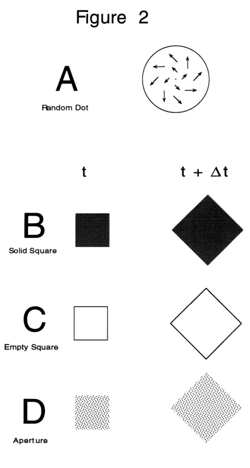
be able to produce stimuli where the instantaneous velocity field is presented without additional cues, in order to study its processing in isolation. This can be done, for example, by presenting the system under study with just two successive image frames, as three frames are obviously required to represent information about acceleration. Alternatively, with the random dot displays used in our lab to study MSTd (Graziano et al, 1994; see Chapter Two of this thesis), we simulated the approach to a vertical plane but removed all cues except the positional velocity field. Components of acceleration were eliminated by displacing the dots every frame in accordance with the same vector field. Although the velocity field produced is consistent with an approaching plane, because the velocity field does not change with time the plane remains a fixed simulated distance away from the observer.
If the nervous system only has access to the velocity field and does not process higher order information such as acceleration, or if such a system is deprived of these cues in the stimulus, ambiguities in the optical flow arise. The general belief that the human visual system is poor in detecting acceleration makes the former situation highly relevant. One such ambiguity that has received considerable attention is that the instantaneous velocity field produced by rectilinear (straight) translation in the presence of eye rotation is identical to curvilinear motion with the eyes stationary. Being able to resolve this ambiguity is important behaviorally; therefore information about acceleration is needed to make this distinction based on flow information alone (Warren et al, 1991; Longuet-Higgins, 1986). Alternatively, other modalities (vestibular input, an extra-retinal signal for eye movement) could also resolve these ambiguities.
Patterns of retinal flow can be quite complex, particularly in animals not limited to a terrestrial setting. These types of patterns are all effective stimuli for units in MSTd. The simplest case occurs when the observer makes a head or eye movement with no observer translation and no motion in the environment. In this situation, the retinal flow is a homogeneous
Chapter 1: Cortical Area MST and Optical Flow: A Review
array of velocity vectors all with the same orientation and length. The pattern of flow is completely independent of environmental structure, including relative depth. Recti-linear translation parallel to the line of sight, in the absence of eye rotation, produces either expansion for forward motion (all velocity vectors pointed away from the direction of heading), or contraction for backward motion. The spatial arrangement of the velocity vectors, as well as their orientation, is independent of environmental structure and is determined entirely by observer translation. However, the lengths of these vectors varies depending on the layout of the environment, e.g. those vectors closer to the observer have greater magnitude than those further away, for the same retinal eccentricity. For the special case of recti-linear movement orthogonal to the line of site (with no eye rotation), a translational pattern of flow similar to that described for eye rotation results, but the magnitude of the vectors is a function of environmental layout.
Under natural conditions, we generally track features in the environment while moving forward. If the feature is exactly in the direction that we are heading, or if the feature is very far away (the horizon), no eye rotation is required to maintain fixation and a pure expansion flow pattern is produced. However, if the feature we are fixating is nearby (a mark on the sidewalk in front of us), we induce a retinal flow pattern determined by adding the motion vectors that would have been produced separately from the translation and the rotation. Adding the radial flow field to the rotational flow field produces a spiral pattern of flow, assuming some depth variation is present in the image. The exact pattern of the spiral (ratio of expansion/contraction to rotation) is determined by the structure of the environment. As the depth variation in the environment approaches zero, the instantaneous velocity field approaches that of a pure radial (expansion) pattern, appearing like a pure observer translation. Because of this, we have a situation where the optical flow can be ambiguous if the image flow is not allowed to evolve with time or if only the instantaneous velocity field is considered. Finally, because our movement trajectories are commonly not
Chapter 1: Cortical Area MST and Optical Flow: A Review
perfectly straight, we often produce hyperbolic flow patterns during periods of curvilinear motion. Even more complex flow patterns are possible, as when, for example, a monkey is swinging through a tree, moving his eyes and head, all independently. Although it is mathematically trivial to predict these flow patterns by adding the different motion contributions separately, going in the opposite direction is much more difficult, and even ill-posed, especially when higher order information such as acceleration is deprived. When there is movement in the environment, such as a tree blowing in the wind, this further distorts the flow pattern, at least locally. We can see that there is a tremendous amount of potential information present in the global motion pattern falling on the retina, but it is a nontrivial task to disentangle all this conflicting information.
Direction Of Heading Determination
Gibson (1950) was the first to recognize that information about the direction of observer translation could be determined from an examination of the optical flow. He correctly identified the global focus of expansion in these patterns as corresponding to the direction of heading. This is true for all recti-linear movement, independent of whether or not there are simultaneous rotations of the eye or head. Unfortunately, the nervous system does not have direct access to optical flow, and the focus of expansion with respect to retinal flow, to which the observer does have direct access, only corresponds to the direction of heading when there is no simultaneous eye or head rotation. Because MSTd responds to the types of flow patterns produced by observer translation, this region has been implicated as the location where direction of heading computations are processed (Judge, 1990). Many workers in MST have labeled it the "ego-motion" center, and we will address the strengths and weaknesses of this proposal before considering alternative roles that this cortical area might play. For convenience, we will use the label ego-motion to refer specifically to direction of heading determination and distinguish this function from related issues such as the sensation of vection and the motor
Chapter 1: Cortical Area MST and Optical Flow: A Review
compensations related to ego-motion such as postural stabilization. These additional phenomena will be considered subsequently.
The determination of direction of heading from optical flow has received considerable attention over the past 15 years, particularly in the computational and psychophysical literatures (Prazdny et al, 1980; Poggio et al 1991). Far less attention has been given to the issue of ego-speed, possibly because this parameter is unavailable from optical flow in the absence of information about the absolute depth of environmental features (Larish and Flach, 1990). The problem of determining direction of heading can be formalized as follows: The observer has six potential degrees of freedom for movement, three rotational and three translational. Alternatively, Chasles's theorem states that every movement can be decomposed into a translation and a rotation around a single axis in a unique manner. These two formulations are formally equivalent; four degrees of freedom are used to determine the proper axis in the latter description of the problem. Cues from multiple modalities, including vestibular, auditory, and somatosensory, may be instrumental in facilitating the determination of these parameters, but this discussion will be limited to information provided by retinal flow, perhaps in combination with an extra-retinal signal about eye rotation. Furthermore, in the analysis that follows, we will assume that any rotational component (about the vertical axis) of the flow is a consequence of eye movement and that the head is fixed relative to the shoulders. All the mechanisms discussed that consider an extra-retinal signal about eye rotation could trivially be extended to include an efference copy signal for head rotation.
As alluded to previously, the task of determining direction of heading for the case of recti-linear translation without eye or head rotation is trivial, at least conceptually: identify the global outflow of expansion. Whether or not the nervous system has components capable of detecting this feature in the flow is another question-and this may not be necessary as other potential cues about heading direction are also available. However, in the case of
Chapter 1: Cortical Area MST and Optical Flow: A Review
translation with eye rotation, some decomposition of the different flow components is necessary before the global focus of outflow, invariantly represented in the optical flow field, can be recovered from the retinal flow (Regan and Beverley, 1992). Before we can consider whether the response characteristics of MST cells are appropriate for ego-motion representation, we need to examine the effect of manipulating the informational content present in flow stimuli both on navigational performance and MST single unit activity. We can then attempt to correlate how these input variables effect psychophysical performance with how they effect response selectivity in MST. A close relation between the two would suggest a functional link. Computation models of DOH determination will also be reviewed, in order to assess whether the response characteristics of neurons in MSTd are appropriate for carrying out this task.
Psychophysical Studies
Early studies had concluded that subjects are quite poor at determining direction of heading based on optical flow alone, with or without eye rotation. However, in the 1980s several groups working independently determined that heading accuracy under a wide range of conditions is approximately 1-2 degrees (Warren et al, 1988), sufficient for the behaviorally important task of obstacle avoidance, e.g. smashing into trees while running. Similar results were obtained even for the more complex case of curvilinear movement (Warren et al, 1991; Turano et al, 1994). One study (Regan and Beverley, 1982) reported heading judgments as accurate as 0.03 degrees for rectilinear translation based on detecting the maximum of divergence in the flow field, but it is now widely agreed that this probably was a consequence of static positional cues in their displays that are not available under naturally occurring situations. Although many of these studies used evolving flow displays with other cues present besides the instantaneous velocity field, high performance was maintained even when acceleration was removed from the stimuli (Warren et al, 1991). When the global orientation of the velocity vectors was kept intact, but the length of
Chapter 1: Cortical Area MST and Optical Flow: A Review
these vectors was randomized (a "direction" field), heading judgments remained accurate, but not when the length of these vectors was left unaltered and the vector orientations were randomized (a "speed" field) (Warren et al, 1991). Interestingly, units in MSTd display a analogous response behavior, showing strong selectivity for direction fields but not for speed fields (Tanaka et al, 1989). Other studies showed that even when considerable noise is added to the stimuli, heading determination remained robust (van den Berg, 1992). In addition, reducing stimulus exposure time had little effect on performance down to 150 msec, much less time than is supplied by the average intervals between saccades. These studies have provided strong support that optical flow is used to determine direction of heading under the limitations imposed under naturally occurring conditions.
Probably the most relevant perceptual data concerning direction of heading determination and the possible necessity of an extra-retinal smooth pursuit signal comes from separate papers by Warren (1988ab; 1990) and Royden, et al (1992). Warren claims human performance is highly accurate for simulated approaches to a horizontal plane, a vertical plane, and a cloud of dots. This was true even when subjects tracked a feature (through smooth pursuit eye movements) that was away from the direction of heading. However, if the rotational component to the retinal flow was added directly to the visual stimulus on the screen, simulating the effects of eye rotation, performance dropped for the approach to a vertical plane, but remained high for the two other conditions. Note that in both the actual and simulated eye rotation sets of conditions, retinal flow was identical. Any performance change had to be a consequence of extra-retinal information about eye movement. Warren concluded that subjects could determine direction of heading from optical flow alone, with or without eye rotation, as long as depth variation was present in the stimulus.
Royden, et al (1992) confirmed many of Warren's findings, but obtained much poorer performance in environments containing depth variation
Chapter 1: Cortical Area MST and Optical Flow: A Review
under the condition of forward translation plus simulated tracking eye movements. He concluded that an extraretinal signal is required to determine direction of heading, even when the environment contains variations in depth. A possible way of reconciling these differences is to consider the pursuit speeds used by the two groups of investigators. Warren used pursuit speeds in the range of 0.3 to 1.2 degrees/sec while Royden used speeds as high as 5 degrees per second (van den Berg, 1993). Perhaps the visual system, using information contained in retinal flow alone, can determine direction of heading when the distortions due to rotation are small compared to the signal from observer translation. With greater angular rotational velocities, an extraretinal signal would be required to facilitate this process. Further complicating the story, another group (van den Berg, 1992) concluded that we can determine direction of heading from retinal flow alone as long as there are features very far away from the observer to use as a reference. For these very distant features, retinal flow depends almost entirely on observer rotation, not on translation, remembering that the rotational component of the flow is independent of feature distance. In this study, performance under simulated rotation conditions was poor for the approach to a cloud of dots whose most distant points were relatively near to the subject. It would be interesting to investigate more quantitatively how the signals from translational and rotational flow interacted under more varied experimental conditions. One study of interest (van den Berg, 1992) looked at direction of heading judgments in the presence of noise added to the veridical flow signal. These results showed that an extra-retinal signal becomes more important as the amount of noise in the display increases. We conclude that under optimal conditions direction of heading computations can be carried out by the human visual system based on optical flow alone but that an extraretinal signal is required as conditions deteriorate either because of a lack of depth variation in the stimulus, noisy conditions, or high rotational eye velocities.
A somewhat analogous situation exists in the psychophysical literature
Chapter 1: Cortical Area MST and Optical Flow: A Review
muscles of the observer are partially paralyzed, subjects have a hard time identifying target location in the dark, but have no trouble in the light (reviewed by Matin, 1986).
Cutting (1992) has taken the stance that Gibson's early approach to ego-motion, emphasizing the importance of optical flow, has resulted in misguided attempts to extract this information, seemingly at all costs, from the pattern of retinal flow which is immediately available to the visual system. He argues that no such recovery process is necessary, and that information available in retinal flow is sufficient for recovering direction of heading. He proposes that we use assymetries in the retinal flow field, such as motion parallax, to guide successive eye movements towards the direction of heading. His proposal has several deficiencies. Warren demonstrated that we can accurately recover direction of heading from displays that contain no motion parallax. Additionally, both Warren and Royden conclusively showed that repeated periods of smooth pursuit followed by a saccade to a new environmental feature were not required for accurate navigation.
Models for Determining DOH
Demonstrating that observers can determine direction of heading from retinal flow without an extra-retinal smooth pursuit signal does not provide an algorithm for how this might be done. Knowing the algorithm used by the visual system to recover DOH would be instrumental in determining whether or not MST is an appropriate candidate for this function.
Despite the non-equivalence of the outflow focus and DOH under conditions of eye rotation (with respect to retinal flow), it is common in the psychophysical and computational literature for authors to defend Gibson's approach. The claim is made that under conditions of eye rotation the nervous system must first decompose the flow into translational and rotational components before using this singularity for navigational purposes. What exactly is meant by this decomposition is not at all clear. Neurons respond to stimulus features, such as (potentially) the global focus
Chapter 1: Cortical Area MST and Optical Flow: A Review
of expansion. If eye rotation is present, this cue is not a reliable indicator of heading direction. While many of the models discussed below, including those based on differential invariants and differential motion, claim to decompose flow, they are really just extracting a feature from the flow field that, unlike the focus of expansion, is invariant to rotation. If a particular region of the brain is determining direction of heading in this manner, we would expect its neurons to respond to these invariant properties rather than the global focus of expansion. Support for these models cannot be considered as vindicating Gibson's hypothesis, as the flow properties being extracted are not derived from, nor dependent on, the stimulus component he proposed. We will consider one model, which uses an extra-retinal signal in combination with the global focus of expansion, that can legitimately be considered a decomposition scheme consistent with Gibson's proposal.
Some models for determining DOH only have relevance in the artificial intelligence community. For example, discrete models rely on measuring the locations and displacements of a small number of points to set up a system of equations to recover the 6 parameters (3 rotational, 3
translational) that will uniquely describe observer motion. Four to seven points in two successive frames are generally sufficient with this class of model (Warren and Hannon, 1990). These algorithms are obviously non-biological, but are useful in setting lower limits on the amount of information required for these computations. They are notoriously prone to noise as they rely on extremely accurate measurements of displacement and position. Another class of models that has been popular in the computational community is characterized by a least squares search for possible surface layouts and observer motions that are consistent with the observed flow. Although these models are generally resistant to noise and degrade gracefully, they often depend on severe assumptions about surface layout in order to narrow the parameter space searched. Recently, T. Poggio proposed an algorithm that takes advantage of Green's theorem to detect the expansion component of optical flow in a positionally invariant way.
Chapter 1: Cortical Area MST and Optical Flow: A Review
Although this model is robust to noise and its implementation is biologically plausible, he does not offer a way to recover direction of heading once these types of flow fields are detected (Poggio et al, 1991).
A second class of models based on computing the differential invariants
(Koenderink and van Doom, 1981; Koenderink, 1986; Regan and Beverley,
1982) divergence (div), curl, and deformation (def), recognize that these
properties of flow fields are invariant with respect to eye rotation. This is because the effect of eye rotation on retinal flow is to add a constant velocity vector on top of every velocity vector representing the optical flow. Spatial derivatives like div, curl, and def, which are dependent only on differences between vectors, are unaffected by such a homogeneous transformation. One of the appeals of this class of model is that information about the structure of the environment is also available from these operations, and many structure from motion algorithms have been shown to use these operators successfully. Unfortunately, for purposes of ego-motion representation this dependence on environmental structure turns out to be a liability. Although these operators are invariant with respect to eye rotation, they do not possess this property with regards to environmental layout. For example, Regan and Beverley (1982) have proposed that we determine direction of heading by detecting the maximum of divergence in the optical flow field, which is not the same as the focus of expansion. Although the maximum of divergence is invariant with respect to gaze direction and eye rotation, it is rarely in the direction of heading. Even if (as they claim) observers are very sensitive to this flow feature, it is not clear how they could use this information to extract information about self motion. In addition to this difficulty, these models are sensitive to noise and require dense flow patterns with smooth depth variations over which to take spatial derivatives. Perceptual experiments discussed above show high levels of performance under noisy conditions, sparse displays, and with environmental layouts such as clouds that preclude the taking of spatial derivatives (Warren et al, 1988). Furthermore, units in MSTd have been
Chapter 1: Cortical Area MST and Optical Flow: A Review
shown to be ineffective in extracting these differential invariants from more complex flow patterns (Orban et al, 1992).
Several models which rely on differential image motion to recover DOH have received considerable attention (Rieger et al, 1985; Hildreth, 1992). Differential motion, which includes both edge parallax and motion parallax, occurs during observer translation when two environmental features which occupy nearby locations in the optic array are at different depths. The magnitudes of the velocity vectors associated with these two features depend on their depth, with image points located closer to the observer having a greater retinal velocity for the same eccentricity. If no head or eye rotation is occurring, the orientation of these two vectors is identical, and they point along the direction of heading. However, with a rotational component to the flow, these two vectors are no longer pointed in the same direction, and neither point in the direction the observer is translating. However, if a new vector is created by subtracting these two vectors from one another, this new vector is oriented such that the direction of heading falls somewhere along an extension of its axis. Two such subtraction vectors at different spatial locations uniquely determine the direction of heading by the intersection of their axes of orientation. This procedure can be repeated across the optical array, creating a new positional vector field, whose focus of expansion is aligned with the direction of heading.
Recently, a related model using the affine coordinate system in place of the Cartesian has been proposed (Beusmans, 1993) which also relies on local variations in depth to recover direction of heading. Affine flow is zero for the approach to a vertical plane; it is easy to show that the entire class of differential motion models fail in environments without depth variation. This is consistent with our previous discussion of the ambiguities present in the positional velocity field representing the approach to a vertical plane. Another problem with differential motion algorithms is that they recover only the axis of translation. They do not indicate the translational direction
Chapter 1: Cortical Area MST and Optical Flow: A Review
along this axis and therefore whether the observer is receding or approaching from the scene. Information about the relative depth of features in the optic array can provide this information. This class of models is appealing because several groups (Regan et al, 1982; Warren et al,
1988) have shown depth variation in the stimulus improves heading
performance when extra-retinal information about eye rotation is absent. As discussed below, to date little sensitivity to differential image motion has been demonstrated for units in MSTd.
An important consideration, under emphasized in the literature, is the fact that we have direct access to optical flow information under a number of situations which leave the eyes stationary relative to their orbits. If these events are frequent enough to effectively guide behavior, further discussion of various decomposition schemes is unnecessary. It has already been mentioned that eye rotation is non-existent when looking in the direction of heading and when fixating on any distant feature. However, such moments of stability also occur for short periods of time immediately following saccades and may even occur briefly during pursuit. If these periods are long enough for the nervous system to detect the focus of expansion and occur frequently enough to provide a behaviorally sufficient sampling rate, we have a very simple solution to our problem. More work needs to be done comparing the frequency and duration of these rotation-free periods to the minimal time required to detect the outflow focus.
MST and direction of heading
One of the strongest indications that MST may be involved in ego-motion analysis is the presence of an extra-retinal smooth pursuit signal that is independent of visual input. All the previously discussed models recovered direction of heading from retinal flow information alone. However, consideration of the psychophysical literature indicates that an extra-retinal eye signal plays an important role in factoring out the flow contribution of eye rotation under non-optimal visual conditions (see above.) From a computation point this task is trivial. Because the effect of eye