HAL Id: hal-03021277
https://hal.archives-ouvertes.fr/hal-03021277
Submitted on 24 Nov 2020
HAL is a multi-disciplinary open access
archive for the deposit and dissemination of
sci-entific research documents, whether they are
pub-lished or not. The documents may come from
teaching and research institutions in France or
abroad, or from public or private research centers.
L’archive ouverte pluridisciplinaire HAL, est
destinée au dépôt et à la diffusion de documents
scientifiques de niveau recherche, publiés ou non,
émanant des établissements d’enseignement et de
recherche français ou étrangers, des laboratoires
publics ou privés.
To cite this version:
Isabelle Lamsoul, Loïc Dupré, Pierre Lutz. Molecular Tuning of Filamin A Activities in the Context
of Adhesion and Migration. Frontiers in Cell and Developmental Biology, Frontiers media, 2020,
�10.3389/fcell.2020.591323�. �hal-03021277�
MINI REVIEW published: 20 November 2020 doi: 10.3389/fcell.2020.591323
Edited by: Lei-Miao Yin, Shanghai University of Traditional Chinese Medicine, China Reviewed by: Susanna Carola Fagerholm, University of Helsinki, Finland Ben Goult, University of Kent, United Kingdom Jon Humphries, The University of Manchester, United Kingdom Massimiliano Baldassarre, University of Aberdeen, United Kingdom *Correspondence: Isabelle Lamsoul Isabelle.Lamsoul@inserm.fr Pierre G. Lutz Pierre.Lutz@inserm.fr Specialty section: This article was submitted to Cell Adhesion and Migration, a section of the journal Frontiers in Cell and Developmental Biology Received: 04 August 2020 Accepted: 05 November 2020 Published: 20 November 2020 Citation: Lamsoul I, Dupré L and Lutz PG (2020) Molecular Tuning of Filamin A Activities in the Context of Adhesion and Migration. Front. Cell Dev. Biol. 8:591323. doi: 10.3389/fcell.2020.591323
Molecular Tuning of Filamin A
Activities in the Context of Adhesion
and Migration
Isabelle Lamsoul
1* , Loïc Dupré
1,2and Pierre G. Lutz
1*
1Centre de Physiopathologie de Toulouse Purpan, INSERM, CNRS, Université de Toulouse, UPS, Toulouse, France, 2Ludwig Boltzmann Institute for Rare and Undiagnosed Diseases, Vienna, Austria
The dynamic organization of actin cytoskeleton meshworks relies on multiple
actin-binding proteins endowed with distinct actin-remodeling activities. Filamin A is a
large multi-domain scaffolding protein that cross-links actin filaments with orthogonal
orientation in response to various stimuli. As such it plays key roles in the modulation
of cell shape, cell motility, and differentiation throughout development and adult life. The
essentiality and complexity of Filamin A is highlighted by mutations that lead to a variety
of severe human disorders affecting multiple organs. One of the most conserved activity
of Filamin A is to bridge the actin cytoskeleton to integrins, thereby maintaining the later
in an inactive state. We here review the numerous mechanisms cells have developed
to adjust Filamin A content and activity and focus on the function of Filamin A as a
gatekeeper to integrin activation and associated adhesion and motility.
Keywords: actin cytoskeleton, filamin, integrin, cell adhesion, cell migration
INTRODUCTION
The filamin protein family is represented in nearly all Metazoa. Phylogenetically, the filamin
genes diverge from a common single ancestral gene between chordate invertebrate and vertebrate
lineages. Filamins comprise a N-terminal binding domain (ABD) composed of two
actin-binding calponin homology (CH) domains followed by immunoglobulin like repeats (IgFLN) of
high sequence similarity (
van der Flier and Sonnenberg, 2001
). All C-terminal filamin repeats of
filamins characterized so far have the property to homodimerize. The number of filamin repeats
differs substantially in invertebrates but is almost constant in vertebrates (
Light et al., 2012
). The
vertebrate genomes contain three filamins, Filamin A, B and C, with an intraspecies sequence
identity of over 64% (
Kesner et al., 2010
). Filamins A and B are ubiquitously expressed, whereas
Filamin C is expressed in smooth and striated muscles. Filamins A and B are localized to the
cortex and stress fibers, whereas Filamin C is localized to the sarcomeric Z-line complex (
van der
Ven et al., 2000
;
Sheen et al., 2002
). Disease-associated mutations and knockout mouse models
suggest that Filamins A and B are critical for various aspects of skeletal, vasculature, cardio, and
cerebral development (
Fox et al., 1998
;
Sheen et al., 2002
;
Robertson et al., 2003
;
Krakow et al.,
2004
;
Farrington-Rock et al., 2006, 2008
;
Feng et al., 2006
;
Hart et al., 2006
;
Lu et al., 2007
;
Zhou
et al., 2007
;
Metais et al., 2018
;
Yamak et al., 2020
), whereas Filamin C is essential for skeletal muscle
and heart development (
Goetsch et al., 2005
;
Vorgerd et al., 2005
;
Dalkilic et al., 2006
;
Duff et al.,
2011
;
Zhou et al., 2020
).
FILAMIN A, A HUB FOR MULTIPLE
BINDING PARTNERS
Filamin A interacts with about a hundred binding-partners, many
of which being involved in the regulation of signaling pathways
converging toward actin cytoskeleton organization (Figure 1).
Filamin A has a dual role in controlling the architecture and
the mechanics of the actin cytoskeleton. Filamin A is an
actin-binding and cross-linking protein whose primary function is to
organize the actin cytoskeleton in orthogonal filament arrays
(
Nakamura et al., 2007
). Importantly, the mechanic properties of
this filamentous actin (F-actin) network is dependent on Filamin
A concentration (
Tseng et al., 2004
;
Gardel et al., 2006
;
Esue et al.,
2009
). At high Filamin A concentration, tighter F-actin bundles
are observed and the F-actin network undergoes stress stiffening
under applied forces (
Schmoller et al., 2009
). In contrast, at
lower relative Filamin A cross-link concentrations, the F-actin
cytoskeleton is more dynamic and can soften in response to stress
(
Tseng et al., 2004
). Furthermore, the non-linear elasticity of the
actin network is attributed to the flexibility of Filamin A (
Kasza
et al., 2009
;
Schmoller et al., 2009
). Filamin A also localizes to
points of intersection between stress fibers and cortical actin
where it plays a role in the isotropic redistribution of applied
forces to focal adhesions (
Kumar et al., 2019
). Three
actin-binding sites (ABS) within the Filamin A ABD were recently
identified (Figure 1;
Iwamoto et al., 2018
). The first one,
ABS-N, located at the N-ter of the CH domain 1 contributes to F-actin
binding while the two others, ABS2 and ABS2
0, facilitate binding
in the groove between adjacent actin subunits (
Iwamoto et al.,
2018
). While the ABD is necessary and sufficient for F-actin
binding (
Razinia et al., 2011
), a domain within a Filamin A
fragment encompassing filamin repeats 9 to 15 is necessary for
high avidity F-actin binding (
Nakamura et al., 2007
).
In mammals, the ABD of filamins is followed by 24 filamin
repeats interrupted by two hinge regions often referred as rod
1 and 2, one between repeats 15 and 16 and another between
repeats 23 and 24 (Figure 1). Filamin A domains can be
divided into four subgroups (A, B, C, and D) based on amino
acid similarities (
Ithychanda et al., 2009b
). Filamin A repeats
of subgroup A (4, 9, 12, 17, 19, 21, and 23) interact with a
set of biologically important ligands including platelet receptor
glycoprotein Ib
α (GPIbα) (
Nakamura et al., 2006
), migfilin
(
Lad et al., 2008
;
Ithychanda et al., 2009a,b
), Cystic Fibrosis
Transmembrane conductance Receptor (CFTR) (
Smith et al.,
2010
), FilGAP (
Nakamura et al., 2009
), Pro-prion (
Li et al., 2010
),
Ankyrin repeat containing protein with a SOCS box 2 alpha
(ASB2
α) (
Lamsoul et al., 2011
) and
β chains of integrins (
Kiema
et al., 2006
;
Takala et al., 2008
). All subgroup A Filamin A repeats
and their binding partners have similar mode of interaction.
Indeed, the CD face of Filamin A repeats represents a common
interface for Filamin A-ligand interaction (
Lad et al., 2007
;
Heikkinen et al., 2009
). Interestingly, Filamin A repeats can also
engage in intramolecular contacts (IgFLNa16-17, IgFLNa18-19,
and IgFLNa20-21) that may become disrupted by binding of one
of the repeats to the integrin
β cytoplasmic tail or by mechanical
forces (
Lad et al., 2007
;
Heikkinen et al., 2009
). Smoothelins A
and B, as well as fimbacin can bind to the cryptic CD cleft of
Filamin A repeat 21 exposed in mechanically activated Filamin
A (
Wang and Nakamura, 2019a,b
). Filamin A can also interact
with small GTPases of the Rho family, Rac, Rho, cdc42, and RalA
(
Ohta et al., 1999
;
Bellanger et al., 2000
;
Vadlamudi et al., 2002
;
Ueda et al., 2003
) and with proteins upstream and downstream of
the GTPases (
Ohta et al., 2006
;
Nakamura et al., 2009
) known to
regulate cytoskeletal dynamics and cell protrusions. In addition,
RhoA activity is downregulated through interactions between
Filamin A and
αIIbβ3 integrins and is critical to proplatelet
formation (
Donada et al., 2019
).
Filamin A is also localized into the nucleus where it plays
roles in DNA repair through interaction with BRCA1 and
BRCA2 (
Yue et al., 2009
;
Velkova et al., 2010
), as well as
transcription through interaction with transcription factors such
as the androgen receptor (
Loy et al., 2003
;
McGrath et al., 2013
;
Savoy et al., 2015
), Smads (
Sasaki et al., 2001
) or PEBP2
β/CBFβ
(
Yoshida et al., 2005
). Filamin A also associates with the MKL1
transcriptional co-activator, stimulating the activity of the Serum
Responsive Factor (SRF) transcription factor and cell migration
(
Kircher et al., 2015
).
How Filamin A integrates the signals triggered by its
multiple binding partners and whether such complex molecular
interactions might be tuned differentially in different cell types
remain key open questions. Nevertheless, the essentiality of
Filamin A is highlighted by variants in the gene
FLNA that
lead to 10 distinct genetic syndromes affecting a wide diversity
of organs (
Wade et al., 2020
). Importantly, pathogenic variants
of
FLNA could contribute to aberrant cytoskeletal regulation
leading either to loss-of-function or gain-of function disorders.
Indeed, variants are found in the two CH domains of the ABD,
CHD1 and 2, in periventricular nodular heterotopia (PH) and
otopalatodigital (OPD) syndromes type 1 and 2, respectively.
Variants within CHD1 are likely to disrupt Filamin A interaction
with actin (
Iwamoto et al., 2018
) whereas variants within CHD2
are likely to constitutively expose the CHD1 ABS to ligand (
Clark
et al., 2009
). Interestingly, a mutation in
FLNA in a male patient
with PH and congenital intestinal pseudoobstruction potentiates
αIIbβ3 integrin activation likely through less binding of mutant
Filamin A to
β3 integrin and facilitated recruitment of Talin by
the
β3 subunit (
Berrou et al., 2017
). The relationship between
other
FLNA pathogenic variants and Filamin A functions is less
understood and remains to be investigated.
FILAMIN A, A NEGATIVE REGULATOR
OF INTEGRINS
Because the filamin domains involved in binding actin and
integrins have the highest content of ancestral residues of
any domains (
Kesner et al., 2010
), integrins are considered
among the most important interaction partners of filamins.
Integrins are heterodimeric transmembrane receptors formed by
α and β subunits. These are single spanning membrane proteins
with a large extracellular ectodomain and a short intracellular
cytoplasmic tail. Integrins mediate cell to extracellular matrix
Lamsoul et al. Filamin A Regulation
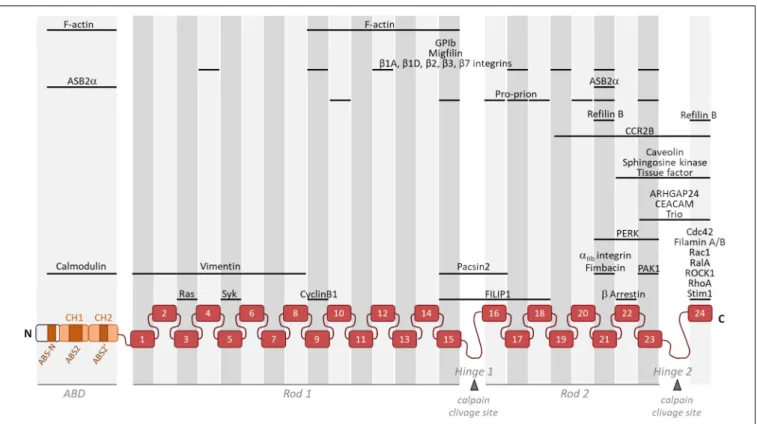
FIGURE 1 | Schematic representation of monomeric Filamin A illustrating its general structure and the binding location of partners involved in actin cytoskeleton organization and/or cell motility. Filamin A consists of an actin-binding domain (ABD) composed of two actin-binding calponin homology (CH) domains followed by Ig-like repeats (1–24). Two intervening calpain-sensitive hinge domains separate the 24 Ig-like repeats into two rod domains. The three ABS (ABS-N, ABS2, and ABS20
) are indicated. Partners involved in actin cytoskeleton organization and/or cell motility but with unknown Filamin A binding domain(s) (14-3-3, ELP1, FILIP1L, p190RhoGAP, and Lck) are not represented.
and cell to cell contacts and integrate external cues to
the actin cytoskeleton and signaling pathways (
Legate and
Fassler, 2009
;
Humphries et al., 2019
;
Kechagia et al., 2019
).
Interactions between integrins and their extracellular ligands
are tightly regulated thanks to integrin activators and integrin
inhibitors. Importantly, switching integrins between inactive
and active conformations is crucial for integrin functions
(
Bouvard et al., 2013
). Integrin activation has largely been
documented (
Kim et al., 2011
;
Sun et al., 2019
). This process is
regulated via either extracellular ligands (outside-in activation)
or intracellular binding partners (inside-out activation).
Integrin-inactivating proteins such as integrin cytoplasmic
domain-associated protein 1 (ICAP1), SHARPIN (SHANK-domain-associated
RH domain-interacting protein) and filamins are required for
integrin inactivation in different settings (
Calderwood et al., 2001
;
Bouvard et al., 2003
;
Rantala et al., 2011
). The physiological
relevance of integrin-inactivating proteins is crucial for integrin
function as exemplified by the phenotypes of mice lacking
integrin inactivators (
Bouvard et al., 2013
). Integrin inactivators
either stabilize the inactive state of integrins or promote integrin
deactivation during cyclical cell-adhesion processes such as
migration. Indeed, a substantial proportion of cell surface
integrins is in a resting state (
Arjonen et al., 2012
). Inactive
integrins are in a closed conformation, in which the binding of
both extracellular ligand and intracellular activators is repressed.
In human, there are several integrin
β subunits that bind filamins
(
Kiema et al., 2006
;
Ithychanda et al., 2009b
).
Filamin A is a major gatekeeper to integrin activation. Since
the discovery of Filamin A as a binding partner of the
β2 integrin
subunit 25-years ago (
Sharma et al., 1995
) and the first evidence
that increased Filamin A-β2 integrin interactions restrict cell
migration (
Calderwood et al., 2001
;
Bouvard et al., 2003
;
Rantala
et al., 2011
), several modes of action of Filamin A as an integrin
inactivator have been proposed. They depend on the identity
of the integrin
α and β chains or could be specific to only a
subset of integrin
αβ heterodimers. First, binding of Filamin
A domains of subgroup A to the C terminus of the integrin
β tail (β1, β2, β3, or β7) results in direct competition with
talin binding by occupying an overlapping binding site (
Kiema
et al., 2006
;
Ithychanda et al., 2009b
). Second, Filamin A forms a
ternary complex engaging the cytoplasmic tails of both integrin
αIIb and β3, thereby stabilizing the inner-membrane clasp and
competing with talin recruitment to the
β subunit cytoplasmic
tail by binding both the C-terminal and membrane-proximal
regions of the
β3 tail (
Liu et al., 2015
). These two modes of
action of Filamin A restrain the integrin in a resting state.
Interestingly, domains within functionally important binding
interfaces of both filamin repeats and integrin subunits have
diverged in critical residues, indicating that filamin isoforms may
bind and regulate integrin
αβ heterodimers differentially. Indeed,
Kesner et al. (2010)
described the substitution in the
β strand
C of the filamin repeat 21, an ancestral Ser/Thr in Filamins B
and C changed to an Ala at residue 2272 in Filamin A during
the mammalian period. Furthermore,
β1 and β7 integrins have
ideally positioned hydrophobic amino acids to bind Filamin
tighter than
β2 and β3 integrins (
Ithychanda et al., 2009b
).
Furthermore, some of the key residues in the
αIIb subunit that are
important for interaction with filamin via their side chains, are
not conserved in all integrin
α subunits, reinforcing the notion
that filamins bind integrins differentially.
Because several Filamin A repeats can bind the cytoplasmic
tails of
β integrins and have the ability to clasp αIIb and
β3 cytoplasmic tails, it seems plausible that they can bind
simultaneously, and such interactions may promote clustering
of inactive integrins (
Ithychanda et al., 2009b
;
Liu et al.,
2015
). Although the biological significance of these Filamin A
clutches remains to be establish, it is tempting to speculate that
upon Filamin A removal, pre-clustered integrins would become
engaged by multivalent ECM and thereby activated.
MULTIPLE REGULATORY MECHANISMS
CONTROLING FILAMIN A ACTIVITIES
AND LEVELS
Integrin activation can be achieved through the binding of
proteins to Filamin A. Indeed, migfilin can bind Filamin A
with a high affinity, uncoupling the Filamin A-integrin link,
sequestering Filamin A away from the
β integrin cytoplasmic tail
and thus counteracting Filamin A-mediated integrin inactivation
(
Lad et al., 2008
;
Ithychanda et al., 2009a
;
Das et al., 2011
). This
allows the binding of integrin activators, talin and kindlins, to
the
β integrin cytoplasmic tail, leading to inside-out integrin
activation (
Tadokoro et al., 2003
;
Tu et al., 2003
;
Kiema et al.,
2006
;
Wegener et al., 2007
;
Moser et al., 2008
). Internally
generated and externally imposed mechanical forces can also
regulate Filamin A interaction with partners by triggering
conformational changes that expose otherwise masked
partner-binding site, thereby leading to integrin activation (
Ehrlicher
et al., 2011
;
Nakamura et al., 2014
). Although evidence for the
mechanosensing function of Filamin A in Drosophila oogenesis
has been provided, its precise role in cell differentiation and
morphogenesis in mammals is still lacking (
Razinia et al.,
2012
;
Huelsmann et al., 2016
). Filamin A is also regulated
by phosphorylation. Several kinases such as protein kinase C
(
Kawamoto and Hidaka, 1984
), ribosomal S6 kinase (
Ohta
and Hartwig, 1996
;
Woo et al., 2004
), p21-activated kinase 1
(PAK1) (
Vadlamudi et al., 2002
;
Hammer et al., 2013
), the
cyclic adenosine monophosphate (cAMP)-dependent protein
kinase A (
Jay et al., 2004
), Akt (
Li et al., 2015
), mTOR2
(
Chantaravisoot et al., 2015
;
Sato et al., 2016
) and the
serine/threonine kinase Ndr2 (
Waldt et al., 2018
) phosphorylate
Filamin A on serine 2152. This phosphorylation event positively
regulates cell migration (
Woo et al., 2004
;
Hammer et al.,
2013
;
Li et al., 2015
;
Sato et al., 2016
). Activation of receptor
tyrosine kinases was shown to trigger cell rounding and
integrin inactivation via increased Filamin A phosphorylation
(
Vial and McKeown-Longo, 2012
;
Mai et al., 2014
). In addition,
G Protein-Coupled Receptors that directly bind Filamin A can
also promote Filamin A phosphorylation (
Tirupula et al., 2015
).
Filamin A phosphorylation by the cAMP-dependent protein
kinase protects Filamin A against proteolysis by calpains (
Chen
and Stracher, 1989
). Phosphorylation of
β2 integrins impairs
Filamin A binding, allowing the binding of the 14-3-3 protein
to the
β2 subunit and adhesion of Jurkat T cells to ICAM-1
(
Takala et al., 2008
).
Tuning the cellular concentration of Filamin A represents
another level of regulation expected to impact integrin activation,
although this has not been formally demonstrated yet (Figure 2).
Filamins are regulated by proteolysis, which provides an
irreversible regulatory mechanism for processes requiring
Filamin removal. Filamin A is cleaved by calpain and caspase
at the two hinge regions, producing a 170 kDa protein
encompassing the ABD and Filamin A repeats 1 to 15 and a
110 kDa protein that is further cleaved to generate a 90 kDa
fragment containing repeats 16 to 23 (Figure 1) (
Gorlin et al.,
1990
;
Browne et al., 2000
). Filamins A and B are also regulated
by proteasomal degradation which represents a fast, reversible,
localized and selective regulatory mechanism that allows cells to
acutely adapt or fine-tune cellular processes. Surprisingly, only
few proteins linked to cytoskeleton dynamics, cell adhesion and
migration have been shown to be regulated by this proteolysis
pathway in non-muscle cells (
Schaefer et al., 2012
). Of interest,
control of the cellular concentration of Filamins A through
ubiquitin-mediated protein degradation represents a seminal
example of proteasomal degradation of an actinbinding and
-crosslinking protein. Using several molecular and cellular biology
approaches, we and others demonstrated that the ASB2
α E3
ubiquitin ligase (E3) triggers ubiquitylation and proteasomal
degradation of Filamins A and B (
Heuze et al., 2008
;
Burande
et al., 2009
;
Lamsoul et al., 2013
;
Razinia et al., 2013
;
Sakane
et al., 2013
;
Spinner et al., 2015
;
Metais et al., 2018
). ASB2α is the
specificity subunit of a multimeric E3 of the Cullin 5-RING Ligase
family involved in the recruitment of proteins to be ubiquitylated
(
Lamsoul et al., 2016
). By degrading Filamins A and B, ASB2
α
regulates cell spreading, adhesion and cell migration (
Heuze
et al., 2008
;
Baldassarre et al., 2009
;
Lamsoul et al., 2011, 2013
;
Spinner et al., 2015
). Furthermore, our recent results support a
model of cardiac cell differentiation that relies on a key role for
ASB2
α in remodeling the actin cytoskeleton through
induced-degradation of Filamin A (
Metais et al., 2018
). Indeed, the
timely controlled removal of Filamin A ensures critical functions
in differentiating cardiac muscle cells suggesting that Filamin
A degradation is necessary to modify the actin cytoskeleton
organization and properties in order to build the sarcomere, and
thus for heartbeats. In addition, the Filamin A interacting protein
(FILIP) interacts with Filamin A and induces its degradation with
impacts on the mode of neuron migration (
Nagano et al., 2004
;
Sato and Nagano, 2005
). More recently, Filamin A expression
was shown to be regulated by a microRNA (miR) and a circular
RNA (circRNA). Indeed, miR-486-3p can bind Filamin A 3
0UTR
thereby reducing Filamin A expression while circFLNA sponges
miR-486-3p resulting in increased Filamin A expression (
Wang
et al., 2019
). Important questions remain: Does Filamin A
Lamsoul et al. Filamin A Regulation
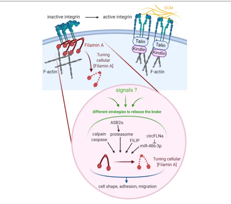
FIGURE 2 | Tuning the cellular concentration of Filamin A represents a pivotal mechanism to regulate integrin-dependent adhesion and migration. Regulation of integrins involves both integrin inhibitors (e.g., Filamin A) and integrin activators (e.g., Talin, Kindlin). Filamin A protein is regulated through cleavages by calpain and caspase, ASB2α-mediated degradation by the proteasome, and interaction with FILIP. Filamin A mRNA is down-regulated by miR-486-3p which is itself sponged by circFLNA. Created with BioRender.com.
degradation directly translate into increased integrin activation?
What is the biological relevance of variable Filamin A levels in
different cell subtypes or at discrete stages of cell differentiation?
Why cells have evolved so many different mechanisms to regulate
Filamin A activity and to up-regulate or down-regulate Filamin A
concentration?
FILAMIN A IN CELL ADHESION AND
MIGRATION
The first evidence for a role of Filamin A in cell motility
was provided in 1992 (
Cunningham et al., 1992
). Indeed, at
the cellular level, Filamin A deficiency in a human melanoma
cell line promotes plasma membrane blebbing and causes
loss of motility. The role of Filamin A in migration was
further supported by the finding that nonsense mutations
in the Filamin A gene are associated with the neuronal
migration disorder periventricular heterotopia (
Fox et al., 1998
).
However, the role of Filamin A in cell motility is more
complex. By providing a physical link between integrins and
the actin cytoskeleton and by negatively regulating integrins,
Filamin A exerts key roles in regulating positively or negatively
cell adhesion and migration according to cell types and/or
conditions. Furthermore, Filamin A binding partners may vary
according to cell types and/or in response to microenvironment
cues such as extracellular matrix components, chemokines
or shear flow. This is likely to influence cell adhesion and
migration. While the loss of Filamins A or B alone has
no effect on cell motility, loss of both filamins following
knockdown/knockout or ASB2
α-mediated degradation have
highlighted the role of filamins in different aspects of cell
motility (
Heuze et al., 2008
;
Baldassarre et al., 2009
;
Lynch
et al., 2011
;
Lamsoul et al., 2013
;
Spinner et al., 2015
). It
is tempting to speculate that the functions of Filamin A in
many cell types may have been missed in assays using Filamin
A single knockout/knockdown cells because of compensation
by Filamin B (
Sheen et al., 2002
;
Baldassarre et al., 2009
).
Filamin-depleted cells exhibit impaired cell spreading (
Heuze
et al., 2008
;
Kim et al., 2008
;
Baldassarre et al., 2009
;
Lynch
et al., 2011
). In addition, increased adhesion of Filamin
A-depleted neutrophils has been described (
Sun et al., 2013
).
Furthermore, Filamin A knockdown or ASB2
α-mediated Filamin
A degradation enhances adhesion of myeloid leukemia cells to
fibronectin (
Lamsoul et al., 2011
). In contrast, Roth et al. found
that Filamin A was dispensable for adhesion of differentiated
HL-60 cells (
Roth et al., 2017
). However, Filamin A depleted
primary murine neutrophils display increased spreading on
and higher adhesion in shear-free conditions to
β2 integrin
ligands, indicating that Filamin A is a negative regulator
of
β2 integrin adhesion in neutrophils (
Uotila et al., 2017
).
Although Filamin A negatively regulates
β2 integrin adhesion
in Jurkat T cells, its absence leads to a reduction of primary
T cell adhesion to integrin ligands under conditions of shear
flow and to a reduced trafficking into lymph nodes and
sites of inflammation (
Moser et al., 2009
;
Savinko et al.,
2018
). Interestingly, Filamin A and vimentin can cooperate
to regulate integrin-mediated cell spreading and cell adhesion
(
Kim et al., 2010a,b
).
Filamins A and B depleted cells exhibit impaired initiation
of migration of fibrosarcoma HT1080 cells (
Baldassarre
et al., 2009
). Filamin A silencing increases cell adhesion
and decreased migration of the bronchial carcinoid H727
cells (
Vitali et al., 2017
). In contrast, silencing of Filamin A
inhibits Snail-induced adhesion and increases migration of
colon adenocarcinoma HT29 cells (
Wieczorek et al., 2017
).
In accordance with these results, Filamin A is required to
mediate SST2 effects on adhesion and migration of the
pancreatic endocrine QGP1 cells (
Vitali et al., 2016
). Filamin
A also positively regulates directional migration of bone
osteosarcoma U-2 OS cells and mouse embryonic fibroblasts
by suppressing Rac 1 activity downstream of
β1 integrins
(
Jacquemet et al., 2013
). Knockdown of Filamins A and B
in fibrosarcoma cells was also shown to augment matrix
metalloproteinase activity increasing their invasive potential
(
Baldassarre et al., 2012
). These results are in agreement with
the fact that ASB2α regulates immature dendritic cell migration
by promoting extracellular matrix proteolysis (
Lamsoul
et al., 2013
). Conversely, Filamin A stabilizes podosomes
in macrophages and is required for their mesenchymal but
not for their amoeboid migration (
Guiet et al., 2012
). In
addition, in the absence of Filamin A, macrophages display
impaired migration associated with reduced atherosclerosis
in mice (
Bandaru et al., 2019
). Several evidences indicate
that Filamin A regulates the intracellular trafficking of
β1
integrins (
Meyer et al., 1998
;
Kim et al., 2010b
). This is
likely to affect
β1 integrin-dependent processes. On the
basis of these scattered observations, it is clear that we still
miss today a unified view of the roles of Filamin A in cell
adhesion and migration.
CONCLUDING REMARKS
As reviewed here, the timely proteolysis and/or removal of
Filamin A have emerged as pivotal mechanisms to regulate
its cellular concentration and integrin-dependent adhesion and
migration. When integrating the knowledge gained about the
function of Filamin A beyond its integrin regulation role, one is
tempted to speculate that this key protein at the interface between
multiple receptors, signaling pathways and the actin cytoskeleton
exerts different and specific cellular functions in response to a
wide-range of environmental cues. As exemplified by the wide
spectrum of developmental malformations and diseases caused
by mutations in its gene, Filamin A indeed stands out as a major
molecular player in different biological processes. In this context,
it will be particularly interesting to further investigate how the
multiple mechanisms able to adjust Filamin A concentration
and activity contribute to its function in different cellular and
physiological settings.
AUTHOR CONTRIBUTIONS
All authors contributed to both the review conceptualization and
the writing process.
FUNDING
Research in our lab related to the subject of this review was
supported by INSERM, CNRS, University of Toulouse and grants
from the Fondation ARC pour la Recherche sur le Cancer, the
Comité Midi-Pyrénées de la Ligue contre le Cancer and Sanofi
iAwards Europe (to PGL) and the CNRS IRP program (grant
“SysTact” to LD).
ACKNOWLEDGMENTS
The authors apologize to their colleagues whose work could not
be cited due to space constraints.
Lamsoul et al. Filamin A Regulation
REFERENCES
Arjonen, A., Alanko, J., Veltel, S., and Ivaska, J. (2012). Distinct recycling of active
and inactive beta1 integrins.Traffic 13, 610–625. doi: 10.1111/j.1600-0854.
2012.01327.x
Baldassarre, M., Razinia, Z., Brahme, N. N., Buccione, R., and Calderwood, D. A. (2012). Filamin A controls matrix metalloproteinase activity and regulates cell invasion in human fibrosarcoma cells.J. Cell Sci 125, 3858–3869. doi: 10.1242/ jcs.104018
Baldassarre, M., Razinia, Z., Burande, C. F., Lamsoul, I., Lutz, P. G., and Calderwood, D. A. (2009). Filamins regulate cell spreading and initiation of cell
migration.PLoS One 4:e7830. doi: 10.1371/journal.pone.0007830
Bandaru, S., Ala, C., Salimi, R., Akula, M. K., Ekstrand, M., Devarakonda, S., et al. (2019). Targeting filamin a reduces macrophage activity and atherosclerosis. Circulation 140, 67–79. doi: 10.1161/circulationaha.119.039697
Bellanger, J. M., Astier, C., Sardet, C., Ohta, Y., Stossel, T. P., and Debant, A. (2000). The Rac1- and RhoG-specific GEF domain of Trio targets filamin to remodel cytoskeletal actin.Nat. Cell Biol. 2, 888–892. doi: 10.1038/35046533
Berrou, E., Adam, F., Lebret, M., Planche, V., Fergelot, P., Issertial, O., et al. (2017). Gain-of-function mutation in filamin a potentiates platelet integrin alphaIIbbeta3 activation.Arterioscler. Thromb. Vasc. Biol. 37, 1087–1097. doi: 10.1161/atvbaha.117.309337
Bouvard, D., Pouwels, J., De Franceschi, N., and Ivaska, J. (2013). Integrin inactivators: balancing cellular functions in vitro and in vivo.Nat. Rev. Mol. Cell Biol. 14, 430–442. doi: 10.1038/nrm3599
Bouvard, D., Vignoud, L., Dupe-Manet, S., Abed, N., Fournier, H. N., Vincent-Monegat, C., et al. (2003). Disruption of focal adhesions by integrin cytoplasmic domain-associated protein-1 alpha.J. Biol. Chem. 278, 6567–6574. doi: 10.1074/ jbc.m211258200
Browne, K. A., Johnstone, R. W., Jans, D. A., and Trapani, J. A. (2000). Filamin (280-kDa actin-binding protein) is a caspase substrate and is also cleaved directly by the cytotoxic T lymphocyte protease granzyme B during apoptosis. J. Biol. Chem. 275, 39262–39266. doi: 10.1074/jbc.c000622200
Burande, C. F., Heuze, M. L., Lamsoul, I., Monsarrat, B., Uttenweiler-Joseph, S., and Lutz, P. G. (2009). A label-free quantitative proteomics strategy to identify
E3 ubiquitin ligase substrates targeted to proteasome degradation.Mol. Cell
Proteomics 8, 1719–1727. doi: 10.1074/mcp.m800410-mcp200
Calderwood, D. A., Huttenlocher, A., Kiosses, W. B., Rose, D. M., Woodside, D. G., Schwartz, M. A., et al. (2001). Increased filamin binding to beta-integrin cytoplasmic domains inhibits cell migration.Nat. Cell Biol. 3, 1060–1068. doi: 10.1038/ncb1201-1060
Chantaravisoot, N., Wongkongkathep, P., Loo, J. A., Mischel, P. S., and Tamanoi, F. (2015). Significance of filamin A in mTORC2 function in glioblastoma.Mol. Cancer 14:127.
Chen, M., and Stracher, A. (1989). In situ phosphorylation of platelet actin-binding protein by cAMP-dependent protein kinase stabilizes it against proteolysis by calpain.J. Biol. Chem. 264, 14282–14289.
Clark, A. R., Sawyer, G. M., Robertson, S. P., and Sutherland-Smith, A. J. (2009). Skeletal dysplasias due to filamin A mutations result from a gain-of-function
mechanism distinct from allelic neurological disorders.Hum. Mol. Genet. 18,
4791–4800. doi: 10.1093/hmg/ddp442
Cunningham, C. C., Gorlin, J. B., Kwiatkowski, D. J., Hartwig, J. H., Janmey, P. A., Byers, H. R., et al. (1992). Actin-binding protein requirement for cortical stability and efficient locomotion.Science 255, 325–327. doi: 10.1126/science. 1549777
Dalkilic, I., Schienda, J., Thompson, T. G., and Kunkel, L. M. (2006). Loss of FilaminC (FLNc) results in severe defects in myogenesis and myotube structure. Mol. Cell Biol. 26, 6522–6534. doi: 10.1128/mcb.00243-06
Das, M., Ithychanda, S. S., Qin, J., and Plow, E. F. (2011). Migfilin and filamin as regulators of integrin activation in endothelial cells and neutrophils.PLoS One 6:e26355. doi: 10.1371/journal.pone.0026355
Donada, A., Balayn, N., Sliwa, D., Lordier, L., Ceglia, V., Baschieri, F., et al. (2019). Disrupted filamin A/alphaIIbbeta3 interaction induces
macrothrombocytopenia by increasing RhoA activity.Blood 133, 1778–1788.
doi: 10.1182/blood-2018-07-861427
Duff, R. M., Tay, V., Hackman, P., Ravenscroft, G., McLean, C., Kennedy, P., et al. (2011). Mutations in the N-terminal actin-binding domain of filamin C cause
a distal myopathy.Am. J. Hum. Genet. 88, 729–740. doi: 10.1016/j.ajhg.2011.
04.021
Ehrlicher, A. J., Nakamura, F., Hartwig, J. H., Weitz, D. A., and Stossel, T. P. (2011). Mechanical strain in actin networks regulates FilGAP and integrin binding to
filamin A.Nature 478, 260–263. doi: 10.1038/nature10430
Esue, O., Tseng, Y., and Wirtz, D. (2009). Alpha-actinin and filamin cooperatively
enhance the stiffness of actin filament networks.PLoS One 4:e4411. doi: 10.
1371/journal.pone.0004411
Farrington-Rock, C., Firestein, M. H., Bicknell, L. S., Superti-Furga, A., Bacino, C. A., Cormier-Daire, V., et al. (2006). Mutations in two regions of FLNB result
in atelosteogenesis I and III.Hum. Mutat. 27, 705–710. doi: 10.1002/humu.
20348
Farrington-Rock, C., Kirilova, V., Dillard-Telm, L., Borowsky, A. D., Chalk, S., Rock, M. J., et al. (2008). Disruption of the Flnb gene in mice phenocopies the
human disease spondylocarpotarsal synostosis syndrome.Hum. Mol. Genet. 17,
631–641. doi: 10.1093/hmg/ddm188
Feng, Y., Chen, M. H., Moskowitz, I. P., Mendonza, A. M., Vidali, L., Nakamura, F., et al. (2006). Filamin A (FLNA) is required for cell-cell contact in vascular
development and cardiac morphogenesis.Proc. Natl. Acad. Sci. U.S.A. 103,
19836–19841. doi: 10.1073/pnas.0609628104
Fox, J. W., Lamperti, Eksioglu, Y. Z., Hong, S. E., Feng, Y., Graham, D. A., et al. (1998). Mutations in filamin 1 prevent migration of cerebral cortical neurons in
human periventricular heterotopia.Neuron 21, 1315–1325. doi:
10.1016/s0896-6273(00)80651-0
Gardel, M. L., Nakamura, F., Hartwig, J. H., Crocker, J. C., Stossel, T. P., and Weitz, D. A. (2006). Prestressed F-actin networks cross-linked by hinged filamins
replicate mechanical properties of cells. Proc. Natl. Acad. Sci. U.S.A. 103,
1762–1767. doi: 10.1073/pnas.0504777103
Goetsch, S. C., Martin, C. M., Embree, L. J., and Garry, D. J. (2005). Myogenic progenitor cells express filamin C in developing and regenerating skeletal muscle.Stem Cells Dev. 14, 181–187. doi: 10.1089/scd.2005.14.181
Gorlin, J. B., Yamin, R., Egan, S., Stewart, M., Stossel, T. P., Kwiatkowski, D. J., et al. (1990). Human endothelial actin-binding protein (ABP-280, nonmuscle filamin): a molecular leaf spring.J. Cell Biol. 111, 1089–1105. doi: 10.1083/jcb. 111.3.1089
Guiet, R., Verollet, C., Lamsoul, I., Cougoule, C., Poincloux, R., Labrousse, A., et al. (2012). Macrophage mesenchymal migration requires podosome stabilization
by filamin A.J. Biol. Chem. 287, 13051–13062. doi: 10.1074/jbc.m111.307124
Hammer, A., Rider, L., Oladimeji, P., Cook, L., Li, Q., Mattingly, R. R., et al. (2013). Tyrosyl phosphorylated PAK1 regulates breast cancer cell motility in
response to prolactin through filamin A.Mol. Endocrinol. 27, 455–465. doi:
10.1210/me.2012-1291
Hart, A. W., Morgan, J. E., Schneider, J., West, K., McKie, L., Bhattacharya, S., et al. (2006). Cardiac malformations and midline skeletal defects in mice lacking
filamin A.Hum. Mol. Genet. 15, 2457–2467. doi: 10.1093/hmg/ddl168
Heikkinen, O. K., Ruskamo, S., Konarev, P. V., Svergun, D. I., Iivanainen, T., Heikkinen, S. M., et al. (2009). Atomic structures of two novel immunoglobulin-like domain pairs in the actin cross-linking protein filamin.J. Biol. Chem. 284, 25450–25458. doi: 10.1074/jbc.m109.019661
Heuze, M. L., Lamsoul, I., Baldassarre, M., Lad, Y., Leveque, S., Razinia, Z., et al.
(2008). ASB2 targets filamins A and B to proteasomal degradation.Blood 112,
5130–5140. doi: 10.1182/blood-2007-12-128744
Huelsmann, S., Rintanen, N., Sethi, R., Brown, N. H., and Ylanne, J. (2016). Evidence for the mechanosensor function of filamin in tissue development.Sci. Rep. 6:32798.
Humphries, J. D., Chastney, M. R., Askari, J. A., and Humphries, M. J. (2019). Signal transduction via integrin adhesion complexes.Curr. Opin. Cell Biol. 56, 14–21. doi: 10.1016/j.ceb.2018.08.004
Ithychanda, S. S., Das, M., Ma, Y. Q., Ding, K., Wang, X., Gupta, S., et al. (2009a). Migfilin, a molecular switch in regulation of integrin activation.J. Biol. Chem. 284, 4713–4722. doi: 10.1074/jbc.m807719200
Ithychanda, S. S., Hsu, D., Li, H., Yan, L., Liu, D., Das, M., et al. (2009b). Identification and characterization of multiple similar ligand-binding repeats in filamin: implication on filamin-mediated receptor clustering and cross-talk. J. Biol. Chem. 284, 35113–35121. doi: 10.1074/jbc.m109.060954
Iwamoto, D. V., Huehn, A., Simon, B., Huet-Calderwood, C., Baldassarre, M., Sindelar, C. V., et al. (2018). Structural basis of the filamin A actin-binding
domain interaction with F-actin.Nat. Struct. Mol. Biol. 25, 918–927. doi: 10.1038/s41594-018-0128-3
Jacquemet, G., Morgan, M. R., Byron, A., Humphries, J. D., Choi, C. K., Chen, C. S., et al. (2013). Rac1 is deactivated at integrin activation sites through
an IQGAP1-filamin-A-RacGAP1 pathway.J. Cell Sci. 126, 4121–4135. doi:
10.1242/jcs.121988
Jay, D., Garcia, E. J., and de la Luz Ibarra, M. (2004). In situ determination of a PKA phosphorylation site in the C-terminal region of filamin.Mol. Cell. Biochem. 260, 49–53. doi: 10.1023/b:mcbi.0000026052.76418.55
Kasza, K. E., Koenderink, G. H., Lin, Y. C., Broedersz, C. P., Messner, W., Nakamura, F., et al. (2009). Nonlinear elasticity of stiff biopolymers connected by flexible linkers.Phys. Rev. E Stat. Nonlin. Soft. Matter. Phys. 79:041928. Kawamoto, S., and Hidaka, H. (1984). Ca2+-activated, phospholipid-dependent
protein kinase catalyzes the phosphorylation of actin-binding proteins. Biochem. Biophys. Res. Commun. 118, 736–742. doi: 10.1016/0006-291x(84) 91456-6
Kechagia, J. Z., Ivaska, J., and Roca-Cusachs, P. (2019). Integrins as biomechanical
sensors of the microenvironment.Nat. Rev. Mol. Cell Biol. 20, 457–473. doi:
10.1038/s41580-019-0134-2
Kesner, B. A., Milgram, S. L., Temple, B. R., and Dokholyan, N. V. (2010). Isoform divergence of the filamin family of proteins.Mol. Biol. Evol. 27, 283–295. doi: 10.1093/molbev/msp236
Kiema, T., Lad, Y., Jiang, P., Oxley, C. L., Baldassarre, M., Wegener, K. L., et al. (2006). The molecular basis of filamin binding to integrins and competition with talin.Mol. Cell 21, 337–347. doi: 10.1016/j.molcel.2006.01.011
Kim, C., Ye, F., and Ginsberg, M. H. (2011). Regulation of integrin activation. Annu. Rev. Cell Dev. Biol. 27, 321–345.
Kim, H., Nakamura, F., Lee, W., Hong, C., Perez-Sala, D., and McCulloch, C. A. (2010a). Regulation of cell adhesion to collagen via beta1 integrins is dependent on interactions of filamin A with vimentin and protein kinase C epsilon.Exp. Cell Res. 316, 1829–1844. doi: 10.1016/j.yexcr.2010.02.007
Kim, H., Nakamura, F., Lee, W., Shifrin, Y., Arora, P., and McCulloch, C. A. (2010b). Filamin A is required for vimentin-mediated cell adhesion and spreading.Am. J. Physiol. Cell Physiol. 298, C221–C236.
Kim, H., Sengupta, A., Glogauer, M., and McCulloch, C. A. (2008). Filamin A regulates cell spreading and survival via beta1 integrins.Exp. Cell Res. 314, 834–846. doi: 10.1016/j.yexcr.2007.11.022
Kircher, P., Hermanns, C., Nossek, M., Drexler, M. K., Grosse, R., Fischer, M., et al. (2015). Filamin A interacts with the coactivator MKL1 to promote the activity of the transcription factor SRF and cell migration.Sci. Signal. 8:ra112. doi: 10.1126/scisignal.aad2959
Krakow, D., Robertson, S. P., King, L. M., Morgan, T., Sebald, E. T., Bertolotto, C., et al. (2004). Mutations in the gene encoding filamin B disrupt vertebral
segmentation, joint formation and skeletogenesis.Nat. Genet. 36, 405–410.
doi: 10.1038/ng1319
Kumar, A., Shutova, M. S., Tanaka, K., Iwamoto, D. V., Calderwood, D. A., Svitkina, T. M., et al. (2019). Filamin A mediates isotropic distribution of applied force across the actin network.J. Cell Biol. 218, 2481–2491. doi: 10.1083/jcb. 201901086
Lad, Y., Jiang, P., Ruskamo, S., Harburger, D. S., Ylanne, J., Campbell, I. D., et al. (2008). Structural basis of the migfilin-filamin interaction and competition
with integrin beta tails.J. Biol. Chem. 283, 35154–35163. doi: 10.1074/jbc.
m802592200
Lad, Y., Kiema, T., Jiang, P., Pentikainen, O. T., Coles, C. H., Campbell, I. D., et al. (2007). Structure of three tandem filamin domains reveals auto-inhibition of
ligand binding.EMBO J. 26, 3993–4004. doi: 10.1038/sj.emboj.7601827
Lamsoul, I., Burande, C. F., Razinia, Z., Houles, T. C., Menoret, D., Baldassarre, M., et al. (2011). Functional and structural insights into ASB2α, a novel regulator
of integrin-dependent adhesion of hematopoietic cells. J. Biol. Chem. 286,
30571–30581. doi: 10.1074/jbc.m111.220921
Lamsoul, I., Metais, A., Gouot, E., Heuze, M. L., Lennon-Dumenil, A. M., Moog-Lutz, C., et al. (2013). ASB2alpha regulates migration of immature dendritic
cells.Blood 122, 533–541. doi: 10.1182/blood-2012-11-466649
Lamsoul, I., Uttenweiler-Joseph, S., Moog-Lutz, C., and Lutz, P. G. (2016). Cullin 5-RING E3 ubiquitin ligases, new therapeutic targets?Biochimie 122, 339–347. doi: 10.1016/j.biochi.2015.08.003
Legate, K. R., and Fassler, R. (2009). Mechanisms that regulate adaptor binding to beta-integrin cytoplasmic tails.J. Cell Sci. 122, 187–198. doi: 10.1242/jcs. 041624
Li, C., Yu, S., Nakamura, F., Pentikainen, O. T., Singh, N., Yin, S., et al. (2010). Pro-prion binds filamin A, facilitating its interaction with integrin beta1, and contributes to melanomagenesis.J. Biol. Chem. 285, 30328–30339. doi: 10.1074/ jbc.m110.147413
Li, L., Lu, Y., Stemmer, P. M., and Chen, F. (2015). Filamin A phosphorylation by Akt promotes cell migration in response to arsenic.Oncotarget 6, 12009–12019. doi: 10.18632/oncotarget.3617
Light, S., Sagit, R., Ithychanda, S. S., Qin, J., and Elofsson, A. (2012). The evolution of filamin-a protein domain repeat perspective.J. Struct. Biol. 179, 289–298. doi: 10.1016/j.jsb.2012.02.010
Liu, J., Das, M., Yang, J., Ithychanda, S. S., Yakubenko, V. P., Plow, E. F., et al. (2015). Structural mechanism of integrin inactivation by filamin.Nat. Struct. Mol. Biol. 22, 383–389. doi: 10.1038/nsmb.2999
Loy, C. J., Sim, K. S., and Yong, E. L. (2003). Filamin-A fragment localizes to the
nucleus to regulate androgen receptor and coactivator functions.Proc. Natl.
Acad. Sci. U.S.A. 100, 4562–4567. doi: 10.1073/pnas.0736237100
Lu, J., Lian, G., Lenkinski, R., De Grand, A., Vaid, R. R., Bryce, T., et al. (2007).
Filamin B mutations cause chondrocyte defects in skeletal development.Hum.
Mol. Genet. 16, 1661–1675. doi: 10.1093/hmg/ddm114
Lynch, C. D., Gauthier, N. C., Biais, N., Lazar, A. M., Roca-Cusachs, P., Yu, C. H., et al. (2011). Filamin depletion blocks endoplasmic spreading and destabilizes force-bearing adhesions.Mol. Biol. Cell 22, 1263–1273. doi: 10.1091/mbc.e10-08-0661
Mai, A., Muharram, G., Barrow-McGee, R., Baghirov, H., Rantala, J., Kermorgant, S., et al. (2014). Distinct c-Met activation mechanisms induce cell rounding or invasion through pathways involving integrins, RhoA and HIP1.J. Cell Sci. 127, 1938–1952. doi: 10.1242/jcs.140657
McGrath, M. J., Binge, L. C., Sriratana, A., Wang, H., Robinson, P. A., Pook, D., et al. (2013). Regulation of the transcriptional coactivator FHL2 licenses activation of the androgen receptor in castrate-resistant prostate cancer.Cancer Res. 73, 5066–5079. doi: 10.1158/0008-5472.can-12-4520
Metais, A., Lamsoul, I., Melet, A., Uttenweiler-Joseph, S., Poincloux, R., Stefanovic, S., et al. (2018). Asb2alpha-Filamin A axis is essential for actin cytoskeleton remodeling during heart development.Circ. Res. 122:e34.
Meyer, S. C., Sanan, D. A., and Fox, J. E. (1998). Role of actin-binding protein
in insertion of adhesion receptors into the membrane.J. Biol. Chem. 273,
3013–3020. doi: 10.1074/jbc.273.5.3013
Moser, M., Bauer, M., Schmid, S., Ruppert, R., Schmidt, S., Sixt, M., et al. (2009). Kindlin-3 is required for beta2 integrin-mediated leukocyte adhesion to endothelial cells.Nat. Med. 15, 300–305. doi: 10.1038/nm.1921
Moser, M., Nieswandt, B., Ussar, S., Pozgajova, M., and Fassler, R. (2008). Kindlin-3 is essential for integrin activation and platelet aggregation.Nat. Med. 14, 325–330. doi: 10.1038/nm1722
Nagano, T., Morikubo, S., and Sato, M. (2004). Filamin A and FILIP (Filamin A-Interacting Protein) regulate cell polarity and motility in neocortical subventricular and intermediate zones during radial migration.J. Neurosci. 24, 9648–9657. doi: 10.1523/jneurosci.2363-04.2004
Nakamura, F., Heikkinen, O., Pentikainen, O. T., Osborn, T. M., Kasza, K. E., Weitz, D. A., et al. (2009). Molecular basis of filamin A-FilGAP interaction and its impairment in congenital disorders associated with filamin A mutations. PLoS One 4:e4928. doi: 10.1371/journal.pone.0004928
Nakamura, F., Osborn, T. M., Hartemink, C. A., Hartwig, J. H., and Stossel, T. P. (2007). Structural basis of filamin A functions.J. Cell Biol. 179, 1011–1025. doi: 10.1083/jcb.200707073
Nakamura, F., Pudas, R., Heikkinen, O., Permi, P., Kilpelainen, I., Munday, A. D., et al. (2006). The structure of the GPIb-filamin A complex.Blood 107, 1925–1932. doi: 10.1182/blood-2005-10-3964
Nakamura, F., Song, M., Hartwig, J. H., and Stossel, T. P. (2014). Documentation and localization of force-mediated filamin A domain perturbations in moving cells.Nat. Commun. 5:4656.
Ohta, Y., and Hartwig, J. H. (1996). Phosphorylation of actin-binding protein 280 by growth factors is mediated by p90 ribosomal protein S6 kinase.J. Biol. Chem. 271, 11858–11864. doi: 10.1074/jbc.271.20.11858
Lamsoul et al. Filamin A Regulation
Ohta, Y., Hartwig, J. H., and Stossel, T. P. (2006). FilGAP, a Rho- and ROCK-regulated GAP for Rac binds filamin A to control actin remodelling.Nat. Cell Biol. 8, 803–814. doi: 10.1038/ncb1437
Ohta, Y., Suzuki, N., Nakamura, S., Hartwig, J. H., and Stossel, T. P. (1999). The small GTPase RalA targets filamin to induce filopodia.Proc. Natl. Acad. Sci. U.S.A. 96, 2122–2128. doi: 10.1073/pnas.96.5.2122
Rantala, J. K., Pouwels, J., Pellinen, T., Veltel, S., Laasola, P., Mattila, E., et al. (2011). SHARPIN is an endogenous inhibitor of beta1-integrin activation.Nat. Cell Biol. 13, 1315–1324. doi: 10.1038/ncb2340
Razinia, Z., Baldassarre, M., Bouaouina, M., Lamsoul, I., Lutz, P. G., and Calderwood, D. A. (2011). The E3 ubiquitin ligase specificity subunit ASB2alpha targets filamins for proteasomal degradation by interacting with the filamin actin-binding domain.J. Cell Sci. 124, 2631–2641. doi: 10.1242/jcs. 084343
Razinia, Z., Baldassarre, M., Cantelli, G., and Calderwood, D. A. (2013). ASB2alpha, an E3 ubiquitin ligase specificity subunit, regulates cell spreading and triggers proteasomal degradation of filamins by targeting the filamin
calponin homology 1 domain.J. Biol. Chem. 288, 32093–30105. doi: 10.1074/
jbc.m113.496604
Razinia, Z., Makela, T., Ylanne, J., and Calderwood, D. A. (2012). Filamins in
mechanosensing and signaling.Annu. Rev. Biophys. 41, 227–246. doi: 10.1146/
annurev-biophys-050511-102252
Robertson, S. P., Twigg, S. R., Sutherland-Smith, A. J., Biancalana, V., Gorlin, R. J., Horn, D., et al. (2003). Localized mutations in the gene encoding the
cytoskeletal protein filamin A cause diverse malformations in humans.Nat.
Genet. 33, 487–491. doi: 10.1038/ng1119
Roth, H., Samereier, M., Begandt, D., Pick, R., Salvermoser, M., Brechtefeld, D., et al. (2017). Filamin A promotes efficient migration and phagocytosis of neutrophil-like HL-60 cells.Eur. J. Cell Biol. 96, 553–566. doi: 10.1016/j.ejcb. 2017.05.004
Sakane, A., Alamir Mahmoud, Abdallah, A., Nakano, K., Honda, K., Kitamura, T., et al. (2013). Junctional Rab13-binding protein (JRAB) regulates cell spreading via filamins.Genes Cells 18, 810–822. doi: 10.1111/gtc.12078
Sasaki, A., Masuda, Y., Ohta, Y., Ikeda, K., and Watanabe, K. (2001). Filamin associates with Smads and regulates transforming growth factor-beta signaling. J. Biol. Chem. 276, 17871–17877. doi: 10.1074/jbc.m008422200
Sato, M., and Nagano, T. (2005). Involvement of filamin A and filamin A-interacting protein (FILIP) in controlling the start and cell shape of radially migrating cortical neurons.Anat. Sci. Int. 80, 19–29. doi: 10.1111/j.1447-073x. 2005.00101.x
Sato, T., Ishii, J., Ota, Y., Sasaki, E., Shibagaki, Y., and Hattori, S. (2016). Mammalian target of rapamycin (mTOR) complex 2 regulates filamin
A-dependent focal adhesion dynamics and cell migration. Genes Cells 21,
579–593. doi: 10.1111/gtc.12366
Savinko, T., Guenther, C., Uotila, L. M., Llort Asens, M., Yao, S., Tojkander, S., et al. (2018). Filamin A is required for optimal T cell integrin-mediated force transmission, flow adhesion, and T cell trafficking.J. Immunol. 200, 3109–3116. doi: 10.4049/jimmunol.1700913
Savoy, R. M., Chen, L., Siddiqui, S., Melgoza, F. U., Durbin-Johnson, B., Drake, C., et al. (2015). Transcription of Nrdp1 by the androgen receptor is regulated
by nuclear filamin A in prostate cancer.Endocr. Relat. Cancer 22, 369–386.
doi: 10.1530/erc-15-0021
Schaefer, A., Nethe, M., and Hordijk, P. L. (2012). Ubiquitin links to cytoskeletal
dynamics, cell adhesion and migration.Biochem. J. 442, 13–25. doi: 10.1042/
bj20111815
Schmoller, K. M., Lieleg, O., and Bausch, A. R. (2009). Structural and viscoelastic properties of actin/filamin networks: cross-linked versus bundled networks. Biophys. J. 97, 83–89. doi: 10.1016/j.bpj.2009.04.040
Sharma, C. P., Ezzell, R. M., and Arnaout, M. A. (1995). Direct interaction of
filamin (ABP-280) with the beta 2-integrin subunit CD18.J. Immunol. 154,
3461–3470.
Sheen, V. L., Feng, Y., Graham, D., Takafuta, T., Shapiro, S. S., and Walsh, C. A. (2002). Filamin A and Filamin B are co-expressed within neurons during
periods of neuronal migration and can physically interact.Hum. Mol. Genet.
11, 2845–2854. doi: 10.1093/hmg/11.23.2845
Smith, L., Page, R. C., Xu, Z., Kohli, E., Litman, P., Nix, J. C., et al. (2010). Biochemical basis of the interaction between cystic fibrosis transmembrane
conductance regulator and immunoglobulin-like repeats of filamin. J. Biol.
Chem. 285, 17166–17176. doi: 10.1074/jbc.m109.080911
Spinner, C. A., Uttenweiler-Joseph, S., Metais, A., Stella, A., Burlet-Schiltz, O., Moog-Lutz, C., et al. (2015). Substrates of the ASB2alpha E3 ubiquitin ligase in dendritic cells.Sci. Rep. 5:16269.
Sun, C., Forster, C., Nakamura, F., and Glogauer, M. (2013). Filamin-A regulates
neutrophil uropod retraction through RhoA during chemotaxis.PLoS One
8:e79009. doi: 10.1371/journal.pone.0079009
Sun, Z., Costell, M., and Fassler, R. (2019). Integrin activation by talin, kindlin and mechanical forces.Nat. Cell Biol. 21, 25–31. doi: 10.1038/s41556-018-0234-9 Tadokoro, S., Shattil, S. J., Eto, K., Tai, V., Liddington, R. C., de Pereda, J. M., et al.
(2003). Talin binding to integrin beta tails: a final common step in integrin activation.Science 302, 103–106. doi: 10.1126/science.1086652
Takala, H., Nurminen, E., Nurmi, S. M., Aatonen, M., Strandin, T., Takatalo, M., et al. (2008). Beta2 integrin phosphorylation on Thr758 acts as a molecular
switch to regulate 14-3-3 and filamin binding.Blood 112, 1853–1862. doi:
10.1182/blood-2007-12-127795
Tirupula, K. C., Ithychanda, S. S., Mohan, M. L., Naga Prasad, S. V., Qin, J., and Karnik, S. S. (2015). G protein-coupled receptors directly bind filamin A with high affinity and promote filamin phosphorylation.Biochemistry 54, 6673–6683. doi: 10.1021/acs.biochem.5b00975
Tseng, Y., An, K. M., Esue, O., and Wirtz, D. (2004). The bimodal role of filamin in controlling the architecture and mechanics of F-actin networks.J. Biol. Chem. 279, 1819–1826. doi: 10.1074/jbc.m306090200
Tu, Y., Wu, S., Shi, X., Chen, K., and Wu, C. (2003). Migfilin and Mig-2 link focal adhesions to filamin and the actin cytoskeleton and function in cell shape
modulation.Cell 113, 37–47. doi: 10.1016/s0092-8674(03)00163-6
Ueda, K., Ohta, Y., and Hosoya, H. (2003). The carboxy-terminal pleckstrin
homology domain of ROCK interacts with filamin-A.Biochem. Biophys. Res.
Commun. 301, 886–890. doi: 10.1016/s0006-291x(03)00048-2
Uotila, L. M., Guenther, C., Savinko, T., Lehti, T. A., and Fagerholm, S. C. (2017). Filamin A regulates neutrophil adhesion, production of reactive oxygen species,
and neutrophil extracellular trap release.J. Immunol. 199, 3644–3653. doi:
10.4049/jimmunol.1700087
Vadlamudi, R. K., Li, F., Adam, L., Nguyen, D., Ohta, Y., Stossel, T. P., et al. (2002). Filamin is essential in actin cytoskeletal assembly mediated by p21-activated kinase 1.Nat. Cell Biol. 4, 681–690. doi: 10.1038/ncb838
van der Flier, A., and Sonnenberg, A. (2001). Structural and functional aspects of
filamins.Biochim. Biophys. Acta 1538, 99–117. doi: 10.1016/s0167-4889(01)
00072-6
van der Ven, P. F., Obermann, W. M., Lemke, B., Gautel, M., Weber, K., and Furst, D. O. (2000). Characterization of muscle filamin isoforms suggests a possible
role of gamma-filamin/ABP-L in sarcomeric Z-disc formation. Cell Motil.
Cytoskeleton 45, 149–162. doi: 10.1002/(sici)1097-0169(200002)45:2<149::aid-cm6>3.0.co;2-g
Velkova, A., Carvalho, M. A., Johnson, J. O., Tavtigian, S. V., and Monteiro, A. N. (2010). Identification of Filamin A as a BRCA1-interacting protein required for efficient DNA repair.Cell Cycle 9, 1421–1433. doi: 10.4161/cc.9.7.11256 Vial, D., and McKeown-Longo, P. J. (2012). Epidermal growth factor (EGF)
regulates alpha5beta1 integrin activation state in human cancer cell lines
through the p90RSK-dependent phosphorylation of filamin A.J. Biol. Chem.
287, 40371–40380. doi: 10.1074/jbc.m112.389577
Vitali, E., Boemi, I., Rosso, L., Cambiaghi, V., Novellis, P., Mantovani, G., et al. (2017). FLNA is implicated in pulmonary neuroendocrine tumors
aggressiveness and progression.Oncotarget 8, 77330–77340. doi: 10.18632/
oncotarget.20473
Vitali, E., Cambiaghi, V., Zerbi, A., Carnaghi, C., Colombo, P., Peverelli, E., et al. (2016). Filamin-A is required to mediate SST2 effects in pancreatic
neuroendocrine tumours.Endocr. Relat. Cancer 23, 181–190. doi:
10.1530/erc-15-0358
Vorgerd, M., van der Ven, P. F., Bruchertseifer, V., Lowe, T., Kley, R. A., Schroder, R., et al. (2005). A mutation in the dimerization domain of filamin c causes a
novel type of autosomal dominant myofibrillar myopathy.Am. J. Hum. Genet.
77, 297–304. doi: 10.1086/431959
Wade, E. M., Halliday, B. J., Jenkins, Z. A., O’Neill, A. C., and Robertson, S. P. (2020). The X-linked filaminopathies: synergistic insights from clinical and
molecular analysis.Hum. Mutat. 41, 865–883. doi: 10.1002/humu.24002
Waldt, N., Seifert, A., Demiray, Y. E., Devroe, E., Turk, B. E., Reichardt, P., et al. (2018). Filamin A phosphorylation at serine 2152 by the serine/threonine kinase Ndr2 controls TCR-induced LFA-1 activation in T Cells.Front. Immunol. 9:2852. doi: 10.3389/fimmu.2018.02852
Wang, L., and Nakamura, F. (2019a). Identification of Filamin A mechanobinding partner I: smoothelin specifically interacts with the Filamin A mechanosensitive
domain 21. Biochemistry 58, 4726–4736. doi: 10.1021/acs.biochem.9b0
0100
Wang, J., and Nakamura, F. (2019b). Identification of Filamin A mechanobinding partner II: fimbacin is a novel actin cross-linking and Filamin A binding
protein.Biochemistry 58, 4737–4743. doi: 10.1021/acs.biochem.9b00101
Wang, J. X., Liu, Y., Jia, X. J., Liu, S. X., Dong, J. H., Ren, X. M., et al. (2019). Upregulation of circFLNA contributes to laryngeal squamous cell carcinoma
migration by circFLNA-miR-486-3p-FLNA axis.Cancer Cell Int. 19:196.
Wegener, K. L., Partridge, A. W., Han, J., Pickford, A. R., Liddington, R. C., Ginsberg, M. H., et al. (2007). Structural basis of integrin activation by talin. Cell 128, 171–182. doi: 10.1016/j.cell.2006.10.048
Wieczorek, K., Wiktorska, M., Sacewicz-Hofman, I., Boncela, J., Lewinski, A., Kowalska, M. A., et al. (2017). Filamin A upregulation correlates with Snail-induced epithelial to mesenchymal transition (EMT) and cell adhesion but its
inhibition increases the migration of colon adenocarcinoma HT29 cells.Exp.
Cell Res. 359, 163–170. doi: 10.1016/j.yexcr.2017.07.035
Woo, M. S., Ohta, Y., Rabinovitz, I., Stossel, T. P., and Blenis, J. (2004). Ribosomal S6 kinase (RSK) regulates phosphorylation of filamin A on an important regulatory site.Mol. Cell Biol. 24, 3025–3035. doi: 10.1128/mcb.24.7.3025-3035. 2004
Yamak, A., Hu, D., Mittal, N., Buikema, J. W., Ditta, S., Lutz, P. G., et al. (2020). Loss of Asb2 impairs cardiomyocyte differentiation and leads to congenital double outlet right ventricle. iScience 23:100959. doi: 10.1016/j.isci.2020.10 0959
Yoshida, N., Ogata, T., Tanabe, K., Li, S., Nakazato, M., Kohu, K., et al. (2005). Filamin A-bound PEBP2beta/CBFbeta is retained in the cytoplasm and prevented from functioning as a partner of the Runx1 transcription factor.Mol. Cell Biol. 25, 1003–1012. doi: 10.1128/mcb.25.3.1003-1012.2005
Yue, J., Wang, Q., Lu, H., Brenneman, M., Fan, F., and Shen, Z. (2009). The cytoskeleton protein filamin-A is required for an efficient recombinational
DNA double strand break repair.Cancer Res. 69, 7978–7985. doi: 10.1158/
0008-5472.can-09-2177
Zhou, X., Tian, F., Sandzen, J., Cao, R., Flaberg, E., Szekely, L., et al. (2007). Filamin B deficiency in mice results in skeletal malformations and impaired microvascular development.Proc. Natl. Acad. Sci. U.S.A. 104, 3919–3924. doi: 10.1073/pnas.0608360104
Zhou, Y., Chen, Z., Zhang, L., Zhu, M., Tan, C., Zhou, X., et al. (2020). Loss of
Filamin C is catastrophic for heart function.Circulation 141, 869–871. doi:
10.1161/circulationaha.119.044061
Conflict of Interest:The authors declare that the research was conducted in the absence of any commercial or financial relationships that could be construed as a potential conflict of interest.
Copyright © 2020 Lamsoul, Dupré and Lutz. This is an open-access article distributed under the terms of the Creative Commons Attribution License (CC BY). The use, distribution or reproduction in other forums is permitted, provided the original author(s) and the copyright owner(s) are credited and that the original publication in this journal is cited, in accordance with accepted academic practice. No use, distribution or reproduction is permitted which does not comply with these terms.